Back to Journals » OncoTargets and Therapy » Volume 12
Propofol Suppresses Proliferation, Migration, Invasion And Promotes Apoptosis By Upregulating microRNA-140-5p In Gastric Cancer Cells
Authors Zhu F, Li Q, Yang Y, Wang L, Wang J ![]()
Received 30 July 2019
Accepted for publication 16 October 2019
Published 25 November 2019 Volume 2019:12 Pages 10129—10138
DOI https://doi.org/10.2147/OTT.S225360
Checked for plagiarism Yes
Review by Single anonymous peer review
Peer reviewer comments 2
Editor who approved publication: Prof. Dr. Geoffrey Pietersz
This paper has been retracted.
Fengbo Zhu, 1,* Qiuxia Li, 1,* Ying Yang, 2 Liangui Wang, 1 Jing Wang 3
1Department of Gastroenterology, Jinan Fifth People’s Hospital, Jinan City, Shandong Province 250022, People’s Republic of China; 2Department of Hyperbaric Oxygen, Jinan Fifth People’s Hospital, Jinan City, Shandong Province 250022, People’s Republic of China; 3Department of Anesthesiology, Jinan Fifth People’s Hospital, Jinan City, Shandong Province 250022, People’s Republic of China
*These authors contributed equally to this work
Correspondence: Jing Wang
Department of Anesthesiology, Jinan Fifth People’s Hospital, No. 24297, Jingshi Road, Jinan City, Shandong Province 250022, People’s Republic of China
Tel +86-18615280204
Email [email protected]
Purpose: This study aimed to investigate the anti-tumor effect of propofol on gastric cancer (GC) and its underlying mechanisms.
Patients and methods: SGC-7901 and MKN45 cells were transfected and divided into the following groups: Control group, Propofol group, Propofol+miR-140-5p inhibitor group and miR-140-5p inhibitor group. Moreover, cell proliferation, apoptosis, migration and invasion of SGC-7901 and MKN45 cells were evaluated by BrdU incorporation assay, Annexin V-FITC/PI double staining assay, wound healing assay and transwell assay, respectively. The mRNA expressions of matrix metalloproteinase 2 (MMP-2) and MMP-9 were detected by qRT-PCR. Cleaved caspase-3, Bcl-2, MMP-2 and MMP-9 expressions were detected by Western blot.
Results: Propofol inhibited cell proliferation, migration and invasion, but promoted cell apoptosis in SGC-7901 and MKN45 cells. Propofol also elevated the expression of miR-140-5p. Suppression of miR-140-5p could reverse the effects of propofol on the biological behavior of SGC-7901 and MKN45 cells. Meanwhile, propofol treatment increased the expression of cleaved caspase-3 but decreased Bcl-2, MMP-2 and MMP-9 in SGC-7901 and MKN45 cells. The expression of cleaved caspase-3 was downregulated while Bcl-2, MMP-2 and MMP-9 were upregulated by miR-140-5p suppression.
Conclusion: Propofol could inhibit cell proliferation, migration and invasion, as well as promote cell apoptosis by upregulating miR-140-5p in gastric cancer cells.
Keywords: propofol, gastric cancer, miR-140-5p, migration, proliferation, apoptosis
Introduction
Gastric cancer (GC) is the sixth common cancer and the fifth leading cause of cancer-related mortality in the world.1,2 Although substantial improvements have been made in the diagnosis and treatment of GC in recent years, the 5-year survival rate of patients still remains only 30% or less.3 Thus, searching for novel and more effective therapeutic medicines will be helpful for GC therapy.
Propofol (2, 6-diisopropylphenol) is a commonly used intravenous anesthetic agent.4 Recently, a growing number of studies have reported that propofol plays an anti-tumor activity in a variety of cancers, including pancreatic cancer,5 lung cancer6 and gastric cancer.7 Cell proliferation and invasion of pancreatic cancer cells are inhibited by propofol via modulating miR-133a.5 Wang et al8 indicated that propofol suppressed proliferation and invasion of GC cells via regulating miR-221. However, little information is available about the anti-tumor effect of propofol in GC cells. Thus, more researches are still needed to further investigate the mechanisms of propofol on suppressing GC growth and metastasis.
MicroRNAs (miRNAs) are a small (18–25 nt) noncoding single-stranded RNA molecule, which plays important roles in post-transcriptional gene regulation.9,10 MiRNAs exert pivotal roles in various physiological and pathological processes such as cell proliferation, apoptosis and metastasis.11–14 Recently, miR-140-5p has been reported to function as a tumor suppressor in various cancers, including GC. Fang et al15 indicated that miR-140-5p suppressed the proliferation and migration of GC via regulating YES1. Zhang et al16 reported that propofol inhibited cell proliferation, migration and invasion through regulation of miR-195 in GC cells. Another study also illustrated that cell proliferation of GC cells is repressed by propofol via regulating miR-451 and matrix metalloproteinase 2 (MMP-2).17 However, the interaction between propofol and miR-140-5p on GC has not been well studied.
In this research, we investigated the effects of propofol on cell proliferation, migration, invasion and apoptosis of GC cells and its related molecular mechanisms. Our results revealed that propofol could inhibit cell proliferation, migration and invasion, as well as promote cell apoptosis by upregulating miR-140-5p in gastric cancer SGC-7901 and MKN45 cells. The findings of our study may provide new theoretical foundation for deeply exploring the treatment of GC by using propofol.
Materials And Methods
Cell Culture And Reagents
Human gastric cancer cells SGC-7901 and MKN45 were obtained from Shanghai Institute of Cell Biology, Chinese Academy of Sciences. All the cells were maintained in Dulbecco’s modified Eagle’s medium (DMEM, Sigma, USA) containing 10% fetal bovine serum (FBS, Gibco, USA) and 1% penicillin/streptomycin, at 37°C in 5% carbon dioxide and 95% air. Propofol was acquired from Sigma-Aldrich and diluted with dimethyl sulfoxide (DMSO, Sigma-Aldrich, USA) for in vitro assays.
Cell Viability Assay
The viability of SGC-7901 and MKN45 cells was tested by using cell counting kit-8 (CCK-8, Dojindo Molecular Technologies, USA) assays. In brief, the cells (1 × 10⁴ cells/well) were plated in 96-well plates and exposed to different concentrations of propofol (0, 1, 5, 10 and 20 μg/mL). After 48 h, 10 μL of CCK-8 solution was added to each well and the cells were cultured for 1 h at 37°C incubator. Finally, the absorbance at 450 nm was measured by using a microplate reader (Bio-Rad, USA).
Cell Transfection Assay
The miR-140-5p inhibitor was purchased from GenePharma (Shanghai, China) and transfected into SGC-7901 and MKN45 cells by Lipofectamine® 2000 Reagent (Invitrogen, USA). Briefly, SGC-7901 and MKN45 cells were added to 6-well plates and incubated with or without propofol for 48 hrs. Afterwards, the cells were transfected with miR-140-5p inhibitor when cells were 80% confluence in the plate well. The transfected SGC-7901 and MKN45 cells were randomly assigned to four groups: Control group (cultured with medium only), Propofol group (cultured with medium containing 10 μg/mL propofol), Propofol+miR-140-5p inhibitor group (cultured with medium containing 10 ug/mL propofol and 50 nM miR-140-5p inhibitor) and miR-140-5p inhibitor group (cultured with medium containing 50 nM miR-140-5p inhibitor). Finally, all the cells were cultured at 37°C incubator for 48 hrs.
Cell Proliferation Assay
The proliferation of SGC-7901 and MKN45 cells was visualized by bromodeoxyuridine (BrdU) kit (Thermo Fisher, USA), a colorimetric immunoassay. In brief, transfected SGC-7901 and MKN45 cells were seeded into 96-well plates (5 × 10³ cells/well) and incubated at 37°C overnight. Then, the cells were labeled with 1 mg/mL BrdU solution for 3 hrs. In addition, the cells were denatured by using the kit’s FixDenta solution for 30 mins and were then incubated for 90 mins by using peroxidase-conjugated anti-BrdU antibody. The number of BrdU-positive cells was observed under a fluorescence microscope (Axioskop 2 Plus, Germany). BrdU-positive cells were counted for five fields per section under a magnification (× 400) in a blinded manner.
Apoptosis Assay
The apoptosis of SGC-7901 and MKN45 cells was measured by Annexin V-FITC/PI apoptosis detection kit (Nanjing Jiancheng, China). Briefly, the transfected SGC-7901 and MKN45 cells were washed three times with phosphate buffer saline (PBS) and resuspended in 1×Binding buffer. Then, Annexin V-FITC and propidium iodide (PI) were utilized to stain SGC-7901 and MKN45 cells for 15 mins. Finally, apoptotic cells were analyzed using a flow cytometer (BD Biosciences, USA).
Wound Healing Assay
Wound healing assay was done as previously described.18 The transfected SGC-7901 and MKN45 cells were inoculated onto 6-well plates (5 × 10⁵ cells/well) and cultivated at 37°C. After 48 hrs, a pipette tip was used to scrape a wound, followed by washing with PBS three times. The cells were then cultured in serum-free medium at 37°C. Subsequently, the widths of scratch wound were measured under a light microscope at 24 hrs.
Transwell Assay
Transwell assay was performed as previously described.19 The transfected SGC-7901 and MKN45 cells (1×10⁵ cells/well) were inoculated to upper chamber (Corning, USA) and the inserts were pre-coated with Matrigel (BD, USA). The complete medium (600 μL) was subsequently added into the bottom chamber. After incubation in 37°C incubator for 24 hrs, the non-migratory cells were removed in upper chamber. The migrated cells were fixed with 4% paraformaldehyde followed by crystal violet staining. The number of migrating cells was counted under an optical microscope at 200 × magnification.
Real-Time Fluorogenic PCR Assay
As recommendation of supplier, total RNA of SGC-7901 and MKN45 cells was extracted using TRIZOL (Invitrogen, USA). Then, total RNA was reverse-transcribed into cDNA by Revert Aid First Strand cDNA Synthesis Kit (ThermoScientific, USA) and measured by using qRT-PCR (Bio-Rad, USA) with SYBR green qPCR Master Mix (Thermo Scientific, USA). Primers used for qRT-PCR analysis were as follows: miR-140-5p (forward): 5′-TGCGGCAGTGGTTTTACCCTATG-3′, (reverse): 5′-CCAGTGCAGGGTCCGAGGT-3′; U6 (forward): 5′-TGCGGGTGCTCGCTTCGGCAGC-3′, (reverse): 5′-CCAGTGCAGGGTCCGAGGT-3′; MMP-2 (forward): 5′-CGCTCAGATCCGTGGTGAGAT-3′, (reverse): 5′-CGGGAGCTCAGGCCAGAATG-3′; MMP-9 (forward): 5′-GTGCTGGGCTGCTGCTTTGCTG-3′, (reverse): 5′-TGGGGTTCGCATGGCCTTCA-3′; GAPDH (forward): 5′-ACTCGTCATACTCCTGCT-3′, (reverse): 5′-GAAACTACCTTCAACTCC-3′.
Western Blot Analysis
Total proteins were extracted by RIPA lysis buffer (Beyotime Biotechnology, China). Protein samples (30 μg) were loaded and separated by 10% sodium dodecyl sulfate-polyacrylamide gel electrophoresis and then transferred into polyvinylidene difluoride membrane. The membranes were washed with PBS and blocked with 5% skim milk, followed by incubated in the following primary antibodies at 4°C overnight: cleaved caspase-3 (1:1000, Cell Signaling, USA), Bcl-2 (1:1000, Sigma Aldrich, USA), MMP-2 (1:1000, Thermo Fisher, USA), MMP-9 (1:1000, Thermo Fisher, USA), β-actin (1:2000, Cell Signal, USA). Afterwards, the peroxidase-labeled secondary antibody (anti-rabbit IgG, 1:5000, Cell Signal, USA) was used for incubation for 2 hrs. The protein blots were visualized with an enhanced chemiluminescence (ECL) kit. Finally, the density of Western blot bands was analyzed using Quantity One 1-D Analysis Software (Bio-Rad, USA).
Statistical Analysis
All statistical analyses were performed using SPSS19.0 Statistical Software (Chicago, IL). The results were presented in the form of mean ± standard deviation. The differences between various groups were analyzed by one-way ANOVA followed by the Tukey’s post hoc test, and the data of the two groups were assessed using the Student t test. P < 0.05 was considered to be statistically significant. Three independent events were done in all experiments.
Results
Propofol Inhibited Cell Viability Of MKN45 And SGC-7901 Cells
The function of propofol on cell viability was tested by using CCK-8 assay in both MKN45 and SGC-7901 cells (Figure 1). When compared with propofol 0 μg/mL group (control), cell viability was significantly decreased in 5 μg/mL group, 10 μg/mL group and 20 μg/mL group (all P < 0.05) in both MKN45 and SGC-7901 cells and in a dose-dependent manner. However, no significant difference was found between 0 μg/mL group and 1 μg/mL group (P > 0.05). When the cells were incubated with 10 μg/mL propofol, cell viability was reduced to almost 50%. Therefore, 10 μg/mL of propofol was selected for use in the subsequent experiments.
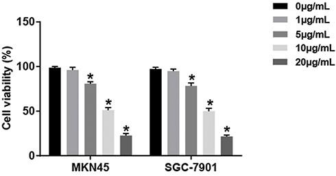 |
Figure 1 The effects of different concentrations of propofol on cell viability of MKN45 and SGC-7901 cells were measured by CCK-8 assay. *P < 0.05, vs. 0 μg/mL group. The values correspond to the mean ± standard deviation obtained from three independent experiments. |
miR-140-5p Was Upregulated By Propofol In MKN45 And SGC-7901 Cells
As determined by qRT-PCR in Figure 2, the expression of miR-140-5p in Propofol group was significantly increased compared with Control group (P < 0.05). Furthermore, miR-140-5p expression was significantly decreased in miR-140-5p inhibitor group compared with Control group (P < 0.05). When compared with Propofol group, the expression of miR-140-5p was significantly reduced in Propofol+miR-140-5p inhibitor group (P < 0.05). Those above results suggested that propofol might upregulate miR-140-5p expression in MKN45 and SGC-7901 cells.
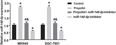 |
Figure 2 Relative miR-140-5p mRNA expression in MKN45 and SGC-7901 cells was detected by qRT-PCR. *P < 0.05, vs. Control group; #P < 0.05, vs. Propofol group, &P < 0.05, vs. miR-140-5p inhibitor group. The values correspond to the mean ± standard deviation obtained from three independent experiments. |
Propofol Suppressed Proliferation And Promoted Apoptosis Of MKN45 And SGC-7901 Cells By Upregulating miR-140-5p
The effects of propofol on cell proliferation of MKN45 and SGC-7901 cells were assessed by using BrdU incorporation assay (Figure 3A). The cell proliferation ability was significantly inhibited in Propofol group and increased in miR-140-5p inhibitor group compared with Control group (P < 0.05). When compared with Propofol group, cell proliferation ability was significantly increased in Propofol+miR-140-5p inhibitor group (P < 0.05). The cell proliferation ability was significantly decreased in Propofol+miR-140-5p inhibitor group than that in miR-140-5p inhibitor group (P < 0.05). All results above indicated that propofol could suppress cell proliferation of MKN45 and SGC-7901 cells by upregulating miR-140-5p.
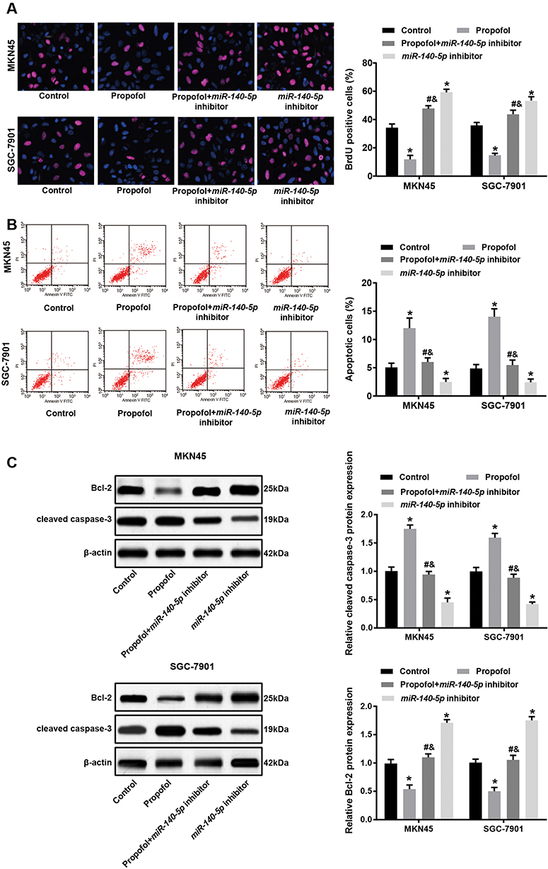 |
Figure 3 miR-140-5p participated in the effects of propofol on MKN45 and SGC-7901 cell proliferation and apoptosis. (A) Cell proliferation was measured by BrdU incorporation assay (× 400). (B) Cell apoptosis was measured by Annexin V-FITC/PI double staining assay. (C) The protein expressions of cleaved caspase-3 and Bcl-2 were detected by Western blot. *P < 0.05, vs. Control group; #P < 0.05, vs. Propofol group, &P < 0.05, vs. miR-140-5p inhibitor group. The values correspond to the mean ± standard deviation obtained from three independent experiments. |
To determine the effects of propofol on cell apoptosis of MKN45 and SGC-7901 cells, we performed Annexin V-FITC/PI double staining assay. The results of Figure 3B showed that the rates of MKN45 and SGC-7901 apoptosis cells were significantly increased in Propofol group compared with Control group (P < 0.05). When compared with Propofol group, the cell apoptosis was significantly decreased in Propofol+miR-140-5p inhibitor group (P < 0.05). But the cell apoptosis was significantly increased in Propofol+miR-140-5p inhibitor group compared to miR-140-5p inhibitor group (P < 0.05). Cell apoptosis is regulated by the Bcl-2 family and caspase family proteins. Therefore, the expressions of cleaved caspase-3 and Bcl-2 in MKN45 and SGC-7901 cells were measured by Western blot assay (Figure 3C). The results showed that cleaved caspase-3 expression was increased (P < 0.05) whereas expression of Bcl-2 was decreased (P < 0.05) by propofol compared to Control group. When compared with Propofol group, the expression of cleaved caspase-3 was significantly reduced in Propofol+miR-140-5p inhibitor group, while Bcl-2 expression was significantly increased (P < 0.05). Moreover, the expression level of cleaved caspase-3 was increased (P < 0.05) while Bcl-2 was decreased (P < 0.05) in Propofol+miR-140-5p inhibitor group compared to miR-140-5p inhibitor group. All those results suggested that propofol could promote cell apoptosis of MKN45 and SGC-7901 cells by upregulating miR-140-5p.
Propofol Inhibited Cell Migration And Invasion Of MKN45 And SGC-7901 Cells By Upregulating miR-140-5p
The impacts of propofol on migration and invasion of MKN45 and SGC-7901 cells were measured by using the wound healing assay (Figure 4A) and transwell assay (Figure 4B and C). The results revealed that both cell migration and invasion of MKN45 and SGC-7901 cells were significantly inhibited in Propofol group compared with Control group (P < 0.05). Moreover, cell migration and invasion were significantly higher in miR-140-5p inhibitor group than those in Control group (P < 0.05). When compared with Propofol group, cell migration and invasion were significantly increased in Propofol+miR-140-5p inhibitor group (P < 0.05). The cell migration and invasion were markedly repressed in Propofol+miR-140-5p inhibitor group compared with miR-140-5p inhibitor group (P < 0.05), suggesting Propofol could inhibit cell migration and invasion of MKN45 and SGC-7901 cells through upregulating miR-140-5p.
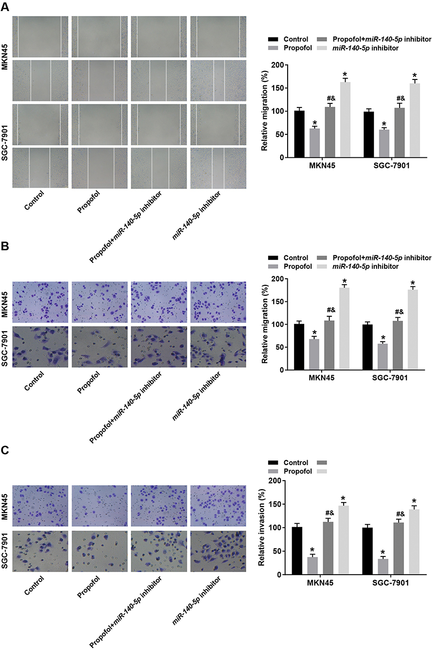 |
Figure 4 miR-140-5p participated in the effects of propofol on MKN45 and SGC-7901 cell migration and invasion. (A) Cell migration ability was measured by Wound healing assay. (B) Cell migration ability was tested by Transwell assay (×200). (C) Cell invasion ability was detected by Transwell assay (×200). *P < 0.05, vs. Control group; #P < 0.05, vs. Propofol group, &P < 0.05, vs. miR-140-5p inhibitor group. The values correspond to the mean ± standard deviation obtained from three independent experiments. |
Propofol Inhibited The Expressions Of MMP-2 And MMP-9 In MKN45 And SGC-7901 Cells By Upregulating miR-140-5p
As indicated by qRT-PCR assay (Figure 5A and B) and Western blot assay (Figure 5C), the expressions of MMP-2 and MMP-9 were decreased in Propofol group compared to Control group (P < 0.05). In addition, MMP-2 and MMP-9 expressions were significantly higher in miR-140-5p inhibitor group than those in Control group (P < 0.05). When compared with Propofol group, MMP-2 and MMP-9 expressions were significantly increased in Propofol+miR-140-5p inhibitor group (P < 0.05). On the contrary, the expressions of MMP-2 and MMP-9 were markedly repressed in Propofol+miR-140-5p inhibitor group compared with miR-140-5p inhibitor group (P < 0.05). All those results suggested that propofol could inhibit the expressions of MMP-2 and MMP-9 in MKN45 and SGC-7901 cells by upregulating miR-140-5p.
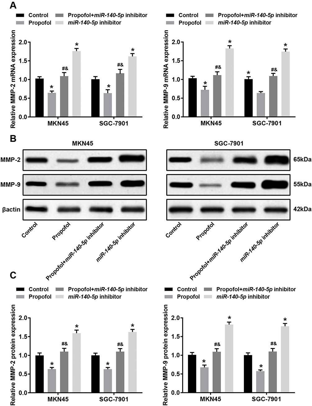 |
Figure 5 Propofol suppressed the expression of MMP-2 and MMP-9 by upregulating miR-140-5p. (A) The mRNA expression of MMP-2 and MMP-9 were tested by qRT-PCR. (B and C) The protein expression levels of MMP-2 and MMP-9 were measured by Western blot. *P < 0.05, vs. Control group; #P < 0.05, vs. Propofol group, &P < 0.05, vs. miR-140-5p inhibitor group. The values correspond to the mean ± standard deviation obtained from three independent experiments. |
Discussion
GC is one of the most common malignant neoplasms around the world.20 Approximately 1 million new cases are identified and almost 850,000 deaths are reported each year.21 It is urgent to explore and discover innovative and effective drugs to better treat this disease. In the current study, we demonstrated that propofol might suppress proliferation, migration, invasion and promote apoptosis by upregulating microRNA-140-5p in GC cells SGC-7901 and MKN45.
Apart from its anesthetic effects, propofol also exerts a number of non-anesthetic effects.22 Recently, increasing evidences have indicated that propofol plays an anti-tumor role in various cancers.23–25 In addition, mounting researches have indicated propofol suppresses proliferation and invasion of cancer cells.26,27 Peng and Yang et al17 reported that propofol could inhibit proliferation and promote apoptosis of GC cells. Wang et al8 also demonstrated that propofol suppressed proliferation and invasion of human GC cells. In our study, we found propofol exerted anti-tumor effect on GC cells by suppressing cell proliferation, migration and invasion, and promoting cell apoptosis. However, the exact anti-tumor mechanism of propofol in GC is still unknown.
Recent studies have indicated that miRNAs are significantly associated with gastric tumorigenesis and development.28–30 Previous studies have reported that miR-647 suppressed cell proliferation and promoted apoptosis in GC cells.31 MiR-1256 inhibited cell proliferation and promoted cell apoptosis in GC.32 miR-206 and miR-140-5p have the ability to inhibit proliferation, migration and invasion of GC cells.15,33 In addition, propofol has been reported to suppress proliferation and invasion of GC cells via regulating miR-2218 and miR-195.16 To sum up, propofol may exert anti-tumor effects by regulating the expression of miRNAs in GC cells. Therefore, we investigated whether propofol might exert anti-tumor effects on GC cells by modulating miR-140-5p.
To further investigate the functional mechanism of propofol on apoptosis of GC cells, the expressions of apoptosis-related factors (cleaved caspase-3 and Bcl-2) were measured. Studies have indicated that cleaved caspase-3 is a potent caspase that plays a crucial role in cell apoptosis.34 Furthermore, the anti-apoptosis protein Bcl-2, a member of the Bcl-2 family, is vital to the intrinsic apoptotic pathway.35 Our results showed that the expression of cleaved caspase-3 was increased but Bcl-2 was decreased by propofol treatment. On the contrary, the expression of cleaved caspase-3 was decreased but Bcl-2 was increased by miR-140-5p inhibitor treatment. The results more accurately confirmed that propofol could promote cell apoptosis in GC cells by upregulating miR-140-5p. It is known that inhibition of GC metastasis is a key step in GC treatment.36 Some researchers have indicated that MMP-2 and MMP-9 play central roles in cancer cell migration.37 In our study, the expressions of MMP-2 and MMP-9 were decreased by propofol treatment. Nevertheless, the expression of MMP-2 and MMP-9 was increased by miR-140-5p inhibitor treatment. These findings further provided evidence that propofol could also inhibit GC cell migration and invasion by upregulating miR-140-5p.
Conclusion
The present study demonstrated that propofol could significantly inhibit cell proliferation, migration and invasion, as well as promote cell apoptosis by upregulating miR-140-5p in SGC-7901 and MKN45 cells of GC. Our research provides an innovatively regulatory mechanism about the propofol in GC cells and points a new way to the treatment of GC.
Data Availability
All data used to support the findings of this study, which are included within the article.
Disclosure
The authors declare that there is no conflict of interest regarding this work.
References
1. Bray F, Ferlay J, Soerjomataram I, et al. Global cancer statistics 2018: GLOBOCAN estimates of incidence and mortality worldwide for 36 cancers in 185 countries. CA Cancer J Clin. 2018;68:394–424. doi:10.3322/caac.21492
2. Ferlay J, Colombet M, Soerjomataram I, et al. Estimating the global cancer incidence and mortality in 2018: GLOBOCAN sources and methods. Int J Cancer. 2019;15:144:1941–1953. doi:10.1002/ijc.31937
3. Liu H, Gao Y, Song D, et al. Correlation between microRNA-421 expression level and prognosis of gastric cancer. Int J Clin Exp Pathol. 2015;8:15128–15132
4. Fleck T, Schubert S, Ewert P, et al. Propofol effect on cerebral oxygenation in children with congenital heart disease. Pediatr Cardiol. 2015;36:543–549. doi:10.1007/s00246-014-1047-7
5. Wang ZT, Gong HY, Zheng F, Liu DJ, Dong TL. Propofol suppresses proliferation and invasion of pancreatic cancer cells by upregulating microRNA-133a expression. Genet Mol Res. 2015;14:7529–7537. doi:10.4238/2015.July.3.28
6. Cui WY, Liu Y, Zhu YQ, et al. Propofol induces endoplasmic reticulum (ER) stress and apoptosis in lung cancer cell H460. Tumour Biol. 2014;35:5213–5217. doi:10.1007/s13277-014-1677-7
7. Yang C, Gao J, Yan N, et al. Propofol inhibits the growth and survival of gastric cancer cells in vitro through the upregulation of ING3. Oncol Rep. 2016;37:587–593. doi:10.3892/or.2016.5218
8. Wang ZT, Gong HY, Zheng F, et al. Propofol suppresses proliferation and invasion of gastric cancer cells via downregulation of microRNA-221 expression. Genet Mol Res. 2015;14:8117–8124.
9. Pu M, Chen J, Tao Z, et al. Regulatory network of miRNA on its target: coordination between transcriptional and post-transcriptional regulation of gene expression. Cell Mol Life Sci. 2019;76:441–451. doi:10.1007/s00018-018-2940-7
10. Squadrito ML, Baer C, Burdet F, et al. Endogenous RNAs modulate microRNA sorting to exosomes and transfer to acceptor cells. Cell Rep. 2014;8:1432–1446. doi:10.1016/j.celrep.2014.07.035
11. He L, Hannon GJ. MicroRNAs: small RNAs with a big role in gene regulation. Nat Rev Genet. 2004;5:522–531. doi:10.1038/nrg1379
12. ZiaSarabi P, Sorayayi S, Hesari A, et al. Circulating microRNA-133, microRNA-17 and microRNA-25 in serum and its potential diagnostic value in gastric cancer. J Cell Biochem. 2019. doi:10.1002/jcb.28503
13. Sun L, Liang J, Wang Q, et al. MicroRNA-137 suppresses tongue squamous carcinoma cell proliferation, migration and invasion. Cell Prolif. 2016;49:628–635. doi:10.1111/cpr.2016.49.issue-5
14. Liu WZ, Liu N. Propofol inhibits lung cancer A549 cells growth and epithelial-mesenchymal transition process by up-regulation of microRNA-1284. Oncol Res. 2018;1–8. doi:10.3727/096504018X15172738893959
15. Fang Z, Yin S, Sun R, et al. miR-140-5p suppresses the proliferation, migration and invasion of gastric cancer by regulating YES1. Mol Cancer. 2017;16:139. doi:10.1186/s12943-017-0708-6
16. Zhang W, Wang Y, Zhu Z, et al. Propofol inhibits proliferation, migration and invasion of gastric cancer cells by up-regulating microRNA-195. Int J Biol Macromol. 2018;120:975–984. doi:10.1016/j.ijbiomac.2018.08.173
17. Peng Z, Zhang Y. Propofol inhibits proliferation and accelerates apoptosis of human gastric cancer cells by regulation of microRNA-451 and MMP-2 expression. Genet Mol Res. 2016;15.
18. Lu D, Yao Q, Zhan C, et al. MicroRNA-146a promote cell migration and invasion in human colorectal cancer via carboxypeptidase M/src-FAK pathway. Oncotarget. 2017;8:22674–22684. doi:10.18632/oncotarget.15158
19. Tiwari A, Pattnaik N, Jaiswal AM, et al. Increased FSHD region gene1 expression reduces in vitro cell migration, invasion, and angiogenesis, ex vivo supported by reduced expression in tumors. Biosci Rep. 2017;37:BSR20171062. doi:10.1042/BSR20171062
20. Faghihloo E, Araei Y, Mohammadi M, et al. The effect of oxamflatin on the E-cadherin expression in gastric cancer cell line. Cancer Gene Ther. 2016;23:396–399. doi:10.1038/cgt.2016.52
21. Bertuccio P, Chatenoud L, Levi F, et al. Recent patterns in gastric cancer: a global overview. Int J Cancer. 2010;125:666–673. doi:10.1002/ijc.24290
22. Vasileiou I, Xanthos T, Koudouna E, et al. Propofol: a review of its non-anaesthetic effects. Eur J Pharmacol. 2009;605:1–8. doi:10.1016/j.ejphar.2009.01.007
23. Wang JW, Cheng WW, Xu T, et al. Propofol induces apoptosis and inhibits the proliferation of rat embryonic neural stem cells via gamma-aminobutyric acid type A receptor. Genet Mol Res. 2015;14:14920. doi:10.4238/2015.November.18.57
24. Du Q, Liu J, Zhang X, et al. Propofol inhibits proliferation, migration, and invasion but promotes apoptosis by regulation of Sox4 in endometrial cancer cells. Braz J Med Biol Res. 2018;51:e6803. doi:10.1590/1414-431x20176803
25. Liu SQ, Zhang JL, Li ZW, et al. Propofol inhibits proliferation, migration, invasion and promotes apoptosis through down-regulating mir-374a in hepatocarcinoma cell lines. Cell Physiol Biochem. 2018;49:2099–2110. doi:10.1159/000493814
26. Ye Z, Jingzhong L, Yangbo L, et al. Propofol inhibits proliferation and invasion of osteosarcoma cells by regulation of microRNA-143 expression. Oncol Res. 2013;21:201–207. doi:10.3727/096504014X13890370410203
27. Miao Y, Zhang Y, Wan H, et al. GABA-receptor agonist, propofol inhibits invasion of colon carcinoma cells. Biomed Pharmacother. 2010;64:583–588. doi:10.1016/j.biopha.2010.03.006
28. Wang J, Chen X, Su L, et al. MicroRNA-126 inhibits cell proliferation in gastric cancer by targeting LAT-1. Biomed Pharmacother. 2015;72:66–73. doi:10.1016/j.biopha.2015.04.001
29. Xiao-Jun X, Jun D, Ya-Wen L, et al. MiR-1271 inhibits cell proliferation, invasion and EMT in gastric cancer by targeting FOXQ1. Cell Physiol Biochem. 2015;36:1382–1394. doi:10.1159/000430304
30. Huang T, Wang-Johanning F, Zhou F, et al. MicroRNAs serve as a bridge between oxidative stress and gastric cancer (review). Int J Oncol. 2016;49:1791–1800. doi:10.3892/ijo.2016.3686
31. Cao W, Wei W, Zhan Z, et al. Role of miR-647 in human gastric cancer suppression. Oncol Rep. 2017;37:1401–1411. doi:10.3892/or.2017.5383
32. Xu Z, Li Z, Wang W, et al. MIR-1265 regulates cellular proliferation and apoptosis by targeting calcium binding protein 39 in gastric cancer and, thereby, impairing oncogenic autophagy. Cancer Lett. 2019;449:226–236. doi:10.1016/j.canlet.2019.02.026
33. Deng M, Qin Y, Chen X, et al. MiR-206 inhibits proliferation, migration, and invasion of gastric cancer cells by targeting the MUC1 gene. Onco Targets Ther. 2019;12:849–859. doi:10.2147/OTT.S180021
34. Ramanjit G, Marc S, Klas B, et al. Role of caspase-3 activation in cerebral ischemia-induced neurodegeneration in adult and neonatal brain. J Cereb Blood Flow Metab. 2002;22:420. doi:10.1097/00004647-200204000-00006
35. Namura S, Zhu J, Fink K, et al. Activation and cleavage of caspase-3 in apoptosis induced by experimental cerebral ischemia. J Neurosci. 1998;18:3659–3668. doi:10.1523/JNEUROSCI.18-10-03659.1998
36. Dulskas A, Bandar MA, Choi YY, et al. A case of gastric cancer metastasis to the breast in a female with BRCA2 germline mutation and literature review. Acta Chir Belg. 2017;1–5.
37. Zoi P, Dimitra M, Konstantina K, et al. Strategies to target matrix metalloproteinases as therapeutic approach in cancer. Methods Mol Biol. 2018;1731:325–348.
 © 2019 The Author(s). This work is published and licensed by Dove Medical Press Limited. The
full terms of this license are available at https://www.dovepress.com/terms
and incorporate the Creative Commons Attribution
- Non Commercial (unported, 3.0) License.
By accessing the work you hereby accept the Terms. Non-commercial uses of the work are permitted
without any further permission from Dove Medical Press Limited, provided the work is properly
attributed. For permission for commercial use of this work, please see paragraphs 4.2 and 5 of our Terms.
© 2019 The Author(s). This work is published and licensed by Dove Medical Press Limited. The
full terms of this license are available at https://www.dovepress.com/terms
and incorporate the Creative Commons Attribution
- Non Commercial (unported, 3.0) License.
By accessing the work you hereby accept the Terms. Non-commercial uses of the work are permitted
without any further permission from Dove Medical Press Limited, provided the work is properly
attributed. For permission for commercial use of this work, please see paragraphs 4.2 and 5 of our Terms.