")
Back to Journals » International Medical Case Reports Journal » Volume 15
Promising Treatment for Twenty-Nail Dystrophy with Combination of Fractional Carbon Dioxide Laser and Topical Therapy: A Case Report
Authors Ruchiatan K , Nuraeni L , Pranata AY, Puspitosari D, Hindritiani R
Received 5 July 2022
Accepted for publication 11 August 2022
Published 31 August 2022 Volume 2022:15 Pages 469—472
DOI https://doi.org/10.2147/IMCRJ.S381192
Checked for plagiarism Yes
Review by Single anonymous peer review
Peer reviewer comments 2
Editor who approved publication: Professor Ronald Prineas
Kartika Ruchiatan, Lita Nuraeni, Achmad Yudha Pranata, Diah Puspitosari, Reti Hindritiani
Department of Dermatology and Venereology, Faculty of Medicine, Universitas Padjadjaran-Dr. Hasan Sadikin Hospital, Bandung, Indonesia
Correspondence: Kartika Ruchiatan, Department of Dermatology and Venereology, Faculty of Medicine, Universitas Padjadjaran – Dr. Hasan Sadikin Hospital, Jl. Pasteur 38, Bandung, West Java, 40161, Indonesia, Tel +62811247932, Email [email protected]
Abstract: Twenty-nail dystrophy (TND) is a trachyonychia affecting all nails which cause aesthetic complaints. The difficulty of topical medication to penetrate through the nail plate brings a great challenge for TND treatment. We reported a case of TND in a 27-year-old woman, with clinical manifestation of trachyonychia, longitudinal ridging, mottled lunula, and subungual hyperkeratosis with ragged cuticle affected all nails. She has suffered from TND for 20 years with no systemic involvement found. Histopathological examination supported the diagnosis of nail lichen planus. The patient received a mixture of topical therapy consist of tacrolimus, urea, and salicylic acid, combined with fractional carbon dioxide (CO2) laser with pulse energy: 160 mJ, pulse duration: 8.0 ms, density level: 17, and depth level: 2. After two sessions of treatment within five weeks interval, clinical improvement was seen as refinement of nail’s texture. Treatment of TND often unsatisfactory due to difficulty of drug penetration through the nail plate. The fractional CO2 laser creates a column of destruction down to the dermis, which aid penetration of topical medication and stimulate nail bed rejuvenation. The use of fractional CO2 laser as a penetration enhancer can be a therapeutic option for the treatment of TND with promising result as shown in this patient. This procedure enabled combination with topical medications as long-term therapy for TND.
Keywords: fractional carbon dioxide laser, penetration enhancer, twenty-nail dystrophy
Introduction
Twenty-nail dystrophy (TND) is a disorder of the entire nail, which is characterized by a rough nail surface or trachyonychia.1,2 TND can cause aesthetic complaints, reduce self-confidence, and affect the patient’s quality of life.1,3 There are several diseases that commonly accompany TND, including alopecia areata, atopic dermatitis, rheumatoid arthritis, psoriasis, and lichen planus.1,2
Lichen planus (LP) is an inflammatory disease that can attack the skin, mucosa, and nails.4 Nail abnormalities in LP can occur without any skin or mucosal lesions.5 Nail LP is reported to be resistant to almost all topical treatments,1 while systemic and long-term use of the drug can cause major side effects, and intralesional injection cause pain during administration which causes discomfort to the patient. Therefore, a therapeutic modality that can aid penetration of the drug on to nail plate was needed, and without causing much discomfort for the patient.6
Case
A 27-year-old woman complained of nail damage on all her fingers and toes accompanied by itching for about 20 years. The patient had already went to several dermatovenereologists and been treated with various topical and oral therapy for months but there were no significant improvements. From physical examination of all nails showed trachyonychia, longitudinal ridge, mottled lunula, and subungual hyperkeratosis (Figure 1). The cuticles appear hyperkeratotic. There were no skin lesions found on other parts of the body. Fungal culture examination was negative. The results of histopathological examination of the left toe nail biopsy showed lichenoid reactions which supported the diagnosis of nail LP.
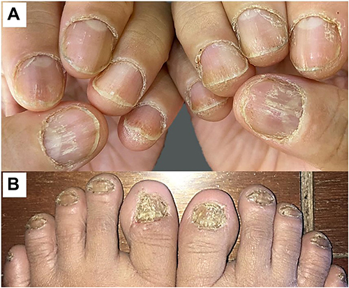 |
Figure 1 Fingernails of hands (A) and feet (B) before combination therapy. |
Patient was diagnosed as TND caused by LP and treated with topical mixture of 0.1% tacrolimus cream, 20% urea cream, and 20% salicylic acid, applied twice daily and combined with fractional CO2 laser. We also gave 10 mg desloratadine orally once daily. The fractional CO2 laser treatment was performed at five weeks intervals without any anesthesia beforehand. Two passes of the laser treatment with pulse energy: 160 mJ, pulse duration: 8.0 ms, density level: 17, depth level: 2, square shape, and array pattern was done over the nail plate until the pixelated frosted area was seen. There was no immediate bleeding or oozing after treatment. Clinical improvement on all nails was seen after two sessions of fractional CO2 laser treatment (Figure 2). The laser procedures were well tolerated. The patient did not feel pain during the procedure and no adverse effects were reported.
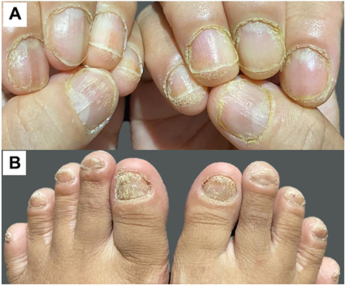 |
Figure 2 One month after 2nd procedure. Note the improvement of fingernails of hands (A) and feet (B). |
Discussion
Intralesional or systemic corticosteroids are the first-line therapy in nail LP.5 Intralesional injection clearly uncomfortable and causes pain, while systemic administration can cause side effects, especially when given for a long time.7 The topical medication has the advantage of not causing pain and does not provide side effects like systemic drugs. Some topically administered medication for nail LP are corticosteroids, tazarotene, calcipotriol, and tacrolimus.1,8 Tacrolimus is an immune modulator that has immunosuppressive effect by inhibiting T cell activation, which is thought to play a role in the pathogenesis of LP.6 This drug interacts with cytoplasmic proteins such as cyclophilin-like and FK506 protein which interferes with calcineurin phosphatase activity thereby inhibiting the transcription of proinflammatory cytokines. Ujiie et al6 reported a better therapeutic response to topical tacrolimus versus topical corticosteroids in five nail of LP patients. Topical tacrolimus has an anti-inflammatory effect with fewer side effects compared to corticosteroids.9 However, nail LP was reported to be resistant to topical treatment, this is thought to be due to the difficulty of the drug penetrating the nail plate.1,8 Therefore, topical application of drugs in the treatment of nails requires penetration enhancers that may facilitate penetration into the nail.
There are three types of penetration enhancers, known as chemical, physical, and mechanical. Keratolytic agents which include urea and salicylic acid acted as chemical penetration enhancers.10 The use of urea and salicylic acid in the mixture with other active ingredients of topical medications can also increase drug penetration into the nails.
Laser is one of the physical penetration enhancers that helps the penetration of drugs by forming small holes in the nails.10 The mechanism of action of the fractional CO2 laser in nail dystrophy is not yet fully understood.3 Fractional CO2 laser can cause tissue ablation in the form of small holes in the dermis layer called microscopic treatment zones (MTZ),3 that will increase the penetration of topical agents.3 In addition, the fractional CO2 laser allows relatively deeper energy penetration into the dermis layer.11 This causes controlled protein denaturation resulting in reactive collagen remodeling which will stimulate nail bed rejuvenation while maintaining epidermal integrity and shortening healing time.3,11 Although laser therapy may help accelerate healing, it is important to avoid over-prosecution of curative effects when the treatment itself may cause excessive clinical damage to the nail bed and deck.11
Due to the physiological nature of nails and the underlying conditions longer treatment should be completed. Laser procedures for nails can be held 3–4 sessions within 4 to 5 weeks interval until the improvement achieved.3 Lim et al3 in 2013 reported satisfactory results in combination fractional CO2 laser therapy in combination with 0.25% dexamethasone lotion in two patients with idiopathic TND. The fractional CO2 laser procedure was performed in three sessions with an interval of four weeks.
In our patient nail abnormalities appeared to have improved after the second laser procedures, characterized by reducing trachyonychia, longitudinal ridging, dystrophy, and subungual hyperkeratosis of the nails. Until this report was written, the patient had received four sessions of fractional CO2 laser procedures on the entire nail. We observed patient for almost two years. The patient still come for follow up and satisfied with the result of the nail condition remain stable.
Conclusion
The use of fractional CO2 laser as a penetration enhancer of topical therapy with suggested mechanism of nail bed rejuvenation can be a therapeutic option for the treatment of TND with promising result. Additional sessions and longer follow up could be done for optimal result and patient satisfaction. The combination of laser with topical or oral medication is recommended for a better outcome in TND.
Ethical Statement
The publications of images were included in the patient consent for publication of the case. Institutional approval to publish the case details has been obtained.
Consent Statement
The authors certify that they have obtained all appropriate patient consent forms. The patient signed a consent form for the publication of the case details and images.
Acknowledgments
The authors would like to thank the staff of Department of Dermatology and Venereology, Faculty of Medicine, Universitas Padjadjaran-Dr. Hasan Sadikin General Hospital, Bandung, West Java, Indonesia.
Funding
There is no funding to report.
Disclosure
The authors report no conflicts of interest in this work.
References
1. Haneke E. Nail disorders. In: Kang S, Amagai M, Bruckner AL, Enk AH, Margolis DJ, McMichael AJ, editors. Fitzpatrick’s Dermatology in General Medicine.
2. Baran D, Baran R. Disorders of nails. In: Burns T, Breathnach S, Cox N, Griffiths C, editors. Rook’s Textbook of Dermatology.
3. Lim EH, Seo YJ, Lee JH, Im M. Onychodystrophy treated using fractional carbon dioxide laser therapy and topical steroids: new treatment options for nail dystrophy. Dermatol Surg. 2013;39(12):1931–1933. doi:10.1111/dsu.12365
4. Iorizzo M, Tosti A, Starace M, et al. Isolated nail lichen planus: an expert consensus on treatment of the classical form. J Am Acad Dermatol. 2020;83:1717–1723. doi:10.1016/j.jaad.2020.02.056
5. Piraccini BM, Saccani E, Starace M, Balestri R, Tosti A. Nail lichen planus: response to treatment and longterm follow-up. Eur J Dermatol. 2010;20(4):489–496. doi:10.1684/ejd.2010.0952
6. Ujiie H, Shibaki A, Akiyama M, Shimizu H. Successful treatment of nail lichen planus with topical tacrolimus. Acta Dermatol Venereol. 2010;90(2):218–219. doi:10.2340/00015555-0814
7. Shanmuganathan H, Kumar R. A case of twenty nail dystrophy affecting a 12 year-old boy. Int J Contemp Pediatr. 2019;6(6):2702. doi:10.18203/2349-3291.ijcp20194759
8. McClanahan DR, English JC. Therapeutics for adult nail psoriasis and nail lichen planus: a guide for clinicians. Am J Clin Dermatol. 2018;19(4):559–584. doi:10.1007/s40257-018-0350-0
9. Lohani B, Kumar G. Medicated nail lacquers-for effective treatment of nail disorders. Indo Am J Pharm Sci. 2018;5(3):1392–1403.
10. Ibrahim O, Dovvrey JS. Fundamental of laser and light-based treatment. In: Kang S, Amagai M, Bruckner AL, Enk AH, Margolis DJ, McMichael AJ, editors. Fitzpatrick’s Dermatology in General Medicine.
11. Ma W, Si C, Carrero LM, et al. Laser treatment for onychomycosis: a systematic review and meta-analysis. Medicine. 2019;98(48):e17948. doi:10.1097/MD.0000000000017948
 © 2022 The Author(s). This work is published and licensed by Dove Medical Press Limited. The full terms of this license are available at https://www.dovepress.com/terms.php and incorporate the Creative Commons Attribution - Non Commercial (unported, v3.0) License.
By accessing the work you hereby accept the Terms. Non-commercial uses of the work are permitted without any further permission from Dove Medical Press Limited, provided the work is properly attributed. For permission for commercial use of this work, please see paragraphs 4.2 and 5 of our Terms.
© 2022 The Author(s). This work is published and licensed by Dove Medical Press Limited. The full terms of this license are available at https://www.dovepress.com/terms.php and incorporate the Creative Commons Attribution - Non Commercial (unported, v3.0) License.
By accessing the work you hereby accept the Terms. Non-commercial uses of the work are permitted without any further permission from Dove Medical Press Limited, provided the work is properly attributed. For permission for commercial use of this work, please see paragraphs 4.2 and 5 of our Terms.