
Back to Journals » Diabetes, Metabolic Syndrome and Obesity » Volume 19
Promising Biomarkers for Early Diagnosis: Advances in Understanding the Pathogenesis of Diabetic Peripheral Neuropathy
Authors Lyu Q, Tan Y, Zeng X  , Peng S, Lee M
Received 10 October 2025
Accepted for publication 27 January 2026
Published 10 February 2026 Volume 2026:19 568751
DOI https://doi.org/10.2147/DMSO.S568751
Checked for plagiarism Yes
Review by Single anonymous peer review
Peer reviewer comments 2
Editor who approved publication: Dr Rebecca Baqiyyah Conway
Qingqin Lyu,1,* Yanyan Tan,2,3,* Xianqi Zeng,2,3,* Siping Peng,2,3 Maosheng Lee2,3
1National Health Data Institute, Shenzhen, 518033, People’s Republic of China; 2The Fourth Clinical Medical College, Guangzhou University of Chinese Medicine, Shenzhen, 518033, People’s Republic of China; 3Department of Endocrinology, Shenzhen Traditional Chinese Medicine Hospital, Shenzhen, 518033, People’s Republic of China
*These authors contributed equally to this work
Correspondence: Siping Peng, Email [email protected] Maosheng Lee, Email [email protected]
Abstract: Diabetic peripheral neuropathy (DPN) is one of the most common chronic complications of diabetes mellitus. The most common type is distal symmetric polyneuropathy, and lesions frequently lead to disabling neuropathic pain and even amputation, increasing the risk of death. At present, the pathogenesis of DPN has not been clarified, and its insidious onset and lack of obvious symptoms in most patients in the early stages often lead to delayed diagnosis, which is unfavourable for prevention and treatment. This article provides an overview of the existing research progress on early diagnostic markers of DPN, especially oxidative stress-related markers, neural tissue damage markers, inflammation-related markers, neurovascular damage markers, and gene-related markers, in the hope that it can provide a reference for early diagnosis and treatment of DPN, and slow down the occurrence and development of DPN.
Keywords: diabetic peripheral neuropathy, diagnostic markers, pathogenesis, diabetes mellitus
Introduction
More than 50% of people with diabetes will develop diabetic neuropathy, which is one of the most common and troublesome complications of both type 1 diabetes mellitus (T1DM) and type 2 diabetes mellitus (T2DM).1–3 Approximately 30%~40% of patients with diabetic neuropathy report experiencing neuropathic pain, and the main neuropathy is diabetic peripheral neuropathy (DPN).3,4 DPN is a primary contributor to diabetic foot complications, including ulceration and Charcot neuroarthropathy, often leading to debilitating neuropathic pain and lower-limb amputation.1,3,5
According to the World Health Organisation, lower limb amputations are 10 times more common in diabetics than in non-diabetics. Many studies have shown a substantial increase in mortality among diabetics who undergo major amputations, with 5-year mortality rates ranging from 44% to 68%.1,3 Patients with DPN also have a significantly higher risk of cardiovascular death than those without DPN. The International Diabetes Federation 2021 report identifies diabetes as a fast-growing global epidemic in the 21st century, with an estimated prevalence rising from 10.5% (536.6 million people) in 2021 to 12.2% (783.2 million) in 2045.3,6,7
The clinical underdiagnosis of DPN stems from its heterogeneous pathogenesis and symptom overlap with other disorders.3,4 This diagnostic challenge underscores the urgent need for novel biomarkers. Furthermore, over 50% of DPN patients remain asymptomatic, which further delays early detection. To mitigate the clinical and socioeconomic burden of DPN, identifying reliable diagnostic markers is critical for enabling early screening and intervention. In this article, we summarize the latest research progress on diagnostic markers for DPN in terms of its pathophysiology as well as the mechanisms by which it may occur, expanding new ideas for the prevention and treatment of DPN.
Pathophysiology of Diabetic Peripheral Neuropathy
DPN pathophysiology involves multifactorial mechanisms, including metabolic dysregulation, mitochondrial dysfunction, and structural nerve damage. The length-dependent lesion of peripheral nerves in DPN occurs first in the toes and progresses to the proximal end. In advanced stages, similar damage may affect the fingers. This nerve damage reflects a “stocking-glove” distribution (Figure 1a), which is closely related to the structure and function of the peripheral nervous system (PNS).2,3,8
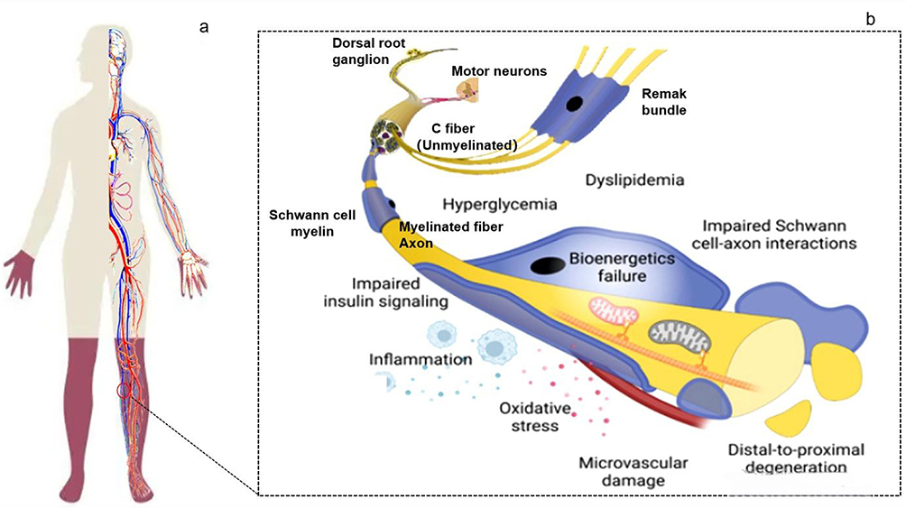 |
Figure 1 PNS structure and pathophysiology of diabetic peripheral neuropathy.2,8 (a) DPN manifests as nerve injury (red color) in a “stocking-glove” pattern. (b) Sustained hyperglycemia triggers oxidative stress and inflammation via the polyol pathway, advanced glycation end-products (AGEs) accumulation, and mitochondrial dysfunction, collectively damaging neurons and glial cells. Schwann cells (SCs)-critical for myelination-exhibit impaired interactions with axons, a hallmark of DPN. Hyperglycemia disrupts SC metabolic homeostasis, causing myelin sheath abnormalities and demyelination of myelinated fibers. It also alters the microenvironment of unmyelinated C-fibers in Remak bundles, promoting degeneration of sensory neurons (eg, dorsal root ganglia) and motor neurons. Inflammatory mediators and oxidative stress further impair axonal transport and and endoneurial blood flow, ultimately leading to pain, paresthesia, and motor deficits. The pathogenic complexity of DPN, rooted in multifactorial interactions, offers actionable insights for precision diagnosis and therapy. |
Peripheral nerves are composed of neurons (cell bodies and axons), supporting glial cells (eg, Schwann cells, SCs), blood vessels, and connective tissues. Each sensory axon (nerve fibre) extends from a dorsal root ganglion (DRG) neuron and usually carries electrical impulses. Sensory and motor neurons transmit afferent and efferent signals through nerve fibers, which are classified as unmyelinated (eg, C fibers) or myelinated (eg, Aδ and Aβ fibers), enabling distinct sensory modalities and conduction velocities.8 Under diabetic conditions (eg, hyperglycemia and dyslipidemia), neural structure is disrupted, accompanied by bioenergetic deficits, impaired insulin signaling, inflammation, oxidative stress, microvascular damage (Figure 1b). These are the typical pathological features of DPN.8–10
Long axons in the limbs require substantial energy to maintain membrane potential and signal transmission, primarily sourced from substrate catabolism via metabolic coupling between SCs and axons. SCs preferentially utilize glucose through glycolysis for adenosine triphosphate (ATP) production, with alternative pathways (eg, fatty acid oxidation) contributing to energy supply. Additionally, bidirectional communication between neurons and SCs occurs via paracrine signaling, extracellular vesicles, and direct molecular exchange.2,8 At the cellular level, mitochondrial dysfunction and oxidative stress in SCs drive neuronal damage, including axonal loss and demyelination, ultimately impairing energy homeostasis (Figure 2).9 Notably, insulin signaling—particularly through the PI3K-Akt pathway—plays a dual role: it not only regulates glucose metabolism but also promotes SC differentiation and remyelination, thereby mitigating DPN progression.9,11–13
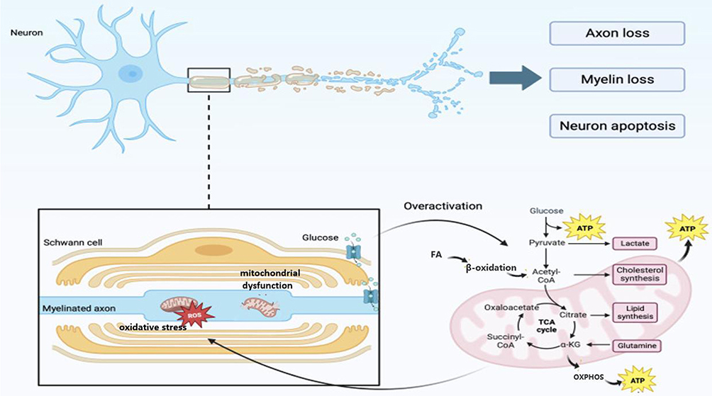 |
Figure 2 Demyelination and axonal damage of DPN.8,9 Energy production in Schwann cells (SCs) relies on substrate catabolism to generate ATP. Glucose is preferentially metabolized via glycolysis to pyruvate, which enters mitochondria through the mitochondrial pyruvate carrier (MPC). Pyruvate fuels the tricarboxylic acid (TCA) cycle, providing substrates for oxidative phosphorylation (OXPHOS) in the inner mitochondrial membrane (IMM). Fatty acid oxidation (FAO) serves as an alternative energy source. SCs also synthesize cholesterol and lipids critical for myelin maintenance. Bidirectional communication between neurons and SCs occurs via paracrine factors (eg, growth factors), gap junctions, and extracellular vesicles (EVs) carrying miRNAs. In DPN, hyperglycemia disrupts SC-axon metabolic coupling, impairing mitochondrial function and ATP synthesis. Concurrently, oxidative stress and TCA cycle dysfunction (eg, altered succinyl-CoA and oxaloacetate levels) exacerbate demyelination and axonal loss. These metabolic deficits misprocessing, drive a degenerative cascade characterized by neuronal apoptosis and motor-sensory deficits. |
DPN induces progressive sensory fiber degeneration and neuropathic pain, mediated by voltage-gated sodium channel (VGSC; Nav1.7, 1.8),8 voltage-gated potassium channels, ligand-gated transduction channels (eg, transient receptor potential cation channel subfamily A member 1, (Nav1.8, TRPA1),14 calcium channels (CAV3.2), and hyperpolarization-activated cyclic nucleotide-gated channels (HCN2).2
In healthy neural states (Figure 3a and b), ion channels sense initial stimuli and transmit them to sensory receptors.2 Subsequently, neural ion channels generate an action potential, which propagates along the axon to the central nervous system (CNS). Finally, in the dorsal horn of the spinal cord, ion channels trigger the release of neurotransmitters, at which time the potential changes of nerve membranes are relatively stable, and nerve cells produce ATP through normal energy metabolism to maintain cell function. Nav1.8 and TRPA1 channels also play a normal role.
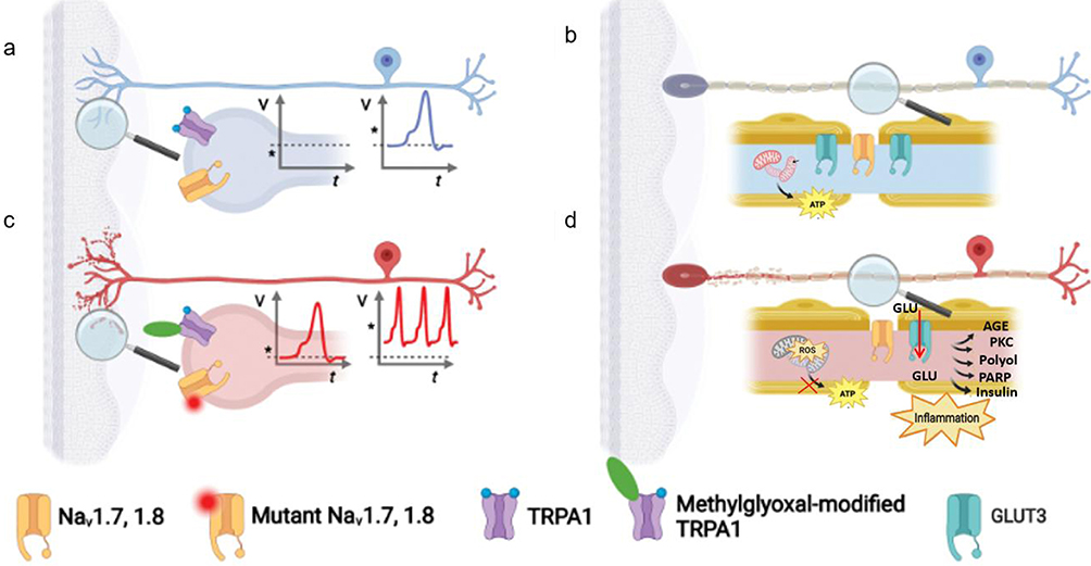 |
Figure 3 Neuropathy of diabetic peripheral neuropathy.2,9 (a) Under normal circumstances, the changes of nerve cell membrane potential are relatively stable; (b) nerve cells produce ATP through normal energy metabolism to maintain cell function, and TRPA1 ion channels play a normal role. (c) DPN reaction substrate and channel changes involve a variety of molecules related to nerve conduction, such as mutated Nav1.7 and 1.8 channels that cause abnormal oscillation of nerve cell membrane potential, methylglyoxal modifies TRPA1 to change its function and participate in the neuropathic process. (d) GLUT3 (glucose transporter 3) function may also be affected, further affecting D-glucose uptake and metabolism of nerve cells, glucose (GLU) metabolism is abnormal, reactive oxygen species (ROS) production is increased, mitochondrial function is affected, and ATP production is blocked. At the same time, a series of signaling pathways are activated, such as AGE (advanced glycosylation end products) -PKC (protein kinase C) pathway, Polyol pathway, PARP (polyADP-ribose polymerase) pathway, leading to inflammation. |
However, in DPN (Figure 3c and d), mutated Nav1.7 and 1.8 channels cause abnormal oscillations in nerve membrane potential. At the same time, TRPA1 is modified by methylglyoxal, which triggers a series of changes, including increased production of reactive oxygen species (ROS), blocked production of ATP, and inflammatory responses. Glucose transporter 3 (GLUT3) function may also be affected, with abnormal glucose (GLU) metabolism in nerve cells. These changes are closely related to the mechanism of abnormal nerve signals and nerve damage in diabetic peripheral neuropathy.8
Hyperglycemia and hyperlipidemia disrupt nerve function via multiple pathways,9 including protein kinase C (PKC) pathway, polyol pathway, advanced glycosylated terminal (AGE) pathway, hexosamine pathway, PARP pathway, insulin pathway, compounded by mitochondrial dysfunction, oxidative stress, inflammatory response, metabolic abnormalities, and microvascular disease.15,16 Neuronal demyelination and neuronal damage (Figure 3c and d) ultimately slows down nerve conduction, which in turn leads to energy loss and metabolic disorders.8
In light of the established pathophysiological mechanisms outlined in previous sections, we systematically evaluate existing diagnostic modalities for DPN and propose novel biomarkers that may enhance early detection and clinical management.
Electrophysiological Diagnosis of Neuropathy
Electrophysiological Indicators
The clinical diagnosis of DPN requires identification of neuropathy signs/symptoms in patients with prediabetes or diabetes mellitus following exclusion of alternative etiologies.4,6,17 Initial evaluation combines medical history, symptom analysis, and physical examination. Electrophysiological testing becomes essential for atypical presentations, with nerve conduction studies (NCS) remaining the diagnostic gold standard.1,18–20 To enhance diagnostic completeness, composite indices that integrate symptoms, signs, and simple tests are invaluable. For instance, the DPN-Check is a succinct, patient-administered screening tool that efficiently combines symptom questionnaires with monofilament and vibration perception tests, offering a rapid clinical assessment. In contrast, the more comprehensive Toronto Clinical Neuropathy Score (TCNS) systematically grades symptoms, sensory test results, and reflex outcomes to quantify both the presence and severity of DPN. The inclusion of such indices provides a structured, multi-modal framework that complements single biomarker findings and aligns with holistic diagnostic approaches.
NCS serve as the cornerstone of electrophysiological diagnostics, evaluating conduction abnormalities in both sensory (eg, sural and superficial peroneal nerves) and motor nerves (eg, tibial and common peroneal nerves) to assess axonal integrity and demyelination in diabetic neuropathy. Clinically, we commonly use electrical diagnostic tests (eg, electromyography, EMG),21–25 which mainly investigate NCS and F-wave abnormality rate or other abnormal parameters, to confirm the severity of nerve damage. NCS combined with F-wave analysis demonstrates high diagnostic accuracy (sensitivity, specificity >80%).25 Patients with extended disease duration (>10 years) exhibited markedly slower motor nerve conduction velocities (NCV) in the median, ulnar, common peroneal, and posterior tibial nerves compared to those with early-stage disease (<1 year)23 accompanied by progressive amplitude reduction.2. This temporal decline in neural function is further corroborated by preclinical evidence: diabetic animal models demonstrated a 44.4% decrease in mean NCV versus non-diabetic controls (P<0.001).26 Integration of HbA1c values with electromyography enhances early detection (98% sensitivity, 94% specificity).24 Significantly, combining NCS with clinical examination increases diagnostic yield from 40.4% to 62.2%, with 14.0% prevalence of neuropathic pain.27 The evidence suggests NCS parameters may serve prognostic functions, as demonstrated by improved conduction velocities following 6–12 weeks of statin therapy.28,29
Conventional nerve conduction studies primarily assess large-fiber neuropathy;2 however, DPN predominantly involves pathologies of small myelinated and unmyelinated nerve fibers, which are critical for nociceptive signal transmission in response to noxious stimuli. Particularly, standard electrophysiological modalities such as NCV testing and EMG exhibit limited sensitivity in detecting small-fiber dysfunction,29 particularly in asymptomatic patients or those with early-stage DPN characterized by subclinical neuropathic alterations.
Small-Fiber Neuropathy (SFN) Diagnostics
Small-fiber neuropathy (SFN), a pivotal contributor to neuropathic pain and a hallmark of early diabetic peripheral neuropathy (DPN),30,31 presents significant diagnostic challenges due to the limited sensitivity of conventional nerve conduction studies (NCS) in detecting small myelinated and unmyelinated fiber pathology.32 To address NCS utility is constrained in detecting SFN, skin biopsy with intraepidermal nerve fiber density (IENFD) quantification has emerged as a complementary diagnostic modality, demonstrating high diagnostic efficacy (88%), positive predictive value (75%), and negative predictive value (90%) for neuropathy.33 Of note, IENFD shows superior sensitivity (51.1%; 95% CI: 43.7–58.5) and specificity (90%; 95% CI: 79.5–96.2) for early SFN detection compared to conventional electrophysiological methods.34 Despite these advantages, IENFD faces criticism due to its reduced sensitivity in identifying mild nerve damage and the inherent challenges in tracking longitudinal neural changes, underscoring the need for more refined quantitative methodologies to enhance its clinical applicability.35 Moreover, skin biopsy is invasive, expensive and lacks diagnostic laboratory capacity.36 These limitations highlight the importance of integrating multimodal diagnostic approaches to optimize SFN assessment and monitoring.
In recent years, corneal confocal microscopy (CCM) has assessed non-invasive assessment of small-fiber integrity.34,36–38 By leveraging high-resolution confocal imaging, CCM enables precise quantification of corneal subepithelial nerve plexus morphology—including nerve fiber length, density, branching complexity, and curvature39—providing a surrogate marker for peripheral small-fiber damage in DPN.31 This conclusion is corroborated by a meta-analysis of 13 studies involving 1680 participants, which confirms the diagnostic utility of CCM in detecting early neural degeneration in DPN.40 This technique not only correlates strongly with IENFD but also facilitates dynamic monitoring of neuropathic progression and therapeutic response.34 CCM provides high-resolution structural assessment of the corneal subepithelial nerve plexus, its utility is restricted to localized evaluation of small-fiber morphology and does not extend to systemic small-fiber pathology. Furthermore, diagnostic interpretation of CCM findings requires careful exclusion of confounding ocular comorbidities (eg, dry eye syndrome, infectious keratitis), as these conditions may artifactually alter nerve architecture and compromise specificity.
Parallel advancements in functional assessments include quantitative sensory testing (QST), which evaluates nociceptive thresholds (eg, thermal and mechanical pain sensitivity).31 Nociceptive hypersensitivity, reflecting early small-fiber dysfunction, exhibits high diagnostic accuracy (sensitivity: 84%; specificity: 81%),41 positioning QST as a valuable tool for identifying subclinical DPN. Although the subjectivity of Quantitative Sensory Testing (QST) is well-recognized, emerging automated thermal threshold testing devices enhance objectivity by standardizing stimulus delivery and response recording.
The comparative strengths and limitations of these diagnostic modalities are summarized in Table 1. While current methods demonstrate clinical utility in detecting DPN across disease stages, their inherent constraints—including invasiveness, operator dependency, and limited sensitivity for early small-fiber pathology—highlight the urgent need to develop and validate novel non-invasive biomarkers with enhanced specificity for neuroaxonal degeneration. Future research should prioritize translational innovations to bridge this diagnostic gap and refine precision medicine strategies in DPN management.
 |
Table 1 Assessing Clinical Detection Method in DPN (Human Clinical Study) |
Neurotissue Damage Factor (NTDF)
Neurofilament Heavy Chain (NF-H)
Emerging biomarkers for small-fiber neuropathy (SFN) highlight axonal degeneration as a critical early pathological feature.42 Neurofilament heavy chain (NF-H), a cytoskeletal protein essential for axonal integrity, undergoes phosphorylation (pNF-H)—a post-translational modification regulating axonal caliber and microtubule interactions. Serum pNF-H exhibits exceptional stability, resisting enzymatic degradation, and serves as a robust biomarker of neuroaxonal injury. Preclinical studies demonstrate that restoring NF homeostasis rescues neuronal ultrastructure and immunohistochemical signatures in diabetic models.43 Clinically, pNF-H levels are elevated in diabetic peripheral neuropathy (DPN) patients (605.99 [IQR: 281.17–1332.78] pg/mL) versus non-DPN controls (311.98 [189.59–634.12] pg/mL; P = 0.007),44 with impaired glucose tolerance (IGT)-SFN subgroups showing intermediate elevations (170.6 [140.0–223.6] pg/mL vs controls: 76.55 pg/mL, IGT-non-SFN: 64.7 pg/mL).32 Multivariate analysis identifies pNF-H (OR = 1.429, 95% CI: 1.315–1.924) and 2-hour plasma glucose (OR = 2.375, 1.157–4.837) as independent SFN predictors,32, establishing pNF-H as a type 2 diabetes-specific risk marker. Prospective validation of its prognostic utility and therapeutic targeting potential is warranted.
Myelin Protein Zero (MPZ)
A prospective observational pilot study3 revealed a novel link between myelin-associated protein mutations and neurodegeneration in DPN. In human Schwann cells and nerve tissue biopsies, myelin protein zero (MPZ) emerged as the most robust biomarker of myelin integrity. Participants with DPN exhibited a marked reduction in circulating MPZ mRNA levels (P < 0.001) alongside elevated serum neurofilament light chain (NFL) protein concentrations (P<0.05). Participants with DPN exhibited a marked reduction in circulating MPZ mRNA levels (quantified by qRT-PCR; P < 0.001) alongside elevated serum neurofilament light chain (NFL) protein concentrations (measured via a high-sensitivity immunoassay; P < 0.05). These alterations correlated significantly with electrophysiological abnormalities, diminished fractional anisotropy on diffusion tensor imaging, and quantitative sensory testing deficits. Longitudinal analysis demonstrated that elevated NFL levels predicted pain hypersensitivity phenotypes (P<0.05), while reduced MPZ mRNA preceded pain alleviation (P<0.001) and heralded progressive nerve dysfunction 24 months in advance (hazard ratio [HR] = 6.519). These findings position NFL and MPZ mRNA as dual biomarkers for distinguishing axonal degeneration (associated with hyperalgesia) from demyelination (linked to hypoalgesia), offering a mechanistic framework for phenotype-specific diagnosis in DPN. The biological rationale for the inclusion of NF-H and MPZ is based on their distinct structural properties: NF-H, as a heavily phosphorylated and cross-linked cytoskeletal component of axons, exhibits remarkable protease resistance and an extended half-life in circulation following axonal injury, while MPZ, as a key structural protein of myelin, is released into serum upon demyelination, with its detection window influenced by its comparatively shorter half-life.
Nerve Growth Factor (NGF)
Axonal degeneration in DPN is mechanistically linked to diminished nerve growth factor (NGF) expression,29,45 driven by microcirculatory impairment and hypoxia-ischemia-induced neuronal injury.46 Beyond its well-established roles in promoting angiogenesis and peripheral nerve repair,47 NGF critically protects against SC apoptosis,48 and sustains the functional integrity of DRG sensory neurons-a dual mechanism essential for mitigating DPN progression.49 A clinical study has further validated the diagnostic utility of NGF in DPN. A serum NGF threshold of 50.25 pg/mL demonstrates high diagnostic efficacy, with sensitivity, specificity, and accuracy reaching 96.9%, 77.3%, and 84.6%, respectively.50 These emerging studies highlight the translational value of NGF-based strategies, offering both therapeutic avenues to attenuate DPN progression and biomarker-driven tools for precision patient stratification. Incorporating comparative data on BDNF or NT-3 would enhance the comprehensiveness of the analysis and help better contextualize the specific role of NGF in diabetic peripheral neuropathy.
Neuron-Specific Enolase (NSE)
NSE, a cytoplasmic glycolytic enzyme predominantly expressed in neuroendocrine and neuronal tissues, is released into systemic circulation following neuronal injury, with a biological half-life of 48 hours.51 Elevated serum NSE levels reflect neuroaxonal damage and correlate with DPN severity.52 In clinical cohorts, NSE concentrations were marginally higher in diabetes patients versus controls (9.1 ± 1.5 vs 8.7±1.7 μg/L, P=0.037), but markedly elevated in DPN subgroups compared to non-neuropathic diabetics (10.8 ± 2.8 vs 9.1 ± 1.5 μg/L, P < 0.001). At a diagnostic threshold of 10.10 μg/L, NSE achieved 66.3% sensitivity and 72.5% specificity for distinguishing DPN,51 outperforming heat shock protein 27 (HSP27) in discriminatory capacity (sensitivity: 92% vs 75%; specificity: 74% vs 71%).53 These findings position NSE as a promising biomarker for neuroaxonal injury in DPN, though therapeutic targeting of NSE-related pathways remains exploratory.
Protein Kinase C (PKC)
PKC, a serine/threonine kinase, mediates bidirectional regulatory effects on neuronal function through its multifunctional signaling pathway. Chronic hyperglycemia promotes glyceraldehyde-3-phosphate accumulation, which is subsequently converted to diacylglycerol (DAG) - a potent activator of neuronal PKC isoforms.54 This metabolic cascade induces PKC hyperactivation, initiating a pathogenic cascade. Clinically, PKC dysregulation correlates with characteristic DPN symptoms including lower extremity burning pain, paresthesia, and numbness. Preclinical studies establish direct links between PKC signaling and peripheral inflammatory pain pathways,55,56 potentially mediated through the TGF-β/PKC/TRPV1 axis.57 Mechanistically, PKC activation reduces Na/K-ATPase activity,58 disrupting ionic homeostasis and impairing NCV and regenerative capacity.59 This dysfunction is further exacerbated by upregulated TRPV1-mediated currents, which correlate with clinical manifestations of sensory abnormalities.60 Therapeutic targeting of this pathway demonstrates clinical promise: PKC inhibitors such as ruboxistaurin significantly restore nerve conduction parameters and microvascular perfusion.30,61 Especially, PKC-δ emerges as a robust diagnostic biomarker, with serum levels >16.6 achieving 89% sensitivity and 100% specificity for distinguishing diabetic patients with neuropathy.58 These findings collectively validate PKC-δ dual role as both a pathogenic mediator and a modifiable therapeutic target in diabetic neuropathy.
Despite the availability of alternative diagnostic modalities for nerve injury assessment, most remain validated against NCS as the clinical reference standard. As summarized in Table 2, emerging biomarkers exhibit variable diagnostic performance, with sensitivities ranging from 51.1% to 96.9% and specificities between 67.4% and 100%. But these novel biomarkers are currently confined to preclinical and early-phase clinical research. To advance their translational potential, some critical steps are required: multicenter trials employing standardized protocols to establish population-specific diagnostic thresholds via ROC curve analysis and comparative studies across diverse genetic backgrounds (eg, Asian vs Caucasian cohorts) to address potential biomarker variability in sensitivity (Δ >15% in pilot studies) and specificity.
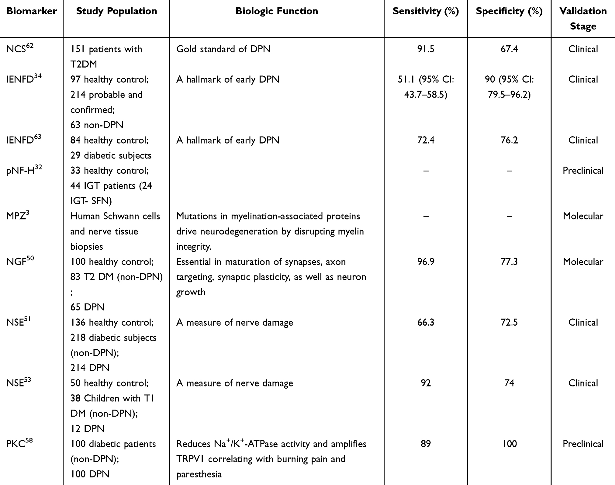 |
Table 2 Sensitivity and Specificity of Diagnostic Markers for Nerve Injury (Human Clinical Study) |
Oxidative Stress-Related Biomarkers
Oxidative stress has emerged as a pivotal pathophysiological mechanism underlying DPN, characterized by disrupted redox homeostasis resulting from excessive ROS generation and impaired antioxidant defense systems.64 This imbalance induces oxidative damage through multiple pathways, including neuronal morphological alterations, protein/nucleic acid denaturation, and ultimately programmed cell death.65 Substantial evidence supports oxidative stress as a primary driver of neuronal dysfunction in DPN.22
Hyperglycemia-driven ROS overproduction predominantly arises from two mechanisms:66
Glucose Autoxidation (animal model, in vitro data): Direct generation of ROS via glucose metabolism intermediates by NOS, NADPH oxidases, xanthine oxidase, and hemeperoxidase enzymes, such as myeloperoxidase;
Non-enzymatic Protein Glycation (animal model, in vitro data): Formation of advanced glycation end-products (AGEs), which amplify oxidative damage through receptor-mediated signaling.
Persistent elevations in ROS and reactive nitrogen species (RNS) contribute to endothelial dysfunction, insulin resistance, and pancreatic β-cell impairment, thereby accelerating both microvascular and macrovascular diabetic complications.66,67 Preclinical studies using DRG neurons exposed to high-glucose conditions reveal a characteristic oxidative stress profile: 3-fold elevation of lipid peroxidation marker malondialdehyde (MDA), coupled with 60% and 50% reductions in GSH and SOD activity, respectively.68
Early clinical investigations confirm these findings in DPN patients:69
Compared to healthy controls (human clinical study): Serum MDA levels are elevated by >2-fold (healthy: 0.55 ±0.05 nmol/L vs DPN: 1.35±0.13 nmol/L; p < 0.01), with GSH levels reduced by 27.7% ± 2.0% (healthy: 52.45 ± 3.22 mg/dL vs DPN: 37.9 ± 1.18 mg/dL);
Compared to diabetics without DPN (human clinical study): DPN patients exhibit a 1.4-fold increase in MDA (non-DPN: 0.96 ± 0.07 nmol/L vs DPN: 1.35 ± 0.13 nmol/L) and a 3.68% ± 1.68% decline in GSH (non-DPN: 39.35 ± 1.73 mg/dL vs DPN: 37.9 ± 1.18 mg/dL).
A meta-analysis by Mallet et al’s meta-analysis of 69 articles56 further solidifies the role of oxidative stress biomarkers in DPN, establishing two critical associations:
- Systemic oxidative burden: Total oxidative status (TOS) and oxidative stress index (OSI) are significantly elevated in DPN patients, and
- Genetic susceptibility: Polymorphisms in antioxidant enzyme genes (eg, glutathione peroxidase, GPx) correlate with increased DPN risk and ROS-induced DNA damage, particularly via hydrogen peroxide-mediated pathways.70
The convergence of evidence presented here not only consolidates oxidative stress as a central mediator of neuropathic pathogenesis but also offer operational thresholds and reference ranges to standardize DPN diagnostic workflows. Recent trials highlight the therapeutic potential of targeting oxidative stress:
After-treatment cohorts exhibit significant reductions in MDA levels, restored SOD activity, elevated total antioxidant capacity (TAOC), as well as improved motor/sensory NCVs compared to before-treatment baselines (P< 0.05).71
Bilirubin (human clinical study): This endogenous antioxidant may mitigate DPN risk by suppressing ROS via inhibition of the PKC-NAD (P)H oxidase pathway.22
NRF-2 (animal model, in vitro data): Preclinical experiments also confirmed that it can improve diabetes peripheral neuropathy by activating the NRF-2 dependent antioxidant system.72–75
N-acetylcysteine (NAC) (human clinical study): High-dose NAC therapy demonstrates clinical efficacy, increasing NRF2 levels by 25.3% (p < 0.05) and glutathione peroxidase (GPx) activity by 100%, while reducing TNF-α levels by 21.45% compared to controls.76 It is critical to note that the canonical activation of NRF2 predominantly occurs through the kinetic inhibition of its repressor, KEAP1, which involves the covalent modification of specific cysteine residues on KEAP1 by electrophiles, thereby stabilizing NRF2; furthermore, emerging pharmacologic strategies are exploring both covalent and non-covalent KEAP1-NRF2 protein–protein interaction inhibitors, which exhibit distinct kinetic and toxicological profiles.
This study expands the therapeutic effect of DPN under the antioxidant system, which may reduce the structural and functional damage to nerve fibers, thus maintaining the integrity of neurons under the condition of diabetes.
Conversely, methylglyoxal, a cytotoxic glycolysis byproduct and AGE precursor, exacerbates peripheral nerve and Schwann cell damage through oxidative pathways.77 Intriguingly, methylglyoxal modulates ion channel function (eg, Nav 1.8, TRPA1), potentially linking oxidative stress to painful DPN phenotypes.8 Mitochondrial dysfunction in Schwann cells (SCs) is not merely an accompanying phenomenon but a central driver of phenotypic transition, myelin maintenance failure, and subsequent axonal degeneration. The hyperglycemic milieu directly impairs SC mitochondrial homeostasis by disrupting critical cellular energy-sensing and metabolic pathways. Dysregulation of the AMPK/PGC-1α signaling axis is considered a pivotal event.78 PGC-1α can inhibit the dedifferentiation of Schwann cells by targeting paraoxonase 1 (PON1), thereby delaying peripheral nerve degeneration. Activation of the AMPK/SIRT1/PGC-1αpathway can alleviate high glucose-induced oxidative stress, ferroptosis, and mitochondrial dysfunction in Schwann cells.79 Furthermore, AMPK signaling is involved in regulating mitophagy, the process of clearing damaged mitochondria, which is crucial for maintaining SC health.80
Although the mechanism of oxidative stress has been confirmed by many research inferences (Figure 4), there are still key challenges in DPN. The specificity and sensitivity of oxidative stress biomarkers (eg, MDA, GSH, NRF2, methylglyoxal (human clinical study)) require rigorous validation in diverse populations. While preclinical/clinical studies show promise, large-scale human trials are lacking. Genetic polymorphisms caused by oxidative stress will also be a new field, which will be summarized and analyzed in the following content.
 |
Figure 4 Possible mechanisms and biomarkers of oxidative stress in DPN.9 Under hyperglycemic conditions, glucose auto-oxidation and non-enzymatic protein glycation impair glycolysis, generating methylglyoxal as a cytotoxic byproduct. Methylglyoxal modulates ion channels (Nav1.8/TRPA1) to induce DPN-associated pain and reduced nerve conduction velocity. Concurrently, hyperglycemia-driven oxidative pathways elevate lipid peroxidation biomarkers (eg, malondialdehyde, MDA), depleting antioxidant defenses (glutathione, GSH; superoxide dismutase, SOD) and exacerbating mitochondrial dysfunction. This dysfunction amplifies superoxide anion and reactive oxygen species (ROS) production, reflected by elevated total oxidative state (TOS) and oxidative stress index (OSI). Hyperglycemia activate protein kinase C (PKC), impairing insulin signaling and promoting insulin resistance. These cascades further increase MDA levels and may influence genetic polymorphisms in antioxidant enzyme-encoding genes (eg, glutathione peroxidase). PKC activation may also affect the production of oxidants and AGEs through the NADPH oxidase complex, causing alterations in cellular functioning. Furthermore, this results in abnormal NSV. The imbalance induces neuronal damage and ultimately programmed cell death. Notably, oxidative stress markers (MDA, TOS, OSI) represent potential early diagnostic indicators for DPN progression. |
Neuro-Vascular Injury Related Markers
DPN is characterized as a symmetrical, length-dependent sensorimotor polyneuropathy resulting from chronic hyperglycemia-associated metabolic derangements that contribute to microvascular dysfunction and cardiovascular comorbidities.6 Cheng et al4 (human clinical study) systematically demonstrated that cardiac troponin T (cTnT), B-type natriuretic peptide (BNP), C-reactive protein (CRP), myeloperoxidase (MPO), and homocysteine (Hcy) may serve as clinically actionable indicators for early diagnosis and risk stratification of DPN.
Hcy- A Dual Role in Oxidative Stress and Neurotoxicity
Hcy (human clinical study) has emerged as a key mediator of neurovascular injury in DPN, with study analyses confirming its elevated serum levels in patients across diverse populations.69,81–83 A study revealed a 23% increase in DPN risk per 1 μmol/L increment in plasma Hcy.84 Consistently, subjects with confirmed DNP exhibited significantly elevated total plasma Hcy levels compared to non-neuropathic controls (12.8 [9.2–14.8] μmol/L vs 8.0 [7.7–9.1] μmol/L, P=0.005), highlighting its potential as a modifiable risk factor in diabetes management.85 In a retrospective study, patients were divided into two groups based on blood Hcy levels, namely the HHcy group and the NHHcy group. The incidence rates of DPN in both groups were 98.5% and 36.2%, respectively.82
Hcy levels >15 μmol/L induce reductions in NCV,82,86 however, one study reported no significant decrease in tibial NCV despite elevated HCY level.87 Mechanistically, this discrepancy may arise from differential susceptibility of nerve subtypes, with high Hcy levels driving synergistic pathways:82,88 (1) oxidative stress, (2) endothelial cell damage, and (3) neurotoxicity damage. While these findings position Hcy as a promising diagnostic candidate, its specificity and sensitivity require validation in multi-ethnic cohorts.
CRP and MPO
Related to inflammation. Elevated levels of CRP and MPO frequently co-occur with pro-inflammatory cytokines tumor necrosis factor-α (TNF-α) and interleukin-6 (IL-6) in experimental diabetic neuropathy models26,89–91 (animal model, in vitro data). Emerging clinical evidence indicates that serum CRP (human clinical study) is significantly increased in patients with DPN,91–93 while higher CRP level is inversely related to NCV (P<0.001),91 suggesting a role for systemic inflammation in axonal dysfunction. Preclinical studies associate MPO with behavioral abnormalities, sciatic nerve demyelination, and NCV impairment.26,90 Clinically (human clinical study), a German prospective cohort established MPO as an independent predictor of DPN progression.94 These suggest the role of CRP and MPO in bridging oxidative stress, inflammation and neural degeneration.
Cardiac Biomarkers (BNP/NT-proBNP, Hs-cTnT)
BNP/NT-proBNP and hs-cTnT (human clinical study) exhibit strong associations with peripheral neuropathy severity.95 In T2DM patients, elevated serum BNP correlates positively with systolic blood pressure, neutrophil-to-lymphocyte ratio (NLR), vibration perception thresholds, and comorbidities including diabetic foot ulcers and nephropathy.96 Furthermore, NT-proBNP levels predict microvascular and macrovascular complication risks in diabetes.97 This relationship may stem from shared microvascular pathophysiology: impaired neural perfusion in DPN parallels myocardial ischemia, with both processes amplifying end-organ damage through hypoxic and oxidative mechanisms. In the context of DPN, confounding cardiovascular comorbidities such as hypertension and dyslipidemia are known to elevate circulating levels of NT-proBNP and cardiac troponin T (cTnT), which can complicate the specific interpretation of these biomarkers for neuropathy risk stratification.
Other Vascular Diagnostic Markers
Vascular endothelial growth factor (VEGF), endothelin-1 (ET-1), nitric oxide (NO) and endothelial nitric oxide synthase (eNOS) are associated with neuromicrovascular markers and are involved in the progression of DPN through mechanisms like endothelial dysfunction, inflammatory infiltration, and microvascular structure destruction.
VEGF (human clinical study), a key player in angiogenesis, has neuroprotective and trophic effects and is crucial for nerve repair and regeneration. However, research on VEGF levels in DPN patients has been inconsistent. A meta-analysis of 14 studies with 1983 participants (807 DPN patients, 808 non-DPN diabetes patients, and 368 healthy controls) showed significantly higher VEGF levels in DPN patients compared to non-DPN diabetes patients (SMD: 2.12 [1.34, 2.90], P < 0.00001) and healthy individuals (SMD: 3.50 [2.24, 4.75], P< 0.00001). Moreover, increased circulating VEGF levels are not linked to a higher DPN risk (OR: 1.02 [0.99, 1.05], P < 0.00001).98 Recent analysis found that elevated serum VEGF - B levels are an independent DPN risk factor in T2D patients, likely related to the VEGF - B - VEGFR1 signaling pathway.99 Cross - sectional studies also supported increased serum VEGF - B levels (P < 0.05).100 VEGF negatively correlates with motor nerve amplitude and positively correlates with NSS and DNE scores. ROC analysis indicated strong diagnostic ability of VEGF (AUC: 0.807), and logistic regression identified VEGF as the only significant predictor (OR: 1.11, 95% CI (1.03, 1.20), P= 0.0092).101
ET-1 (human clinical study) functions differently from VEGF, mainly causing vasoconstriction. Some clinical trials have directly shown increased serum ET-1 levels in DPN patients.69,102 It may act through the AGEs/ET-1/TNF - α/NOS axis in diabetes, leading to abnormal blood glucose and lipids, oxidative stress, and inflammation,103 suggesting it could be a diabetes treatment target.104 Recent study confirmed that ET −1 and Ang - (1–7) form a protective signal network via the physical interaction of MasR and ETBR to counteract vascular damage.105 Decreased AMPK-eNOS bioavailability mediates DPN development through increased apoptosis and reduced autophagy related to oxidative stress.106 These studies offer new perspectives and research targets for DPN - induced vascular diseases.
Inflammatory Biomarkers
Emerging evidence underscores that hyperglycemia-induced oxidative stress, microvascular dysfunction, and cardiovascular comorbidities synergistically exacerbate neuroinflammatory cascades in DPN.1,30 Cross-sectional analyses consistently identify systemic inflammation as a pivotal mediator of DPN pathogenesis, with candidate biomarkers spanning tumor necrosis factor-α (TNF-α)-associated pathways, interleukin (IL) networks, and hematologic indices like neutrophil-to-lymphocyte ratio (NLR).107
Meta-analyses adhering to PRISMA guidelines (15 studies) revealed markedly elevated NLR levels (human clinical study) in DPN cohorts versus diabetic controls.93,108 Independent validation studies further established NLR’s diagnostic potential, demonstrating 75% accuracy for distinguishing DPN (AUC=0.851).109 This validate NLR as a robust inflammatory indicator. Threshold analysis demonstrated that NLR ≥2.66 significantly correlated with DPN risk (OR=1.985, 95% CI:1.29–3.05).110 ROC curve evaluations further highlighted the superior sensitivity of combined HbA1c+NLR detection (97.2%) over individual markers (HbA1c:71.6%; NLR:90%), albeit with moderate specificity (45%) vs individual markers (HbA1c:63.8%%; NLR:50.00%).111
Significantly, Multicohort studies consistently report elevated systemic levels of TNF-α,112–117 IL-6, and CRP114 (human clinical study) are significantly increased in DPN patients. A longitudinal investigation identified baseline plasma TNF-α, IL-6, and intercellular adhesion molecule-1 (ICAM-1) as independent predictors of 5-year DPN incidence in Chinese diabetics.118 Notably, impaired glucoregulation exacerbated TNF-α/IL-6 elevations in neuropathy subgroups compared to non-neuropathic controls.119 US cohort data corroborated these findings, linking hs-CRP, IL-6, TNF-α, IL-1 receptor antagonist (IL-1RA), and ICAM-1 to distal sensory-motor polyneuropathy (DSPN) progression, while reduced lipocalin levels emerged as a novel risk modifier120 (human clinical study). Electrophysiological correlations further substantiate TNF-α’s pathogenic role: serum TNF-α levels inversely correlated with motor/sensory NCV (median, ulnar, peroneal, and posterior tibial nerves) in T2 DM.121
Overall, research on the diagnosis of DPN using inflammatory factors has made some progress, with some inflammatory factors showing good diagnostic potential.122 For example, NLR has shown promising prospects as a clinical diagnostic biomarker for DPN, while TNF-α increases with the progression of the disease. In short-term DPN (T2DM<8 years), TNF-α levels were 43.78±26.68 pg/mL (non-neurological control group: 20.45±11.22 pg/mL; P<0.05); in long-term DPN (T2DM≥8 years), TNF-α levels were 74.07±32.05 pg/mL compared with the short-term group, P<0.01). The course of disease was positively correlated with TNF-α levels (r=0.488, P=0.002; r=0.478, P=0.012).121 Elevated TNF-α can be used to classify the severity of neurological disorders. These accumulated evidence suggest that these inflammatory biomarkers can serve as diagnostic and clinical staging tools for DPN.
The quantitative analysis of inflammatory mediators, especially TNF-α, across DPN staging, holds promise for predictive and diagnostic applications. In order to enhance sensitivity and specificity, it is still necessary to establish large-scale longitudinal studies using multi-system inflammatory biomarkers or multi-combination biomarkers, such as oxidative stress markers (eg, MDA), cardiovascular biomarkers (eg, Hcy), and neurodamage biomarkers (eg, VEGF), in combination to establish critical values for specific stages and optimize sensitivity/specificity indicators for clinical deployment. Evaluating the clinical staging of DPN will be a good research direction.
Gene-Related Markers
Non-Coding RNA
Non-coding RNAs (ncRNAs) are a class of RNAs that are not translated into proteins.123 Based on their functions, they can be categorized into rRNAs, microRNAs (miRNAs), long non-coding RNAs (lncRNAs), circular RNAs (circRNAs), and piwi RNAs (piRNAs). ncRNAs play crucial roles in the regulation of epigenetic modifications, protein transcription, and post-transcriptional regulatory functions. In the progression of DPN, ncRNAs can influence inflammation, oxidative stress, cellular autophagy, or apoptosis processes, and may link it to clinical spheres such as other metabolic and neural pathologies.124 Since some ncRNAs are stably present in the blood of DPN patients, they are considered potential biomarkers that can aid in early clinical diagnosis.123
Using whole-transcriptome sequencing, a systematic analysis was conducted on differentially expressed mRNAs, lncRNAs, and miRNAs in the SCs of DPN rats and controls. It was found that there were 2925 mRNAs, 164 lncRNAs, and 49 miRNAs that were significantly differentially expressed in DPN rat SCs125 (animal model, in vitro data). miRNAs and lncRNAs are two classes of genes that have been more frequently studied.
Spallone et al126 conducted a comprehensive investigation into the expression profiles of various miRNAs in DPN and explored the underlying mechanisms from a genetic perspective. Their research revealed that miRNAs exert significant influence on oxidative stress and nerve damage by modulating multiple critical pathways, including the AGE-RAGE axis, PKC-α/NADP oxidase activity, inflammatory factors (eg, TNF-α and IL-1β), and the NF-κB signaling pathway. For instance, miR-146 (animal model) has been reduced in the sciatic nerves of diabetic mice and rats and is negatively related to inflammatory cytokines and whose mimics produce beneficial structural and functional effects.127 In an innovative therapeutic approach, they utilized nanoparticle-miR-146a-5p (animal model) and observed notable improvements in nerve conduction velocity and reductions in demyelination. These findings underscore the potential of miRNAs as therapeutic targets for DPN. Moreover, to enhance diagnostic accuracy, they employed ROC analysis to identify various miRNAs with higher diagnostic accuracy, specificity, and sensitivity for DPN. Notably, miRNA combinations (eg, miR-128a and miR-155) (animal model) demonstrated high diagnostic efficacy (AUC>0.8) in ROC analysis, supporting their potential use as non-invasive biomarkers.
Recent studies have further elucidated the therapeutic potential of miRNAs in diabetic neuropathy through distinct molecular mechanisms. Notably, miR-503-5p (animal model) was demonstrated to mitigate neuropathic pain in T2DM murine models by targeting SEPT9 to suppress astrocyte activation,128 while miR-186-5p exhibited significant correlations with oxidative stress markers in clinical populations.129 Therefore, miRNAs have been considered as potential biomarkers for diabetes and its vascular complications and neuropathy,46,130 Specific miRNAs may also be new therapeutic targets for the treatment of painful DPN.131 The diagnostic and therapeutic potential of these regulatory molecules is amplified through their natural transport system – exosomes. These nano-vesicles facilitate intercellular communication through autocrine, paracrine, and endocrine mechanisms by shuttling ncRNAs, including functional miRNAs and mRNAs. Experimental evidence reveals their therapeutic capacity in neurological contexts: plasma-derived exosomes were shown to ameliorate peripheral neuropathy in type 1 diabetic rats via the miR-20b-3p/Stat3 axis.132 Furthermore, comprehensive analyses have identified exosomal involvement in multiple T2DM-related pathways, with animal studies systematically evaluating their therapeutic efficacy in DPN management (see Table 3). This converging evidence establishes a strong rationale for pursuing exosomal ncRNAs, particularly pathway-specific miRNAs, as both mechanistic biomarkers and targeted therapeutic vehicles for DPN.133
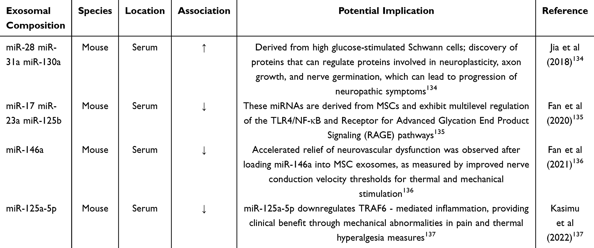 |
Table 3 Role of Exosomal Components in Diabetic Neuropathy in vivo (Satyadev et al (2023))133 |
lncRNAs have also been shown to be involved in DPN. In a diabetic rat model, small interfering RNAs (siRNAs) targeting specific lncRNAs may attenuate DPN by decreasing the production of inflammatory factors. Using microarray analysis, 446 and 1327 differentially expressed lncRNAs were identified between diabetic patients with and without DPN.138 The lncRNA/mRNA co-expression network in DPN patients suggests the involvement of the neurotrophic factor-MAPK signaling pathway. Several lncRNAs (CCNT2-AS1, RP1-249H1.2, CTD-3239E11.2, RP11-51B23.3, STAM-AS1, and LINC00629) have been pointed out as potential interactors in the above signaling pathway139 (animal model).
Recent investigations have pioneered three critical advancements in diabetic peripheral neuropathy (DPN) research: (1) systematic elucidation of ncRNA regulatory mechanisms through exosome-mediated transcellular signaling (eg, miR-146a-5p nanoparticles enhancing nerve repair), (2) integration of multi-omics technologies to decode epigenetic-metabolic networks, and (3) development of nanotherapeutic strategies enabling diagnosis-to-treatment synergy. Notably, context-specific ncRNA axes (eg, miR-503-5p/SEPT9) demonstrate dual utility as mechanistic biomarkers and therapeutic targets. However, to achieve clinical translation, future studies must prioritize human tissue validation using spatial multi-omics, dynamic mapping of ncRNA interactions in neural microenvironments, and functional characterization of underexplored ncRNAs (eg, circRNAs). Addressing these gaps will bridge preclinical discoveries to precision medicine applications for DPN.
DNA Methylation (Human Clinical Study)
DNA methylation refers to the stable and reversible attachment of methyl (CH3) groups to cytosines at DNA palindromes (called CpG islands). DNA methylation is thought to be involved in regulating the expression of key genes that induce DPN. Genome-wide methylation analyses of samples collected from the same diabetic patients 16–17 years apart revealed that key genomic loci associated with diabetic complications of DNA methylation persist over time.140 Hyperglycemia alters the DNA methylation status, thereby inducing altered gene expression associated with DPN. Significant reductions in genome-wide DNA methylation are potential biomarkers for DPN.92,139 Genomic DNA methylation levels were significantly decreased in the DPN group compared with the non-DPN group when the duration is ≥5 years.92 For the first time, a comprehensive analysis of peroneal nerve DNA methylation profiles in DPN patients with significant nerve regeneration and patients with significant neurodegeneration (representing two extreme DPN phenotypes) was performed. A total of 3460 differentially methylated CpG dinucleotides were identified. These differentially methylated CpG-related genes are highly enriched in biological processes related to DPN progression, such as neurodevelopment, neuronal development and axon guidance, glycerophospholipid metabolism, and mitogen-activated protein kinase (MAPK) signaling.141
Other Possible Genetic Diagnoses
Single locus or multigene risk is also gaining interest in the development of DPN. The development of histomics has facilitated genome-wide association studies. Studies on differentially expressed genes (DEGs) and functional enrichment analyses of DPN have also been reported.142–144 These studies have identified co-expressed DEGs that are enriched in lipid metabolism-related pathways, signaling pathways, vascular regulation, inflammatory oxidative stress, and immunomodulatory pathways.5,145 Many recently identified genes, including (human clinical study) the aldose reductase (AKR1B1) gene, VEGF,5,10, methylenetetrahydrofolate reductase (MTHFR) enzyme variants, APOE, and angiotensin-converting enzyme (ACE) genes, play important roles in the polyol pathway, vascular endothelial cell value-addition, and oxidative stress in endothelial cells induced by hyperhomocysteinemia. These factors are thought to be important for the pathogenesis of diabetic neuropathy.19 Vitamin B12, an essential cofactor for the degradation of methylmalonic acid and the synthesis of methionine from homocysteine, exerts intrinsic antioxidant properties both in vivo and in vitro. The presence of oxidative stress in vitamin B12-deficient patients suggests that the neurological changes observed in patients with DPN may be caused by cellular B12 deficiency.146 Emerging evidence suggests PARP-1-targeted siRNA (animal model) therapy attenuates diabetic peripheral neuropathy in streptozotocin-induced rats by coordinately modulating oxidative-inflammatory-apoptotic axes. This intervention significantly reduces oxidative stress (MDA, P < 0.001) while restoring antioxidant defenses (GSH, CAT, SOD, P < 0.001), and concurrently suppresses pro-inflammatory mediators (NF-κB, IL-6, IL-1β, TNF-α, TGF-β) and apoptotic executors (Caspase-3/9, Bak, Bax) with statistical significance (P < 0.01).147
All of these target genes may be candidate biomarkers for DPN. Other possible genes will not be further excavated here. However, regardless of the type of genes, there is still a lack of in-depth studies with large sample data.
Metabolic Diagnostic Markers (Human Clinical Study)
Metabolic dysregulation is increasingly recognized as a critical contributor to the progression of DPN. Comprehensive profiling of diabetes-associated metabolites in biological matrices using analytical chemistry techniques has revealed potential biomarkers for early DPN diagnosis, including amino acids, organic acids, fatty acids, lipids, and carbohydrates.
Comparative studies demonstrate distinct lipid profile alterations in T2DM patients with DPN. Serum triglycerides and alanine aminotransferase levels are significantly elevated, whereas C-peptide and total cholesterol levels are reduced compared to non-neuropathic controls.148 Hypertriglyceridemia is independently associated with both the loss of myelinated peroneal nerve fiber density144 and an increased risk of lower limb amputation in T2DM.143 Although reduced HDL-cholesterol has been implicated as a DPN risk factor in some studies,149 meta-analytic evidence indicates this association is significant only in T1DM-related neuropathy (P<0.05), with no statistically significant difference observed in T2DM cohorts.150 Beyond lipid metabolism, A 1H-NMR metabolomics study revealed significantly reduced serum formate levels in DPN patients versus T2DM controls (P<0.001, AUC=0.981), showing negative correlations with HbA1c, uric acid (UA), and cystatin C (P<0.05), and positive association with albumin. These findings position formate as a biomarker implicating mitochondrial dysfunction and gut dysbiosis in DPN pathogenesis, with dual diagnostic and therapeutic potential.151 Additionally, Pre-analytical variables such as fasting status, sample processing time, and storage conditions can critically influence the stability and measured concentrations of metabolites, potentially introducing significant bias into biomarker studies for DPN.
Liquid chromatography-based metabolomic analysis of T2DM patients with distal symmetrical sensorimotor polyneuropathy (DSPN) identified 16 plasma metabolites and 3 cholesterol derivatives as discriminative markers. Among these (human clinical study), phenylalanine, alanine, lysine, tryptophan, and SMC16:0 exhibited the highest diagnostic potential for distinguishing DSPN from non-DSPN cases (P<0.05).152
Challenge and Discussion
DPN is diagnosed based on clinical symptoms and ancillary tests such as electrophysiological studies. Per the Expert Consensus on Diabetic Neuropathy (2021), diagnostic criteria require: Confirmed diabetes history; Neuropathy onset post-diabetes diagnosis; Neuropathic symptoms (eg, pain, numbness) plus ≥1 abnormal neurological test (eg, impaired ankle reflex, sensory perception); Asymptomatic cases require ≥2 abnormal test results.31
The clinical diagnosis of DPN currently relies on electrophysiological methods, which are operationally complex, cost-intensive, and prone to diagnostic inaccuracies. While existing diagnostic approaches and biomarkers show potential, their clinical utility is limited by inconsistent methodologies, lack of standardized diagnostic staging criteria, and susceptibility to confounding variables. For instance, oxidative stress markers such as MDA69,71 exhibit population-specific variability and may present aberrant levels in non-diabetic neuropathies, undermining their diagnostic specificity. Similarly, NSE, though indicative of neural injury, demonstrates limited specificity due to elevations in diverse neurological disorders and malignancies.153,154 The absence of harmonized detection protocols and validated diagnostic thresholds further complicates cross-study comparability. Critically, single-marker analyses fail to capture the multifactorial progression of DPN, as exemplified by TNF-α, which reflects inflammatory processes but provides no insight into concurrent neurovascular or metabolic dysfunction.
A multimodal approach integrating functional, structural, and molecular biomarkers could enhance diagnostic precision. Combining nerve conduction studies (eg, NCV, F-wave parameters) with neural injury markers (eg, NF-H, MPZ) enables simultaneous assessment of functional impairment and structural degeneration. Similarly, pairing oxidative stress markers (MDA, GSH) with inflammatory indices (NLR, TNF-α) may provide a holistic evaluation of disease mechanisms. Machine learning approaches offer powerful tools for integrating multimodal data in DPN biomarker discovery, with convolutional neural networks (CNNs) and random forests (RFs) representing two distinct paradigms. CNNs excel at analyzing high-dimensional, spatially structured data like corneal confocal microscopy images or nerve ultrasound, automatically extracting complex hierarchical features for superior pattern recognition. In contrast, RFs are particularly effective for processing structured, tabular data—such as combining clinical scores, biochemical parameters, and quantitative sensory thresholds—by identifying non-linear interactions and providing interpretable feature importance rankings. While CNNs achieve high accuracy in image-based diagnostics, their “black-box” nature contrasts with the inherent explainability of RFs, which can reveal key predictive biomarkers from heterogeneous clinical datasets. Current evidence highlights promising candidates such as Hcy, NLR, and TNF-α for monitoring DPN progression, while oxidative stress markers and genetic biomarkers require further validation of their diagnostic thresholds and specificity.
As illustrated in Table 4, we have integrated the potential biomarkers for Diabetic Peripheral Neuropathy (DPN). This integration underscores a pivotal conceptual advancement: DPN is not the result of a single pathway disruption. Instead, it emerges from a complex “pathological network” formed by the dynamic interaction of multiple systems, including oxidative stress, inflammation, and neurovascular injury.
 |
Table 4 An Integrated View: Crosstalk Among Oxidative Stress, Inflammation, and Neurovascular Dysfunction |
Translation of these biomarkers into clinical practice necessitates three critical steps: establishing large-scale, multi-ethnic cohort studies to elucidate the molecular pathways of biomarker neuropathy associations, establishing population specific diagnostic thresholds through ROC curve analysis using standardized multicenter trials, and conducting comparative studies in different genetic backgrounds (eg Asian and Caucasian cohorts) to address potential biomarker sensitivity variability (Δ>15% in pilot studies) and specificity. Current evidence suggests that to achieve a clinical application sensitivity/specificity of ≥80%, validation is required in cohorts of over 5000 participants from more than 3 races, supplemented by longitudinal data on biomarker stability and disease progression correlation (r > 6).
Emerging technologies, particularly artificial intelligence (eg, CNN-LSTM deep learning architectures), could accelerate biomarker discovery by enabling multimodal data integration and early detection of subclinical abnormalities during the impaired glucose tolerance phase. This review systematically categorizes biomarkers by pathogenic mechanism (neural conduction deficits, oxidative stress, inflammation, neurovascular injury, metabolic dysregulation, and genetic factors) to provide clinicians with a framework for interpreting complex DPN pathophysiology (Figure 5). We fully acknowledge the inherent heterogeneity among studies, whether clinical, animal, or cellular. It is undeniable that direct, point-by-point comparisons of outcomes across all cited studies are impractical due to significant variations in study design, population characteristics, and experimental models, which inevitably influence the reported parameters. Nevertheless, the biomarkers discussed in this article provide valuable reference points for guiding more targeted future research. This is particularly true for findings that demonstrate consistency across different research models. For instance, the specific alterations in markers of oxidative stress, neurovascular injury, and certain inflammatory pathways have been robustly observed in both animal models of DPN and in human patients. These convergent points across species should garner focused attention from researchers, as they may illuminate the most promising pathways for future clinical translation.
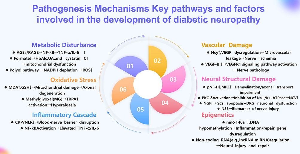 |
Figure 5 Pathogenesis Mechanisms Key pathways and factors involved in the development of diabetic neuropathy. |
Future diagnostic strategies should prioritize the development and validation of multimodal biomarker panels that integrate complementary pathophysiological information from metabolic, inflammatory, and neurotrophic pathways, as such panels are expected to outperform single biomarkers in sensitivity, specificity, and their ability to guide personalized therapeutic interventions for DPN. While current biomarkers remain preclinical, their clinical adoption requires rigorous validation across diverse populations and standardized implementation. Only through such efforts can we achieve the target diagnostic ensitivity/specificity of ≥80% essential for cost-effective, stage-specific DPN management.
Conclusion
Current research on DPN biomarkers encompasses a diverse array of candidates, including oxidative stress mediators, neural injury proteins, inflammatory cytokines, vascular dysfunction markers, metabolic intermediates, and genetic/epigenetic regulators; however, their clinical translation remains constrained by a predominant reliance on cross-sectional studies, lack of standardized diagnostic thresholds, and insufficient validation in large, multi-ethnic prospective cohorts.
Data Sharing Statement
Data sharing is not applicable to this article as no data were created or analysed in this study.
Acknowledgments
Qingqin Lyu, Yanyan Tan and Xianqi Zeng contributed equally to this paper and should be considered co-first authors.
Author Contributions
ML: Writing–original draft, Writing–review and editing, Conceptualization, Data curation, Funding acquisition, Investigation, Methodology. QL, YT, XZ: Data curation, Formal analysis, Methodology, Writing–original draft. SP: Conceptualization, Investigation, Project administration, Supervision, Writing–review and editing. All authors gave final approval of the version to be published; have agreed on the journal to which the article has been submitted; and agree to be accountable for all aspects of the work.
Funding
National Natural Science Foundation of China (82205042), Shenzhen Natural Science Foundation General Project (JCYJ20220531092004009, JCYJ20230807094612026). Sanming Project of Medicine in Shenzhen (No. SZZYSM202411016).
Disclosure
The authors declare that the research was conducted in the absence of any commercial or financial relationships that could be construed as a potential conflict of interest. The authors have no conflicts of interest to declare.
References
1. Selvarajah D, Kar D, Khunti K, et al. Diabetic peripheral neuropathy: advances in diagnosis and strategies for screening and early intervention. Lancet Diabetes Endocrinol. 2019;7(12):938–23. doi:10.1016/S2213-8587(19)30081-6
2. Elafros MA, Andersen H, Bennett DL, et al. Towards prevention of diabetic peripheral neuropathy: clinical presentation, pathogenesis, and new treatments. Lancet Neurol. 2022;21(10):922–936. doi:10.1016/S1474-4422(22)00188-0
3. Morgenstern J, Groener JB, Jende J, et al. Neuron-specific biomarkers predict hypo- and hyperalgesia in individuals with diabetic peripheral neuropathy. Diabetologia. 2021;64(12):2843–2855. doi:10.1007/s00125-021-05557-6
4. Cheng MK, Guo YY, Kang XN, et al. Advances in cardiovascular-related biomarkers to predict diabetic peripheral neuropathy. World J Diabetes. 2023;14(8):1226–1233. doi:10.4239/wjd.v14.i8.1226
5. Chen X, Liu Q, Chen N, et al. Diagnostic biomarker for type 2 diabetic peripheral neuropathy via comprehensive bioinformatics analysis. J Diabetes. 2024;16(3):e13506. doi:10.1111/1753-0407.13506
6. Atmaca A, Ketenci A, Sahin I, et al. Expert opinion on screening, diagnosis and management of diabetic peripheral neuropathy: a multidisciplinary approach. Front Endocrinol. 2024;15:1380929. doi:10.3389/fendo.2024.1380929
7. Sun H, Saeedi P, Karuranga S, et al. Erratum to “IDF Diabetes Atlas: global, regional and country-level diabetes prevalence estimates for 2021 and projections for 2045” [Diabetes Res. Clin. Pract. 183 (2022) 109119]. Diabet Res Clin Pract. 2023;204:110945. doi:10.1016/j.diabres.2023.110945
8. Eid SA, Rumora AE, Beirowski B, et al. New perspectives in diabetic neuropathy. Neuron. 2023;111(17):2623–2641. doi:10.1016/j.neuron.2023.05.003
9. Zhu J, Hu Z, Luo Y, et al. Diabetic peripheral neuropathy: pathogenetic mechanisms and treatment. Front Endocrinol. 2023;14:1265372. doi:10.3389/fendo.2023.1265372
10. Mizukami H, Osonoi S. Pathogenesis and molecular treatment strategies of diabetic neuropathy collateral glucose-utilizing pathways in diabetic polyneuropathy. Int J Mol Sci. 2020;22(1):94. doi:10.3390/ijms22010094
11. Kobayashi M, Zochodne DW. Diabetic neuropathy and the sensory neuron: new aspects of pathogenesis and their treatment implications. J Diabetes Investig. 2018;9(6):1239–1254. doi:10.1111/jdi.12833
12. Ogata T, Iijima S, Hoshikawa S, et al. Opposing extracellular signal-regulated kinase and Akt pathways control Schwann cell myelination. J Neurosci. 2004;24(30):6724–6732. doi:10.1523/JNEUROSCI.5520-03.2004
13. Huang TJ, Verkhratsky A, Fernyhough P. Insulin enhances mitochondrial inner membrane potential and increases ATP levels through phosphoinositide 3-kinase in adult sensory neurons. Mol Cell Neurosci. 2005;28(1):42–54. doi:10.1016/j.mcn.2004.08.009
14. Souza MDAD, Nassini R, Geppetti P, et al. TRPA1 as a therapeutic target for nociceptive pain. Expert Opin Ther Targets. 2020;24(10):997–1008. doi:10.1080/14728222.2020.1815191
15. Bodman MA, Dreyer MA, Varacallo MA. Diabetic Peripheral Neuropathy. 2025.
16. Patel S, Khan H, Majumdar A. Crosstalk between Sirtuins and Nrf2: SIRT1 activators as emerging treatment for diabetic neuropathy. Metab Brain Dis. 2022;37(7):2181–2195. doi:10.1007/s11011-022-00956-z
17. American Diabetes Association Professional Practice Committee. 12. Retinopathy, neuropathy, and foot care: standards of care in diabetes-2024. Diabetes Care. 2024;47(Suppl 1):S231–S243. doi:10.2337/dc24-S012
18. England JD, Gronseth GS, Franklin G, et al. Distal symmetrical polyneuropathy: a definition for clinical research. A report of the American Academy of Neurology, the American Association of Electrodiagnostic Medicine, and the American Academy of Physical Medicine and Rehabilitation. Arch Phys Med Rehabil. 2005;86(1):167–174. doi:10.1016/j.apmr.2004.09.011
19. Kallinikou D, Soldatou A, Tsentidis C, et al. Diabetic neuropathy in children and adolescents with type 1 diabetes mellitus: diagnosis, pathogenesis, and associated genetic markers. Diabetes Metab Res Rev. 2019;35(7):e3178. doi:10.1002/dmrr.3178
20. Feng Y, Schlosser FJ, Sumpio BE. The Semmes Weinstein monofilament examination as a screening tool for diabetic peripheral neuropathy. J Vasc Surg. 2009;50(3):675–682. doi:10.1016/j.jvs.2009.05.017
21. Perez HM, Calderon VA, Aguilar CS, et al. Electroacupuncture efficacy in diabetic polyneuropathy: study protocol for a double-blinded randomized controlled multicenter clinical trial. BMC Complement Med Ther. 2024;24(1):90. doi:10.1186/s12906-024-04375-8
22. Abe K, Maeda Y, Matsuzaki C, et al. Bilirubin is inversely related to diabetic peripheral neuropathy assessed by sural nerve conduction study. J Diabetes Investig. 2021;12(11):2028–2035. doi:10.1111/jdi.13568
23. Sun WL. Clinical value of neuroelectromyography in the diagnosis of peripheral neuropathy in diabetic patients with different course of disease. Fertil Health. 2024;30(12):43–45.
24. Rao D. Application value of HbA1C combined with neuromyography in early diagnosis of diabetic peripheral neuropathy. J Snake. 2024;36(02):213–216.
25. Qiu JM, Xie HH, Xian S. Value of combined electrophysiological examination in early diagnosis of diabetic peripheral neuropathy. Guangzhou Med J. 2022;53(01):137–140.
26. Srilatha K, Reddy KP. Sciatic nerve structural and functional recovery with extract of Phyllanthus amarus and Esculetin in STZ-induced hyperglycemic rats. Ann Neurosci. 2019;26(3–4):17–29. doi:10.1177/0972753120911840
27. Erbas T, Ertas M, Yucel A, et al. Prevalence of peripheral neuropathy and painful peripheral neuropathy in Turkish diabetic patients. J Clin Neurophysiol. 2011;28(1):51–55. doi:10.1097/WNP.0b013e3182051334
28. Zangiabadi N, Shafiee K, Alavi KH, et al. Atorvastatin treatment improves diabetic polyneuropathy electrophysiological changes in non-insulin dependent diabetic patients: a double blind, randomized clinical trial. Minerva Endocrinol. 2012;37(2):195–200.
29. Hernandez-Ojeda J, Roman-Pintos LM, Rodriguez-Carrizalez AD, et al. Effect of rosuvastatin on diabetic polyneuropathy: a randomized, double-blind, placebo-controlled Phase IIa study. Diabetes Metab Syndr Obes. 2014;7:401–407. doi:10.2147/DMSO.S65500
30. So WZ, Qi WN, Tan HC, et al. Diabetic corneal neuropathy as a surrogate marker for diabetic peripheral neuropathy. Neural Regen Res. 2022;17(10):2172–2178. doi:10.4103/1673-5374.327364
31. Yu Y. Gold Standard for Diagnosis of DPN. Front Endocrinol. 2021;12:719356. doi:10.3389/fendo.2021.719356
32. Li YP, Yan ZQ, Han LP, et al. The association between Phosphorylated Neurofilament Heavy Chain (pNF-H) and Small Fiber Neuropathy (SFN) in patients with impaired glucose tolerance. Diabetes Ther. 2020;11(1):71–81. doi:10.1007/s13300-019-00716-w
33. McArthur JC, Stocks EA, Hauer P, et al. Epidermal nerve fiber density: normative reference range and diagnostic efficiency. Arch Neurol. 1998;55(12):1513–1520. doi:10.1001/archneur.55.12.1513
34. Gylfadottir SS, Itani M, Kristensen AG, et al. Assessing corneal confocal microscopy and other small fiber measures in diabetic polyneuropathy. Neurology. 2023;100(16):e1680–e1690. doi:10.1212/WNL.0000000000206902
35. Badian RA, Ekman L, Pripp AH, et al. Comparison of novel wide-field in vivo corneal confocal microscopy with skin biopsy for assessing peripheral neuropathy in type 2 diabetes. Diabetes. 2023;72(7):908–917. doi:10.2337/db22-0863
36. Burgess J, Frank B, Marshall A, et al. Early detection of diabetic peripheral neuropathy: a focus on small nerve fibres. Diagnostics. 2021;11(2):165. doi:10.3390/diagnostics11020165
37. Cosmo E, Midena G, Frizziero L, et al. Corneal confocal microscopy as a quantitative imaging biomarker of diabetic peripheral neuropathy: a review. J Clin Med. 2022;11(17):5130. doi:10.3390/jcm11175130
38. Alam U, Jeziorska M, Petropoulos IN, et al. Diagnostic utility of corneal confocal microscopy and intra-epidermal nerve fibre density in diabetic neuropathy. PLoS One. 2017;12(7):e180175. doi:10.1371/journal.pone.0180175
39. Bondugulapati LN, Rao NN. Corneal confocal microscopy: potential usage in the context of diabetes mellitus. Pract Diabetes. 2021;38(2).
40. Jiang MS, Yuan Y, Gu ZX, et al. Corneal confocal microscopy for assessment of diabetic peripheral neuropathy: a meta-analysis. Br J Ophthalmol. 2016;100(1):9–14. doi:10.1136/bjophthalmol-2014-306038
41. Ponirakis G, Odriozola MN, Odriozola S, et al. NerveCheck: an inexpensive quantitative sensory testing device for patients with diabetic neuropathy. Diabet Res Clin Pract. 2016;113:101–107. doi:10.1016/j.diabres.2015.12.023
42. Casanova-Molla J, Morales M, Garrabou G, et al. Mitochondrial loss indicates early axonal damage in small fiber neuropathies. J Peripher Nerv Syst. 2012;17(2):147–157. doi:10.1111/j.1529-8027.2012.00396.x
43. Abdelrahman SA, Samak MA, Shalaby SM. Fluoxetine pretreatment enhances neurogenic, angiogenic and immunomodulatory effects of MSCs on experimentally induced diabetic neuropathy. Cell Tissue Res. 2018;374(1):83–97. doi:10.1007/s00441-018-2838-6
44. Qiao X, Zhang S, Zhao W, et al. Serum phosphorylated neurofilament-heavy chain, a potential biomarker, is associated with peripheral neuropathy in patients with type 2 diabetes. Medicine. 2015;94(44):e1908. doi:10.1097/MD.0000000000001908
45. Kim HC, Cho YJ, Ahn CW, et al. Nerve growth factor and expression of its receptors in patients with diabetic neuropathy. Diabet Med. 2009;26(12):1228–1234. doi:10.1111/j.1464-5491.2009.02856.x
46. Simeoli R, Fierabracci A. Insights into the role of MicroRNAs in the onset and development of diabetic neuropathy. Int J Mol Sci. 2019;20(18):4627. doi:10.3390/ijms20184627
47. Li R, Ma J, Wu Y, et al. Dual delivery of NGF and bFGF Coacervater ameliorates diabetic peripheral neuropathy via inhibiting Schwann cells apoptosis. Int J Biol Sci. 2017;13(5):640–651. doi:10.7150/ijbs.18636
48. Li R, Wu Y, Zou S, et al. NGF attenuates high glucose-induced ER stress, preventing schwann cell apoptosis by activating the PI3K/Akt/GSK3beta and ERK1/2 pathways. Neurochem Res. 2017;42(11):3005–3018. doi:10.1007/s11064-017-2333-6
49. Nori SL, Rocco ML, Florenzano F, et al. Increased nerve growth factor signaling in sensory neurons of early diabetic rats is corrected by electroacupuncture. Evid Based Complement Alternat Med. 2013;2013:652735. doi:10.1155/2013/652735
50. Sun Q, Tang DD, Yin EG, et al. Diagnostic significance of serum levels of nerve growth factor and brain derived neurotrophic factor in diabetic peripheral neuropathy. Med Sci Monit. 2018;24:5943–5950. doi:10.12659/MSM.909449
51. Li J, Zhang H, Xie M, et al. NSE, a potential biomarker, is closely connected to diabetic peripheral neuropathy. Diabetes Care. 2013;36(11):3405–3410. doi:10.2337/dc13-0590
52. Anju M, Ummer VS, Maiya AG, et al. Effect of photobiomodulation on serum neuron specific enolase (NSE) among patients with diabetic peripheral neuropathy - A pilot study. Diabetes Metab Syndr. 2020;14(5):1061–1063. doi:10.1016/j.dsx.2020.06.065
53. Abo HA, Abd ENS, Allam ET, et al. Promising predictors of diabetic peripheral neuropathy in children and adolescents with type 1 diabetes mellitus. Ital J Pediatr. 2024;50(1):215. doi:10.1186/s13052-024-01774-y
54. Feldman EL, Nave KA, Jensen TS, et al. New horizons in diabetic neuropathy: mechanisms, bioenergetics, and pain. Neuron. 2017;93(6):1296–1313. doi:10.1016/j.neuron.2017.02.005
55. Lyu Q. Cellular Mechanisms underlying the Effects of MrgC Receptors on CFA-Evoked Inflammatory Pain. Fujian Normal University; 2013.
56. Wang D, Wang P, Jiang J, et al. Activation of Mas Oncogene-Related G Protein-Coupled Receptors Inhibits Neurochemical Alterations in the Spinal Dorsal Horn and Dorsal Root Ganglia Associated with Inflammatory Pain in Rats. J Pharmacol Exp Ther. 2015;354(3):431–439. doi:10.1124/jpet.115.225672
57. Zan Y, Kuai CX, Qiu ZX, et al. Berberine ameliorates diabetic neuropathy: TRPV1 modulation by PKC pathway. Am J Chin Med. 2017;45(8):1709–1723. doi:10.1142/S0192415X17500926
58. Fattah SA, Elmadani M, Abo-Elmatty DM, et al. Genetic variants of ALR (−106C --> T /-12C --> G) and serum PKC-delta are associated with peripheral neuropathy in Egyptian diabetic patients with impaired handwriting. J Diabetes Metab Disord. 2022;21(1):557–565. doi:10.1007/s40200-022-01008-0
59. Samaddar S, Koneri R. Polyphenols of marine red macroalga Symphyocladia latiuscula ameliorate diabetic peripheral neuropathy in experimental animals. Heliyon. 2019;5(5):e1781. doi:10.1016/j.heliyon.2019.e01781
60. Pabbidi MR, Premkumar LS. Role of transient receptor potential channels Trpv1 and Trpm8 in diabetic peripheral neuropathy. J Diabetes Treat. 2017;2017(4).
61. Mansoor H, Tan HC, Lin MT, et al. Diabetic Corneal Neuropathy. J Clin Med. 2020;9(12):3956. doi:10.3390/jcm9123956
62. Shehab DK, Al-Jarallah KF, Abraham M, et al. Back to basics: ankle reflex in the evaluation of peripheral neuropathy in type 2 diabetes mellitus. QJM. 2012;105(4):315–320. doi:10.1093/qjmed/hcr212
63. Umapathi T, Tan WL, Loke SC, et al. Intraepidermal nerve fiber density as a marker of early diabetic neuropathy. Muscle Nerve. 2007;35(5):591–598. doi:10.1002/mus.20732
64. Lin Q, Li K, Chen Y, et al. Oxidative stress in diabetic peripheral neuropathy: pathway and mechanism-based treatment. Mol Neurobiol. 2023;60(8):4574–4594. doi:10.1007/s12035-023-03342-7
65. Iqbal Z, Azmi S, Yadav R, et al. Diabetic peripheral neuropathy: epidemiology, diagnosis, and pharmacotherapy. Clin Ther. 2018;40(6):828–849. doi:10.1016/j.clinthera.2018.04.001
66. Sifuentes-Franco S, Pacheco-Moises FP, Rodriguez-Carrizalez AD, et al. The role of oxidative stress, mitochondrial function, and autophagy in DiabeticP olyneuropathy. J Diabetes Res. 2017;2017:1673081. doi:10.1155/2017/1673081
67. Bandeira SD, Da Fonseca LJ, Guedes GD, et al. Oxidative stress as an underlying contributor in the development of chronic complications in diabetes mellitus. Int J Mol Sci. 2013;14(2):3265–3284. doi:10.3390/ijms14023265
68. Magadmi R, Borouk K, Youssef D, et al. Neuroprotective Effect of Red Sea Marine Sponge Xestospongia testudinaria extract using in vitro and in vivo diabetic peripheral neuropathy models. Pharmaceuticals. 2022;15(11):1309. doi:10.3390/ph15111309
69. El Boghdady NA, Badr GA. Evaluation of oxidative stress markers and vascular risk factors in patients with diabetic peripheral neuropathy. Cell Biochem Funct. 2012;30(4):328–334. doi:10.1002/cbf.2808
70. Mallet ML, Hadjivassiliou M, Sarrigiannis PG, et al. The role of oxidative stress in peripheral neuropathy. J Mol Neurosci. 2020;70(7):1009–1017. doi:10.1007/s12031-020-01495-x
71. Jia Y, Li Y. Analysis of MDA, SOD, TAOC, MNCV, SNCV, and TSS scores in patients with diabetes peripheral neuropathy. Open Life Sci. 2024;19(1):20220945. doi:10.1515/biol-2022-0945
72. Abdelkader NF, Ibrahim SM, Moustafa PE, et al. Inosine mitigated diabetic peripheral neuropathy via modulating GLO1/AGEs/RAGE/NF-kappaB/Nrf2 and TGF-beta/PKC/TRPV1 signaling pathways. Biomed Pharmacother. 2022;145:112395.
73. Gao N, Ma B, Jia H, et al. Translocator protein alleviates allodynia and improves Schwann cell function against diabetic peripheral neuropathy via activation of the Nrf2-dependent antioxidant system and promoting autophagy. Diabet Med. 2023;40(6):e15090. doi:10.1111/dme.15090
74. Li J, Hu X, Liang F, et al. Therapeutic effects of moxibustion simultaneously targeting Nrf2 and NF-kappaB in diabetic peripheral neuropathy. Appl Biochem Biotechnol. 2019;189(4):1167–1182. doi:10.1007/s12010-019-03052-8
75. Yang X, Yao W, Liu H, et al. Tangluoning, a traditional Chinese medicine, attenuates in vivo and in vitro diabetic peripheral neuropathy through modulation of PERK/Nrf2 pathway. Sci Rep. 2017;7(1):1014. doi:10.1038/s41598-017-00936-9
76. Emara SM, Fahmy SF, AbdelSalam MM, et al. Effect of high-dose N-acetyl cysteine on the clinical outcome of patients with diabetic peripheral neuropathy: a randomized controlled study. Diabetol Metab Syndr. 2025;17(1):79. doi:10.1186/s13098-025-01624-9
77. Shillo P, Sloan G, Greig M, et al. Painful and painless diabetic neuropathies: what is the difference? Curr Diab Rep. 2019;19(6):32. doi:10.1007/s11892-019-1150-5
78. Chowdhury SK, Smith DR, Fernyhough P. The role of aberrant mitochondrial bioenergetics in diabetic neuropathy. Neurobiol Dis. 2013. doi:10.1016/j.nbd.2012.03.016
79. Hu M, Jiang W, Ye C, et al. Honokiol attenuates high glucose-induced peripheral neuropathy via inhibiting ferroptosis and activating AMPK/SIRT1/PGC-1α pathway in Schwann cells. Phytother Res. 2023;37(12):5787–5802.
80. Xu B, Zhou Z, Fang J, et al. Exosomes derived from schwann cells alleviate mitochondrial dysfunction and necroptosis after spinal cord injury via AMPK signaling pathway-mediated mitophagy. Free Radic Biol Med. 2023;208:319–333.
81. Chen T, Xiao S, Chen Z, et al. Risk factors for peripheral artery disease and diabetic peripheral neuropathy among patients with type 2 diabetes. Diabet Res Clin Pract. 2024;207:111079. doi:10.1016/j.diabres.2023.111079
82. Li M, Wu K, Chang J, et al. A retrospective study on the time in range of blood glucose and type 2 diabetic peripheral neuropathy. Biomed Res Int. 2022;2022:2743679. doi:10.1155/2022/2743679
83. Zheng LQ, Zhang HL, Guan ZH, et al. Elevated serum homocysteine level in the development of diabetic peripheral neuropathy. Genet Mol Res. 2015;14(4):15365–15375. doi:10.4238/2015.November.30.14
84. Gonzalez R, Pedro T, Martinez-Hervas S, et al. Plasma homocysteine levels are independently associated with the severity of peripheral polyneuropathy in type 2 diabetic subjects. J Peripher Nerv Syst. 2012;17(2):191–196. doi:10.1111/j.1529-8027.2012.00408.x
85. Jianbo L, Yuche C, Ming S, et al. Association of homocysteine with peripheral neuropathy in Chinese patients with type 2 diabetes. Diabet Res Clin Pract. 2011;93(1):38–42. doi:10.1016/j.diabres.2011.03.020
86. Leishear K, Ferrucci L, Lauretani F, et al. Vitamin B12 and homocysteine levels and 6-year change in peripheral nerve function and neurological signs. J Gerontol a Biol Sci Med Sci. 2012;67(5):537–543. doi:10.1093/gerona/glr202
87. Lv N, Jia L, Liu F, et al. Elevated circulating homocysteine concentrations delayed nerve conduction velocity and increase the risk of diabetic kidney disease in patients with type 2 diabetes. Front Endocrinol. 2024;15:1451758. doi:10.3389/fendo.2024.1451758
88. Real JT, Folgado J, Molina MM, et al. [Plasma homocysteine, Lp(a), and oxidative stress markers in peripheral macroangiopathy in patients with type 2 diabetes mellitus]. Clin Investig Arterioscler. 2016;28(4):188–194. French. doi:10.1016/j.arteri.2016.05.007
89. Tang C, Han R, Wu J, et al. Effects of baicalin capsules combined with alpha-lipoic acid on nerve conduction velocity, oxidative stress and inflammatory injury in patients with diabetic peripheral neuropathy. Am J Transl Res. 2021;13(4):2774–2783.
90. Li M, Huang D, Liu X, et al. Tang-Tong-Fang confers protection against experimental diabetic peripheral neuropathy by reducing inflammation. Evid Based Complement Alternat Med. 2015;2015:574169. doi:10.1155/2015/574169
91. Rashad NM, El-Shabrawy RM, Sabry HM, et al. Interleukin-6 and hs-CRP as early diagnostic biomarkers for obesity-related peripheral polyneuropathy in non-diabetic patients. Egypt J Immunol. 2018;25(2):153–165.
92. Zhang HH, Han X, Wang M, et al. The association between genomic DNA methylation and diabetic peripheral neuropathy in patients with type 2 diabetes mellitus. J Diabetes Res. 2019;2019:2494057. doi:10.1155/2019/2494057
93. Allwright M, Karrasch JF, O’Brien JA, et al. Machine learning analysis of the UK Biobank reveals prognostic and diagnostic immune biomarkers for polyneuropathy and neuropathic pain in diabetes. Diabet Res Clin Pract. 2023;201:110725. doi:10.1016/j.diabres.2023.110725
94. Herder C, Kannenberg JM, Huth C, et al. Myeloperoxidase, superoxide dismutase-3, cardiometabolic risk factors, and distal sensorimotor polyneuropathy: the KORA F4/FF4 study. Diabetes Metab Res Rev. 2018;34(5):e3000. doi:10.1002/dmrr.3000
95. Hicks CW, Wang D, Daya NR, et al. Associations of cardiac, kidney, and diabetes biomarkers with peripheral neuropathy among older adults in the Atherosclerosis Risk in Communities (ARIC) Study. Clin Chem. 2020;66(5):686–696. doi:10.1093/clinchem/hvaa051
96. Yan P, Wan Q, Zhang Z, et al. Association between circulating B-type natriuretic peptide and diabetic peripheral neuropathy: a cross-sectional study of a Chinese type 2 diabetic population. J Diabetes Res. 2020;2020:3436549. doi:10.1155/2020/3436549
97. Birukov A, Eichelmann F, Kuxhaus O, et al. Opposing associations of NT-proBNP with risks of diabetes and diabetes-related complications. Diabetes Care. 2020;43(12):2930–2937. doi:10.2337/dc20-0553
98. Ding R, Zhu S, Zhao X, et al. Vascular endothelial growth factor levels in diabetic peripheral neuropathy: a systematic review and meta-analysis. Front Endocrinol. 2023;14:1169405. doi:10.3389/fendo.2023.1169405
99. Zhou R, Xue Y, Zhu Z, et al. VEGF-B is involved in diabetic peripheral neuropathy in patients with type 2 diabetes. Growth Factors. 2024;42(3):101–110. doi:10.1080/08977194.2024.2377553
100. Amelia R, Wahyuni AS, Yunanda Y, et al. Early detection of diabetic peripheral neuropathy in diabetic patients: a cross-sectional study. Curr Diabetes Rev. 2024;21(2):e1151354199.
101. Mohanraj PS, Das A, Sen A, et al. Evaluating the diagnostic potential of serum vascular endothelial growth factor and adiponectin in diabetic peripheral neuropathy. Cureus. 2024;16(1):e53017. doi:10.7759/cureus.53017
102. Motawi TK, Rizk SM, Ibrahim IA, et al. Alterations in circulating angiogenic and anti-angiogenic factors in type 2 diabetic patients with neuropathy. Cell Biochem Funct. 2014;32(2):155–163. doi:10.1002/cbf.2987
103. Shati AA, Maarouf A, Dawood AF, et al. Lower extremity arterial disease in type 2 diabetes mellitus: metformin inhibits femoral artery ultrastructural alterations as well as vascular tissue levels of AGEs/ET-1 axis-mediated inflammation and modulation of vascular iNOS and eNOS expression. Biomedicines. 2023;11(2):361. doi:10.3390/biomedicines11020361
104. Jain A, Coffey C, Mehrotra V, et al. Endothelin-1 traps as a potential therapeutic tool: from diabetes to beyond? Drug Discov Today. 2019;24(9):1937–1942. doi:10.1016/j.drudis.2019.07.008
105. Montezano AC, Kuriakose J, Hood KY, et al. Ang-(1-7) and ET-1 Interplay Through Mas and ET(B) receptor interaction defines a novel vasoprotective mechanism. Hypertension. 2025;82(2):267–281. doi:10.1161/HYPERTENSIONAHA.124.22693
106. Chung YC, Lim JH, Oh HM, et al. Calcimimetic restores diabetic peripheral neuropathy by ameliorating apoptosis and improving autophagy. Cell Death Dis. 2018;9(12):1163. doi:10.1038/s41419-018-1192-7
107. Panou T, Gouveri E, Papazoglou D, et al. The role of novel inflammation-associated biomarkers in diabetic peripheral neuropathy. Metabol Open. 2024;24:100328. doi:10.1016/j.metop.2024.100328
108. Rezaei SA, Arsenault G, Nabipoorashrafi SA, et al. Relationship between neutrophil to lymphocyte ratio and diabetic peripheral neuropathy: a systematic review and meta-analysis. Eur J Med Res. 2023;28(1):523. doi:10.1186/s40001-023-01479-8
109. Upadhyayula SK, Ubaru S, Raajeshwi P, et al. Neutrophil-lymphocyte ratio and urine albumin-creatinine ratio as indicators of microvascular complications in type 2 diabetes mellitus: a cross-sectional study. Cureus. 2024;16(12):e75196. doi:10.7759/cureus.75196
110. Li J, Wang X, Jia W, et al. Association of the systemic immuno-inflammation index, neutrophil-to-lymphocyte ratio, and platelet-to-lymphocyte ratio with diabetic microvascular complications. Front Endocrinol. 2024;15:1367376. doi:10.3389/fendo.2024.1367376
111. Lou B, Hu YL, Jiang ZH. Predictive value of combined HbA1c and neutrophil-to-lymphocyte ratio for diabetic peripheral neuropathy in type 2 diabetes. Med Sci Monit. 2024;30:e942509. doi:10.12659/MSM.942509
112. Mu ZP, Wang YG, Li CQ, et al. Association between tumor necrosis factor-alpha and diabetic peripheral neuropathy in patients with type 2 diabetes: a meta-analysis. Mol Neurobiol. 2017;54(2):983–996. doi:10.1007/s12035-016-9702-z
113. Sen A, Mohanraj PS, Ranjan A, et al. Unraveling the role of tumor necrosis factor-alpha in diabetic peripheral neuropathy: a systematic review and meta-analysis. Cureus. 2023;15(12):e49926. doi:10.7759/cureus.49926
114. Ge S, Xie J, Zheng L, et al. Associations of serum anti-ganglioside antibodies and inflammatory markers in diabetic peripheral neuropathy. Diabet Res Clin Pract. 2016;115:68–75. doi:10.1016/j.diabres.2016.02.005
115. Duksal T, Tiftikcioglu BI, Bilgin S, et al. Role of inflammation in sensory neuropathy in prediabetes or diabetes. Acta Neurol Scand. 2016;133(5):384–390. doi:10.1111/ane.12474
116. Xu H, Wang Q, Wang Q, et al. Clinical significance of apelin in the treatment of type 2 diabetic peripheral neuropathy. Medicine. 2021;100(17):e25710. doi:10.1097/MD.0000000000025710
117. Long P, Guo C, Wen T, et al. Therapeutic effects of Mudan granules on diabetic retinopathy: mitigating fibrogenesis caused by FBN2 deficiency and inflammation associated with TNF-alpha elevation. J Ethnopharmacol. 2025;337(Pt 3):118963. doi:10.1016/j.jep.2024.118963
118. Zheng H, Sun W, Zhang Q, et al. Proinflammatory cytokines predict the incidence of diabetic peripheral neuropathy over 5 years in Chinese type 2 diabetes patients: a prospective cohort study. EClinicalMedicine. 2021;31:100649. doi:10.1016/j.eclinm.2020.100649
119. Li X, Zhu J, Liu N, et al. TNF-alpha in peripheral neuropathy patients with impaired glucose regulation. J Diabetes Res. 2017;2017:7024024. doi:10.1155/2017/7024024
120. Herder C, Kannenberg JM, Huth C, et al. Proinflammatory cytokines predict the incidence and progression of distal sensorimotor polyneuropathy: KORA F4/FF4 Study. Diabetes Care. 2017;40(4):569–576. doi:10.2337/dc16-2259
121. Hussain G, Rizvi SA, Singhal S, et al. Serum levels of TNF-alpha in peripheral neuropathy patients and its correlation with nerve conduction velocity in type 2 diabetes mellitus. Diabetes Metab Syndr. 2013;7(4):238–242. doi:10.1016/j.dsx.2013.02.005
122. Baka P, Escolano-Lozano F, Birklein F. Systemic inflammatory biomarkers in painful diabetic neuropathy. J Diabetes Complications. 2021;35(10):108017. doi:10.1016/j.jdiacomp.2021.108017
123. He Y, Qu L. Non-coding RNAs in diabetic peripheral neuropathy: their role and mechanisms underlying their effects. Metabolism. 2024;154:155833. doi:10.1016/j.metabol.2024.155833
124. Meydan C, Uceyler N, Soreq H. Non-coding RNA regulators of diabetic polyneuropathy. Neurosci Lett. 2020;731:135058. doi:10.1016/j.neulet.2020.135058
125. Wang C, Xu X, Chen J, et al. The construction and analysis of lncRNA-miRNA-mRNA competing endogenous RNA network of schwann cells in diabetic peripheral neuropathy. Front Bioeng Biotechnol. 2020;8:490. doi:10.3389/fbioe.2020.00490
126. Spallone V, Ciccacci C, Latini A, et al. What is in the field for genetics and epigenetics of diabetic neuropathy: the role of MicroRNAs. J Diabetes Res. 2021;2021:5593608. doi:10.1155/2021/5593608
127. Feng Y, Chen L, Luo Q, et al. Involvement of microRNA-146a in diabetic peripheral neuropathy through the regulation of inflammation. Drug Des Devel Ther. 2018;12:171–177. doi:10.2147/DDDT.S157109
128. Guo Y, Zeng J, Zhuang Y, et al. MiR-503-5p alleviates peripheral neuropathy-induced neuropathic pain in T2DM mice by regulating SEPT9 to inhibit astrocyte activation. Sci Rep. 2024;14(1):14361. doi:10.1038/s41598-024-65096-z
129. Guo B, Xu X, Chi X, et al. Relationship of lncRNA FTX and miR-186-5p levels with diabetic peripheral neuropathy in type 2 diabetes and its bioinformatics analysis. Ir J Med Sci. 2024;193(5):2293–2299. doi:10.1007/s11845-024-03720-7
130. La Marca V, Fierabracci A. Insights into the diagnostic potential of extracellular vesicles and their miRNA signature from liquid biopsy as early biomarkers of diabetic micro/macrovascular complications. Int J Mol Sci. 2017;18(9):1974. doi:10.3390/ijms18091974
131. Bali KK, Gandla J, Rangel DR, et al. A genome-wide screen reveals microRNAs in peripheral sensory neurons driving painful diabetic neuropathy. Pain. 2021;162(5):1334–1351. doi:10.1097/j.pain.0000000000002159
132. Li J, Wu G, Li W, et al. Plasma exosomes improve peripheral neuropathy via miR-20b-3p/Stat3 in type I diabetic rats. J Nanobiotechnology. 2023;21(1):447. doi:10.1186/s12951-023-02222-5
133. Satyadev N, Rivera MI, Nikolov NK, et al. Exosomes as biomarkers and therapy in type 2 diabetes mellitus and associated complications. Front Physiol. 2023;14:1241096. doi:10.3389/fphys.2023.1241096
134. Jia L, Chopp M, Wang L, et al. Exosomes derived from high-glucose-stimulated Schwann cells promote development of diabetic peripheral neuropathy. FASEB J. 2018;32(12):fj201800597R. doi:10.1096/fj.201800597R
135. Fan B, Li C, Szalad A, et al. Mesenchymal stromal cell-derived exosomes ameliorate peripheral neuropathy in a mouse model of diabetes. Diabetologia. 2020;63(2):431–443.
136. Fan B, Chopp M, Zhang ZG, et al. Treatment of diabetic peripheral neuropathy with engineered mesenchymal stromal cell-derived exosomes enriched with microRNA-146a provide amplified therapeutic efficacy. Exp Neurol. 2021;341:113694. doi:10.1016/j.expneurol.2021.113694
137. Kasimu A, Apizi X, Talifujiang D, et al. miR-125a-5p in astrocytes attenuates peripheral neuropathy in type 2 diabetic mice through targeting TRAF6. Endocrinol Diabetes Nutr. 2022;69(1):43–51. doi:10.1016/j.endinu.2021.01.007
138. Luo L, Ji LD, Cai JJ, et al. Microarray analysis of long noncoding RNAs in female diabetic peripheral neuropathy patients. Cell Physiol Biochem. 2018;46(3):1209–1217. doi:10.1159/000489071
139. Jankovic M, Novakovic I, Nikolic D, et al. Genetic and epigenomic modifiers of diabetic neuropathy. Int J Mol Sci. 2021;22(9):4887. doi:10.3390/ijms22094887
140. Chen Z, Miao F, Paterson AD, et al. Epigenomic profiling reveals an association between persistence of DNA methylation and metabolic memory in the DCCT/EDIC type 1 diabetes cohort. Proc Natl Acad Sci U S A. 2016;113(21):E3002–E3011. doi:10.1073/pnas.1603712113
141. Guo K, Elzinga S, Eid S, et al. Genome-wide DNA methylation profiling of human diabetic peripheral neuropathy in subjects with type 2 diabetes mellitus. Epigenetics. 2019;14(8):766–779. doi:10.1080/15592294.2019.1615352
142. Cho NR, Yu Y, Oh CK, et al. Neuropeptide Y: a potential theranostic biomarker for diabetic peripheral neuropathy in patients with type-2 diabetes. Ther Adv Chronic Dis. 2021;12:364090512. doi:10.1177/20406223211041936
143. Yang Y, Wang Q. Three genes expressed in relation to lipid metabolism considered as potential biomarkers for the diagnosis and treatment of diabetic peripheral neuropathy. Sci Rep. 2023;13(1):8679. doi:10.1038/s41598-023-35908-9
144. Tu Y, Chen Z, Zhang F, et al. Gene expression profiling of the sciatic nerve in streptozotocin-induced diabetic rats with peripheral neuropathy. J Diabetes Res. 2020;2020:5283284. doi:10.1155/2020/5283284
145. Liang J, Gong X, Hu X, et al. Integrated genetic analysis of diabetic complications: bioinformatics insights into foot ulcers, neuropathy and peripheral artery disease. Int Wound J. 2024;21(2):e14748. doi:10.1111/iwj.14748
146. Schleicher E, Didangelos T, Kotzakioulafi E, et al. Clinical pathobiochemistry of Vitamin B(12) deficiency: improving our understanding by exploring novel mechanisms with a focus on diabetic neuropathy. Nutrients. 2023;15(11):2597. doi:10.3390/nu15112597
147. Moqbel RM, MG H, Samaddar S, et al. siRNA targeting PARP-1 alleviates diabetic peripheral neuropathy in a streptozotocin-induced rat model. J Drug Target. 2025;33(3):424–435. doi:10.1080/1061186X.2024.2431316
148. Shao MM, Xiang HJ, Lu H, et al. Candidate metabolite markers of peripheral neuropathy in Chinese patients with type 2 diabetes. Am J Transl Res. 2022;14(8):5420–5440.
149. Callaghan BC, Xia R, Banerjee M, et al. Metabolic syndrome components are associated with symptomatic polyneuropathy independent of glycemic status. Diabetes Care. 2016;39(5):801–807.
150. Cai Z, Yang Y, Zhang J. A systematic review and meta-analysis of the serum lipid profile in prediction of diabetic neuropathy. Sci Rep. 2021;11(1):499.
151. Xu W, Xue W, Zhou Z, et al. Formate might be a novel potential serum metabolic biomarker for type 2 diabetic peripheral neuropathy. Diabetes Metab Syndr Obes. 2023;16:3147–3160.
152. Chang KH, Chen CM, Lin CN, et al. Identification of blood metabolic biomarkers associated with diabetic distal symmetric sensorimotor polyneuropathy in patients with type 2 diabetes mellitus. J Peripher Nerv Syst. 2023;28(4):651–663.
153. Zhu C, Huang J, Jin X, et al. The predictive value of delta-like3 and serum NSE in evaluating chemotherapy response and prognosis in patients with advanced small cell lung carcinoma: an observational study. Medicine. 2024;103(23):e38487.
154. Liu D, Wu D, Ni J, et al. NSE and ProGRP are promising markers for diagnosis, efficacy evaluation, follow-up monitoring, and prognosis of small cell esophageal carcinoma. Thorac Cancer. 2025;16(4):e70026.
 © 2026 The Author(s). This work is published and licensed by Dove Medical Press Limited. The
full terms of this license are available at https://www.dovepress.com/terms
and incorporate the Creative Commons Attribution
- Non Commercial (unported, 4.0) License.
By accessing the work you hereby accept the Terms. Non-commercial uses of the work are permitted
without any further permission from Dove Medical Press Limited, provided the work is properly
attributed. For permission for commercial use of this work, please see paragraphs 4.2 and 5 of our Terms.
© 2026 The Author(s). This work is published and licensed by Dove Medical Press Limited. The
full terms of this license are available at https://www.dovepress.com/terms
and incorporate the Creative Commons Attribution
- Non Commercial (unported, 4.0) License.
By accessing the work you hereby accept the Terms. Non-commercial uses of the work are permitted
without any further permission from Dove Medical Press Limited, provided the work is properly
attributed. For permission for commercial use of this work, please see paragraphs 4.2 and 5 of our Terms.