
Back to Journals » International Journal of Nanomedicine » Volume 21
Progress in the Development of Metal-Organic Frameworks (MOFs) for Medical Imaging of Tumors: Prospects and Challenges
Received 1 February 2026
Accepted for publication 17 June 2026
Published 15 July 2026 Volume 2026:21 600440
DOI https://doi.org/10.2147/IJN.S600440
Checked for plagiarism Yes
Review by Single anonymous peer review
Peer reviewer comments 3
Editor who approved publication: Dr Kamakhya Prakash Misra
Xin Li, Fei Teng
Department of Radiology, The Second Hospital of Jilin University, Changchun, 130000, People’s Republic of China
Correspondence: Fei Teng, Department of Radiology, The Second Hospital of Jilin University, Changchun, 130000, People’s Republic of China, Email [email protected]
Abstract: Metal-organic frameworks (MOFs) are an emerging class of porous nanomaterials for tumor imaging owing to their tunable porosity, exceptionally high surface area, confinement effect, and high density of active sites, which collectively enable superior loading efficiency, signal amplification, and multifunctionality compared with conventional nanoplatforms. This review is a general overview of the advances that have been conducted in the development of imaging agents based on MOFs, with the perspective of their use in different imaging modalities such as magnetic resonance imaging (MRI), positron emission tomography (PET), fluorescence imaging, and computed tomography (CT). All the peculiarities of MOFs, including the possibility to introduce a metal, adjust the surface, and combine different imaging methods into one are described in detail. We also discuss the application of MOFs to tumor targeting and drug delivery and how this system can be functionalized to allow targeting of tumors and the possibility of personalized therapy. Despite these advantages, major barriers including complex synthesis, limited physiological stability, potential toxicity, scalability issues, and regulatory constraints continue to widen the bench-to-bedside gap and hinder the clinical translation of MOF-based imaging systems. New technologies in the design of MOFs, including hybrid and multifunctional MOFs, present new possibilities to improve imaging sensitivity, resolution, and therapeutic success. Future perspectives including theranostic integration and potential clinical applications of MOF-based imaging platforms are also discussed. Finally, despite the presence of multiple challenges, MOFs can be used to revolutionize the field of tumor imaging and enhance the process of cancer diagnosis and treatment with more sensitive, targeted, and personalized strategies.
Keywords: metal-organic frameworks, tumor imaging, multimodal imaging, drug delivery, personalized medicine
Introduction
Early tumor detection remains a major cause of cancer prognosis and therapeutic success, as diagnosis at advanced stages is frequently associated with metastasis, limited treatment options, and reduced survival rates.1–3 Current clinical imaging modalities, including computed tomography (CT), magnetic resonance imaging (MRI), positron emission tomography (PET), and ultrasound, play essential roles in tumor diagnosis and monitoring. However, they still suffer from important limitations such as inadequate sensitivity for micro-invasive lesions, limited molecular specificity, radiation exposure, high operational cost, and insufficient differentiation between benign and malignant tissues.4–9 These limitations highlight the growing need for advanced imaging platforms capable of improving tumor-selective detection, signal sensitivity, and multimodal diagnostic performance.
Recently, metal–organic frameworks (MOFs) gained a lot of interest in different fields like catalysis, imaging, and drug delivery.10–13 Especially MOFs have attracted considerable attention for overcoming the limitations of conventional imaging systems due to tunable porosity, exceptionally high surface area, confinement effect, structural versatility, and high density of active sites. These properties enable efficient loading of imaging agents, enhanced signal amplification, and multifunctional integration within a single nanosystem.12,13 Furthermore, MOFs can be engineered with imaging-active metals, fluorescent probes, targeting ligands, and stimuli-responsive components, making them attractive platforms for MRI, PET, CT, fluorescence imaging, and multimodal tumor imaging applications.14–20 The advantages and disadvantages of imaging modalities are given in Table 1.
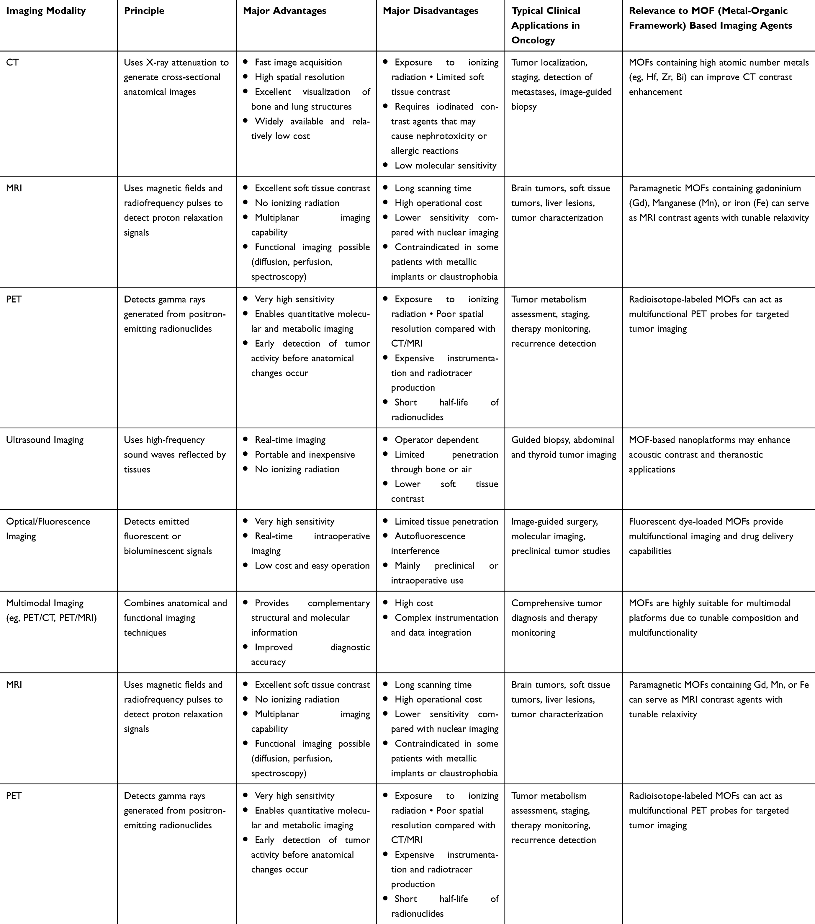 |
Table 1 Comparison of Major Medical Imaging Modalities for Tumor Diagnosis and Monitoring |
Even though these imaging modalities have been found to be effective in the detection of tumors, these modalities have limitations in regard to detecting tumors at an early stage, specificity and in certain cases sensitivity especially when the tumors are in the micro-invasive or minimal stages of development. Consequently, a vast clinical gap exists in the need to apply more sophisticated methods, which can enhance tumor early detection and define its biology better. A potential area of future research is the application of molecular imaging agents with the ability to give both anatomical and functional information on tumors on a molecular basis. As an example, molecular imaging biomarkers have demonstrated the capability to identify early-stage cancers and an option of detecting tumors at subclinical disease stage, which could enable interventions to be performed at earlier stages.4
The latter advanced methods such as the implementation of MOFs are actively being studied to address the shortcomings of the existing imaging technologies.12,13 MOFs possess high surface area, tunable structures, and favorable functionalization capability, making them promising platforms for tumor imaging and drug delivery applications. Recent developments in MOF-based imaging agents demonstrate that they have the potential to increase the sensitivity and specificity of tumor detection and could help to overcome some of the major limitations of the traditional imaging modalities.14 In the last couple of years, MOFs were identified as a promising imaging platform because they feature tunable metal-organic coordination, high porosity, and large surface areas in addition to the capacity to introduce imaging-active metals or functional linkers.15 MOF-based nanoplatforms have demonstrated considerable potential as contrast agents for MRI, PET, fluorescence, and hybrid imaging applications.16 Analysis of these publications shows a significant increase in MOFs publications in the last five years and demonstrates imaging as one of the most popular areas of its application, indicating an increase in interest and a possible direction of translation.17
Despite the rapid expansion of MOF-related literature, most existing reviews primarily focus on general theranostic applications, drug delivery systems, or broad biomedical uses of MOFs. Comparatively, fewer reviews critically evaluate the specific progress of MOF-based platforms in tumor imaging from the perspective of multimodal imaging integration, imaging-active metal engineering, and clinical translation challenges. In addition, limited attention has been given to the relationship between MOF structural design, imaging performance, biosafety, and translational feasibility.
Therefore, the present review specifically focuses on the recent progress of MOF-based systems for tumor imaging across MRI, PET, CT, fluorescence, and multimodal imaging applications, with particular emphasis on structure–property relationships, imaging-active metal incorporation, smart surface functionalization, and translational barriers limiting clinical implementation. Unlike previous reviews, this article also highlights the emerging bench-to-bedside challenges associated with biosafety evaluation, large-scale synthesis, reproducibility, regulatory approval, and long-term clinical applicability of MOF-based imaging agents.
MOFs: Structure and Properties
MOFs are crystalline porous materials obtained by the coordination of metal nodes (ions or clusters) and organic linkers (ligands) into a repeating network structure with known porosity and a high surface area,21 as illustrated in Figure 1. The connectivity centers become the metal nodes which are commonly known as the secondary building units (SBUs) and the organic linkers connect the nodes in one, two or three dimensions to create the extended structures.22 The size and shape of the pore, their connection, and the topology of the whole MOF can be adjusted to diverse metal ions/clusters, different linker molecules, and modified post-synthetically.23 In common MOFs, the metal nodes may be isolated metal ions (eg, Zn2+, Cu2+, Fe3+) or metal oxo/hydroxide clusters (eg, Zr6O4(OH)4).24,25 The organic linkers tend to be carboxylate or imidazolate or phosphonate or other multidentate ligands that bind to the metal centers to determine the topology of the structure.26
 |
Figure 1 Diagrammatic representation of synthesis and structure of a MOF. The MOF itself is built by self-assembling two key elements, namely the Metal Ions or Metal Clusters (symbolized by the yellow circles) and the Organic Linkers (symbolized by the grey rods). The obtained structure is a highly organized, porous and three-dimensional MOF in which metal centers serve as nodes and organic linkers serve as connecting struts. |
The versatility of MOFs is that by changing the metal node or the linker, or both, the properties (chemical stability, pore environment, functional groups, guest-binding capacity) of the material can be changed.23 Zirconium-based MOFs (Zr-MOFs) have been found to be an especially strong and highly studied category due to the strong Zr–O interactions, excellent hydrolytic stability, and good biocompatibility.16,24 Examples include the UiO-series (UiO −66, UiO −67), MOF −808, and the NU −1000, all of which are Zr-based scaffolds that find applications in biomedical applications.24 Copper based MOFs (Cu-MOFs) (eg, HKUST −1) are also widespread; copper paddlewheel units have been employed due to their flexibility of coordination and the availability of open metal sites.27 Fe based MOFs (Fe-MOFs) are also being investigated to be used in biomedicine because of magnetic/paramagnetic properties of Fe that can be used in imaging and therapies.28,29
The metal node type also determines stability, geometry of coordination as well as imaging-relevant characteristics (such as paramagnetism to take advantage of MRI, metal X-ray absorption to take advantage of CT contrast, radionuclide compatibility to take advantage of PET). Likewise, the targeting moieties, fluorescent dyes, responsive groups (pH, enzyme), or radiolabels can be installed on the organic linker based on its choice and functionalization. The linkers also determine the size of the pore, uptake of the guests, surface chemistry and biocompatibility in general.30 Besides, topology (connectivity of nodes and linkers) and pore environment (size, shape, functionality) determine the interaction between the guests (imaging agents, drugs, contrast molecules) and the framework. MOFs have been optimized to biomedical imaging by post-synthetic modifications (eg, ligand exchange, metal-node doping, surface coating).31 The synthesis approaches for MOFs with imaging capabilities are shown in Figure 2.12 The main properties of MOFs relevant to tumor imaging are given in Table 2.23,25,32–39
 |
Figure 2 Principal approaches for the fabrication of MOFs with imaging functionalities: (a) in situ encapsulation of imaging agents during MOF synthesis; (b) integration of imaging agents as intrinsic structural components, including organic linkers, metal ions, or metal clusters; (c) postsynthetic incorporation of imaging agents within the porous architecture of the MOF; and (d) postsynthetic surface functionalization of MOF particles with imaging units.12 |
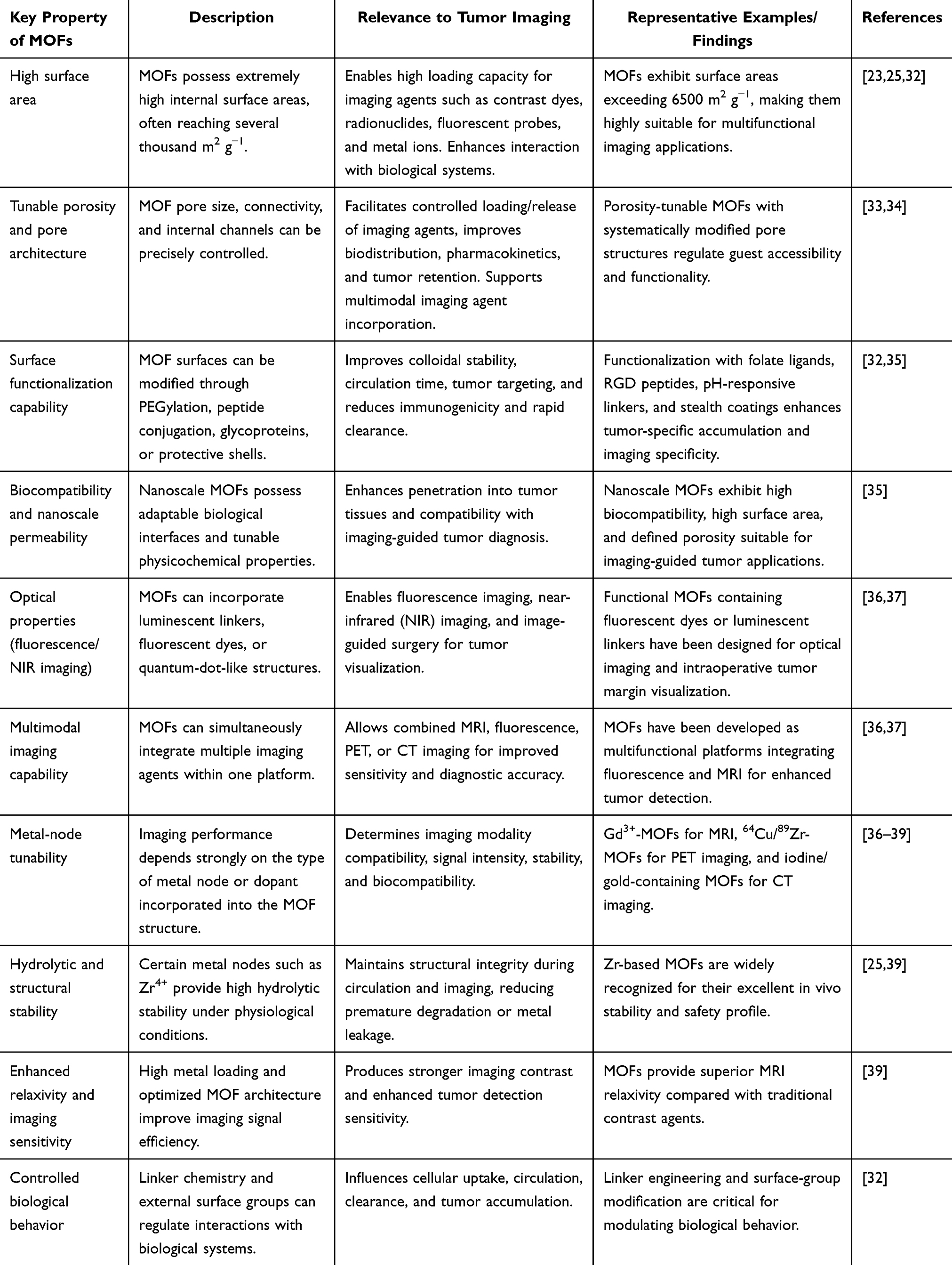 |
Table 2 Key Properties of MOFs Relevant to Tumor Imaging |
Applications of MOFs in Tumor Imaging
Role in Different Imaging Modalities
MRI
MRI remains a central non-invasive modality for tumor diagnosis due to its high soft-tissue contrast and absence of ionizing radiation. However, clinically used T1 contrast agents, predominantly Gd3⁺-based chelates, suffer from limited relaxivity, non-specific biodistribution, and potential nephrogenic systemic toxicity in patients with impaired renal clearance.40,41 These limitations have encouraged the development of MOF-based contrast agents as high-density and structurally tunable MRI platforms. The efficacy of MR contrast agent is dictated fundamentally by the Solomon-Bloembergen-Morgan theory, where proton relaxivities (r1 and r2) are described based on various molecular and electronic properties, including the number of inner-sphere water molecules (q), water lifetimes (τM), rotational correlation times (τR), and electronic relaxation times (T1e and T2e). SBM theory dictates that optimal tuning of τR and τM is vital towards improving T1-weighted contrast efficacy. The same principles are used in MOFs, where these relaxation properties can be adjusted using the rigidity, architecture, and metal-node coordination environment of the framework.42–44 However, it should be noted that relaxivity is highly sensitive to the intensity of the magnetic field, and all the obtained values for r1 need to be clearly defined as depending on field strength (eg, at 1.5 T, 3.0 T, or higher). It can be observed that r1 values for Gd-based compounds are lower as the magnetic field increases because of decreased relaxation efficiency.41,43,45
MOFs have an edge over traditional chelates owing to the high capacity for metal ions, the ability to tune porosity, and the elongation of water diffusion paths, all of which contribute to the increased ability of paramagnetic centers to interact with water molecules, resulting in better efficiency in proton relaxation processes. For instance, PEGylated Mn-MOF-74 nanocrystals showed r1 values ranging from 8.08 to 13.5 M−1s−1 along with strong signal intensity enhancement in tumor tissue using T1-weighted MRI images.46 However, recent developments show that rational pore engineering boosts the MRI capabilities even more. The hierarchical pores in Gd MOFs facilitate diffusion of water molecules to internal coordinating sites. As a result, the effective amount of water molecules available to participate in the process is increased, thus improving τM.47 In the same way, multifunctional Gd-MOF nanosystems have also been prepared for microwave-responsive MRI guided treatment of cancer patients exhibiting high contrast enhancement owing to optimal structure and composition of such multifunctional nanosystems.48 Metal ion selection is very important in terms of relaxivity and safety. Fe containing MOF compounds are gaining more attention as safer substitutes to Gd based compounds because of their good biocompatibility and natural metabolism, however, low magnetic moment necessitates structural optimization.35 Furthermore, surface modification techniques like PEGylation and targeting using specific ligands increase circulation time, minimize uptake by the reticuloendothelial system, and promote selective uptake of the agent in tumors, thereby increasing the local relaxivity of the agent indirectly.
Despite these advances, clinical translation is limited by physicochemical instability under physiological conditions. MOF degradation can alter SBM-relevant parameters such as τM and q through metal ion leaching or framework collapse, resulting in reduced relaxivity and potential toxicity. Moreover, aggregation in biological media can unpredictably alter τR, leading to inconsistent imaging performance. Therefore, reproducible synthesis, stability optimization, and standardized reporting of relaxivity under defined magnetic field strengths are essential for future clinical translation. The MOFs developed as dual or even multimodal imaging (MRI and fluorescence or PET) facilitate the provision of anatomic and functional information, enhancing the detection, delineation and response to therapy are illustrated in Figure 3.49 Moreover, a hypoxia-resistant and persistent O2 self-supplementing nanoplatform was developed to achieve enhanced synergistic phototherapy under the guidance of Fluorescence/Multispectral Optoacoustic Tomography/CT/MR quadruple-modal imaging. The system was constructed using Pt-decorated gold nanoparticles with MOFs serving as the inner template is shown in Figure 4.50
 |
Figure 3 Schematic depiction of MOF-based nanotherapeutics for cancer theranostic.49 Abbreviations: MRI, Magnetic Resonance Imaging; CT, Computed Tomography; PET, Positron Emission Tomography; OI, Optical Imaging; PAI, Photoacoustic Imaging. |
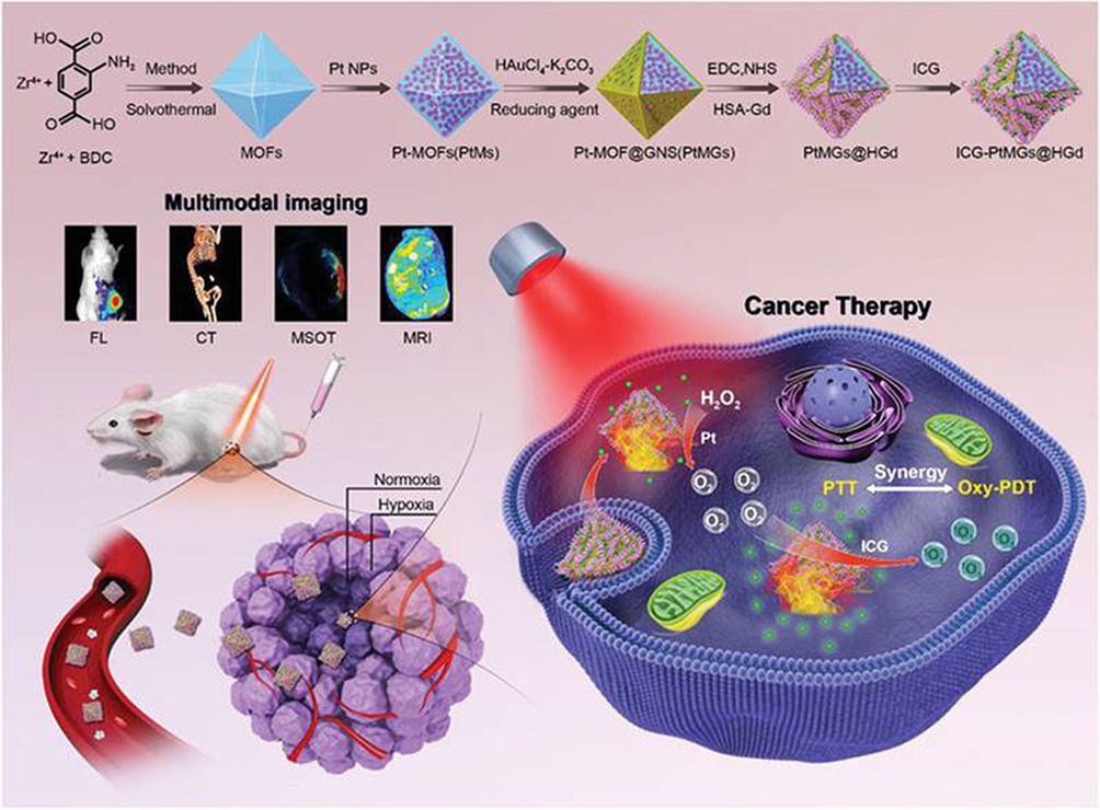 |
Figure 4 Schematic illustration of the ICG-PtMGs@HGd nanoplatforms as H2O2-driven oxygenator for Fluorescence/Multispectral Optoacoustic Tomography/CT/MRI multimodal imaging guided enhanced PDT and PTT synergistic therapy in a solid tumor.50 PDT; photodynamic therapy, PTT; photothermal therapy, ICG-PtMGs@HGd; Pt-decorated MOF@GNSs with human serum albumin-chelated gadolinium and loaded with indocyanine green. |
Positron Emission Tomography (PET)
PET is a highly sensitive molecular imaging modality that enables quantitative visualization of tumor metabolism and biological activity through positron-emitting radionuclides such as Fluorine-18 (18F), 64Cu, and 89Zr.15,51 Conventional PET tracers, however, often suffer from rapid systemic clearance, limited tumor specificity, and suboptimal tumor-to-background ratios, particularly for small-molecule agents and some antibody fragments.15 In recent years, MOFs have emerged as promising nanoplatforms for PET imaging due to their tunable porosity, high surface area, and modular architecture enabling radionuclide incorporation via intrinsic or extrinsic strategies.52,53 Intrinsic radiolabeling involves direct incorporation of radionuclides into the MOF framework during synthesis, often through coordination with metal nodes or secondary building units (SBUs). For example, 89Zr integration into Zr-based MOFs has been demonstrated via framework coordination, improving radiochemical stability and reducing radionuclide leaching under physiological conditions.52 In contrast, extrinsic labeling typically relies on post-synthetic surface modification using chelators such as DOTA, NOTA, or desferrioxamine (DFO), followed by radionuclide complexation (notably 89Zr–DFO systems widely used in immunoPET). Although post-synthetic strategies provide synthetic flexibility and targeting versatility, in vivo stability can be compromised due to transchelation with endogenous metal-binding ligands.51
Recent studies suggest that MOF-based PET probes may improve tumor accumulation and prolong circulation time compared to some small-molecule tracers due to enhanced retention and multivalent loading capacity.53 However, reported extremely high tumor-to-background enhancements should be interpreted cautiously, as PET signal intensity is primarily governed by radionuclide-specific activity and radiochemical stability rather than total metal loading. Thus, the key advantage of MOFs is not simply high metal content, but rather their ability to achieve high radioactivity-per-particle loading, while enabling multifunctional surface engineering and multimodal imaging integration.53 Radiochemical stability remains a major translational challenge. In particular, 89Zr dissociation from unstable frameworks can lead to nonspecific bone uptake due to strong affinity of free Zr ions for phosphate-rich tissues, a well-recognized phenomenon in Zr-based immunoPET chemistry.51 Similarly, acidic and reductive tumor microenvironments can accelerate partial MOF degradation and radionuclide leakage, emphasizing the importance of stability testing in serum, plasma, and competitive biological media for accurate PET interpretation.52 Targeted PET imaging using antibody- or peptide-functionalized MOFs is increasingly explored for tumor-specific delivery and enhanced imaging contrast.15 However, MOF nanoparticles (often 50–200 nm) are significantly larger than classical immune PET constructs such as 89Zr-trastuzumab, which may alter pharmacokinetics.
Fluorescence Imaging
The high-resolution, real-time visualisation of tumors or tumor margins through fluorescence contrasts agents is possible using fluorescence imaging. Recent developments have shown that MOFs have the potential to be highly promising fluorescence agents due to their special internal architecture, tunable pore structure and ability to incorporate or encapsulate fluorescent moieties such as quantum dots (QDs). MOF-based fluorescence agents are characterised by the combination of the merits of MOF scaffolds, namely, high surface area, structural stability, and flexibility of functionalisation, and the optical characteristics of fluorescent dyes, lanthanide centres or QDs, thereby improving the sensitivity and specificity of tumour imaging.12,54,55
MOFs functionalised with QDs or using QDs concepts (so-called QD-MOF hybrids) have been designed to use as tumour imaging contrast agents. As an illustration, a survey on QD which comprises MOF hybrid systems shows how the aggregation of QDs in MOF matrices can be reduced, they can be made more photostable, and more efficient tumour-imaging agents.56 In these systems, localisation and stabilisation of the fluorescent QDs are performed by the MOF in a biocompatible environment, which allows an increase in fluorescence emission and tumour-targeting performance (with surface functionalisation, eg, peptides, antibodies).
The advantages of MOF based fluorescence agents in fluorescence guided surgery are especially attractive. In surgery, FGS can be used to see tumour margins in real time under the fluorescence light during the resection of tumours. Tumour-targeting ligand (folate, RGD peptides) functionalised MOFs will accumulate within tumour tissue and lead to a tumour-to-background fluorescence ratio that helps to better excise the target tumour tissue and minimise the amount of tumour tissue remaining.57 According to a more recent review of MOF applications in imaging-guided tumour diagnosis, MOFs have the ability to enhance tumour targeting, image delineation and be utilised as dual or multi-modal fluorescence-based agents.35
Moreover, MOF based fluorescence agents tend to have greater photostability, increased payload of fluorophore and controlled release kinetics as compared to free dyes.58 Since fluorophores can typically be rapidly photobleached or distributed non-specifically by themselves, the photostability and biodegradation of dyes and fluorophores in MOF pores can be used to preserve dyes and fluorophores, and dyes from quenching and degradation, and design stimuli-controlled release (eg, pH, enzyme) to tumour microenvironment specificity.59 Moreover, the modularity of MOF synthesis can allow both combinations of fluorescence imaging with therapeutic and other imaging modalities (eg, MRI, CT). A MOF platform could, eg, have a fluorescent QD moiety and a paramagnetic metal center attached to it, thus supporting intraoperative fluorescent guidance and preoperative MRI imaging.60,61 Such a multimodal property is beneficial in tumor therapy - a single imaging agent (the MOF) performs many functions, enhancing clinical practice and diagnostic efficiency.62
Nevertheless, along with these encouraging properties, there are still some challenges to clinical translation of MOF based fluorescence imaging agents. These are the possible toxicity of metal constituents or QDs (particularly, heavy metal based QDs), the biocompatibility and clearance, the absence of aggregation or instability in physiological media, the efficient tumor targeting and retention, and the in vivo safety with the application of FGS. It is noted in literature that reproducibility of nanoscale synthesis, stable surface modification, and depth of fluorescence penetration in vivo (particularly of near-infrared emission) are major challenges to next-generation clinical usage.35 The schematic illustration of phosphate-terminal DNA aptamer conjugation onto a Zr-MOF nanoparticle quencher for target-induced imaging and photodynamic therapy and synthesis procedure, drug-loading process, and receptor-mediated endocytosis pathway of targeted UCNPs@MOF core–shell nanocomposites (NCs) is shown in Figure 5.62
 |
Figure 5 (a) Schematic illustration of phosphate-terminal DNA aptamer conjugation onto a Zr-MOF nanoparticle quencher for target-induced imaging and photodynamic therapy. (b) Schematic illustration of the synthesis procedure, drug-loading process, and receptor-mediated endocytosis pathway of targeted UCNPs@MOF core–shell nanocomposites (NCs). The insets show enlarged views of the porous cavities of MIL-100(Fe) and its partial coordination structure, respectively.62 |
Fluorescence imaging in metal–organic frameworks (MOFs) is increasingly categorized into two optical windows: the first near-infrared window (NIR-I, 650–900 nm) and the second near-infrared window (NIR-II, 1000–1700 nm). NIR-I fluorophores have been widely used due to relatively mature dye chemistry and detector availability; however, their clinical utility is limited by significant photon scattering, shallow tissue penetration, and high tissue autofluorescence, which collectively reduce imaging contrast in deep-seated tumors. In contrast, NIR-II imaging provides substantially improved spatial resolution, higher signal-to-noise ratio, and deeper tissue penetration due to reduced photon scattering and lower tissue absorption at longer wavelengths.63–68 Recent advances demonstrate that MOF-based fluorescent platforms can be engineered to incorporate NIR-II emitters or act as nanocarriers for NIR-II dyes, enabling enhanced tumor visualization beyond several centimeters of tissue depth. This transition from NIR-I to NIR-II systems is particularly important for accurate tumor delineation and real-time intraoperative imaging.69,70 Importantly, integrating NIR-II functionality into MOFs also facilitates multimodal imaging when combined with MRI or PET-active components, supporting theranostic applications in oncology.71
CT
CT is a technique of imaging that is used in clinical practice with the highest frequency, allowing high-resolution images of internal structures without invasiveness.72 It is based on X-ray absorption to produce high-resolution cross-sectional body images. Although CT imaging is widely used, there are some limitations to the imaging technology especially the contrast resolution and specificity of the imaging of the soft tissue or tumor. MOFs are new promising materials in improving CT imaging because of their distinctive structural characteristics, composition manageability and combination of diverse imaging modalities. The potential use of MOFs as CT contrast agent is gaining more attention due to the bonding of metal ions that have high X-ray absorption properties.73 Metals with a high atomic number (Z) tantalum (Ta), iodine, barium (Ba), and gadoninium (Gd) have a high level of X-ray absorption, which is very required in enhancing contrast in CT images.74,75
Tunable porosity and surface functionality is one of the key benefits to using MOFs as CT contrast agents. It is possible to design MOFs to ensure a regulated release of metal ions into the bloodstream to have long-lasting and high-contrast imaging of tumors. Likewise, Gd-based MOFs have been investigated as dual-modal contrast agents, with the possibility to have both CT and MRI images of the same system.62,76–78 This Gd ions addition improves the X-ray absorption properties of the MOF and also allows the MRI contrast to be improved, providing the benefits of multimodal imaging. The metal ions that are embedded in MOFs are critical towards the improvement of their work as CT contrast agents. These metal ions have a decreasing X-ray absorption depending on Z with atoms of higher values of Z attenuating X-ray better resulting in better image contrast.79 MOFs made of metal of higher atomic numbers like Ta, iodine, and Ba have a better X-ray attenuation than the traditional iodine based agents.80,81
Clinically used iodinated contrast agents typically show a CT attenuation efficiency on the order of ~20–40 HU per mg I/mL (or equivalent iodine molar concentrations) at standard clinical tube potentials (120 kVp), with the exact value depending strongly on scanner calibration, beam hardening, filtration, and spectrum shape. This dependence arises from the strong energy dependence of iodine’s K-edge (~33.2 keV) and the polychromatic nature of clinical X-ray beams.82,83 By comparison, the high-Z nanomaterials provide improved X-ray absorption owing to their higher atomic number and mass attenuation coefficients. For instance, gold nanoparticles are known to offer excellent CT contrast, usually giving about 4.8–5.4 HU/mM (equivalent to a few tens of HU per mg/mL depending on various parameters and voltage), particularly at clinical voltages of 120 kVp.82,84 In a similar way, bismuth nanoparticles have excellent X-ray attenuation properties because of the high atomic number of Bi (Z = 83). It is shown both experimentally and in vivo that bismuth nanoparticles possess very effective CT contrast properties (hundreds of HU from clinically feasible doses), usually outperforming standard iodinated contrast agents for the same amount of injected material.83 Regarding CT contrast agents based on MOFs, it is clear that the effective attenuation properties will be greatly determined by the use of high atomic number metals as nodes, including hafnium (Hf), Zr, Ta, and bismuth (Bi). The reason is that, thanks to the porosity of the MOFs and their very high metal content, MOFs can attain enhanced effective attenuation due to their concentration levels compared to traditional iodine-based materials. However, CT signal intensity is influenced not only by concentration but also by particle dispersion, aggregation state, hydration environment, and X-ray energy dependence.85–88
An interesting development in these recent works is the shift from traditional monofunctional MOFs to hybrid MOFs that are capable of performing multiple functions or responding to environmental stimuli. Prior work has concentrated mainly on zirconium-containing MOFs due to their superior hydrolytic resistance, stability, and higher loading efficiency, which makes them suitable for biomedical applications.16 However, in recent years, research tends to be focused on the utilization of hybrid and biomimetic MOFs by including various additives such as polymers, cell membranes, peptides, or responsive linkers, addressing concerns like the immune system response, lack of sufficient specificity toward cancer cells, and inadequate stability.24 In addition, iron and manganese based MOFs also received significant attention due to their improved biocompatibility and decreased toxicity in comparison to gadolinium MOFs. It is interesting that Mn based MOFs can exhibit excellent redox property and produce Mn2⁺, thus realizing MRI and degradation-dependent imaging.24 Fe-based MOFs are also extensively investigated due to their good degradation properties in biological systems, providing potential for clinical use in drug delivery and imaging applications.16 Such trends are evident in other recent literature studies that focus on developing translational MOFs with improved biocompatibility, imaging capabilities, multimodality, and personalized theranostics, among others.89
Multimodal Imaging Using MOFs
The development of MOFs-based integrated metal-organic structures in combination with several imaging modalities has become a groundbreaking solution in tumor diagnostics. Researchers are obtaining improved diagnostic accuracy and improved clinical results by incorporation of MOFs into other imaging modalities, which include MRI, PET, and fluorescence imaging. The high surface area, biocompatibility, and customizable porous structures of MOFs make them the best to be used in multimodal imaging. They can be readily conjugated with contrast agents or radionuclides into imaging agents that are more comprehensive in analyzing the tumor and accurately localizing it.90,91
One application of MOFs has the potential to be in dual-modality imaging, where two or more imaging modalities are used in series to exploit the complementary nature of the two. As an example, MOFs like palladium (Pd)-hafnium-based (Pd@Hf-EDB) have been able to improve CT and photoacoustic imaging (PAI) at the same time. This dual-modality system increases contrast in addition to facilitating the precise tracing of tumor boundaries thereby increasing the overall diagnostic accuracy.90 This technology is also revolutionizing the manner in which tumors are detected, staged, and monitored because it provides both structural and functional understanding about tumors.
Furthermore, recent developments, as well, resulted in development of hybrid imaging agents, in which MOFs are used alongside other nanoparticle platforms in multimodal applications. Namely, nanoparticles that have been created using core-shell MOFs have been utilized to enhance the stability and bioavailability of the contrast agents. These bimodal systems have the ability to apply in both MRI and fluorescence imaging, which allows them to track the tumors in real-time, as well as deliver therapeutic agents that are targeted.92 The flexibility of MOFs in these hybrid designs has not limited MOFs to imaging but also cane be used in other fields like theranostics, where diagnostics and therapy are on the same platform.
The use of MOFs in theranostics has been particularly able to be useful in cancer therapy. Porphyrin-based MOFs are being integrated into imaging systems to detect tumors and at the same time perform photodynamic or photothermal therapy by their nature of inherent fluorescence. Such MOFs as those based on Zr-based porphyrin scaffolds not only enable visualization of tumors using fluorescence but also permit the delivery of energy to cancer cells using this scaffold to achieve therapeutic effects.93 The possibility to monitor the actual performance of the treatment and at the same time deliver therapeutic agents is a major breakthrough in individual cancer therapy.
Moreover, MOFs integration along with PET is an effective way of molecular imaging of cancer. Recently, MOF-based PET imaging using radiolabeled metal centers has been developed, which offers high resolution pictures on tumor location at the molecular scale.94 The associated benefits of these PET-MOF agents are that they can identify small tumors or metastasis that may be overlooked by other conventional methods of imaging and therefore an early detection and intervention are feasible.
Nevertheless, no matter how promising all these developments are, a number of obstacles are still present in the process of extending the MOF-based multimodal imaging systems beyond the laboratory to the clinical environment. The problems of the stability, biodegradation and possible toxicity of MOFs should be resolved to make them safe to use in human beings.95 It is important to note that the growth of biocompatible MOFs, as well as control release mechanisms of both imaging agents and therapeutic drugs, is essential to their clinical success. Moreover, a mass production of MOFs that will streamline its synthesis to enhance efficiency and reduce the cost of production will be necessary to enable massive clinical usage.
Tumor Targeting and Drug Delivery Potential
MOF Functionalization for Tumor Specificity
The surface modification of MOFs is critical in increasing their selectivity to tumor therapy and diagnosis. It is feasible to enhance the selective tumor marker binding and minimise off-target effects on tumor cells, by functionalizing MOFs with tumor-specific ligands, including peptides, antibodies, and small molecules. Tumor-associated antigens that are overexpressed have become a target of MOF functionalization because smaller molecules (peptides) can readily access these antigens. As an example, a recent study has emphasized targeting fibroblast activation protein (FAP) and transforming growth factor beta (TGF) as peptides therapy and imaging tools of tumors that can be upregulated in several cancers.96,97 When conjugated to MOFs, these peptides can be used to achieve better targeting and longer retention at the tumor locations thereby facilitating more effective drug delivery and tumor imaging.
Antibodies are also a great potential in tumor targeting as they are very specific with considerable affinity towards the cancer antigens. Both the diagnostic and therapeutic uses of antibody-conjugated MOFs have been pioneered through the development of the antibody-conjugated MOFs. This was demonstrated in a study by Anderluzzi et al, 2024 which showed that antibody-based MOF functionalization targeted the tumor vasculature, such as the membrane-bound enzyme CD13.98 In addition to making the targeting process more efficient, this method also boosted the therapeutic activity of the conjugated drugs using the biological characteristics of MOF materials.
Besides, small molecules like epidermal growth factor receptor (EGFR) and integrin 21 beta 3 have been incorporated into MOFs to target them more effectively in tumor targeting. They enable the accurate targeting of chemotherapeutic agents as recent research demonstrates the ability of MOF-based systems to deliver specific types of tumors.99,100 The targeted delivery of such molecules, reduction of systemic toxicity as well as the pharmacokinetics of encapsulated drugs can be achieved through functionalization of MOFs with these molecules.
The presence of tumor-targeting peptides and small molecules in MOFs further increases the ability of the material to serve in selective targeting. The recent advances of the application of peptide-related approaches, including those demonstrated by Mathieu et al, 2024, reflect the benefits of supramolecular assembly of peptides onto MOFs to reach high specificity to tumor-associated antigens, including HER2, which is a powerful marker in breast cancer.101 This method leads to a higher bioavailability of drugs in the tumor site and is much more beneficial to the outcome of therapeutic effect. Moreover, these peptide-functionalized MOFs have high tumor penetration and retention, proving their applications in the further treatment and diagnostics of cancer.102
Combination of MOFs and tumor-targeting ligands will transform cancer treatment, offering highly specific, highly effectual and versatile platform of drug delivery and diagnostic imaging. Promising opportunities of MOF-based systems in precision medicine can be maximized by further studies on surface modification methods and ligand choices.
MOFs as Drug Delivery Carriers in Tumor Imaging
The exceptional structural properties of MOFs – their high porosity, large internal surface-areas, tuneable pore sizes and surface chemistries – have made them very desirable as delivery vectors of therapeutic agents or imaging probes in tumor therapy and imaging. MOFs are an optimal system to encapsulate or adsorb drugs or contrast agents as reviewed articles have highlighted and therefore combining diagnostic imaging with therapeutic delivery (theranostics) in a single system.103
MOFs have been engineered to carry out small-molecule chemotherapeutics or photosensitizers, radionuclides, fluorescent probes or magnetic contrast agents, in the tumour imaging and therapy context. As an example, Yang et al, 2021 discuss nano-MOFs as drug carriers and indicate that the high drug containing capacity is due to the large pore volume and the versatile chemical nature of the structure.104,105
The loading may be carried out via physical encapsulation (via pores or voids), via adsorption on surfaces, via covalent conjugation to MOF linkers, or via post-synthetic alteration of the surface. Khafaga et al, 2024 remark that MOF functionalisation allows better stability, drug solubility and bioavailability.106 In tumour imaging, imaging moieties can also be conjugated to such drug-loaded MOFs such that the distribution, accumulation and release of the therapeutic agent can be tracked in vivo. The article by Patil et al, 2024 about the stimuli-responsive MOFs that integrate targeted delivery and imaging and cargo release.20
pH‑Responsive Drug Release
One of the most important processes through which MOFs release cargos in tumour environments takes advantage of the acidity of the tumour microenvironment (pH) in comparison with normal tissues (eg, pH = 6.57.0 vs pH = 7.4). MOFs, like zeolitic imidazolate frameworks (ZIFs), have been known to degrade when subjected to acidic pH and release drugs. As an example, dihydroartemisinin (DHA) was delivered in a tumour-therapy setting using an acid-responsive ZIF-8 system.107,108 To validate the utility of pH-responsive MOFs in general, Guillen et al, 2022 showed a pH-stimuli thin film of MIL-88B(Fe) to release ibuprofen.109
More broadly, Yan et al, 2021 provide an overview of environmental-sensitive MOFs that prevent the premature release of drug into the blood and instead, cause release of their cargo only at the tumour locations upon stimuli, eg, low pH.108 This mechanism assistance in tumour imaging/therapy encompasses enhancement of tumour accumulation (through the enhanced permeability and retention effect) and local delivery of contrast or therapeutic agent, thus enhancing imaging specificity and decreasing systemic side effects. Due to their porous structures, MOFs exhibit high drug-loading capacity, facile surface functionalization, and favorable biocompatibility. As a result, numerous MOF-based stimuli-responsive drug delivery systems have been developed, leading to considerable advances in recent years. Schematic illustration of MOFs-based stimuli-responsive system for drug delivery is shown in Figure 6.110
 |
Figure 6 Schematic illustration of MOFs-based stimuli-responsive system for drug delivery.110 |
Enzyme-Responsive Drug Delivery
Besides the pH, endogenous enzymes or tumor-overexpressed biomolecules can serve as stimuli to MOF-based release systems. A growing review is enzyme-immobilised MOFs, in which tumor microenvironment triggers enzymatic catalysis of drug release by MOF carriers.111 Indicatively, a clickable PEG phosphate ligand attached to a Zr based MOF (UiO 66) allowed the release of drug to be controlled in the presence of certain enzymes.112 These enzyme responsive systems will be able to enhance selectivity of release (only in tumour or peritumour regions of high enzyme concentration) and hence enhance contrast between tumour and normal tissue in imaging or better therapeutic index in therapy.
Specific Drug Discharge Processes
In addition to passive accumulation and release of tumours, MOFs have been engineered to be actively targeted to tumour cells or tumour vessels. It is done through conjugation of ligands (eg, antibodies, aptamers, peptides) to the MOF surface, which identify tumour specific markers. As an example, Wang et al showed aptamer-functionsalised Fe-based MOF, which targeted tumour cells.113 In a different report, an aptamer-functionalised Fe3O4 at MOF nanocarrier facilitated simultaneous fluorescence imaging and targeted delivery of drugs to triple-negative breast cancer cells.114 Such targeting in the imaging setting enhances the concentration of the imaging/therapeutic payload in tumor tissue, thereby enhancing contrast and potentially decreasing off-target background. Kong et al, 2024 present a review of MOFs in tumor-targeted drug delivery systems, including the focus on active targeting.115
Acquisition of Tumour Imaging
The delivery of the therapeutics, release, and accumulation of the payload of imaging probes into tumors can be imaged on the same platform when therapy or imaging probes are loaded into MOFs. A study explains the combination of imaging (eg, MRI, fluorescence, CT) with drug delivery of such theranostic MOFs, which allows them to monitor the release and therapeutic response in real-time.116,117 The Advances, applications, and challenges of MOFs in tumor imaging: from single-modal systems to multifunctional theranostic platforms are given in Table 3.12,35,37,38,118–120
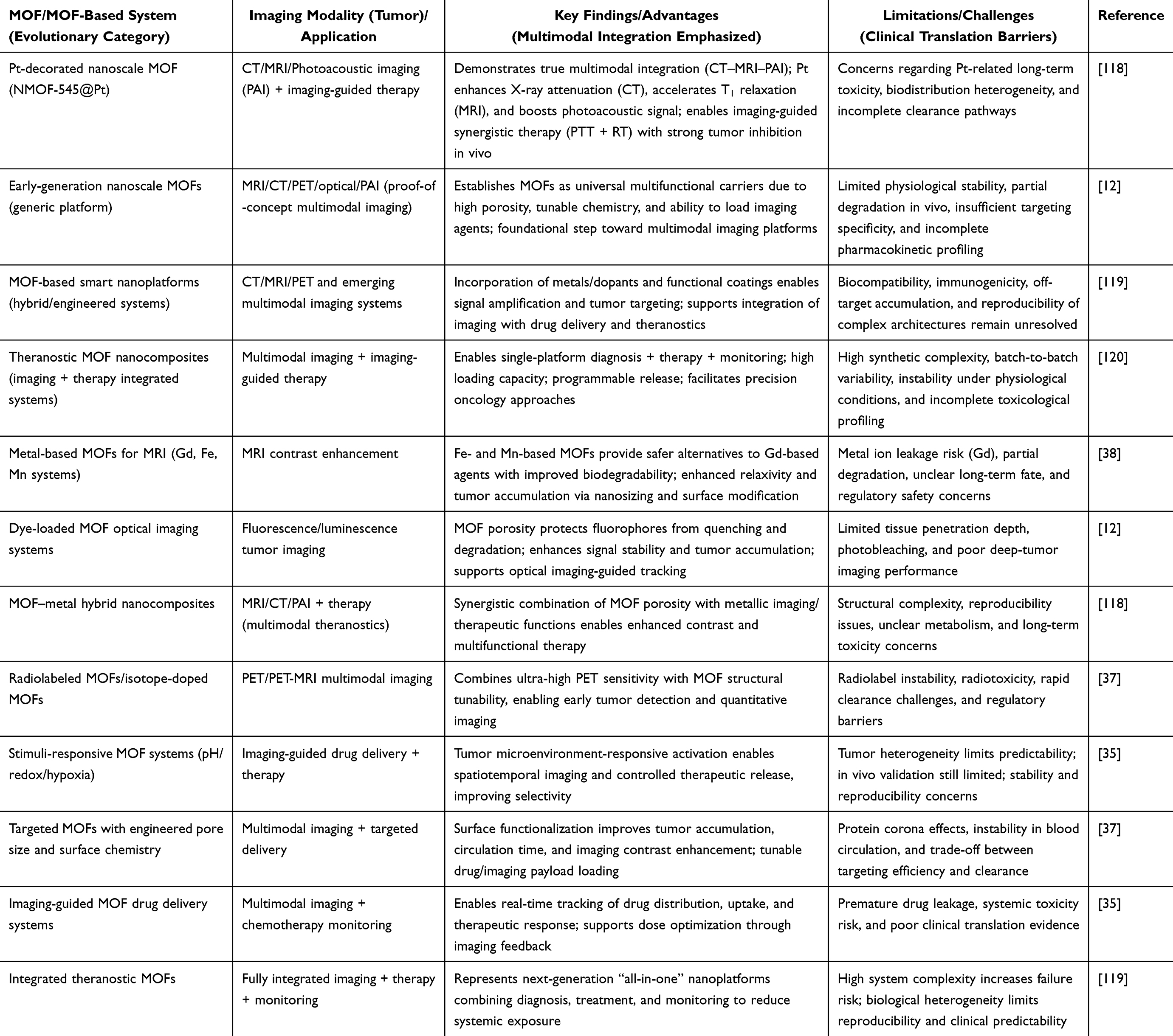 |
Table 3 Advances, Applications, and Challenges of MOFs in Tumor Imaging: From Single-Modal Systems to Multifunctional Theranostic Platforms |
Prospects of MOFs in Tumor Imaging
Enhanced Sensitivity and Resolution
A highly attractive application of MOFs to tumour imaging is that it has the potential to show improved sensitivity and resolution relative to established contrast or imaging agents. This is because of their characteristic structural, chemical and functional tunabilities, which all combine to generate superior signal generation, superior contrast-noise and spatial resolution in imaging modalities.121–123
To begin with, MOFs have the potential to deliver higher amounts of imaging-active (eg, paramagnetic metal ions to be used in MRI, heavy elements to be used in CT, fluorophores to be used in optical imaging) amounts of imaging-active components per particle than many other conventional agents. For example, nanoscale MOFs (NMOFs) have large internal pore volumes, high density of metal-ion group and adjustable organic linkers, which enable high concentrations of imaging moieties to be loaded or incorporated.12,38,62 The high loading can be translated to the higher signal per nanoparticle or dose which can increase the sensitivity ie the capacity to detect low concentration of target tissue or small tumours.
Secondly, the tunable surface chemistry and structure of MOFs facilitate enhanced targeting and retention in tumour tissues, which enhances signal specificity and resolution of tumour versus background.103,124 The fact that it is possible to functionalize MOF surfaces with targeting ligands (eg, peptides, antibodies) or effectively use enhanced permeability and retention effects implies that imaging contrast can be better localized to malignant tissue, enhancing tumor to normal tissue contrast and hence increasing effective resolution.35,62 Further, the porosity of MOFs allows imaging agents to be stabilized by the porous structure and thus not lost by quenching or degradation during their transit to the tumor site, retaining signal integrity.12
Thirdly, the MOFs enable multimodal imaging, which brings together various imaging modalities (eg, MRI + optical + CT) within a single nanoparticle platform. This multimodality has the capability to offer complementary contrast modalities (eg, high spatial resolution MRI, high sensitivity optical, or high-density CT) and hence can enhance the overall resolution: anatomical, functional or molecular.38,125 An example is a MOF particle that can contain a paramagnetic metal cluster to provide MRI and a fluorophore to provide fluorescence imaging, to allow a macro-scale imaging of the particle and microscopic localization.37
Moreover, the fine size and shape control of MOFs at the nanoscale can have a direct effect on imaging resolution. Particles between 20–200nm can extravasate into tumour interstitium yet not be quickly eliminated by the kidney or mononuclear phagocyte system. Tumor penetration and uniform distribution, which can be improved by adjusting MOF size, morphology and surface charge, are factors that can lead to high -resolution imaging of tumor heterogeneity. As it is observed in the review by Duman et al, 2021 NMOFs do tend to have tunable sizes and structures as compared to other nanocarriers.12
Moreover, mechanistically, MOFs permit amplification of signals based on principles of clustering of contrast agents, confinement and improved relaxivity (in MRI). To give an example, the high local concentration of paramagnetic centres in a MOF can be used to provide higher relaxivities (ie a larger effect per unit metal) than free ionic contrast agents in MRI applications.12,38 The increased relaxivity implies that smaller doses can be used to produce the same (or better) contrast and this improves sensitivity and reduces background interference.
Lastly, the background and off-target signal are also reduced with the help of MOFs which indirectly leads to better resolution. Since MOFs may be passively loaded or passively functionalized to stay silent until they arrive at the tumor microenvironment (as in stimuli-responsiveness), the difference in tumor and normal tissue is better defined, and small lesions may be detected.31,108,126 Even though this mechanism is adjacent to those of drug-delivery, it plays a role in imaging towards the future of high spatial resolution, and the detection limits of imaging. According to reviews by Hou et al 2024, MOFs are important such that they are crucial to tumor cell targeting, identification, imaging, among other things in the tumor-diagnostic scheme.35
Biocompatibility and Safety
Tumor-imaging translation of MOFs biocompatibility and safety profile is a major consideration. The biocompatibility of MOFs is highlighted in a number of recent reviews to be dependent upon a variety of design factors - the type of metal ion used, ligand chemistry, particle-size distribution, surface functionalization, colloidal stability and the degradation/clearance routes of these particles in the biological host.28,127,128
MOFs constructed with biologically favorable metals (like Fe, Zn or Mg), ligands that are benign should have reduced cytotoxicity and have more favorable biodistribution properties. As one example, MOFs containing what is deemed as an essential metal in the body eg, Fe, Zn and Mg have been reported by Singh et al, 2021 to have a better biocompatibility.127 Further approaches to minimise immune recognition, oxidative stress and damage of healthy tissues are surface functionalization (eg, PEGylation) and size/shape control.13,128
In terms of in vivo stability and clearance, MOFs should be stable enough to fulfill the imaging task (contrast generation and tumor targeting) but degradable to achieve clearance to prevent accumulation and toxicity over time.35,129,130 Framework stability (strength of metal-ligand bonds), particle size, and vulnerability to physiological circumstances (eg, pH, ionic strength, enzymatic environment) determine biodegradability of nano-MOFs. Singh et al, 2021 synthesis reported in-vivo biodistribution, organ accumulation, clearance dynamics and immune responses of different MOFs.127
It has been demonstrated that some MOFs can be eliminated through a renal or hepatobiliary clearance route depending on size and surface characteristics. Nevertheless, most of the studies are at the small-animal scale, and long-term destiny (weeks to months) is less characterized. A 2024 study by Zhuang et al, 2025 points out that nanoscale MOFs multiscale profiling of biocompatibility, toxicity, inflammation and organ damage is yet to be explored.131
Having a tumor-imaging carrier, MOFs have a favorable safety profile when well-designed (compared to traditional imaging agents), and thus may have a lower systemic toxicity profile, in particular, should they release toxic contrast particles or non-specifically accumulate.12,120 This justifies one of the major opportunities of MOFs in tumor imaging: that these carriers can combine high imaging capability with biological risk tolerability. Nevertheless, complete translation requires standardized toxicity assays, scalable and repeatable production in good-manufacturing-practice (GMP) conditions, and strict in-vivo clearance/accumulation information. Schematic illustration of the primary mechanisms underlying MOF-induced toxicity is shown in Figure 7.132
 |
Figure 7 Schematic illustration of the major mechanisms associated with MOF-induced toxicity. MOF-related toxicity may arise during cellular internalization processes, including endocytosis, diffusion, and membrane adsorption, potentially leading to membrane damage. Following internalization, MOF nanoparticles (MOF NPs) may also induce intracellular toxicity through reactive oxygen species (ROS) generation, thereby affecting cellular organelles such as mitochondria and the nucleus.132 |
Scalability and Cost-Effectiveness
The viability of the implementation of MOF based imaging agents in clinical applications is based on not just on the performance and safety but also on the cost of production, scalability and economic feasibility. One of the key benefits of MOFs is their modular chemistry and tunable synthesis, which theoretically can be cost-effectively manufactured as opposed to less complex tailored nanoparticle systems.23 However, there is still a problem of practical up-scaling.
The recent large-scale MOF production analyses suggest that although it is possible to produce at pilot-scale (kilogram) scale, production at multi-kilogram to ton scale is still limited by the design of the reactor, its yield, cost of purification/activation, solvent consumption, and shaping/housing of materials.131 To illustrate, Chakraborty et al, 2024 outline the so-called techno-economic analyses and life-cycle assessments that MOFs should undergo in order to reach commercial status.133
MOFs have high active contrast agent loading per particle (lowering dose) and can incorporate many functions (theranostic) which could lower the overall cost of the agents and enhance cost-efficiency per diagnostic result.78,134 Tunability of MOF chemistry can potentially allow the use of relatively cheap metals/ligands instead of the rare earth elements or highly engineered QDs, lowering the cost of raw materials.25,135
The modular assembly has a potential of simplifying regulatory and manufacturing pipelines (when standardized), which in the longer term may help to lower the unit-cost relative to a custom nano-agent.
Nevertheless, the cost-effectiveness of real clinical implementation will remain to be based on the requirements of reproducible purity, batch-to-batch reproducibility, stability, shelf-life, regulatory, and imaging efficiency. Summary: although MOFs have potential in the cost-effective and scalable production of imaging-agents, scalable and GMF-compatible production of imaging-agents on a large scale (biomedical imaging) has yet to be demonstrated.
Recent Advances and Innovations in MOF Development
Advancements in MOF Synthesis Techniques
In the recent past, there has been a significant positive shift in synthetic methods to develop biocompatible and stable MOFs, specifically designed to be used in biomedical imaging. As an illustration, mechanochemical, microwave-assisted and sonochemical pathways permit more reproducible and faster MOF generation that requires fewer solvents and less severe conditions and enhances scalability and minimizes toxic by-products.32,136
At the same time, machine-learning-supported design pipelines have recently been developed to predict MOF biocompatibility and toxicity by metal-ligand combinations that allow pre-selection of MOFs to be used in vivo.137
Together with these synthetic improvements, modifications in post-synthetic functionalization (eg, PEG-ylation, biomolecule-anchoring, surface-coating) have improved colloidal stability, bio-distribution and circulatory lifetime of MOFs in physiological media.138,139 The surface engineering is needed to avoid premature aggregation or forming of protein corona both of which worsen imaging performance. These developments, together, empower the synthesis toolkit to allow MOFs with enhanced crystallinity, size/shape control, and increased adaptability to tumor-imaging applications.
Novel MOF Structures to Improve Imaging
MOF structural design innovations have brought in hybrid and multifunctional structures that enhance imaging ability. An example is hybrid MOFs, in which two or more different metal nodes are combined, or in which the core and shell are MOFs and the core can offer a contrasting feature (eg, MRI-active metal) and the shell can include a fluorophore or imaging probe.35,140,141
Multimodal MOFs have been designed to accommodate multimodal imaging agents eg, to incorporate in a single MOF scaffold, MRI, fluorescence and CT contrast. This allows co-registration of imaging, superior fidelity of tumor delineation and functional/molecular imaging. The biomedical MOF domain is a field of research where reviews highlight these imaging and therapy-integration design advances.142 Besides, the stability of MOFs in physiological conditions has been enhanced by choosing metal-linker nodes with high stability (eg, Zr based MOFs) or through surfaces of biocompatible shells, which increases the usability of MOF in tumor imaging in vivo.22
Nanotechnology Interrelation
Combination of MOFs with other nanotechnology components (eg, nanoparticles, quantum dots, upconversion nanoparticles, liposomes) has provided additional opportunities to carry out targeted tumor imaging with increased precision and resolution. To illustrate, MOF shell hybrids, in which a MOF shell surrounds or decorates a magnetic or plasmonic nanoparticle result in combined functionalities: improved MR contrasting of the nanoparticle with a MOF shell and high payload and functionalization capacity of the MOF cage. According to the review by Chandra et al, 2025 the so-called smart nano-hybrid MOFs are the systems that combine MOFs and nanoparticles to deliver drugs, catalyze and image in cancer.143
Similarly, hybrid platforms with up-conversion nanoparticles (UCNPs) and MOFs enable near-infrared excitation and deep-tissue imaging, in which the MOF scaffold increases stability and surface functionalization.35,144 These incorporations allow better tumour targeting (through ligand functionalisation of the hybrid surface), enhance signal amplification (through a high agent loading within the MOF) as well as imaging resolution (through synergistic contrast mechanisms). MOFs chemistry and nanotechnology is offering potent platforms that are awaiting further development to advanced tumor imaging, namely; high contrast, multi-modal read-out, and smart targeting, although the transition of these complex systems into clinical practice has not yet been realized.
Challenges and Limitations in MOF-Based Tumor Imaging
There are a number of challenges and limitations associated with the application of MOFs to medical imaging of tumors. To start with, the biocompatibility and stability of nanoscale MOFs in physiological conditions is a problem: nanoparticles can be readily degraded or aggregated in blood, and this can cause unpredictable clearance, off-target retention, and imprecise imaging contrast. As an example, Liu et al, 2022 emphasize that MOFs showed desirable properties in terms of porosity and tunable composition to be used in bioimaging but, at the same time, their behavior in biological fluids needs powerful control.62 MOF-based imaging probes have low biological targets (eg, endothelial transport, tumor interstitial pressure, reticuloendothelial system uptake) which limit targeting efficiency and tumor accumulation. In spite of surface functionalisation, several MOF-based systems fail to reach sufficient tumor uptake over background tissue and this lowers imaging specificity and sensitivity. According to Hou et al, 2024 MOF-nanocomposites have been used in tumor diagnosis with a tendency to lack optimal delivery to the tumor location.35
Despite the encouraging relaxivity improvement and multiple functionalization of MOFs as MRI contrast agents, however, there are still some problems left to solve. Most MOFs that contain gadolinium, for instance, pose certain health risks due to metal ion leaching, because free Gd3⁺ is known to cause nephrogenic systemic fibrosis.40,145–148 Similarly, Mn- and Fe-based MOFs may undergo partial degradation in physiological environments, resulting in instability, altered relaxivity, and unpredictable pharmacokinetic behavior.88,149–151
Another serious problem is the possibility of aggregating nanoparticles in biological media, which can destabilize the dispersion of MOFs and facilitate their clearance by the RES, thus reducing tumor targeting. In addition, the reproducibility of the imaging signals from magnetic MOFs highly depends on particle size, surface chemistry, and metal coordination geometry. Despite recent advances in surface modification to improve stability and circulating time, the lack of long-term toxicity data continues to be an issue.88,151–153
Although the advantages offered by PET imaging with MOF nanoparticles include efficient radiolabeling and multimodal imaging, some practical and biological challenges hinder their clinical applications.88,152–154 Radiolabeled MOFs might undergo radiolabels detachment or disintegration under physiological conditions, causing background signals or radiation exposure.88,119,155 Furthermore, since the use of PET tracers like 64Cu or 89Zr demands fast labeling and injection processes, it becomes difficult to develop large-scale protocols for the mentioned materials.154,156 Although surface modification could increase the stability of MOFs in vivo, issues connected with their safety, biodistribution, hepatotoxicity, or clearance mechanisms still raise questions.119,153,157 Besides that, introducing radioactivity into multi-functional hybrid MOFs makes the material structure more complicated and decreases its reproducibility.125,152,154
CT-oriented MOFs containing high-atomic-number elements such as Hf, Zr, iodine, or gold demonstrate improved X-ray attenuation and imaging contrast; however, several limitations hinder their biomedical translation.132,158–160 The incorporation of heavy-metal components may increase the risk of long-term metal accumulation and organ toxicity, particularly in the liver and spleen following repeated administration.132,159–161 Additionally, several of the CT-responsive MOFs are known to have comparatively slower rates of biodegradation, which could further complicate their clearance. As compared to other techniques such as MRI and PET, CT is known to be less sensitive at the molecular level, necessitating a higher dose of nanoparticles for obtaining adequate levels of contrast enhancement.158–160
There are manufacturability and reproducibility problems. It is not easy to synthesis MOFs in the nanoscale with even size, uniform surface chemistry and stability; translation could be compromised by batch-to-batch variations. According to the review by Patil et al, 2024 there are difficulties in the reliable engineering of stimuli-responsive MOFs to use them in cancer applications.20 Lastly, there is a long way to translation to clinical use due to regulatory, scalability, cost and long-term evaluation challenges. While many proof‑of‑concept studies exist, the path from bench to bedside for MOF‑based tumor imaging is limited by the lack of standardization and comprehensive in vivo safety/imaging data (eg, biodistribution, pharmacokinetics).37
The chance of degradation products of MOFs in vivo is important in establishing their biosafety properties, especially when MOFs are utilized for biomedical purposes that demand systemic delivery. After MOFs are degraded in vivo, they produce metal ions and organic linkers whose distribution in the body is greatly influenced by their tendency to bind to particular tissues in the body. For example, the metal ions from the degradation of Zr-MOFs have a strong tendency to bind to biological tissues containing phosphates.16,24,162 This is due to the effect of strong coordination of Zr-O-P, which may lead to retention and even skeletal deposition, as has been seen before in cases involving free Zr in the context of nuclear imaging.163,164 Apart from the change in metal ions’ distribution, the structure of the MOF nanoparticles might trigger an immunological response. Nano-crystals have the ability to activate intrinsic immunity, such as macrophage uptake and activation of inflammasome, especially if their structures allow lysosomal retention.165 These interactions can result in either temporary or permanent inflammation due to the presence of cytokine production and the accumulation of immune cells in the vicinity.165,166 More significantly, studies have reported that surface treatments, such as PEGylation and biomimetic coating, effectively limit protein absorption and hence immunogenicity.167,168
Future Directions and Opportunities
Considering the advancements being made in the realm of tumor imaging using MOF materials, a few possibilities for future research in this area can be pointed out. In the first place, the advancement of surface modification or functionalization appears as a highly interesting area for achieving improved tumor targeting abilities of MOFs. By adding some specific targeting ligands (such as antibodies or peptides) which recognize tumor markers, the ability of the MOFs to interact selectively with cancerous tissues is enhanced, thus leading to an increase in accuracy and lower off-target effects.38,130,169 The next potential trend is stimuli-responsive MOFs, where imaging agents or therapeutic drugs can be released in response to biological stimuli, eg, pH, redox potential or enzyme activity within tumor microenvironment. This innovation can greatly enhance the accuracy of imaging and treatment by making sure that imaging agent is only activated at the tumor site instead of systemic toxicity and specificity of imaging. The development of MOFs capable of responding to stimuli like acidity or hypoxia in tumors is on the increase with research indicating that such systems can provide high-resolution imaging and significantly reduce adverse effects.170,171
The other promising area of research is hybrid MOFs, the integration of MOFs with other nanomaterials (graphene, carbon nanotubes, or gold nanoparticles). These hybrids can provide the synergistic advantages, including increased stability, multimodal imaging features, and better therapeutic effect. As an example, the hybrid MOFs have been generated to enhance the biocompatibility and stability of the MOFs in addition to enabling greater variety of imaging methods to be used, including PET in conjunction with MRI.172,173 The potential opportunity is the development of multifunctional MOFs. Scientists are now working on the development of MOFs that combines imaging with therapy into a unified system, which is commonly known as theranostic agents. By enabling the simultaneous imaging and therapy of tumors (eg, chemotherapy, photothermal therapy or gene therapy), these multifunctional platforms can save the patient several treatments.174,175 Such a two-fold role can contribute to the increased individualization of cancer treatment and its effectiveness. The recent literature has also attracted attention to theranostic MOFs, the potential of which extends to imaging and photodynamic therapy, which can be employed to personalize medicine.176
The important direction is to nanostructure MOFs in an attempt to increase their stability and minimize possible toxicity. To enable MOFs to make a jump towards a clinical use, they have to be reproducibly synthesized, and their biodegradation needs to be predictable and safe. It will be essential to advance in the preparation of stable, biocompatible MOF nanoparticles that will not be subject to aggregation and degradation under physiological conditions. Furthermore, to develop MOFs that break down in the body without acting as long-term negatives, scientists are looking into the utilization of biodegradable links and metal nodes.177 MOF-based tumor imaging has potential that can be achieved through clinical translation. The barriers to entry of regulatory bodies, scalability and a better standardization of MOF production will play a key role in their usage in clinical settings. The forthcoming research needs to be directed toward the large-scale preclinical studies, multicenter clinical studies as well to determine the long-term safety, efficacy, and functioning of MOFs in real-life medical conditions.
Conclusion
MOFs have emerged as highly adaptable nanoplatforms for tumor imaging because their tunable metal–organic interfaces enable precise control over contrast performance, payload loading, biodegradability, and targeting behavior. While many traditional contrast agents usually suffer a compromise between signal enhancement ability and potential toxicity issues, MOFs provide an inherently programmable modality that combines high sensitivity with controllable drug delivery, selective targeting of cancer tissues, and multifunctional theranostics. In this way, MOFs can be considered to have a unique role in clinical practice, especially with regard to multimodal and image-guided oncology. Recent research suggests that MOFs are shifting from the traditional single-component form to more stable hybrids with increased biocompatibility. Among these approaches, hybrid MOFs with biomimetic and surface modifications show promise in reaching the clinic earlier by addressing significant hurdles such as rapid clearance and nontargeted accumulation in the body. At the same time, Fe/Mn-based MOFs are replacing Gd-containing MOFs due to greater biodegradability. Despite substantial progress, several limitations continue to hinder clinical implementation, including reproducibility of synthesis, long-term safety evaluation, pharmacokinetic uncertainty, and regulatory standardization. Overall, this review highlights that future advances in MOF-based tumor imaging will depend not only on improved imaging performance, but also on the development of clinically translatable, scalable, and biologically predictable nanoplatforms supported by rigorous interdisciplinary validation.
Data Sharing Statement
Data sharing is not applicable to this article as no new data were created or analyzed in this study.
Ethical Approval and Consent to Participate
Not Applicable. This is a review paper and does not involve direct research on humans or animals.
Author Contributions
All authors made a significant contribution to the work reported, whether that is in the conception, study design, execution, acquisition of data, analysis and interpretation, or in all these areas; took part in drafting, revising or critically reviewing the article; gave final approval of the version to be published; have agreed on the journal to which the article has been submitted; and agree to be accountable for all aspects of the work. All the authors listed meet the criteria for authorship as per the ICMJE guidelines, read the final manuscript and agree to publish this work.
Funding
This study was supported by the Scientific Research Project of the Department of Education of Jilin Province (No. JJKH20261545KJ).
Disclosure
The authors declare that they have no competing interests financial or non-financial or any other interests that might be perceived to influence the results and/or discussion reported in this paper.
References
1. Crosby D, Bhatia S, Brindle KM, et al. Early detection of cancer. Science. 2022;375(6586):eaay9040. doi:10.1126/science.aay9040
2. Malik MMUD, Alqahtani MM, Hadadi I, et al. Molecular imaging biomarkers for early cancer detection: a systematic review of emerging technologies and clinical applications. Diagnostics. 2024;14(21):2459. doi:10.3390/diagnostics14212459
3. Chacko N, Ankri R. Non-invasive early-stage cancer detection: current methods and future perspectives. Clin Exp Med. 2024;25(1):17. doi:10.1007/s10238-024-01513-x
4. Komenan Y. The role of magnetic resonance imaging in early detection of cancer: present and prospective challenges for future research. J Cancer Tumor Int. 2024;14(1):40–29. doi:10.9734/jcti/2024/v14i1249
5. Cerdas MG, Farhat J, Elshafie SI, et al. Exploring the evolution of breast cancer imaging: a review of conventional and emerging modalities. Cureus. 2025;17(4).
6. Kashif M. Advancements in diagnostic imaging techniques for early cancer detection. Premier J Sci. 2024. doi:10.70389/PJS.100021
7. Schwenck J, Sonanini D, Cotton JM, et al. Advances in PET imaging of cancer. Nat Rev Cancer. 2023;23(7):474–490. doi:10.1038/s41568-023-00576-4
8. Garg P, Singhal G, Horne D, et al. Molecular PET imaging: unlocking the secrets of cancer metabolism. Biochem Pharmacol. 2025;242:117202. doi:10.1016/j.bcp.2025.117202
9. Men X, Liu F, Gong M, et al. Activatable fluorescent probes for imaging and diagnosis of hepatocellular carcinoma. J Innovat Optical Health Sci. 2025;18(03):2530004. doi:10.1142/S1793545825300046
10. Feng W, Tie X, Duan X, et al. Covalent immobilization of phosphotungstic acid and amino acid on metal-organic frameworks with different structures: acid-base bifunctional heterogeneous catalyst for the production of biodiesel from insect lipid. Renewable Energy. 2023;210:26–39. doi:10.1016/j.renene.2023.03.120
11. Nie Y, Li D, Peng Y, et al. Metal organic framework coated MnO2 nanosheets delivering doxorubicin and self-activated DNAzyme for chemo-gene combinatorial treatment of cancer. Int J Pharm. 2020;585:119513. doi:10.1016/j.ijpharm.2020.119513
12. Duman FD, Forgan RS. Applications of nanoscale metal–organic frameworks as imaging agents in biology and medicine. J Mat Chem B. 2021;9(16):3423–3449. doi:10.1039/D1TB00358E
13. Abánades Lázaro I, Chen X, Ding M, et al. Metal–organic frameworks for biological applications. Nat Rev Method Primers. 2024;4(1):42. doi:10.1038/s43586-024-00320-8
14. Miller SE, Teplensky MH, Moghadam PZ, et al. Metal-organic frameworks as biosensors for luminescence-based detection and imaging. Interface Focus. 2016;6(4):20160027. doi:10.1098/rsfs.2016.0027
15. Gulati S, Choudhury A, Mohan G, et al. Metal–organic frameworks (MOFs) as effectual diagnostic and therapeutic tools for cancer. J Mat Chem B. 2023;11(29):6782–6801. doi:10.1039/D3TB00706E
16. Gupta DK, Kumar S, Wani MY. MOF magic: zirconium-based frameworks in theranostic and bio-imaging applications. J Mat Chem B. 2024;12(11):2691–2710. doi:10.1039/D3TB02562D
17. Niu H, Du H, Ji Z, et al. Global trends and characteristics of metal–organic frameworks in cancer research: a machine-learning-based bibliometric analysis. Discover Oncol. 2025;16(1):978. doi:10.1007/s12672-025-02716-8
18. Sun W, Li S, Tang G, et al. Recent progress of nanoscale metal-organic frameworks in cancer theranostics and the challenges of their clinical application. Int J Nanomed. 2019;14:10195–10207. doi:10.2147/IJN.S230524
19. Feng C, Liang X, Fan R, et al. Metal-organic frameworks-based nanomedicines to promote cancer immunotherapy: recent advances and future directions. MedComm Biomater Applicat. 2024;3(4):e96. doi:10.1002/mba2.96
20. Patil JH, Patel JK, Shah UA, et al. A comprehensive review on metal-organic frameworks for stimuli-responsive-based drug delivery: recent advances and future trends. Nano Biomed Eng. 2024;16(3):285–308. doi:10.26599/NBE.2024.9290078
21. Abubakar AS, Ahmad B, Ahmad N, et al. Physicochemical evaluation, structural characterization, in vitro and in vivo bioactivities of water-soluble polysaccharides from Luobuma (Apocynum L.) tea. Food Chem. 2024;460:140453. doi:10.1016/j.foodchem.2024.140453
22. Chalermnon M, Thomas SR, Chin JM, et al. Rational design of metal–organic frameworks (MOFs) as hosts for nanoparticles in catalytic applications: concepts, strategies, and emerging trends. Inorg Chem Front. 2025;12(21):6435–6459. doi:10.1039/D5QI01201E
23. Li D, Yadav A, Zhou H, et al. Advances and applications of metal-organic frameworks (MOFs) in emerging technologies: a comprehensive review. Global Challenges. 2024;8(2):2300244. doi:10.1002/gch2.202300244
24. Chattopadhyay K, Mandal M, Maiti DK. A review on zirconium-based metal–organic frameworks: synthetic approaches and biomedical applications. Mater Adv. 2024;5(1):51–67. doi:10.1039/D3MA00735A
25. Zohra Zeggai F, Ait-Touchente Z, Bachari K, Elaissari A. Investigation of metal-organic frameworks (MOFs): synthesis, properties, and applications-an in-depth review. Chem Phys Impact. 2025;10:100864.
26. Bhardwaj R, Basu T. Properties and Factors Affecting the Preparation of Metal-Organic Frameworks, in Advanced Functional Metal-Organic Frameworks. CRC Press; 2023:35–64.
27. Wikipedia. HKUST-1. 2025; Available from: https://en.wikipedia.org/wiki/HKUST-1.
28. Wang A, Walden M, Ettlinger R, et al. Biomedical metal–organic framework materials: perspectives and challenges. Adv Funct Mater. 2024;34(43):2308589. doi:10.1002/adfm.202308589
29. Sezgin P, Gulcay-Ozcan E, Vučkovski M, et al. Biomedical applications of metal–organic frameworks revisited. Ind Eng Chem Res. 2025;64(4):1907–1932. doi:10.1021/acs.iecr.4c03698
30. Torbina VV, Belik YA, Vodyankina OV. Effect of organic linker substituents on properties of metal–organic frameworks: a review. React Chem Eng. 2025;10(6):1197–1215. doi:10.1039/D4RE00570H
31. Cai M, Yang Y, Kong J, et al. Nanoscale metal-organic frameworks as a versatile platform for synergistic combination tumor therapy. J Nanobiotechnol. 2025;23(1):601. doi:10.1186/s12951-025-03625-2
32. Yusuf VF, Malek NI, Kailasa SK. Review on metal–organic framework classification, synthetic approaches, and influencing factors: applications in energy, drug delivery, and wastewater treatment. ACS omega. 2022;7(49):44507–44531. doi:10.1021/acsomega.2c05310
33. Laeim H, Molahalli V, Prajongthat P, et al. Porosity tunable metal-organic framework (MOF)-based composites for energy storage applications: recent progress. Polymers. 2025;17(2):130. doi:10.3390/polym17020130
34. Zhang W, Taheri-Ledari R, Saeidirad M, et al. Regulation of porosity in MOFs: a review on tunable scaffolds and related effects and advances in different applications. J Environ Chem Eng. 2022;10(6):108836. doi:10.1016/j.jece.2022.108836
35. Hou Y, Zhu C, Ban G, et al. Advancements and challenges in the application of metal-organic framework (MOF) nanocomposites for tumor diagnosis and treatment. Int J Nanomed. 2024;Volume 19:6295–6317. doi:10.2147/IJN.S463144
36. Pala R, Pattnaik S, Zeng Y, Busi S, Nauli SM, Liu G. Functional MOFs as Molecular Imaging Probes and Theranostics, in Metal-Organic Frameworks for Biomedical Applications. Elsevier; 2020:425–443.
37. Zhu M, Ke M, Zhao W, et al. Application of metal-organic frameworks in imaging-guided therapy. Analysis Sensing. 2024;4(1):e202300052. doi:10.1002/anse.202300052
38. Bunzen H, Jirak D. Recent advances in metal–organic frameworks for applications in magnetic resonance imaging. ACS Appl Mater Interfaces. 2022;14(45):50445–50462. doi:10.1021/acsami.2c10272
39. Prasuna DN, Rao AV, Anjani Devi K, et al. A review on versatile roles of MOFs: bridging biological applications and environmental technologies. Result Chem. 2025;17:102567. doi:10.1016/j.rechem.2025.102567
40. Vymazal J, Rulseh AM. MRI contrast agents and retention in the brain: review of contemporary knowledge and recommendations to the future. Insights Into Imaging. 2024;15(1):179. doi:10.1186/s13244-024-01763-z
41. Caravan P, Ellison JJ, McMurry TJ, et al. Gadolinium (III) chelates as MRI contrast agents: structure, dynamics, and applications. Chem Rev. 1999;99(9):2293–2352. doi:10.1021/cr980440x
42. Zhou Z, Yang L, Gao J, et al. Structure–relaxivity relationships of magnetic nanoparticles for magnetic resonance imaging. Adv Mater. 2019;31(8):1804567. doi:10.1002/adma.201804567
43. Boccalon M, Leone L, Marino G, et al. Underlining the importance of peripheral protic functional groups to enhance the proton exchange of Gd-based MRI contrast agents. Inorganic Chemistry. 2021;60(17):13626–13636. doi:10.1021/acs.inorgchem.1c01927
44. Tóth É, Helm L, Merbach A. Relaxivity of gadolinium (III) complexes: theory and mechanism. The chemistry of contrast agents in medical magnetic resonance imaging. 2013;25–81.
45. Terreno E, Castelli DD, Viale A, et al. Challenges for molecular magnetic resonance imaging. Chem Rev. 2010;110(5):3019–3042. doi:10.1021/cr100025t
46. Iki N, Nakane R, Masuya-Suzuki A, et al. MRI contrasting agent based on Mn-MOF-74 nanoparticles with coordinatively unsaturated sites. Mol Imag Biol. 2023;25(5):968–976. doi:10.1007/s11307-023-01801-0
47. Yu Y, Cui T, Liu C, et al. Tunable hierarchically porous gadolinium-based metal-organic frameworks for bacteria-targeting magnetic resonance imaging and in situ anti-bacterial therapy. Adv Sci. 2025;12(15):2415209. doi:10.1002/advs.202415209
48. Cui H, Zhao -Y-Y, Wu Q, et al. Microwave-responsive gadolinium metal-organic frameworks nanosystem for MRI-guided cancer thermotherapy and synergistic immunotherapy. Bioact Mater. 2024;33:532–544. doi:10.1016/j.bioactmat.2023.11.010
49. Qin Q, Yang M, Shi Y, et al. Mn-doped Ti-based MOFs for magnetic resonance imaging-guided synergistic microwave thermal and microwave dynamic therapy of liver cancer. Bioact Mater. 2023;27:72–81. doi:10.1016/j.bioactmat.2023.03.019
50. You Q, Zhang K, Liu J, et al. Persistent regulation of tumor hypoxia microenvironment via a bioinspired Pt-based oxygen nanogenerator for multimodal imaging-guided synergistic phototherapy. Adv Sci. 2020;7(17):1903341. doi:10.1002/advs.201903341
51. Failla M, Floresta G, Abbate V. Peptide-based positron emission tomography probes: current strategies for synthesis and radiolabelling. RSC Med Chem. 2023;14(4):592–623. doi:10.1039/D2MD00397J
52. Sohrabi H, Javanbakht S, Oroojalian F, et al. Nanoscale metal-organic frameworks: recent developments in synthesis, modifications and bioimaging applications. Chemosphere. 2021;281:130717. doi:10.1016/j.chemosphere.2021.130717
53. Sun Z, Li T, Mei T, et al. Nanoscale MOFs in nanomedicine applications: from drug delivery to therapeutic agents. J Mat Chem B. 2023;11(15):3273–3294. doi:10.1039/D3TB00027C
54. Liu M, Ren X, Meng X, et al. Metal-Organic frameworks-based fluorescent nanocomposites for bioimaging in living cells and in vivo †. Chin J Chem. 2021;39(2):473–487. doi:10.1002/cjoc.202000410
55. Meng S, Liu J, Yang Y, et al. Lanthanide MOFs based portable fluorescence sensing platform: quantitative and visual detection of ciprofloxacin and Al3+. Sci Total Envir. 2024;922:171115. doi:10.1016/j.scitotenv.2024.171115
56. Chand A, Kim K. Current advances in the cancer therapeutic applications of metal–organic frameworks associated with quantum dots. J Nanotheranostics. 2025;6(2):13. doi:10.3390/jnt6020013
57. Mieog JSD, Achterberg FB, Zlitni A, et al. Fundamentals and developments in fluorescence-guided cancer surgery. Nat Rev Clin Oncol. 2022;19(1):9–22. doi:10.1038/s41571-021-00548-3
58. Svechkarev D, Mohs AM. Organic fluorescent dye-based nanomaterials: advances in the rational design for imaging and sensing applications. Curr Med Chem. 2019;26(21):4042–4064. doi:10.2174/0929867325666180226111716
59. Zhang Q, Yan S, Yan X, et al. Recent advances in metal-organic frameworks: synthesis, application and toxicity. Sci Total Envir. 2023;902:165944. doi:10.1016/j.scitotenv.2023.165944
60. Gong T, Ullah Z, Roy S, et al. Implication of bimodal magnetic resonance and fluorescence imaging probes in advanced healthcare: enhancing disease diagnosis and targeted therapy. Int J Nanomed. 2025;20:9473–9503. doi:10.2147/IJN.S524454
61. Yin C, Hu P, Qin L, et al. The current status and future directions on nanoparticles for tumor molecular imaging. Int J Nanomed. 2024;19:9549–9574. doi:10.2147/IJN.S484206
62. Liu Y, Jiang T, Liu Z. Metal-organic frameworks for bioimaging: strategies and challenges. Nanotheranostics. 2022;6(2):143. doi:10.7150/ntno.63458
63. Schmidt EL, Ou Z, Ximendes E, et al. Near-infrared II fluorescence imaging. Nat Rev Method Primers. 2024;4(1):23. doi:10.1038/s43586-024-00301-x
64. Wang F, Zhong Y, Bruns O, et al. In vivo NIR-II fluorescence imaging for biology and medicine. Nat Photonics. 2024;18(6):535–547. doi:10.1038/s41566-024-01391-5
65. Zhang Z, Du Y, Shi X, et al. NIR-II light in clinical oncology: opportunities and challenges. Nat Rev Clin Oncol. 2024;21(6):449–467. doi:10.1038/s41571-024-00892-0
66. Chen H-J, Wang L, Zhu H, et al. NIR-II fluorescence imaging for in vivo quantitative analysis. ACS Appl Mater Interfaces. 2024;16(22):28011–28028. doi:10.1021/acsami.4c04913
67. Roy S, Bag N, Bardhan S, et al. Recent progress in NIR-II fluorescence imaging-guided drug delivery for cancer theranostics. Adv Drug Delivery Rev. 2023;197:114821. doi:10.1016/j.addr.2023.114821
68. Lin B, Dai R, Liu Z, et al. Dual-targeting lanthanide-ICG-MOF nanoplatform for cancer Theranostics: NIR II luminescence imaging guided sentinel lymph nodes surgical navigation. J Photochem Photobiol B Biol. 2023;245:112731. doi:10.1016/j.jphotobiol.2023.112731
69. Zou M, Wang Y, Jiao J, et al. Advances in the near-infrared II for in vivo fluorescence imaging applications: a review. Talanta. 2026;297:128630. doi:10.1016/j.talanta.2025.128630
70. Ullah Z, Roy S, Gu J, et al. NIR-II fluorescent probes for fluorescence-imaging-guided tumor surgery. Biosensors. 2024;14(6):282. doi:10.3390/bios14060282
71. Yu H, Lin G, Mi P. Recent advances of metal-organic frameworks (MOFs) for drug delivery, cancer imaging and theranostics. Theranostics. 2026;16(9):4980. doi:10.7150/thno.128435
72. Jung H. Basic physical principles and clinical applications of computed tomography. Prog Med Phys. 2021;32(1):1–17. doi:10.14316/pmp.2021.32.1.1
73. Robison L, Zhang L, Drout RJ, et al. A bismuth metal–organic framework as a contrast agent for X-ray computed tomography. ACS Appl Bio Mater. 2019;2(3):1197–1203. doi:10.1021/acsabm.8b00778
74. De La Vega JC, Häfeli UO. Utilization of nanoparticles as X-ray contrast agents for diagnostic imaging applications. Contrast Media Mol Imag. 2015;10(2):81–95. doi:10.1002/cmmi.1613
75. Mutreja I, Maalej N, Kaushik A, et al. High atomic number nanoparticles to enhance spectral CT imaging aspects. Mater Adv. 2023;4(18):3967–3988. doi:10.1039/D3MA00231D
76. Amini SM, Baniasadipour B, Bagheri F, Berenji M. Recent advances in magnetic resonance imaging using metal-organic frameworks. Chin J Academic Radiol. 2025;1–20.
77. Hadwani K, Chaturvedi S, Kumaran SS, et al. Breaking the gadolinium mold: a metal–organic framework contrast agent for magnetic resonance imaging. ACS Appl Bio Mater. 2025;8(9):7989–8009. doi:10.1021/acsabm.5c00975
78. Chowdhury MA. Metal-organic-frameworks as contrast agents in magnetic resonance imaging. ChemBioEng Rev. 2017;4(4):225–239. doi:10.1002/cben.201600027
79. Zhao D, Zhang W, Yu S, et al. Application of MOF-based nanotherapeutics in light-mediated cancer diagnosis and therapy. J Nanobiotechnol. 2022;20(1):421. doi:10.1186/s12951-022-01631-2
80. Jakhmola A, Anton N, Vandamme TF. Inorganic nanoparticles based contrast agents for X-ray computed tomography. Adv Healthcare Mater. 2012;1(4):413–431. doi:10.1002/adhm.201200032
81. Liang Y, Zhu J-Z, Jin S-Y, et al. X-ray luminescent metal–organic frameworks: design strategies and functional applications. J Mater Chem C. 2025;13(10):4836–4860. doi:10.1039/D4TC05299D
82. Bernstein AL, Dhanantwari A, Jurcova M, et al. Improved sensitivity of computed tomography towards iodine and gold nanoparticle contrast agents via iterative reconstruction methods. Sci Rep. 2016;6(1):26177. doi:10.1038/srep26177
83. Cormode DP, Naha PC, Fayad ZA. Nanoparticle contrast agents for computed tomography: a focus on micelles. Contrast Media Mol Imag. 2014;9(1):37–52. doi:10.1002/cmmi.1551
84. Oumano M, Russell L, Salehjahromi M, et al. CT imaging of gold nanoparticles in a human-sized phantom. J Appl Clin Med Phys. 2021;22(1):337–342. doi:10.1002/acm2.13155
85. Wang J, Pan J, Tang Y, et al. Advances of hafnium based nanomaterials for cancer theranostics. Front Chem. 2023;11:1283924. doi:10.3389/fchem.2023.1283924
86. Ahmad MY, Liu S, Tegafaw T, et al. Heavy metal-based nanoparticles as high-performance x-ray computed tomography contrast agents. Pharmaceuticals. 2023;16(10):1463. doi:10.3390/ph16101463
87. Lai J, Luo Z, Chen L, et al. Advances in nanotechnology-based targeted-contrast agents for computed tomography and magnetic resonance. Sci Prog. 2024;107(1):00368504241228076. doi:10.1177/00368504241228076
88. Ding M, Liu W, Gref R. Nanoscale MOFs: from synthesis to drug delivery and theranostics applications. Adv Drug Delivery Rev. 2022;190:114496. doi:10.1016/j.addr.2022.114496
89. Yasir M, Mishra R, Tripathi AS, et al. Theranostics: a multifaceted approach utilizing nano-biomaterials. Discover Nano. 2024;19(1):35. doi:10.1186/s11671-024-03979-w
90. Chen Y-C, Yu Y-S, Wang Y-K, et al. Development of Pd-Loaded Hf-Based Metal–Organic framework as a dual-modal contrast agent for photoacoustic imaging and computed tomography. ACS Biomater Sci Eng. 2025;11(6):3634–3648. doi:10.1021/acsbiomaterials.5c00169
91. Lai X, Jiang H, Wang X. Biodegradable metal organic frameworks for multimodal imaging and targeting theranostics. Biosensors. 2021;11(9):299. doi:10.3390/bios11090299
92. Khan S, Falahati M, Cho WC, et al. Core–shell inorganic NP@ MOF nanostructures for targeted drug delivery and multimodal imaging-guided combination tumor treatment. Adv Colloid Interface Sci. 2023;321:103007. doi:10.1016/j.cis.2023.103007
93. Guan L, Liu F, Zhang C, et al. Porphyrin-based metal-organic frameworks for cancer theranostics. Adv Sensor Energy Materials. 2024;3(4):100123. doi:10.1016/j.asems.2024.100123
94. Zhao J, Zhang Y, Luo Y, Zheng W, Xu X, Luo F. Amorphous metal–organic frameworks: pioneering frontiers in cancer diagnostics and therapeutics. Chem Eng J. 2024;492:152295.
95. Ettlinger R, Lächelt U, Gref R, et al. Toxicity of metal–organic framework nanoparticles: from essential analyses to potential applications. Chem Soc Rev. 2022;51(2):464–484. doi:10.1039/D1CS00918D
96. Sun X, Wu Y, Wang X, et al. Beyond small molecules: antibodies and peptides for fibroblast activation protein targeting radiopharmaceuticals. Pharmaceutics. 2024;16(3):345. doi:10.3390/pharmaceutics16030345
97. Li Y, Zhao H, Hu S, et al. PET imaging with [68Ga]-labeled TGFβ-targeting peptide in a mouse PANC-1 tumor model. Front Oncol. 2023;13:1228281. doi:10.3389/fonc.2023.1228281
98. Anderluzzi G, Ghitti M, Gasparri AM, et al. A novel aminopeptidase N/CD13 inhibitor selectively targets an endothelial form of CD13 after coupling to proteins. Cell Mol Life Sci. 2024;81(1):68. doi:10.1007/s00018-023-05102-1
99. Rodrigues Toledo C, Tantawy AA, Lima Fuscaldi L, et al. EGFR-and integrin αVβ3-targeting peptides as potential radiometal-labeled radiopharmaceuticals for cancer theranostics. Int J Mol Sci. 2024;25(15):8553. doi:10.3390/ijms25158553
100. Chen X, Wang D, Jiang Y-B, et al. Design of cell-specific targeting peptides for cancer therapy. Targets. 2024;2(3):186–201. doi:10.3390/targets2030011
101. Mathieu C, Ghosh S, Draussin J, et al. Supramolecular heterodimer peptides assembly for nanoparticles functionalization. Adv Healthcare Mater. 2024;13(15):2304250. doi:10.1002/adhm.202304250
102. Wang H, Fang T, Wang J, et al. Adaptive size evolution of an MOFs-in-MOF nanovehicle for enhanced nucleus-targeted tumor chemotherapy. Nano Lett. 2024;24(34):10605–10613. doi:10.1021/acs.nanolett.4c02817
103. Cai M, Chen G, Qin L, et al. Metal organic frameworks as drug targeting delivery vehicles in the treatment of cancer. Pharmaceutics. 2020;12(3):232. doi:10.3390/pharmaceutics12030232
104. Yang J, Wang H, Liu J, et al. Recent advances in nanosized metal organic frameworks for drug delivery and tumor therapy. RSC Adv. 2021;11(6):3241–3263. doi:10.1039/D0RA09878G
105. Nirosha yalamandala B, Shen W-T, Min S-H, et al. Advances in functional metal-organic frameworks based on-demand drug delivery systems for tumor therapeutics. Adv NanoBiomed Res. 2021;1(8):2100014. doi:10.1002/anbr.202100014
106. Khafaga DS, El-Morsy MT, Faried H, et al. Metal–organic frameworks in drug delivery: engineering versatile platforms for therapeutic applications. RSC Adv. 2024;14(41):30201–30229. doi:10.1039/D4RA04441J
107. Yan X, Zhao X, Fan M, et al. Acidic environment-responsive metal organic framework-mediated dihydroartemisinin delivery for triggering production of reactive oxygen species in drug-resistant lung cancer. Int J Nanomed. 2024;Volume 19:3847–3859. doi:10.2147/IJN.S451042
108. Yan C, Jin Y, Zhao C. Environment responsive metal–organic frameworks as drug delivery system for tumor therapy. Nanoscale Res Lett. 2021;16(1):140. doi:10.1186/s11671-021-03597-w
109. Guillen SG, Parres-Gold J, Ruiz A, et al. pH-Responsive metal–organic framework thin film for drug delivery. Langmuir. 2022;38(51):16014–16023. doi:10.1021/acs.langmuir.2c02497
110. Cai W, Wang J, Chu C, et al. Metal–organic framework-based stimuli-responsive systems for drug delivery. Adv Sci. 2019;6(1):1801526. doi:10.1002/advs.201801526
111. Prakash O, Verma D, Singh PC. Exploring enzyme-immobilized MOFs and their application potential: biosensing, biocatalysis, targeted drug delivery and cancer therapy. J Mat Chem B. 2024;12(40):10198–10214. doi:10.1039/D4TB01556H
112. Carrillo-Carrión C, Comaills V, Visiga AM, et al. Enzyme-responsive Zr-based metal–organic frameworks for controlled drug delivery: taking advantage of clickable PEG-phosphate ligands. ACS Appl Mater Interfaces. 2023;15(23):27600–27611. doi:10.1021/acsami.3c03230
113. Wang X, Chen Q, Lu C. Aptamer-functionalized iron-based metal–organic frameworks (MOFs) for synergistic cascade cancer chemotherapy and chemodynamic therapy. Molecules. 2022;27(13):4247. doi:10.3390/molecules27134247
114. Alijani H, Noori A, Faridi N, et al. Aptamer-functionalized Fe3O4@ MOF nanocarrier for targeted drug delivery and fluorescence imaging of the triple-negative MDA-MB-231 breast cancer cells. J Solid State Chem. 2020;292:121680. doi:10.1016/j.jssc.2020.121680
115. Kong J, Cai M, Zhu R, et al. The utilization of metal-organic frameworks in tumor-targeted drug delivery systems. J Sci. 2024;9(3):100770.
116. Liu Y, Zhao Y, Chen X. Bioengineering of metal-organic frameworks for nanomedicine. Theranostics. 2019;9(11):3122. doi:10.7150/thno.31918
117. Zong Z, Tian G, Wang J, et al. Recent advances in metal–organic-framework-based nanocarriers for controllable drug delivery and release. Pharmaceutics. 2022;14(12):2790. doi:10.3390/pharmaceutics14122790
118. Ma Y, Mao J, Qin H, et al. Nano-Metal–Organic framework decorated with pt nanoparticles as an efficient theranostic nanoprobe for CT/MRI/PAI imaging-guided radio-photothermal synergistic cancer therapy. Front Bioeng Biotechnol. 2022;10:927461. doi:10.3389/fbioe.2022.927461
119. Munawar J, Khan MS, Zehra Syeda SE, et al. Metal-organic framework-based smart nanoplatforms for biosensing, drug delivery, and cancer theranostics. Inorg Chem Commun. 2023;147:110145. doi:10.1016/j.inoche.2022.110145
120. Shano LB, Karthikeyan S, Kennedy LJ, et al. MOFs for next-generation cancer therapeutics through a biophysical approach—a review. Front Bioeng Biotechnol. 2024;12:1397804. doi:10.3389/fbioe.2024.1397804
121. Ye Z, Bao Y, Chen Z, et al. Recent advances in the metal/organic hybrid nanomaterials for cancer theranostics. Coord Chem Rev. 2024;504:215654.
122. Vodyashkin AA, Sergorodceva AV, Kezimana P, et al. Metal-organic framework (MOF)—a universal material for biomedicine. Int J Mol Sci. 2023;24(9):7819. doi:10.3390/ijms24097819
123. Asadian E, Ahmadi M, Keçili R, Ghorbani-Bidkorbeh F. Emerging Metal-Organic Framework Nanomaterials for Cancer Theranostics. In: Cancer Nanotheranostics. Springer;2021:231–274
124. Khan MU, Alissa M, Inam M, et al. Comprehensive overview of utilizing metal-organic frameworks (MOFs) for precise cancer drug delivery. Microchem J. 2024;204:111056. doi:10.1016/j.microc.2024.111056
125. Abdelhamid HN. Metal-Organic Frameworks (MOFs) as a Unique Theranostic Nanoplatforms for Therapy and Imaging, in Inorganic Nanosystems. Elsevier; 2023:323–350.
126. Harvey PD, Plé J. Recent advances in nanoscale metal–organic frameworks towards cancer cell cytotoxicity: an overview. J Inorg Organomet Polym Mater. 2021;31(7):2715–2756. doi:10.1007/s10904-021-02011-3
127. Singh N, Qutub S, Khashab NM. Biocompatibility and biodegradability of metal organic frameworks for biomedical applications. J Mat Chem B. 2021;9(30):5925–5934. doi:10.1039/D1TB01044A
128. Benny A, Pai SD, Pinheiro D, Chundattu SJ. Metal organic frameworks in biomedicine: innovations in drug delivery. Result Chem. 2024;7:101414.
129. Wu MX, Yang YW. Metal–organic framework (MOF)-based drug/cargo delivery and cancer therapy. Adv Mater. 2017;29(23):1606134. doi:10.1002/adma.201606134
130. Lu K, Aung T, Guo N, et al. Nanoscale metal–organic frameworks for therapeutic, imaging, and sensing applications. Adv Mater. 2018;30(37):1707634. doi:10.1002/adma.201707634
131. Zhuang Y, Mendes BB, Menon D, et al. Multiscale profiling of nanoscale metal-organic framework biocompatibility and immune interactions. Adv Healthcare Mater. 2025;14(29):e01809. doi:10.1002/adhm.202501809
132. Wiśniewska P, Haponiuk J, Saeb MR, et al. Mitigating metal-organic framework (MOF) toxicity for biomedical applications. Chem Eng J. 2023;471:144400. doi:10.1016/j.cej.2023.144400
133. Chakraborty D, Yurdusen A, Mouchaham G, et al. Large-scale production of metal–organic frameworks. Adv Funct Mater. 2024;34(43):2309089. doi:10.1002/adfm.202309089
134. Gautam S, Lakhanpal I, Sonowal L, et al. Recent advances in targeted drug delivery using metal-organic frameworks: toxicity and release kinetics. Next Nanotechnol. 2023;3:100027. doi:10.1016/j.nxnano.2023.100027
135. Rubio-Martinez M, Hadley TD, Batten MP, et al. Scalability of continuous flow production of metal–organic frameworks. ChemSusChem. 2016;9(9):938–941. doi:10.1002/cssc.201501684
136. Saif MS, Batool S, Majeed Y, et al. Metal organic frameworks (MOFs) synthesis and their use as a loading agent in oxidative stress-based diseases. J Environ Chem Eng. 2025;13(5):118725. doi:10.1016/j.jece.2025.118725
137. Menon D, Fairen-Jimenez D. Guiding the rational design of biocompatible metal-organic frameworks for drug delivery. Matter. 2025;8(3):101958. doi:10.1016/j.matt.2025.101958
138. Rajeev S, Panicker UG. Polymer-metal organic frameworks: recent advances in synthesis strategies and applications. J Inorg Organomet Polym Mater. 2025;1–32.
139. Pillai NG, Rhee KY, A A, et al. PEGylation of a shell over core–shell MOFs—a novel strategy for preventing agglomeration and synergism in terms of physicochemical and biological properties. J Mat Chem B. 2023;11(44):10665–10677. doi:10.1039/D3TB01125A
140. Rananaware P, Pandit P, Brahmkhatri V. Gold nanoparticle encapsulated hybrid MOF: synthesis, characterization, and co-drug delivery of 5-fluorouracil and curcumin. Discover Nano. 2024;19(1):201. doi:10.1186/s11671-024-04152-z
141. Zarei-Shokat S, Abedi-Banaei S, Kashtiaray A, et al. A review of core–shell metal–organic frameworks: preparation and biomedical applications. Mater Adv. 2025;6(13):4174–4196. doi:10.1039/D5MA00090D
142. Nasri N, Azad M, Mehrabi Z, et al. Metal–organic frameworks for biomedical applications: bridging materials science and regenerative medicine. RSC Adv. 2025;15(41):34481–34509. doi:10.1039/D5RA05337D
143. Chandra DK, Kumar A, Mahapatra C. Smart nano-hybrid metal-organic frameworks: revolutionizing advancements, applications, and challenges in biomedical therapeutics and diagnostics. Hybrid Adv. 2025;100406.
144. Abodunrin TO, Egharevba GO, Owoeye FD, et al. Metal organic frameworks in photodynamic therapy of cancer: functional roles of active metal centres and integrated and loaded photosensitizers in the framework. Mater Adv. 2025;6(5):1554–1577. doi:10.1039/D4MA00425F
145. Domingo JL, Semelka RC. Gadolinium toxicity: mechanisms, clinical manifestations, and nanoparticle role. Arch Toxicol. 2025;99(10):3897–3916. doi:10.1007/s00204-025-04124-x
146. Rogosnitzky M, Branch S. Gadolinium-based contrast agent toxicity: a review of known and proposed mechanisms. Biometals. 2016;29(3):365–376. doi:10.1007/s10534-016-9931-7
147. Khawaja AZ, Cassidy DB, Al Shakarchi J, et al. Revisiting the risks of MRI with Gadolinium based contrast agents—review of literature and guidelines. Insights Into Imaging. 2015;6(5):553–558. doi:10.1007/s13244-015-0420-2
148. Reiter T, Ritter O, Prince MR, et al. Minimizing risk of nephrogenic systemic fibrosis in cardiovascular magnetic resonance. J Cardiovasc Magn Reson. 2012;14(1):29. doi:10.1186/1532-429X-14-31
149. Liu Y, Gong CS, Dai Y, et al. In situ polymerization on nanoscale metal-organic frameworks for enhanced physiological stability and stimulus-responsive intracellular drug delivery. Biomaterials. 2019;218:119365. doi:10.1016/j.biomaterials.2019.119365
150. Christodoulou I, Serre C, Gref R. Metal-Organic Frameworks for Drug Delivery: Degradation Mechanism and in Vivo Fate, in Metal-Organic Frameworks for Biomedical Applications. Elsevier; 2020:467–489.
151. Bhatia I, Khanchandani S. Stimuli-Responsive Iron-Based Metal-Organic Frameworks (Fe-MOFs) for drug delivery and theranostics in breast cancer: a critical review. J Drug Delivery Sci Technol. 2025;114:107387. doi:10.1016/j.jddst.2025.107387
152. Zhu W, Zhao J, Chen Q, et al. Nanoscale metal-organic frameworks and coordination polymers as theranostic platforms for cancer treatment. Coord Chem Rev. 2019;398:113009. doi:10.1016/j.ccr.2019.07.006
153. Pandey A, Dhas N, Deshmukh P, et al. Heterogeneous surface architectured metal-organic frameworks for cancer therapy, imaging, and biosensing: a state-of-the-art review. Coord Chem Rev. 2020;409:213212.
154. Zhang Z, Sang W, Xie L, et al. Metal-organic frameworks for multimodal bioimaging and synergistic cancer chemotherapy. Coord Chem Rev. 2019;399:213022. doi:10.1016/j.ccr.2019.213022
155. Ahmadi M, Khoramjouy M, Dadashzadeh S, et al. Pharmacokinetics and biodistribution studies of [99mTc]-Labeled ZIF-8 nanoparticles to pave the way for image-guided drug delivery and theranostics. J Drug Delivery Sci Technol. 2023;81:104249. doi:10.1016/j.jddst.2023.104249
156. Goel S, England CG, Chen F, et al. Positron emission tomography and nanotechnology: a dynamic duo for cancer theranostics. Adv Drug Delivery Rev. 2017;113:157–176. doi:10.1016/j.addr.2016.08.001
157. Zhao H, Serre C, Dumas E, Steunou N. Functional MOFs as Theranostics, in Metal-Organic Frameworks for Biomedical Applications. Elsevier; 2020:397–423.
158. Luo Z, Fan S, Gu C, et al. Metal–organic framework (MOF)-based nanomaterials for biomedical applications. Curr Med Chem. 2019;26(18):3341–3369. doi:10.2174/0929867325666180214123500
159. Simon-Yarza T, Mielcarek A, Couvreur P, et al. Nanoparticles of metal-organic frameworks: on the road to in vivo efficacy in biomedicine. Adv Mater. 2018;30(37):1707365. doi:10.1002/adma.201707365
160. Horcajada P, Chalati T, Serre C, et al. Porous metal–organic-framework nanoscale carriers as a potential platform for drug delivery and imaging. Nature Mater. 2010;9(2):172–178. doi:10.1038/nmat2608
161. Liu C-N, Morin J, Dokmanovich M, et al. Nanoparticle contrast-enhanced micro-CT: a preclinical tool for the 3D imaging of liver and spleen in longitudinal mouse studies. J Pharmacol Toxicol Method. 2019;96:67–77. doi:10.1016/j.vascn.2019.02.003
162. Wong H-L, Tsang C-Y, Beyer S. Metal–organic frameworks (MOFs) and their composites as emerging biomaterials for osteoarthritis treatment. Biomimetics. 2023;8(1):97. doi:10.3390/biomimetics8010097
163. He Y, Gao Y, Ma Q, et al. Nanotopographical cues for regulation of macrophages and osteoclasts: emerging opportunities for osseointegration. J Nanobiotechnol. 2022;20(1):510. doi:10.1186/s12951-022-01721-1
164. Zhao X, Chen Z, Zhang S, et al. Application of metal-organic frameworks in infectious wound healing. J Nanobiotechnol. 2024;22(1):387. doi:10.1186/s12951-024-02637-8
165. Khan R, Khan R. Functionalized nanomaterials targeting NLRP3 inflammasome driven immunomodulation: friend or Foe. Nanoscale. 2023;15(39):15906–15928. doi:10.1039/D3NR03857B
166. Khatoon N, Zhang Z, Zhou C, et al. Macrophage membrane coated nanoparticles: a biomimetic approach for enhanced and targeted delivery. Biomater Sci. 2022;10(5):1193–1208. doi:10.1039/D1BM01664D
167. Hou Y, Wu R, Zhou Y, et al. Macrophage membrane-coated nanoparticles in inflammatory diseases: from bioinspired design to translational potential. J Nanobiotechnol. 2025;24(1). doi:10.1186/s12951-025-03921-x
168. Qiu S, Zhu F, Tong L. Application of targeted drug delivery by cell membrane-based biomimetic nanoparticles for inflammatory diseases and cancers. Eur J Med Res. 2024;29(1):523. doi:10.1186/s40001-024-02124-8
169. Chen X, Argandona SM, Melle F, et al. Advances in surface functionalization of next-generation metal-organic frameworks for biomedical applications: design, strategies, and prospects. Chem. 2024;10(2):504–543. doi:10.1016/j.chempr.2023.09.016
170. Pantwalawalkar J, Mhettar P, Nangare S, et al. Stimuli-responsive design of metal–organic frameworks for cancer theranostics: current challenges and future perspective. ACS Biomater Sci Eng. 2023;9(8):4497–4526. doi:10.1021/acsbiomaterials.3c00507
171. Li S, Tan L, Meng X. Nanoscale metal-organic frameworks: synthesis, biocompatibility, imaging applications, and thermal and dynamic therapy of tumors. Adv Funct Mater. 2020;30(13):1908924. doi:10.1002/adfm.201908924
172. Chedid G, Yassin A. Recent trends in covalent and metal organic frameworks for biomedical applications. Nanomaterials. 2018;8(11):916. doi:10.3390/nano8110916
173. Cimen Z, Mutlutürk E, Gencoglu-Katmerlikaya T, et al. Overview of the Current Nano-Materials, Synthesis, Properties. In: Functionalized Smart Nanomaterials for Point-of-Care Testing. 2023;1.
174. Liu J, Huang J, Zhang L, et al. Multifunctional metal–organic framework heterostructures for enhanced cancer therapy. Chem Soc Rev. 2021;50(2):1188–1218. doi:10.1039/D0CS00178C
175. Yang J, Dai D, Zhang X, et al. Multifunctional metal-organic framework (MOF)-based nanoplatforms for cancer therapy: from single to combination therapy. Theranostics. 2023;13(1):295. doi:10.7150/thno.80687
176. Lismont M, Dreesen L, Wuttke S. Metal-organic framework nanoparticles in photodynamic therapy: current status and perspectives. Adv Funct Mater. 2017;27(14):1606314. doi:10.1002/adfm.201606314
177. Feng L, Wang K-Y, Day GS, et al. Destruction of metal–organic frameworks: positive and negative aspects of stability and lability. Chem Rev. 2020;120(23):13087–13133. doi:10.1021/acs.chemrev.0c00722
 © 2026 The Author(s). This work is published and licensed by Dove Medical Press Limited. The
full terms of this license are available at https://www.dovepress.com/terms
and incorporate the Creative Commons Attribution
- Non Commercial (unported, 4.0) License.
By accessing the work you hereby accept the Terms. Non-commercial uses of the work are permitted
without any further permission from Dove Medical Press Limited, provided the work is properly
attributed. For permission for commercial use of this work, please see paragraphs 4.2 and 5 of our Terms.
© 2026 The Author(s). This work is published and licensed by Dove Medical Press Limited. The
full terms of this license are available at https://www.dovepress.com/terms
and incorporate the Creative Commons Attribution
- Non Commercial (unported, 4.0) License.
By accessing the work you hereby accept the Terms. Non-commercial uses of the work are permitted
without any further permission from Dove Medical Press Limited, provided the work is properly
attributed. For permission for commercial use of this work, please see paragraphs 4.2 and 5 of our Terms.