
Back to Journals » Journal of Inflammation Research » Volume 18


Prognostic Significance of Systemic Immune-Inflammation Index in Patients with de Novo non‑M3 Acute Myeloid Leukemia
Authors Nan H  , Yang P, Wang L, Li W, Liu Y, Huang F, Xu F, Shi M , Bai Y
Received 10 July 2025
Accepted for publication 1 November 2025
Published 21 November 2025 Volume 2025:18 Pages 16331—16346
DOI https://doi.org/10.2147/JIR.S552797
Checked for plagiarism Yes
Review by Single anonymous peer review
Peer reviewer comments 2
Editor who approved publication: Dr Subhasis Chattopadhyay
Video abstract of “Immune-Inflammation Index in Patients With myeloid leukemia” [552797].
Views: 27
Huijie Nan,1,* Peiyao Yang,1,* Lijie Wang,2,* Wenqian Li,1 Yuanyuan Liu,2 Fei Huang,3 Fangfang Xu,4 Mingyue Shi,1,* Yanliang Bai1,*
1Department of Hematology, Zhengzhou University People’s Hospital and Henan Provincial People’s Hospital, Zhengzhou, Henan, People’s Republic of China; 2Department of Hematology, Henan University People’s Hospital and Henan Provincial People’s Hospital, Zhengzhou, Henan, People’s Republic of China; 3Department of General Surgery, Handan Iron and Steel Group Hospital, Handan, Hebei, People’s Republic of China; 4Department of Geriatric Medicine, Key Laboratory of Geriatric Medicine, Institute of Geriatric Medicine, Henan Provincial People’s Hospital, People’s Hospital of Zhengzhou University, Zhengzhou, Henan, People’s Republic of China
*These authors contributed equally to this work
Correspondence: Mingyue Shi, Zhengzhou University People’s Hospital and Henan Provincial People’s Hospital, Zhengzhou, Henan, 450003, People’s Republic of China, Email [email protected] Yanliang Bai, Zhengzhou University People’s Hospital and Henan Provincial People’s Hospital, Zhengzhou, Henan, 450003, People’s Republic of China, Email [email protected]
Purpose: The systemic immune-inflammation index (SII), integrating peripheral neutrophil, platelet, and lymphocyte counts, demonstrates proven prognostic value across malignancies. Its clinical utility in de novo non-M3 acute myeloid leukemia (AML) remains underexplored.
Patients and Methods: This retrospective study established the prognostic significance of SII in 262 non-M3 AML patients (diagnosed April 2016–April 2021) and developed a validated survival nomogram. Optimal SII stratification thresholds were determined by X-tile analysis. Clinical characteristics, induction response, overall survival (OS) and event-free survival (EFS) were analyzed. Multivariable Cox regression identified independent prognostic factors for nomogram construction. Model validation included C-index, calibration curves, time-dependent ROC analysis, and decision curve assessment (DCA).
Results: X-tile identified 43.3× 10⁹/L as the optimal SII cutoff. High SII (≥ 43.3× 10⁹/L) correlated with inferior median OS (11 vs 66 months; P< 0.001) and EFS (8 vs 20 months; P< 0.001) versus low SII. Subgroup analysis revealed the adverse prognostic impact of high SII was particularly evident in patients aged < 60 years, those with favorable 2022 ELN risk, and non-transplant recipients. The multivariate analysis pinpointed SII≥ 43.3× 10⁹/L (Hazard Ratio [HR]=1.781, 95% Confidence Interval [CI]:1.264– 2.509, P=0.001) as a significant independent predictor of OS. Additionally, age, white blood cell (WBC) count ≥ 100× 109/L, 2022 ELN risk classification, and receiving HSCT were also independent predictors of OS. Based on these independent predictors, we developed a prognostic nomogram. The nomogram demonstrated good discriminatory ability (C-index: 0.715), accurate calibration, and provided clinical net benefit in DCA. Moreover, the time-dependent ROC AUCs for 1-, 2-, and 3-year OS were 0.781, 0.752 and 0.781, respectively.
Conclusion: SII is an independent prognostic marker in de novo non-M3 AML. The proposed nomogram, incorporating both SII and established prognostic variables, enables precise and individualized survival prediction. This tool has the potential to inform risk-adapted treatment strategies and guide decision-making in clinical practice.
Keywords: inflammatory biomarker, hematologic malignancy, risk stratification, survival prediction model
Introduction
Acute myeloid leukemia (AML) is a clonal hematopoietic malignancy characterized by the uncontrolled proliferation of immature myeloid progenitor cells, leading to ineffective hematopoiesis and bone marrow failure. This disease presents with various clinical manifestations including anemia, increased bleeding risks, and susceptibility to infections.1 Standard management typically includes induction chemotherapy followed by consolidation therapy, with consideration of hematopoietic stem cell transplantation (HSCT) according to patient risk stratification and treatment response.2
Despite therapeutic advancements, the long-term prognosis of AML remains unsatisfactory. Many patients relapse, and overall survival (OS) rates remain suboptimal.3 Currently, treatment decisions for AML are primarily guided by the prognostic risk stratification defined by the 2022 European Leukemia Net (ELN) criteria, which are based mainly on cytogenetic abnormalities and selected molecular markers.4 However, the current classification does not fully capture the heterogeneity within each risk group, and the prognostic value of many molecular abnormalities remains uncertain. Therefore, identifying additional, cost-effective prognostic biomarkers is essential to improve risk stratification, guide individualized treatment, and optimize clinical outcomes.
Emerging evidence highlights the critical role of the tumor immune microenvironment (TIME) as an essential element of the tumor microenvironment, in shaping leukemic progression and therapeutic response.5,6 Previous studies have shown that inflammatory processes are closely intertwined with cancer development and are reflected in peripheral blood immune cell profiles.7 The systemic immune-inflammation index (SII)—calculated from neutrophil, platelet, and lymphocyte counts—has been introduced as a composite biomarker that reflects both immune and inflammatory status. Its advantages include low cost, stability, and ease of acquisition from routine blood tests, making it an attractive candidate for use in clinical prognostication.8,9
Elevated SII has been shown to be associated with poor outcomes in a variety of malignancies, including resectable pancreatic ductal adenocarcinoma (PDAC),10 breast cancer,11 and colorectal cancer (CRC).12 In hematologic malignancies, high SII levels have been demonstrated to be a poor prognostic marker in lymphoma,13 multiple myeloma (MM),14 and polycythemia vera (PV).15 However, data on the prognostic value of SII in AML, particularly in newly diagnosed non-M3 cases, remain limited and inconclusive.
In this study, we retrospectively analyzed a cohort of 262 patients with de novo non-M3 AML to explore the prognostic significance of SII. We further aimed to construct and validate a prognostic nomogram incorporating SII and other clinical variables to improve individualized risk prediction and support treatment decision-making.
Materials and Methods
Ethics Approval
All research was performed in adherence to the principles outlined in the Declaration of Helsinki (1964) and was approved by the Ethics Committee of Henan Provincial People’s Hospital. As this was a retrospective, non-interventional study involving only the review of existing medical records, the requirement for obtaining individual patient informed consent was waived by the Ethics Committee. The waiver was granted on the grounds that the study posed no additional risk to the patients and could not practicably be carried out without the waiver. All patient data were anonymized to ensure confidentiality.
Patients
This investigation enrolled de novo AML patients from Henan Province People’s Hospital (April 2016-April 2021). Exclusion criteria comprised: prior hematological disorders, therapy-related AML, concurrent malignancies, or acute promyelocytic leukemia (APL, FAB M3). AML diagnoses followed the 2022 European Leukemia Net (ELN) guidelines and World Health Organization (WHO) classification for hematolymphoid neoplasms.4,16
Clinical and Laboratory Parameters
Baseline demographic and clinical data were collected, including age, sex, and laboratory parameters obtained within 24 hours of admission: white blood cell (WBC) count, hemoglobin (Hb), platelet (PLT) count, neutrophil count, lymphocyte count, and lactate dehydrogenase (LDH). Bone marrow blast percentage was determined via Wright-Giemsa–stained marrow smears. The systemic immune-inflammation index (SII) was calculated as:

Immunophenotyping and Genetic Testing
Bone marrow aspirates were analyzed for immunophenotype using a BD FACS Canto™ II flow cytometer, providing precise and detailed insights into the cellular characteristics present in the bone marrow. The analysis of chromosomes in bone marrow cells at the time of diagnosis was conducted using standard methodologies, including the R-banding technique at an approximate level of 400 bands. At least 20 metaphases were examined for each patient. Molecular analysis was performed using next-generation sequencing (NGS) and quantitative polymerase chain reaction (qPCR) to assess gene transcription, including AML-MLL, as well as recurrent gene mutations. The methods for these analyses followed those outlined in previously published studies.17
Chemotherapy and Follow‑Up
Following confirmed diagnosis, patients received risk-stratified induction chemotherapy regimens according to disease status and prognostic assessment. Fit patients received standard 3+7 induction chemotherapy with anthracyclines for 3 days, plus cytarabine as a continuous infusion for 7 days. Conversely, patients assessed as unfit were treated with the CAG±D regimen, which included cytarabine, aclarubicin, and granulocyte colony-stimulating factor (G-CSF), with the optional addition of decitabine or venetoclax alongside azacitidine or decitabine. Dosages were moderately adjusted based on the individual patient’s condition.
Bone marrow evaluation was conducted between days 21–28 of the first and second induction cycles. Complete remission (CR) was achieved in 160 patients (61.1%). 189 patients received either re-induction or salvage therapies, while 73 patients failed to achieve CR after ≥2 treatment cycles. Patients attaining CR received high-dose cytarabine-based consolidation. Additionally, 59 patients (22.5%) received hematopoietic stem cell transplantation (HSCT) following completion of their induction chemotherapy. All patients were monitored throughout the study, with the final follow-up conducted on February 1, 2024.
Treatment Response and Survival Time Definitions
Complete Remission (CR)
Complete remission (CR): <5% blasts in bone marrow, no circulating blasts or Auer rods, absence of extramedullary disease, neutrophils >1.0×10⁹/L, and platelets >100×109/L.
Partial Remission (PR)
Partial remission (PR): All hematologic CR criteria met, with marrow blasts 5–25% and ≥50% reduction from baseline.
Non-Remission (NR)
Non-remission (NR): Failure to meet criteria for CR or PR.
Overall Survival (OS)
Overall survival (OS): Time from diagnosis to death from any cause or last follow-up.
Event-Free Survival (EFS)
Event-free survival (EFS): Time from diagnosis to relapse, treatment failure, or death.
Prognostic Model Construction and Validation
A prognostic nomogram for 1-, 2-, and 3-year OS prediction was developed by incorporating independent risk factors identified in multivariate Cox regression. The model’s calibration was evaluated using calibration plots comparing predicted versus observed survival. Model discrimination was assessed via bootstrapping to derive the concordance index (C-index) and time-dependent receiver operating characteristic area under the curve (AUC). Additionally, we applied decision curve analysis (DCA) to evaluate the clinical utility of different models by computing net benefits at various threshold probabilities for the nomograms.
Statistical Analysis
X-tile software (Yale University) was used to determine the optimal cutoff value for SII, stratifying patients into SII-high and SII-low groups. Continuous variables were analyzed using independent t-tests or Mann–Whitney U-tests; categorical variables were compared with chi-square or Fisher’s exact tests. Survival outcomes were estimated using Kaplan–Meier curves and compared via the Log rank test. Cox proportional hazards regression was used for univariate and multivariate analyses; variables with P < 0.05 in the univariate analysis were included in the multivariate model.
All visualization outputs—including Kaplan-Meier curves, nomograms, calibration plots, receiver operating characteristic (ROC) curves, and decision curve analysis (DCA) graphs—were produced using R software (v4.2.1). Non-visual statistical procedures were executed in SPSS (v26.0), with two-tailed testing employed throughout and significance defined as P<0.05.
Results
Baseline Characteristics and the Optimal SII Cutoff
This study analyzed a cohort of 262 de novo non-M3 acute myeloid leukemia (AML) patients (151 male, 111 female) with a median age of 47.5 years (range 5–79). Key laboratory medians were: white blood cells (WBC) 13.65×10⁹/L (0.72–348.46), platelets 38×109/L (2–508), hemoglobin 77 g/L (35–149), bone marrow blasts 59.2% (9.6–94.8%), and peripheral blood blasts 40% (0–97%).
The distribution of subtypes according to the French-American-British (FAB) classification was as follows: 4 patients (1.5%) with M0, 7 (2.7%) with M1, 161 (61.5%) with M2, 44 (16.8%) with M4, 42 (16.0%) with M5, 1 (0.4%) with M7, and 3 (1.1%) unclassified. According to the risk stratification established by the 2022 European Leukemia Net (ELN), 95 patients (36.3%) fell into the favorable-risk category, 91 patients (34.7%) into the intermediate-risk category, and 76 patients (29.0%) into the adverse-risk category. A total of 59 patients (22.5%) underwent hematopoietic stem cell transplantation (HSCT) following chemotherapy, guided by ELN risk stratification (Table 1).
 |
Table 1 Patient Demographics and Baseline Characteristics |
A total of 262 eligible patients were stratified into SII-low and SII-high groups using the optimal cutoff identified by X-tile. The brightest pixel on the X-tile plot corresponded to the most significant separation (p<0.001) and determined an SII threshold of 43.3×10⁹/L. Accordingly, 158 patients with SII<43.3×10⁹/L were assigned to the SII-low group and 104 patients with SII≥43.3×10⁹/L to the SII-high group. Compared with the SII-high group, patients in the SII-low group were younger (p < 0.001) and had lower WBC counts (p = 0.031), lower platelet counts (p < 0.001), lower neutrophil counts (p < 0.001), and higher lymphocyte counts (p = 0.035). The distribution of FAB subtypes also differed significantly, with a higher proportion of AML-M2 in the SII-low group (p=0.005). By contrast, sex distribution, lactate dehydrogenase (LDH) levels, percentages of bone marrow or peripheral blood blasts, 2022 ELN risk categories, and receipt of HSCT did not differ significantly between groups (all p>0.05; Table 1).
Correlation of SII with Immunophenotype and Treatment Response
The association between SII levels and immunophenotypic markers is summarized in Table S1. Among patients in the SII-low group, 131 of 158 (82.9%) were CD34-positive, compared to 66 of 104 (63.5%) in the SII-high group, indicating a significantly higher expression rate of CD34 in the SII-low group (p<0.001). Additionally, patients categorized within the SII-low level group demonstrated a greater likelihood of elevated expression levels of CD38 (p=0.046), CD117 (p=0.002), CD56 (p=0.040), CD4 (p=0.009), and CD14 (p=0.023). In contrast, the expression of other markers, including CD13, CD33, CD123, myeloperoxidase (MPO), CD64, CD15, CD7, CD11b, CD71, CD19, CD2, CD22, and CD20, did not show any significant differences between the two groups (all p>0.05).
Treatment responses following induction chemotherapy are summarized in Table 2. Following the first cycle of chemotherapy, 100 patients (63.3%) in the SII-low group achieved complete remission (CR or CRi), compared to 60 patients (57.7%) in the SII-high group (p=0.363). After the second cycle, 120 patients (75.9%) in the SII-low group attained CR or CRi, whereas 69 patients (66.3%) in the SII-high group reached the same outcomes (p=0.090). Although the percentage of CR/CRi was marginally higher for AML patients in the SII-low group than in the SII-high group, no statistically notable difference was observed between the two groups. Additionally, there were no noteworthy disparities in the rates of partial response (PR) or non-response (NR) across the entire cohort of patients.
 |
Table 2 Treatment Response to First-Cycle and Second-Cycle Induction Chemotherapy Between Two Group |
Relationship Between SII and Clinical Outcomes
We evaluated the prognostic value of the systemic immune-inflammation index (SII) by comparing overall survival (OS) and event-free survival (EFS) between the SII-high and SII-low groups. As anticipated, individuals with higher SII levels demonstrated a shorter median OS (11 vs 66 months, p<0.001) and median EFS (8 vs 20 months, p<0.001), compared to those with lower SII levels (Figure 1A and B).
 |
Figure 1 Kaplan–Meier curves of overall survival (OS) and event-free survival (EFS) for different SII levels (A and B) in AML patients. |
In the subset of patients with cytogenetically normal AML (CN-AML), we observed that those in the SII-high group experienced significantly poorer outcomes in median OS (11 months vs not reached, p<0.001) and median EFS (9 vs 18 months, p=0.041) than their counterparts in the SII-low group (Figure 2A and B).
 |
Figure 2 Kaplan-Meier curves of overall survival (OS) and event-free survival (EFS) for different SII levels (A and B) in the CN-AML patients. |
Subgroup analyses were performed to evaluate whether SII could further refine prognostic stratification within specific clinical contexts. Among patients classified as favorable risk, the SII-low group exhibited improved median OS (not reached vs 14 months; p<0.001) and median EFS (31 vs 11 months; p=0.037) (Figure 3A and B). In the intermediate-risk category, the SII-low group also had superior median OS (32 vs 11 months; p=0.046) (Figure 3C), although no significant difference in EFS was found (Figure 3D). Conversely, for those in the adverse risk classification, SII levels did not significantly affect OS or EFS (Figure 3E and F). When stratified by age (<60 vs ≥60 years), patients under 60 years in the SII-low group had notably longer OS (median not reached vs 18 months, p=0.003) and EFS (median 24 vs 10 months, p=0.037) compared to their high-SII counterparts (Figure 4A and B). In patients aged 60 years and older, the SII-low group maintained a significantly better OS (median 14 vs 8 months, p=0.025), although EFS did not differ significantly between the groups (Figure 4C and D). Furthermore, SII levels showed strong prognostic value in patients who did not undergo hematopoietic stem cell transplantation (HSCT), with the SII-low group exhibiting markedly better OS (median 33 vs 9 months, p<0.001) and EFS (median 15 vs 6 months, p<0.001) (Figure 5A and B). However, in patients who received HSCT, no significant differences in OS or EFS were observed between the SII-high and SII-low groups (Figure 5C and D).
 |
Figure 3 Kaplan–Meier curves of overall survival (OS) and event-free survival (EFS) according to different SII levels in AML patients with favorable risk (A and B), intermediate risk (C and D), and adverse risk (E and F) stratified by 2022 ELN risk classification. |
 |
Figure 4 Kaplan–Meier curves of overall survival (OS) and event-free survival (EFS) according to different SII levels in AML patients<60 years old (A and B) and patients≥60 years old (C and D) stratified by age. |
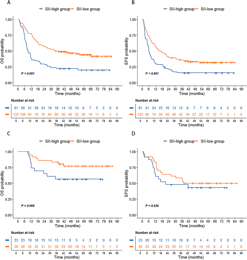 |
Figure 5 Kaplan–Meier curves of overall survival (OS) and event-free survival (EFS) according to different SII levels in AML patients without HSCT (A and B) and patients with HSCT (C and D). |
Collectively, these findings indicate that high SII is associated with poorer survival outcomes across multiple clinically relevant subgroups, particularly in patients with favorable or intermediate ELN risk, younger age, or those not receiving HSCT. In contrast, SII appears to have limited prognostic value in adverse-risk or post-transplant settings.
Univariate and Multivariate Analysis of OS
High SII levels were significantly associated with inferior OS compared to lower SII values. To further elucidate the prognostic value of SII, both univariate and multivariate Cox proportional hazards regression analyses were performed. As shown in Table 3, univariate analysis identified several adverse prognostic factors, including age (p<0.001), WBC count ≥100×109/L (p<0.001), intermediate/adverse ELN risk classification (p<0.001), and SII ≥43.3×10⁹/L (p<0.001). Conversely, receiving hematopoietic stem cell transplantation (HSCT) was associated with improved survival (p<0.001).
 |
Table 3 Univariate and Multivariate Analysis of Factors Correlating to Overall Survival |
In multivariate analysis, five variables remained independently associated with OS. These included: age (hazard ratio [HR]: 1.012; 95% confidence interval [CI]: 1.001–1.023; p=0.032), WBC ≥100×109/L (HR: 2.508; 95% CI: 1.629–3.861; p<0.001), intermediate/adverse ELN risk (HR: 1.786, 95% CI: 1.206–2.644, p=0.004), HSCT (HR: 0.320; 95% CI: 0.191–0.534; p<0.001), and SII ≥43.3×10⁹/L (HR: 1.781; 95% CI: 1.264–2.509; p=0.001). These findings underscore the independent prognostic significance of SII in predicting overall survival in patients with de novo non-M3 AML.
Univariate and Multivariate Analysis of EFS
Consistent with the OS findings, patients with high SII levels experienced significantly shorter EFS compared to those with low SII levels. In univariate analysis, several factors were significantly associated with reduced EFS, including age (p<0.001), WBC count ≥100×109/L (p=0.037), intermediate/adverse ELN risk classification (p=0.005), and SII ≥43.3×10⁹/L (p=0.001). Conversely, receiving hematopoietic stem cell transplantation (HSCT) was associated with improved survival (P < 0.001).
Multivariate analysis confirmed four variables as independent predictors of EFS: age (HR: 1.011; 95% CI: 1.002–1.021; p=0.019), WBC ≥100×109/L (HR: 1.613; 95% CI: 1.064–2.447; p=0.024), HSCT (HR: 0.525; 95% CI: 0.350–0.785; p=0.002), and SII ≥43.3×10⁹/L (HR: 1.483; 95% CI: 1.090–2.019; p=0.012) (Table 4). These results further highlight the prognostic significance of SII in predicting not only overall survival but also event-free survival in de novo non-M3 AML patients.
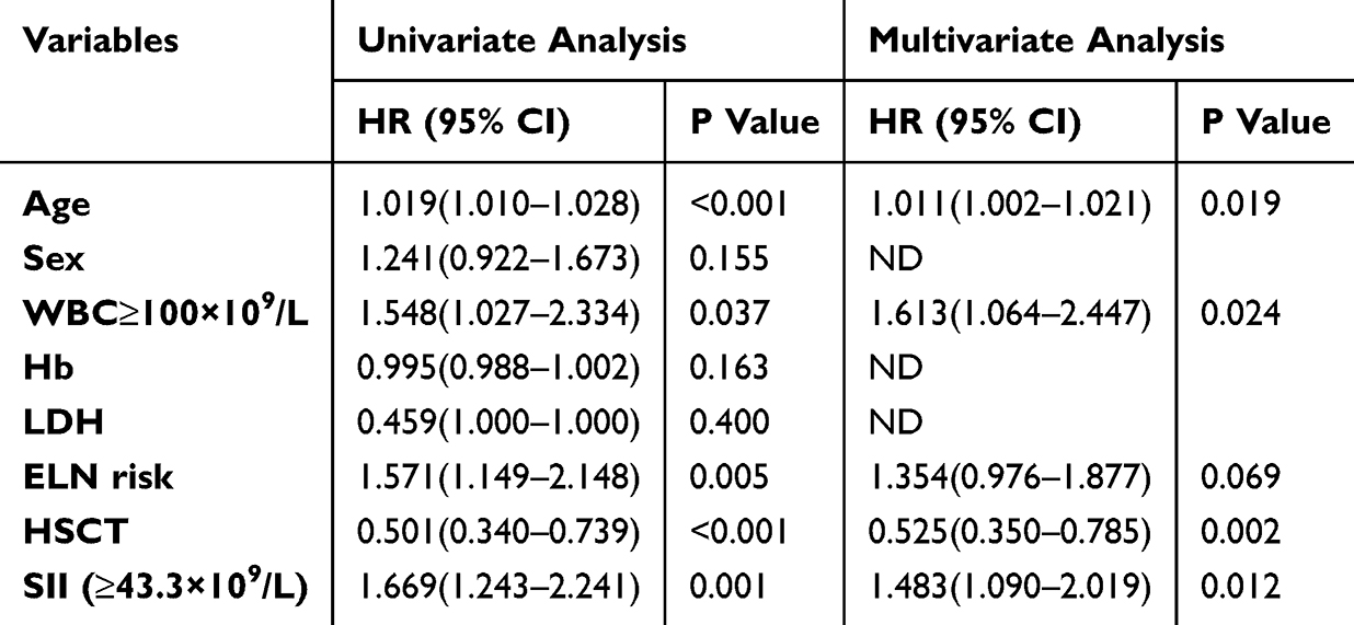 |
Table 4 Univariate and Multivariate Analysis of Factors Correlating to Event-Free Survival |
Development and Validation of a New Nomogram for Predicting Survival
We constructed a prognostic nomogram incorporating five statistically significant predictors derived from multivariate Cox regression to estimate OS. These factors include age, WBC of 100×109/L or higher, ELN risk classification, hematopoietic stem cell transplantation (HSCT), and the systemic immune-inflammation index (SII). Among these factors, HSCT emerged as the most significant contributor to the nomogram’s predictive capability, followed in importance by WBC levels, patient age, ELN risk, and SII (Figure 6). The calibration curves for the nomogram-predicted 1-, 2-, and 3-year survival probabilities indicated a strong linear relationship with the actual observed outcomes (Figure 7A–C). Furthermore, the concordance between predictions based on the established ELN risk stratification (favorable, intermediate, adverse) and the observed results was also evaluated (Figure 7D–F).
 |
Figure 6 Nomogram for predicting 1-, 2- and 3-year overall survival rates. A new nomogram for predicting OS was constructed by the four significant prognostic factors (age, WBC, ELN risk status, HSCT and SII) determined by the Cox regression analysis. |
 |
Figure 7 Performance of the prognostic model. Calibration curve showing predicted and actual 1-, 2- and 3-year survival probabilities (A–C). Calibration curves showed predicted and actual 1-, 2- and 3- year survival probabilities by the ELN risk stratification (favorable, intermediate, and adverse risk) (D–F). |
The C-index for this nomogram was recorded at 0.715 (95% CI 0.677–0.754), surpassing the C-index of the 2022 ELN risk stratification alone, which stood at 0.590 (95% CI 0.545–0.635). This indicates that the nomogram provided a better fit for this cohort. Time-dependent receiver operating characteristic (ROC) analysis further confirmed the nomogram’s predictive performance, with area under the curve (AUC) values of 0.781, 0.752, and 0.781 for 1-, 2-, and 3-year OS, respectively (Figure 8A). Decision curve analysis (DCA) compared the clinical utility and net benefit of the SII-integrated nomogram against the 2022 ELN risk subgrouping model and a model excluding SII. Across a range of threshold probabilities, the SII-integrated nomogram demonstrated higher net clinical benefit, supporting its superior prognostic value in guiding individualized treatment decisions (Figure 8B).
 |
Figure 8 Validation of the nomogram. The area under the ROC curve (AUC) showing the discriminative ability of the prognostic model (A). Decision curve analysis in prediction of de novo non-M3 AML (B). |
Discussion
Acute myeloid leukemia (AML) is a clonal hematopoietic malignancy characterized by uncontrolled proliferation, impaired differentiation, and apoptosis resistance of immature myeloid cells, leading to bone marrow failure and systemic disease manifestations.18 Current prognostic stratification and treatment strategies are largely guided by the 2022 European Leukemia Net (ELN) criteria, which rely on cytogenetic and molecular markers. However, in resource-limited settings, the widespread application of these advanced diagnostic tools is often constrained by cost, infrastructure, and turnaround time.19,20 This highlights the urgent need for simple, cost-effective, and readily accessible biomarkers that can supplement existing risk stratification systems. In this context, our study investigated the prognostic value of the systemic immune-inflammation index (SII)—a non-invasive and widely available hematologic marker—as a complementary tool in the management of AML.
In a cohort of 262 patients with de novo non-M3 AML, we identified elevated baseline SII as an independent predictor of both inferior OS and EFS. Multivariate Cox regression confirmed the prognostic significance of SII alongside established clinical factors such as age, WBC count ≥100×109/L, ELN risk category, and received of HSCT. These findings align with prior studies in solid tumors and hematologic malignancies. For instance, a cohort of 224 patients with DLBCL, which revealed that higher SII levels were linked to shorter OS and progression-free survival (PFS).13 Additionally, Li Chen et al discovered a significant association between SII levels and OS as well as disease-free survival (DFS) among 262 breast cancer patients, particularly in those who did not exhibit lymph vessel invasion.11 Furthermore, a meta-analysis aggregating data from 15 studies, encompassing 2438 cases, highlighted that elevated SII was predictive of responses to immune checkpoint inhibitors (ICIs) and linked to unfavorable survival outcomes in various cancers, although it did not specifically include data from AML patients.21
Subgroup analyses further supported the utility of SII in refining risk classification. Among patients with favorable ELN risk, younger age (<60 years), or without HSCT, high SII was significantly associated with worse OS and EFS. Notably, SII retained prognostic relevance even within traditionally low-risk groups, suggesting it could help identify patients who may benefit from intensified treatment or closer surveillance. To facilitate individualized risk prediction, we constructed a prognostic nomogram incorporating age, WBC count, ELN risk, HSCT status, and SII. This model demonstrated strong discriminatory capacity, with a C-index of 0.715, outperforming the ELN system alone (C-index: 0.590). Calibration curves confirmed high agreement between predicted and observed survival outcomes at 1, 2, and 3 years. Additionally, time-dependent ROC analyses showed favorable predictive performance (AUCs: 0.781, 0.752, and 0.781), and decision curve analysis indicated superior net clinical benefit compared to models lacking SII or relying solely on ELN risk categories. These findings affirm the added value of integrating SII into multivariable prognostic models for AML.
From a biological perspective, inflammation plays a well-recognized role in tumor progression and immune evasion. In AML, the leukemic microenvironment is shaped by a dysregulated balance of pro- and anti-inflammatory cytokines that influence hematopoietic stem cell (HSC) function and promote leukemic cell proliferation, survival, and resistance to therapy.22,23 Immune and inflammatory cells constitute key components of host defense mechanisms. Cell types detectable in peripheral circulation—including neutrophils, monocytes, lymphocytes, and platelets—may facilitate cancer invasion and metastasis.24,25 Neutrophils secrete a variety of pro-tumorigenic mediators, including IL-6, VEGF, MMPs, and FGF, facilitating angiogenesis and leukemic cell dissemination.26,27 Platelets have the ability to interact with tumor cells, offering them mechanical support during various stages of cancer development.28 Research conducted by Xu et al demonstrated that platelets are equipped with factors that promote growth and angiogenesis, aiding in the advancement of cancer. Additionally, they interact with immune cells, including natural killer cells and neutrophils, to help cancer cells evade detection by the immune system.29 Lymphocytes represent essential cellular components of the immune system, playing a pivotal role in activating robust antitumor responses,30 and reduced lymphocyte counts are often associated with impaired immune surveillance and poor prognosis.31–33 Thus, the SII—which integrates neutrophil, platelet, and lymphocyte counts—offers a composite index reflecting both inflammation and immune status. Its elevation likely represents an immunosuppressive, pro-inflammatory state that favors disease progression and treatment resistance.
The primary strength of this study lies in its novel identification of SII as a robust prognostic biomarker in non-M3 AML. Our nomogram integrates patient-specific and disease-related variables to provide accurate, individualized survival predictions. Rigorous validation using C-index, calibration plots, time-dependent ROC curves, and decision curve analysis further supports its clinical applicability. However, several limitations should be acknowledged. First, the study did not account for co-occurring gene mutations beyond those included in the ELN classification, potentially overlooking synergistic prognostic effects. Second, variability in treatment regimens may have introduced confounding factors. Third, as a single-center retrospective study, our findings require external validation in larger, multicenter prospective cohorts to confirm generalizability.
Conclusion
In conclusion, the SII is an accessible biomarker with independent prognostic value in AML. A nomogram incorporating SII and established prognostic factors enables individualized survival prediction and may inform risk-adapted treatment, particularly where access to advanced molecular diagnostics is limited. Prospective studies are warranted to confirm its clinical utility and explore its potential in guiding therapy selection.
Acknowledgments
Huijie Nan, Peiyao Yang and Lijie Wang share first authorship for this study. Mingyue Shi and Yanliang Bai share last authorship for this study. We thank the patients and their families who participated in our study as well as all the investigators who collected and provided the research data.
Funding
This study was funded by Department of Science and Technology of Henan Province (No. 222102310101, H20240205, 212102310205 and LHGJ20230023).
Disclosure
The authors report no conflicts of interest in this work.
References
1. Azenkot T, Jonas BA. Clinical impact of measurable residual disease in acute myeloid leukemia. Cancers. 2022;14(15):3634. doi:10.3390/cancers14153634
2. Demircan MB, Mgbecheta PC, Kresinsky A, et al. Combined activity of the redox-modulating compound setanaxib (GKT137831) with cytotoxic agents in the killing of acute myeloid leukemia cells. Antioxidants. 2022;11(3):513. doi:10.3390/antiox11030513
3. Tomaz V, Griesi-Oliveira K, Puga RD, et al. Molecular characterization of a first-in-human clinical response to nimesulide in acute myeloid leukemia. Front Oncol. 2022;12:874168. doi:10.3389/fonc.2022.874168
4. Döhner H, Wei AH, Appelbaum FR, et al. Diagnosis and management of AML in adults: 2022 recommendations from an international expert panel on behalf of the ELN. Blood. 2022;140(12):1345–1377. doi:10.1182/blood.2022016867
5. Qian L, Lai X, Gu B, Sun X. An immune-related gene signature for predicting neoadjuvant chemoradiotherapy efficacy in rectal carcinoma. Front Immunol. 2022;13:784479. doi:10.3389/fimmu.2022.784479
6. Nishida A, Andoh A. The role of inflammation in cancer: mechanisms of tumor initiation, progression, and metastasis. Cells. 2025;14(7):488. doi:10.3390/cells14070488
7. Lattanzi R, Severini C, Miele R. Prokineticin 2 in cancer-related inflammation. Cancer Lett. 2022;546:215838. doi:10.1016/j.canlet.2022.215838
8. Salazar-Valdivia FE, Valdez-Cornejo VA, Ulloque-Badaracco JR, et al. Systemic immune-inflammation index and mortality in testicular cancer: a systematic review and meta-analysis. Diagnostics. 2023;13(5):843. doi:10.3390/diagnostics13050843
9. Islam MM, Satici MO, Eroglu SE. Unraveling the clinical significance and prognostic value of the neutrophil-to-lymphocyte ratio, platelet-to-lymphocyte ratio, systemic immune-inflammation index, systemic inflammation response index, and delta neutrophil index: an extensive literature review. Turk J Emerg Med. 2024;24(1):8–19. doi:10.4103/tjem.tjem_198_23
10. Jomrich G, Gruber ES, Winkler D, et al. Systemic Immune-Inflammation Index (SII) predicts poor survival in pancreatic cancer patients undergoing resection. J Gastrointest Surg. 2020;24(3):610–618. doi:10.1007/s11605-019-04187-z
11. Chen L, Kong X, Wang Z, Wang X, Fang Y, Wang J. Pre-treatment systemic immune-inflammation index is a useful prognostic indicator in patients with breast cancer undergoing neoadjuvant chemotherapy. J Cell Mol Med. 2020;24(5):2993–3021. doi:10.1111/jcmm.14934
12. Yatabe S, Eto K, Haruki K, et al. Signification of systemic immune-inflammation index for prediction of prognosis after resecting in patients with colorectal cancer. Int J Colorectal Dis. 2020;35(8):1549–1555. doi:10.1007/s00384-020-03615-w
13. Wang Z, Zhang J, Luo S, Zhao X. Prognostic significance of systemic immune-inflammation index in patients with diffuse large B-cell lymphoma. Front Oncol. 2021;11:655259. doi:10.3389/fonc.2021.655259
14. Mikulski D, Robak P, Mirocha G, et al. An elevated systemic inflammation index is related to an inferior response to pomalidomide and dexamethasone treatment in patients with relapsed and refractory multiple myeloma. Contemp Oncol. 2023;27(3):139–146. doi:10.5114/wo.2023.133506
15. Krecak I, Lekovic D, Arsenovic I, et al. Systemic inflammatory index in polycythemia vera and its prognostic implications. J Clin Med. 2024;13(15):4459. doi:10.3390/jcm13154459
16. Khoury JD, Solary E, Abla O, et al. The 5th edition of the World Health Organization Classification of Haematolymphoid Tumours: myeloid and Histiocytic/Dendritic Neoplasms. Leukemia. 2022;36(7):1703–1719. doi:10.1038/s41375-022-01613-1
17. Yu J, Li Y, Li T, et al. Gene mutational analysis by NGS and its clinical significance in patients with myelodysplastic syndrome and acute myeloid leukemia. Exp Hematol Oncol. 2020;9:2. doi:10.1186/s40164-019-0158-5
18. Narayanan D, Weinberg OK. How I investigate acute myeloid leukemia. Int J Lab Hematol. 2020;42(1):3–15. doi:10.1111/ijlh.13135
19. Girard R, Li Y, Stadnik A, et al. A roadmap for developing plasma diagnostic and prognostic biomarkers of cerebral cavernous angioma with symptomatic hemorrhage (CASH). Neurosurgery. 2021;88(3):686–697. doi:10.1093/neuros/nyaa478
20. Voronova V, Glybochko P, Svistunov A, et al. Diagnostic value of combinatorial markers in colorectal carcinoma. Front Oncol. 2020;10:832. doi:10.3389/fonc.2020.00832
21. Kou J, Huang J, Li J, Wu Z, Ni L. Systemic immune-inflammation index predicts prognosis and responsiveness to immunotherapy in cancer patients: a systematic review and meta‑analysis. Clin Exp Med. 2023;23(7):3895–3905. doi:10.1007/s10238-023-01035-y
22. Wuz CF, Wu CY, Chiou RY, et al. The anti-cancer effects of a zotarolimus and 5-fluorouracil combination treatment on a549 cell-derived tumors in BALB/c nude mice. Int J Mol Sci. 2021;22(9). doi:10.3390/ijms22094562
23. Yun JP, Choi EK, Han KD, et al. Risk of atrial fibrillation according to cancer type: a nationwide population-based study. JACC CardioOncol. 2021;3(2):221–232. doi:10.1016/j.jaccao.2021.03.006
24. Jiang S, Wang S, Wang Q, et al. Systemic Inflammation Response Index (SIRI) independently predicts survival in advanced lung adenocarcinoma patients treated with first-generation EGFR-TKIs. Cancer Manag Res. 2021;13:1315–1322. doi:10.2147/cmar.S287897
25. He K, Si L, Pan X, et al. Preoperative Systemic Immune-Inflammation Index (SII) as a superior predictor of long-term survival outcome in patients with stage i-ii gastric cancer after radical surgery. Front Oncol. 2022;12:829689. doi:10.3389/fonc.2022.829689
26. Mao Z, Zhang J, Shi Y, et al. CXCL5 promotes gastric cancer metastasis by inducing epithelial-mesenchymal transition and activating neutrophils. Oncogenesis. 2020;9(7):63. doi:10.1038/s41389-020-00249-z
27. Wu L, Awaji M, Saxena S, Varney ML, Sharma B, Singh RK. IL-17-CXC chemokine receptor 2 axis facilitates breast cancer progression by up-regulating neutrophil recruitment. Am J Pathol. 2020;190(1):222–233. doi:10.1016/j.ajpath.2019.09.016
28. Benbrook DM, Hocker JRS, Moxley KM, Hanas JS. Sera protein signatures of endometrial cancer lymph node metastases. Int J Mol Sci. 2022;23(6):3277. doi:10.3390/ijms23063277
29. Xu L, Li X, Li X, et al. RNA profiling of blood platelets noninvasively differentiates colorectal cancer from healthy donors and noncancerous intestinal diseases: a retrospective cohort study. Genome Med. 2022;14(1):26. doi:10.1186/s13073-022-01033-x
30. Zhang SL, Han B, Mao YQ, et al. Lacticaseibacillus paracasei sh2020 induced antitumor immunity and synergized with anti-programmed cell death 1 to reduce tumor burden in mice. Gut Microbes. 2022;14(1):2046246. doi:10.1080/19490976.2022.2046246
31. Zhang X, Wang X, Li W, Dang C, Diao D. Effectiveness of managing suspected metastasis using plasma D-dimer testing in gastric cancer patients. Am J Cancer Res. 2022;12(3):1169–1178.
32. Gu X, Wei S, Chen B, Zhang W, Zheng S. Development and validation of a combined ferroptosis- and pyroptosis-related gene signatures for the prediction of clinical outcomes in lung adenocarcinoma. Am J Cancer Res. 2022;12(8):3870–3891.
33. Sheng H, Huang Y, Xiao Y, et al. ATR inhibitor AZD6738 enhances the antitumor activity of radiotherapy and immune checkpoint inhibitors by potentiating the tumor immune microenvironment in hepatocellular carcinoma. J Immunother Cancer. 2020;8(1):e000340. doi:10.1136/jitc-2019-000340
 © 2025 The Author(s). This work is published and licensed by Dove Medical Press Limited. The
full terms of this license are available at https://www.dovepress.com/terms
and incorporate the Creative Commons Attribution
- Non Commercial (unported, 4.0) License.
By accessing the work you hereby accept the Terms. Non-commercial uses of the work are permitted
without any further permission from Dove Medical Press Limited, provided the work is properly
attributed. For permission for commercial use of this work, please see paragraphs 4.2 and 5 of our Terms.
© 2025 The Author(s). This work is published and licensed by Dove Medical Press Limited. The
full terms of this license are available at https://www.dovepress.com/terms
and incorporate the Creative Commons Attribution
- Non Commercial (unported, 4.0) License.
By accessing the work you hereby accept the Terms. Non-commercial uses of the work are permitted
without any further permission from Dove Medical Press Limited, provided the work is properly
attributed. For permission for commercial use of this work, please see paragraphs 4.2 and 5 of our Terms.