Back to Journals » Cancer Management and Research » Volume 11
Prevention effect of low-temperature atomization inhalation for radiation induced oral mucositis in patients with head and neck cancer undergoing radiotherapy
Authors Bai XH, Chen ZM, Ma LH, He Z, Li G
Received 27 February 2019
Accepted for publication 18 April 2019
Published 9 May 2019 Volume 2019:11 Pages 4327—4333
DOI https://doi.org/10.2147/CMAR.S206921
Checked for plagiarism Yes
Review by Single anonymous peer review
Peer reviewer comments 2
Editor who approved publication: Dr Antonella D'Anneo
Xing-Hua Bai, Zi-Min Chen, Liang-Hua Ma, Zheng He, Guang Li
Department of Radiation Oncology, The First Affiliated Hospital of China Medical University, Shenyang, People’s Republic of China
Purpose: To investigate the prevention effect of low-temperature atomization inhalation for radiation induced oral mucositis (OM) in patients with head and neck cancer (HNC) undergoing radiotherapy.
Patients and methods: A total of 68 patients with HNC (including nasopharyngeal cancer) undergoing radiotherapy were divided into an intervention group (33 cases) and a control group (35 cases). During radiotherapy, the intervention group received low-temperature (between 4°C and 8°C) atomization inhalation; while the control group received normal temperature (between 18°C and 24°C) atomization inhalation. Atomization inhalation was performed twice a day, 20 minutes per time, using distilled water. The incidence and severity of OM was evaluated every week during radiotherapy. The comparation was made between the two groups.
Results: The two groups were comparable among age, sex, Eastern Cooperative Oncology group (ECOG) score, body mass index (BMI) before radiotherapy, BMI loss during radiotherapy, original tumor site, pathological type, TNM stage, and mean oral cavity irradiated dose. There was a significant difference in the incidence of OM between the two groups (P<0.05). There were fewer patients with severe OM in the intervention group compared to the control group (P<0.05). The onset time of OM in the intervention group was delayed by about 4 days compared to that in the control group (P<0.05). Low-temperature atomization inhalation helped to avoid radiotherapy interruption in the intervention group. No patient in the intervention group suffered any adverse reaction for low-temperature atomization inhalation treatment.
Conclusions: Low-temperature atomization inhalation can reduce the incidence and severity of OM, and slow down the progression process of it. It can be used as a new prevention method during radiotherapy, and should be promoted in clinical practice.
Keywords: low-temperature atomization, head and neck cancer, radiotherapy, radiation induced oral mucositis
Introduction
As we all know, radiotherapy is one of the most important treatment modalities for patients with head and neck cancer (HNC). An acute radiation induced oral mucositis (OM) is one of the most common adverse reactions during radiotherapy for patients with HNC, and can be characterized by dry mouth, pain of mouth and pharynx, oral inflammation, and pseudomembrane formation.1,2 Oral mucous membranes are easy to be broken, to bleed, and to form ulceration during radiotherapy. This will impair the ability to eat, influence the patients seriously, and even lead to the interruption of radiotherapy.3 Therefore, finding effective prevention and treatment methods for radiation induced OM become great concerns for clinical practice. It is well known that cryogenic treatment plays an important role in dealing with OM.4 Low temperature can inhibit the inflammatory response, reduce mucosal edema, and decrease pain for the patients.5 An in vitro study shows that tissue-engineered oral mucosal models incubated at 20°C can increase cell viability and reduce interleukin (IL)-6 and tumor necrosis factor (TNF)-ɑ production compared to the models treated with 5-fluorouracil (5-Fu) incubated at 35°C.6 However, another study can’t detect any difference in cytokine IL-6 level and find significant systolic pressure increase after oral cooling in healthy volunteers.7 Also, quadruple fluid aerosol inhalation can be used for the radiation induced OM in patients undergoing radiotherapy via inhibiting the secretion of EGF in the saliva.8 Hence, we considered to make a combination of cryogenic treatment and fluid aerosol inhalation called low-temperature atomization inhalation as a new method to enhance the prevention effect. Our hypothesis of this study was that the combination of cryogenic treatment and fluid aerosol inhalation would reduce the injury of oral mucosa caused by radiotherapy dramatically through inducing vasoconstriction and then decreasing the secretion of IL-6, TNF-ɑ, and EGF. We enrolled 68 patients with HNC undergoing radiotherapy to assess the prevention effect of low-temperature atomization inhalation for radiation-induced OM.
Patients and methods
Patient characteristics
The study protocol was approved by the Ethics Committee at the Institutional Review Board in the First Hospital of China Medical University, in accordance with the Declaration of Helsinki. A total of 68 patients with HNC (including nasopharyngeal cancer) were enrolled in this study. All patients provided written informed consent for attending this study. These patients received continuous radiotherapy at our hospital between March 2015 and December 2015. They were divided into two groups. The intervention group included 33 cases, and the control group included 35 cases. The inclusion criteria consisted of: 1) pathological diagnosis of HNC with Karnofsky Performance Status (KPS) 70 points or higher; 2) patients were required to quit smoking and drinking from the beginning of radiotherapy; 3) patients could tolerate low-temperature air without airway hyper-responsiveness; 4) no pre-existing inflammation in the oral mucosa; 5) no induction and/or concurrent chemotherapy; and 6) patients were able to cooperate with the treatment as required.
Radiotherapy treatment
All patients received definitive simultaneous integrated boost intensity-modulated radiotherapy (SIB – IMRT): gross tumor target (GTV) 2.12 Gy/fraction, and high-risk clinical target 1.82 Gy/fraction, with a total fraction of 33; low-risk clinical target 1.82 Gy/fraction, with a total fraction of 28. The radiotherapy was delivered five times per week from Monday to Friday.
Procedure for atomization inhalation
The standard operating procedure for atomization inhalation included steps as follows: 1) Make sure the connecting parts were closely connected, and all the switches were on the apparatus; 2) Add 250 mL distilled water into the bottom of the tank to form a 3 cm liquid level immersing the perforated film of the spray; 3) After inspecting for leakage, add 30–50 mL of the distilled water to the atomization tank, place it back onto the water tank, and cover tightly; 4) Ensure the patients were in a comfortable position, and instruct them to breathe deeply; 5) Increase the intensity of fog gradually, from medium to heavy, avoiding respiratory discomfort of the patients stimulated by the instant cold fog; and 6) Keep delivering the treatment for 20 minutes.
Evaluation of OM
According to the Common Terminology Criteria for Adverse Events (CTCAE) version 4.03 combining with the Radiation Therapy Oncology Group (RTOG) criteria, we evaluated the incidence and severity of radiation induced OM every week during the course of radiotherapy until completion. The severity of OM was classified to five grades via CTCAE: Grade 1: Asymptomatic or mild symptoms; intervention not indicated. Grade 2: Moderate pain; not interfere with oral intake; modified diet indicated. Grade 3: Severe pain; interfering with oral intake. Grade 4: Life-threatening consequences; urgent intervention indicated. Grade 5: Death.9 It was also classified to five grades via RTOG criteria: Grade 0: Basically no change. Grade 1: Irritation/may experience mild pain not requiring analgesic. Grade 2: Patchy mucositis that may produce an inflammatory serosanguinous discharge/may experience moderate pain requiring analgesia. Grade 3: Confluent fibrinous mucositis/may include severe pain requiring narcotic. Grade 4: Ulcer, hemorrhage, or necrosis.10 Patients with OM of Grade 1 (asymptomatic) (CTCAE) and/or Grade 0 (RTOG criteria) were defined as a 0 group. Patients with OM of Grade 1 (mild symptoms) to Grade 2 (CTCAE) and/or Grade 1 to Grade 2 (RTOG criteria) were defined as a mild group. Patients with OM of Grade 3 to Grade 5 (CTCAE) and/or Grade 3 to Grade 4 (RTOG criteria) were defined as a severe group.
Atomization inhalation and treatment for OM
The intervention group received distilled water atomization inhalation at a low temperature (the distilled water was kept in the medical cryogenic refrigerator at a temperature between 4°C and 8°C), whereas the control group received distilled water atomization inhalation at room temperature (the distilled water was between 18°C and 24°C). Atomization inhalation was performed twice a day, 20 minutes per time, from the beginning of radiotherapy until completion. All patients were given regular oral care before and after radiotherapy and requested to rinse the mouth after eating. There was no additional treatment to patients in the 0 group. Patients in the mild group were treated by the solution containing 500 mL physiological saline, 10 mg dexamethasone, 160,000 U gentamicin, and 10 mL lidocaine 3 times/day. Patients in the serve group might be treated by oxycontin and parenteral nutrition.
Evaluation of dryness of oral cavity
Dryness of the oral cavity was evaluated via RTOG acute radiation morbidity scoring criteria to the salivary gland at the completion of radiotherapy.11 Grade 0: No change over baseline. Grade 1: Mild mouth dryness/slightly thickened saliva/may have slightly altered taste such as metallic taste/these changes were not reflected in alteration in baseline feeding behavior, such as increased use of liquids with meals. Grade 2: Moderate-to-complete dryness/thick, sticky saliva/markedly altered taste.
Statistical analysis
Data analysis was conducted with SPSS, version 19.0. The proportions of patients in different groups were compared using Pearson’s chi-square test (or Fisher’s exact test, when appropriate). Mean resources were compared using independent-samples t-test. A P-value of <0.05 (two-tailed) was considered to be statistically significant.
Results
Patient characteristics
As shown in Table 1, there was no significant difference between the intervention group and the control group among age, sex, Eastern Cooperative Oncology group (ECOG) score, body mass index (BMI) before radiotherapy, BMI loss during radiotherapy, original tumor site, pathological type, TNM stage, or mean oral cavity irradiated dose.
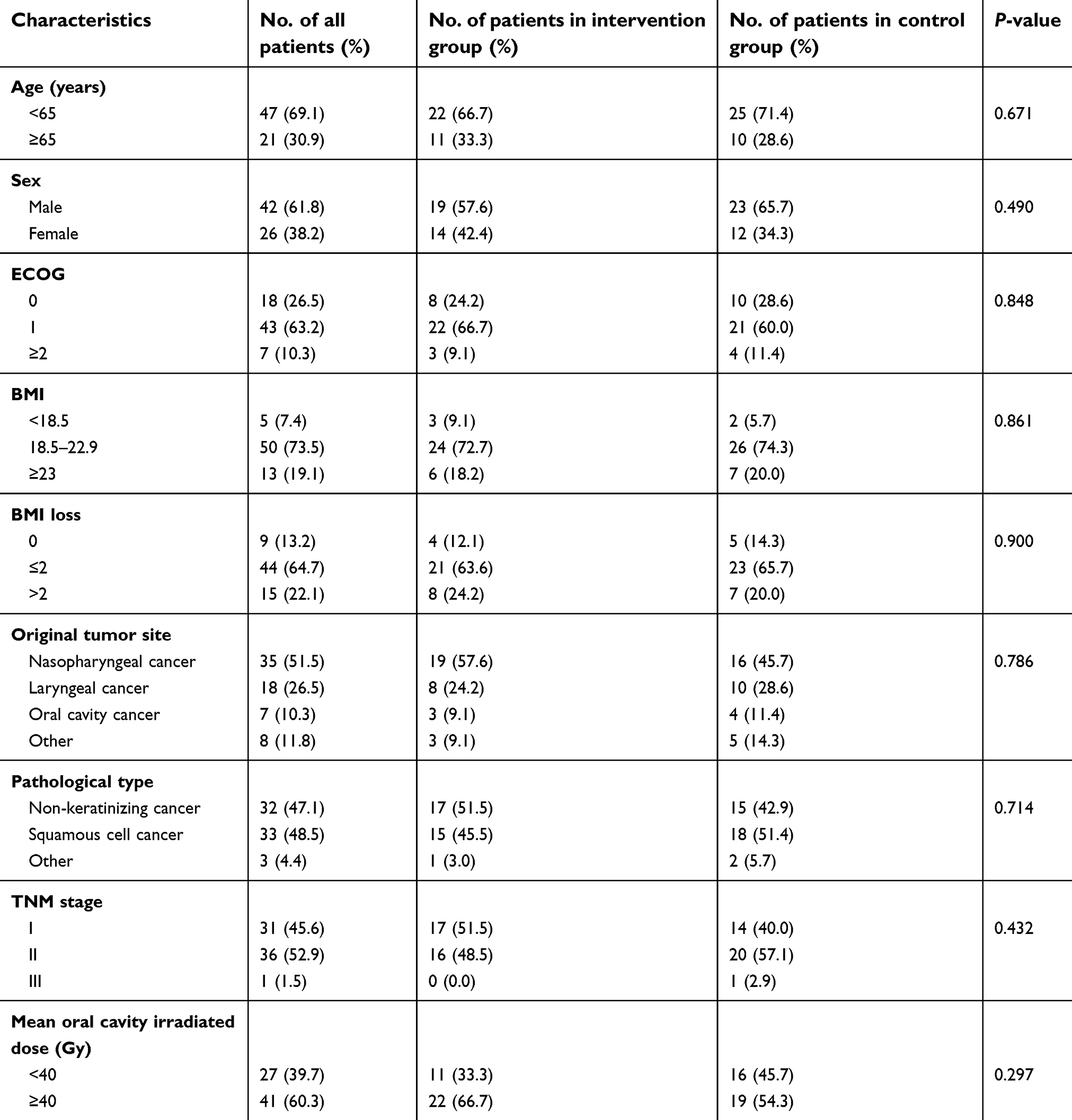 | Table 1 Patient characteristics and comparison between the two groups |
Incidence and severity of OM
As shown in Table 2, there was a significant difference in the incidence of OM between the two groups when the total radiation dose reached 30 Gy (42.4% in the intervention group vs 68.6% in the control group, P<0.05), as well as when the radiotherapy was completed (78.8% in the intervention group vs 91.4% in the control group, P<0.05). There were fewer patients with severe OM in the intervention group compared to the control group both at the 30 Gy radiation dose and at the completion of radiotherapy (6.0% and 18.2% in the intervention group vs 22.9% and 51.4% in the control group respectively, P<0.05). In the intervention group, all patients achieved the radiotherapy as planned without any interruption, while two patients had to interrupt the radiotherapy due to the severe OM in the control group.
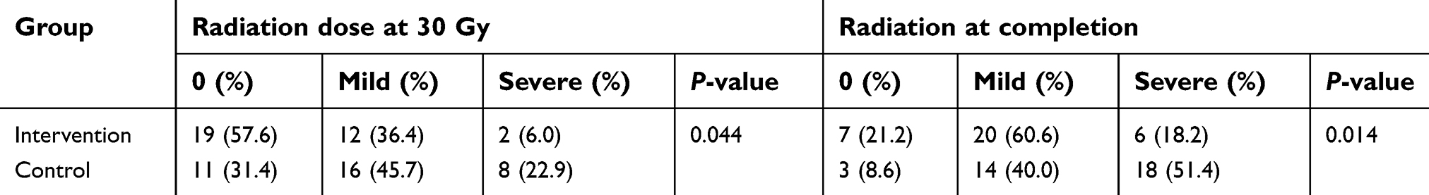 | Table 2 Incidence and severity of OM between the two groups |
The onset time of OM
As shown in Table 3, the onset time of OM in the intervention group was delayed about 4 days compared to that in the control group (16.55 days in the intervention group vs 12.63 days in the control group, P<0.05). Furthermore, no patient in the intervention group suffered any adverse reaction for low-temperature atomization inhalation treatment.
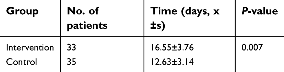 | Table 3 The onset time of OM between the two groups |
Incidence and severity of dryness of oral cavity
As shown in Table 4, patients suffering from Grade 1 and Grade 2 dryness of oral cavity made up 75.8% and 15.2% in the intervention group and 68.6% and 20.0% in the control group, respectively. There was no significant difference on the incidence and severity of dryness of the oral cavity between the two groups (P>0.05).
 | Table 4 Incidence and severity of dryness of the oral cavity between the two groups |
Discussion
Oral mucositis refers to inflammation of the oral mucosa resulting from cancer therapy typically manifesting as atrophy, swelling, erythema, and ulceration.12 It can be induced by either chemotherapy or radiotherapy. There are different characteristics of OM from different causes. Chemotherapy induced mucositis often appears very quickly after drug administration and peaks within 2 weeks, while radiation-induced mucositis has a more gradual clinical course. Usually, when the radiation doses reach 15 Gy, radiation-induced mucositis begins to appear and becomes severe gradually. It will last for weeks or even months.12 In clinical practice, concurrent chemo-radiotherapy will increase the incidence and severity of OM in HNC patients. Vera-Llonch et al13 found that 83% of HNC patients developed OM undergoing radiotherapy, especially in patients with nasopharyngeal or oropharyngeal tumors, and those who received total doses over 50 Gy or with chemotherapy. So in this study, most patients we enrolled were with AJCC (2010) stage I and II who underwent definitive radiotherapy and didn’t need to do induction chemotherapy and/or concurrent chemoradiotherapy. Only one patient with stage III who refused chemotherapy was also included. Because we were with the only interest to radiation induced OM.
Besides chemotherapy, some other factors were also associated with the incidence of OM, including total radiation doses and volume, nutritional status, changes of body weight, smoking, and drinking.14 In this study, we excluded patients who underwent surgery plus postoperative radiotherapy. Because the total radiation doses were not equal between postoperative radiotherapy and definitive radiotherapy. Therefore, all 68 patients in our study underwent definitive radiotherapy with 69.96 Gy to the gross tumor target. The volume of irradiated tissue varied from different types of cancer, as well as different patients. Orlandi et al15 found that mean oral cavity irradiated dose was the most important factor affecting OM. Hence, we were trying to use the mean oral cavity irradiated dose instead of the volume of irradiated tissue. There was no significant difference between the intervention group and the control group. According to Gu et al,16 we applied BMI before radiotherapy to represent the nutrition status of patients, and BMI loss to represent changes of body weight during radiotherapy. We found that both BMI before radiotherapy and BMI loss during radiotherapy were with no significant difference between the two groups. The inclusion criteria of this study included that patients were required to quit smoking and drinking from the beginning of radiotherapy. So we didn’t analyze the impact of smoking and drinking. Furthermore, the two groups were comparable among age, sex, ECOG score, original tumor site, pathological type, and TNM stage. In this case, the different incidence of OM between the two groups should come from the prevention effect of low-temperature atomization inhalation.
In another study, Elting et al17 found that OM occurred in 91% of 204 HNC patients treated by radiotherapy with or without chemotherapy, which would lead an incremental cost of $1,700–$6,000 in the US. So we needed to find preventive strategies to reduce the excess cost.17 Therefore, we developed this study to assess the prevention effect of a new combination of cryogenic treatment and fluid aerosol inhalation. Fortunately, we found significant differences in the incidence of OM between the two groups: 42.4% in the intervention group vs 68.6% in the control group when the radiation doses reached 30 Gy, and 78.8% in the intervention group vs 91.4% in the control group when the radiotherapy was completed. Moreover, besides the incidence, the severity of OM was obviously decreased. The intervention group had less severe OM both at the 30 Gy radiation dose and at the completion of radiotherapy. All these results inferred that our new method of low-temperature atomization inhalation manifested a good prevention effect for radiation-induced OM.
Until now, although the incidence of OM was very high, there was no general consensus on the prevention and treatment of it. Cryogenic treatment, involving the placement of ice chips in the mouth, was recommended to prevent OM in patients receiving bolus 5-Fu and high-dose melphalan.18 The mechanism might be the vasoconstriction induced by the low temperature ice chips in the mouth. However, some patients felt a physically uncomfortable sensation while holding ice in the mouth for 30 minutes or longer.18 Therefore, we modified cryogenic treatment to low-temperature atomization inhalation, which would decrease the uncomfortable sensation significantly. The patients in the intervention group accepted low-temperature atomization inhalation comfortably, and there was no adverse reaction to it. This means that low-temperature atomization inhalation is an acceptable and safe treatment method.
As another option, low-level laser therapy (LLLT) was recommended for the prevention of OM in adult patients receiving hematopoietic stem cell transplantation conditioned with high-dose chemotherapy in the condition of wavelength at 650 nm, power of 40 mW, and each square centimeter treated with the required time to a tissue energy dose of 2 J/cm2 (2 s/point), and for OM in patients with HNC undergoing radiotherapy, without concomitant chemotherapy in the condition of wavelength around 632.8 nm.19 Gautam et al20 also found that LLLT was an effective treatment method to reduce the incidence of severe OM in patients with HNC undergoing concurrent chemoradiotherapy. However, LLLT required expensive equipment and specialized training. So it was difficult to conduct.19 Nevertheless, comparing to LLLT, low-temperature atomization inhalation was very cheap and easy to operate for clinical applications, associated with high patient compliance. It could reduce the incidence and severity of OM and relieve pain for patients. Therefore, low-temperature atomization inhalation should be promoted in clinical practice.
The progression process of radiation-induced OM included five overlapping stages: initiation, up-regulation, amplification, ulceration, and healing. In the initiation stage, many cytokines, including IL-6 and TNF, were secreted to initiate the damage.21 Low temperature could reduce the blood flow and decrease the secretion of IL-6 and TNF-ɑ.6 So the progression process in patients receiving low-temperature atomization inhalation was slowed down. In this study, we found that the onset time for OM in the intervention group was about a 4 days delay than that in the control group. This also manifested the prevention effect of low-temperature atomization inhalation. Although we didn’t test the change of cytokines in saliva, we thought low-temperature atomization inhalation must decrease the injury of oral mucosal cells through inducing vasoconstriction and then decreasing the secretion of IL-6, TNF-ɑ, and EGF, in accordance with the references.6,8
Dryness of the oral cavity is usually accompanied with OM during radiotherapy, mainly due to the irradiation to the salivary glands. In this study, we also included the incidence and severity of dryness of the oral cavity at the completion of radiotherapy as another index. The result showed that, at the end of radiotherapy, the incidence and severity were similar between the two groups (P>0.05). This meant that low-temperature atomization inhalation had no obvious affection on the dryness of the oral cavity.
Receiving radiotherapy as planned without any interruption was very important for a good prognosis in patients with HNC. Interrupting the radiotherapy and prolonging the total treatment time were associated with a poor prognosis. In a study concerning patients with nasopharyngeal cancer undergoing concurrent chemo-radiotherapy, Yao et al22 found that patients with an interruption over 5 days during radiotherapy would have a significantly lower rate of local relapse-free survival compared to those with an interruption of less than 5 days (83% vs 97%). Therefore, they recommended to pay special attention to radiotherapy interruption. In our study, two patients suffered from severe OM interrupted radiotherapy in the control group, while all patients in the intervention group achieved radiotherapy as planned without any interruption. The interruption of radiotherapy was associated with great damage to the patients’ physical and psychological statement, and indicates that the radiotherapy can’t administer smoothly. This confirmed that low-temperature atomization inhalation was helpful to administer radiotherapy as planned, and might be associated with good treatment results.
In summary, our results confirmed that low-temperature atomization inhalation was a simple and effective method for the prevention of radiation-induced OM without any adverse reaction for patients with HNC undergoing radiotherapy.
Conclusions
Low-temperature atomization inhalation can reduce the incidence and severity of OM, and slow down the progression process. It can be used as a new prevention method during radiotherapy and should be promoted in clinical practice.
Disclosure
The authors report no conflicts of interest in this work.
References
1. Trotti A, Bellm LA, Epstein JB, et al. Mucositis incidence, severity and associated outcomes in patients with head and neck cancer receiving radiotherapy with or without chemotherapy: a systematic literature review. Radiother Oncol. 2003;66(3):253–262.
2. Feller L, Essop R, Wood NH, et al. Chemotherapy- and radiotherapy-induced oral mucositis: pathobiology, epidemiology and management. SADJ. 2010;65(8):372–374.
3. Dörr W, Hamilton CS, Boyd T, Reed B, Denham JW. Radiation-induced changes in cellularity and proliferation in human oral mucosa. Int J Radiat Oncol Biol Phys. 2002;52(4):911–917.
4. Wodzinski A. Potential benefits of oral cryotherapy for chemotherapy-induced mucositis. Clin J Oncol Nurs. 2016;20(5):462–465. doi:10.1188/16.CJON.462-465
5. Idayu Mat Nawi R, Lei Chui P, Wan Ishak WZ, Hsien Chan CM. Oral cryotherapy: prevention of oral mucositis and pain among patients with colorectal cancer undergoing chemotherapy. Clin J Oncol Nurs. 2018;22(5):555–560. doi:10.1188/18.CJON.555-560
6. Walladbegi J, Smith SA, Grayson AK, Murdoch C, Jontell M, Colley HE. Cooling of the oral mucosa to prevent adverse effects of chemotherapeutic agents: an in vitro study. J Oral Pathol Med. 2018;47(5):477–483. doi:10.1111/jop.12696
7. Svanberg A, Ohrn K, Broström H, Birgegård G. The effect of cryotherapy on oral mucosa: a study in healthy volunteers. Med Oncol. 2012;29(5):3587–3591. doi:10.1007/s12032-012-0230-z
8. Chen J. [Effect of quadruple fluid aerosol inhalation on oral mucositis induced by radiotherapy in nasopharyngeal carcinoma and its mechanism]. Zhong Nan Da Xue Xue Bao Yi Xue Ban. 2007;32(3):527–530.
9.
10. Tao Z, Gao J, Qian L, et al. Factors associated with acute oral mucosal reaction induced by radiotherapy in head and neck squamous cell carcinoma: a retrospective single-center experience. Medicine (Baltimore). 2017;96(50):e8446. doi:10.1097/MD.0000000000008446
11. Cox JD, Stetz J, Pajak TF. Toxicity criteria of the Radiation Therapy Oncology Group (RTOG) and the European Organization for Research and Treatment of Cancer (EORTC). Int J Radiat Oncol Biol Phys. 1995;31(5):1341–1346. doi:10.1016/0360-3016(95)00060-C
12. Raber-Durlacher JE, Elad S, Barasch A. Oral mucositis. Oral Oncology. 2010;46(6):452–456. doi:10.1016/j.oraloncology.2010.07.001
13. Vera-Llonch M, Oster G, Hagiwara M, Sonis S. Oral mucositis in patients undergoing radiation treatment for head and neck carcinoma. Cancer. 2006;106(2):329–336. doi:10.1002/cncr.21622
14. Radvansky LJ, Pace MB, Siddiqui A. Prevention and management of radiation-induced dermatitis, mucositis, and xerostomia. Am J Health Syst Pharm. 2013;70(12):1025–1032. doi:10.2146/ajhp120467
15. Orlandi E, Iacovelli NA, Rancati T, et al. Multivariable model for predicting acute oral mucositis during combined IMRT and chemotherapy for locally advanced nasopharyngeal cancer patients. Oral Oncol. 2018;86:266–272. doi:10.1016/j.oraloncology.2018.10.006
16. Gu W-S, Fang W-Z, Liu C-Y, et al. Prognostic significance of combined pretreatment body mass index (BMI) and BMI loss in patients with esophageal cancer. Cancer Management and Research. 2019;11:3009–3020. doi:10.2147/CMAR
17. Elting LS, Cooksley CD, Chambers MS, Garden AS. Risk, outcomes, and costs of radiation-induced oral mucositis among patients with head-and-neck malignancies. Int J Radiat Oncol Biol Phys. 2007;68(4):1110–1120. doi:10.1016/j.ijrobp.2007.01.053
18. Peterson DE, Ohrn K, Bowen J, et al. Systematic review of oral cryotherapy for management of oral mucositis caused by cancer therapy. Support Care Cancer. 2013;21(1):327–332. doi:10.1007/s00520-012-1562-0
19. Migliorati C, Hewson I, Lalla RV, et al. Systematic review of laser and other light therapy for the management of oral mucositis in cancer patients. Support Care Cancer. 2013;21(1):333–341. doi:10.1007/s00520-012-1605-6
20. Gautam AP, Fernandes DJ, Vidyasagar MS, Maiya AG, Vadhiraja BM. Low level laser therapy for concurrent chemoradiotherapy induced oral mucositis in head and neck cancer patients – a triple blinded randomized controlled trial. Radiother Oncol. 2012;104(3):349–354. doi:10.1016/j.radonc.2012.06.011
21. Howlader D, Singh V, Mohammad S, Gupta S, Pal US, Pal M. Effect of topical application of pure honey in chemo-radiation-induced mucositis and its clinical benefits in improving quality of life in patients of oral squamous cell carcinoma. J Maxillofac Oral Surg. 2019;18(1):73–79. doi:10.1007/s12663-017-1077-9
22. Yao JJ, Jin YN, Wang SY, et al. The detrimental effects of radiotherapy interruption on local control after concurrent chemoradiotherapy for advanced T-stage nasopharyngeal carcinoma: an observational, prospective analysis. BMC Cancer. 2018;18(1):740. doi:10.1186/s12885-018-4242-8
 © 2019 The Author(s). This work is published and licensed by Dove Medical Press Limited. The
full terms of this license are available at https://www.dovepress.com/terms
and incorporate the Creative Commons Attribution
- Non Commercial (unported, 3.0) License.
By accessing the work you hereby accept the Terms. Non-commercial uses of the work are permitted
without any further permission from Dove Medical Press Limited, provided the work is properly
attributed. For permission for commercial use of this work, please see paragraphs 4.2 and 5 of our Terms.
© 2019 The Author(s). This work is published and licensed by Dove Medical Press Limited. The
full terms of this license are available at https://www.dovepress.com/terms
and incorporate the Creative Commons Attribution
- Non Commercial (unported, 3.0) License.
By accessing the work you hereby accept the Terms. Non-commercial uses of the work are permitted
without any further permission from Dove Medical Press Limited, provided the work is properly
attributed. For permission for commercial use of this work, please see paragraphs 4.2 and 5 of our Terms.