Back to Journals » Clinical, Cosmetic and Investigational Dermatology » Volume 16
Pretibial Pruritic Papular Dermatitis: A Case Report and Emphasis on Effective Treatment with Pentoxifylline
Authors Sakpuwadol N , Harnchoowong S , Suchonwanit P
Received 10 May 2023
Accepted for publication 16 June 2023
Published 21 June 2023 Volume 2023:16 Pages 1589—1593
DOI https://doi.org/10.2147/CCID.S420726
Checked for plagiarism Yes
Review by Single anonymous peer review
Peer reviewer comments 2
Editor who approved publication: Dr Anne-Claire Fougerousse
Nawara Sakpuwadol, Sarawin Harnchoowong, Poonkiat Suchonwanit
Division of Dermatology, Department of Medicine, Faculty of Medicine, Ramathibodi Hospital, Mahidol University, Bangkok, Thailand
Correspondence: Poonkiat Suchonwanit, Division of Dermatology, Department of Medicine, Faculty of Medicine, Ramathibodi Hospital, Mahidol University, 270 Rama VI Road, Ratchathewi, Bangkok, 10400, Thailand, Tel +66-2-2011141, Fax +66-2-201-1211 Ext 4, Email [email protected]
Abstract: Pretibial pruritic papular dermatitis (PPPD) is a distinctive skin disorder in response to persistent pretibial manipulation. Clinically, it manifests as multiple discrete, pruritic, flesh-colored-to-erythematous papules and plaques confined to the pretibial area. The histological hallmark of PPPD comprises irregular epidermal psoriasiform hyperplasia with parakeratosis and spongiosis, dermal fibrosis, and lymphohistiocytic infiltration. Due to its rarity and underrecognition, the prevalence and standard treatment of the disease have yet to be well elucidated. Here, we present a case of PPPD in a 60-year-old female presenting with numerous pruritic, erythematous-to-brownish papules and plaques on bilateral pretibial areas for 1.5 years. The lesions were significantly improved after 1 month of additional treatment with oral pentoxifylline. In this report, we aim to raise awareness in recognizing PPPD since it manifests unique clinical, dermoscopic, and histological features, representing pretibial skin’s response to chronic rubbing. In addition, we proposed a novel effective therapy for the disease using pentoxifylline.
Keywords: persistent, pruritus, rubbing, shin, varicosity, venous insufficiency
Introduction
Pretibial pruritic papular dermatitis (PPPD) is a skin disorder clinically characterized by multiple discrete pruritic, smooth-surface, erythematous pretibial papules that may coalesce into plaques. It was first described by Annessi et al in the clinicopathologic retrospective study of 44 cases presenting with peculiar papules erupted on pretibial skin.1 Initially, all patients were misdiagnosed with other pretibial skin diseases; however, the histopathology revealed unique characteristics that were incongruent with the initial differential diagnosis. Since then, the term PPPD was introduced to define this distinctive cutaneous manifestation. Although the pathogenesis of PPPD has not been well elucidated, the disease was hypothesized to be one of the skin reactions secondary to persistent gentle rubbing, supported by its dermoscopic and histopathological findings.1,2 To date, little attention has been paid to this disease, and there remains limited evidence on its prevalence, dermoscopic features, and effective treatment. Herein, we report a case of a 60-year-old woman with PPPD on bilateral shins who was successfully treated with topical corticosteroids and additional oral pentoxifylline.
Case Presentation
A 60-year-old Thai female presented numerous pruritic, erythematous papules and plaques that erupted on her shins for 1.5 years. Initially, only a few itchy papules developed on each leg; after prolonged scratching, the number of papules gradually increased and coalesced into plaques. The patient was previously treated with topical corticosteroids and oral antihistamines; however, there was no improvement regarding the lesions or pruritus. Thus, the patient decided to visit our hospital for further investigation and treatment. She had hypertension and dyslipidemia for 5 years, which were well controlled with losartan 100 mg daily and rosuvastatin 10 mg daily. Her current occupation is a housewife. She denied a former history of chemical exposure, skin infection, or trauma on the affected skin.
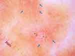
Dermatological examination revealed multiple discrete indurated erythematous-to-brownish papules, some coalescing into plaques on the pretibial skin. Excoriation was seen without lichenification. Varicose veins were also present on both legs (Figure 1). Other systems were unremarkable. The dermoscopic evaluation found dotted and globular vessels over pinkish-white and brownish backgrounds with hemorrhagic crusts (Figure 2).
 |
Figure 2 Dermoscopic features: dotted and globular vessels (blue arrows) over pinkish-white and brownish backgrounds with hemorrhagic crusts (green stars) (original magnification x20). |
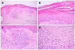
Due to disease persistence, a biopsy was performed for a definitive diagnosis. The histopathological section demonstrated hyperkeratosis with parakeratosis and epidermal hyperplasia with flattening of the rete ridge. Dermis showed clusters of plump endothelial cell-blood vessels with dense superficial perivascular and sparse interstitial cell infiltration, predominantly lymphocytes with few eosinophils. Numerous stellate fibroblasts and some Langerhans cell granulomas were also observed (Figure 3).
Based on the patient’s history, physical examination, and histopathological findings, the diagnosis of PPPD was made. The patient was prescribed 0.05% betamethasone dipropionate, applied twice daily, and oral antihistamines. One month later, her itch was mildly alleviated, but papules and plaques remained unchanged. Thus, we tried intralesional triamcinolone acetonide 5 mg/mL injected in a few lesions and added oral pentoxifylline 400 mg twice daily for her leg varicosity. Interestingly, only 1 month after, all lesions, including intralesional corticosteroid injected and non-injected lesions, flattened into patches, and all symptoms almost resolved (Figure 4). Our patient was delighted with the treatment, which improved not only the skin lesions but also her self-esteem and quality of life.
 |
Figure 4 One month after additional oral pentoxifylline, (A) all lesions flattened into patches; close-up pictures of the right (B) and the left legs (C). |
Discussion
Chronic scratching or rubbing of the skin leads to variable cutaneous disorders.2 PPPD is a recently recognized entity as a distinctive response to persistent gentle rubbing of pretibial skin.1 After the first introduction by Annessi et al in 2006,1 4 cases of PPPD were subsequently reported in the literature.2–4 The condition commonly presents as multiple, intensely pruritic, discrete, smooth-surface, flesh-colored to erythematous papules that may subsequently coalesce into plaques and result in a cobblestone appearance.1,2,4,5 However, in 2019, Flores et al broadened the clinical and histological spectrum of PPPD in 38 patients, demonstrating its epidermal changes.6 Lesions typically limit to unilateral or bilateral shins. Its prevalence is considered underestimated since the lack of disease recognition and misdiagnosis may occur.
Errichetti and Stinco have described dermoscopic features helpful for diagnosing PPPD, compatible with our findings.3 Dotted and globular vessels represent dilated vertically arranged papillary dermal vessels that follow skin inflammation, traumatized, or in skin-overlying stasis,7,8 while the pinkish-white background results from superficial dermal inflammation and fibrosis.8 The particular dermoscopic features indicate chronic cutaneous manipulation and inflammation. The lack of excoriation and lichenification determine that the stimuli were not in a vigorous way. Up to now, little is known regarding specific dermoscopic features of PPPD, and further studies are warranted to verify the findings.
Principal differential diagnoses of PPPD include a disease group presenting papules/plaques in response to chronic scratching, namely lichen simplex chronicus (LSC), prurigo nodularis (PN), and lichen amyloidosis (LA). LSC lesions usually aggregate into indistinctly, thickening and scaling, red-to-purplish plaques with lichenification and hyperpigmentation,9,10 while severe pruritic, hyperkeratotic, dome-shaped nodules characterize PN.11,12 LA is an eruption of discrete, keratotic papules/nodules with amyloid deposits in the dermis.13 Albeit PPPD originates from similar causes to these diseases, it displays distinctive histopathological findings. Histological hallmarks of PPPD are the presence of superficial perivascular and interstitial lymphohistiocytic infiltration, epidermal changes of spongiosis and parakeratosis, and numerous stellate and multinucleated fibroblasts in the dermis.1,2,6 The histological features that indicate persistent rubbing, consisting of uneven psoriasiform epidermal hyperplasia, marked compact orthokeratosis, hypergranulosis, and coarse bundles of collagen arranged in vertical streaks, are absent in PPPD,1,2,6 and the lack of amyloid deposits in the dermis distinguishes PPPD from LA.1,13 Despite unique PPPD histopathology, its pathogenesis has yet to be fully understood. Persistent rubbing could induce chronic skin inflammation and result in the development of PPPD; however, there is insufficient evidence to explain the primary cause of pretibial pruritus that provoked patients to start scratching.1
There is no current standard treatment for PPPD. Previous studies reported the efficacy of topical corticosteroids in improving PPPD lesions.1,2 In contrast, our case failed to address PPPD by topical corticosteroid application. However, additional oral pentoxifylline 400 mg twice daily was impressively effective in our patient since all papules and plaques (not limited to intralesional corticosteroid injected lesions) were significantly resolved within 1 month. The treatment has also shown high efficacy in alleviating an itchy sensation. Hence, this good response could result from pentoxifylline, a methyl-xanthine derivative used in various dermatological diseases, most notably varicosity.14 We hypothesized that, apart from its hemorheological effects in varicosity, pentoxifylline plays a role in the treatment of PPPD by; (1) inhibition of interleukin (IL)-1, IL-6, and tumor necrosis factor-α; (2) suppression of neutrophils, T cells, and B cells; (3) decreased expression of endothelial adhesion molecules; and (4) inhibition of fibroblast biosynthesis.14 This finding may lead to the hypothesis that leg varicosity or chronic venous insufficiency may involve in the mechanism of this condition, guiding further research for investigating the pathogenesis of PPPD. Nevertheless, this hypothesis may be limited by the fact that venous stasis in our patient has yet to be appropriately evaluated.
Conclusion
PPPD represents a unique cutaneous response to chronic gentle rubbing with respect to its clinical, dermoscopic, and histopathological presentations. Due to its rarity, PPPD has not been well known throughout previous literature. In this report, we underlined the importance of PPPD recognition, describing its corresponding histopathology and dermoscopic findings. We also support the utilization of a dermoscopy to distinguish PPPD from other mimicking conditions, thus, reducing the number of skin biopsies. Lastly, we first advocated the effective therapeutic modality for PPPD using pentoxifylline, which improves the treatment outcome and patients’ quality of life. Besides, this finding leads to the hypothesis that chronic venous insufficiency may play a role in the pathogenesis of PPPD.
Ethics Approval and Consent to Participate
This article was performed in accordance with the principles of Declaration of Helsinki. Ethical review and approval was not required to publish the case details in accordance with the local legislation and institutional requirements. Written informed consent was obtained from the patient for publication of this case report and any accompanying images as per our standard institutional rules.
Funding
No sources of funding were used to prepare this manuscript.
Disclosure
The authors declare that this manuscript was prepared in the absence of any commercial or financial relationships that could be construed as a potential conflict of interest.
References
1. Annessi G, Petresca M, Petresca A. Pretibial pruritic papular dermatitis: a distinctive cutaneous manifestation in response to chronic rubbing. Am J Dermatopathol. 2006;28(2):117–121. doi:10.1097/01.dad.0000200017.37082.e4
2. Kecelj B, Kecelj Leskovec N, Žgavec B. A case report and differential diagnosis of pruritic pretibial skin lesions. Acta Dermatovenerol Alp Pannonica Adriat. 2020;29(3):157–159.
3. Errichetti E, Stinco G. Dermoscopy for improving the diagnosis of pretibial pruritic papular dermatitis. Australas J Dermatol. 2018;59(1):e74–e75. doi:10.1111/ajd.12610
4. Noakes R, Mellick N. Case of pretibial pruritic papular dermatitis. Letters to the editor. Australas J Dermatol. 2010;51(3):215–216. doi:10.1111/j.1440-0960.2010.00667.x
5. Suchonwanit P, McMichael AJ. Alopecia in association with malignancy: a review. Am J Clin Dermatol. 2018;19(6):853–865. doi:10.1007/s40257-018-0378-1
6. Flores S, Wada DA, Florell SR, Bowen AR. Pretibial pruritic papular dermatitis: a comprehensive clinical and pathologic review of cases at a single institution. Am J Dermatopathol. 2020;42(1):16–19. doi:10.1097/dad.0000000000001460
7. Vázquez-López F, Kreusch J, Marghoob AA. Dermoscopic semiology: further insights into vascular features by screening a large spectrum of nontumoral skin lesions. Br J Dermatol. 2004;150(2):226–231. doi:10.1111/j.1365-2133.2004.05753.x
8. Errichetti E, Stinco G. Dermoscopy in general dermatology: a practical overview. Dermatol Ther (Heidelb). 2016;6(4):471–507. doi:10.1007/s13555-016-0141-6
9. Suchonwanit P, Kositkuljorn C, Pomsoong C. Alopecia areata: an autoimmune disease of multiple players. Immunotargets Ther. 2021;10:299–312. doi:10.2147/itt.S266409
10. Ju T, Vander Does A, Mohsin N, Yosipovitch G. Lichen simplex chronicus itch: an update. Acta Derm Venereol. 2022;102:adv00796. doi:10.2340/actadv.v102.4367
11. Williams KA, Huang AH, Belzberg M, Kwatra SG. Prurigo nodularis: pathogenesis and management. J Am Acad Dermatol. 2020;83(6):1567–1575. doi:10.1016/j.jaad.2020.04.182
12. Suchonwanit P, Iamsumang W, Leerunyakul K. Topical finasteride for the treatment of male androgenetic alopecia and female pattern hair loss: a review of the current literature. J Dermatolog Treat. 2022;33(2):643–648. doi:10.1080/09546634.2020.1782324
13. Weyers W, Weyers I, Bonczkowitz M, Diaz-Cascajo C, Schill WB. Lichen amyloidosus: a consequence of scratching. J Am Acad Dermatol. 1997;37(6):923–928. doi:10.1016/s0190-9622(97)70066-5
14. Balazic E, Axler E, Konisky H, Khanna U, Kobets K. Pentoxifylline in dermatology. J Cosmet Dermatol. 2023;22(2):410–417. doi:10.1111/jocd.15445
 © 2023 The Author(s). This work is published and licensed by Dove Medical Press Limited. The full terms of this license are available at https://www.dovepress.com/terms.php and incorporate the Creative Commons Attribution - Non Commercial (unported, v3.0) License.
By accessing the work you hereby accept the Terms. Non-commercial uses of the work are permitted without any further permission from Dove Medical Press Limited, provided the work is properly attributed. For permission for commercial use of this work, please see paragraphs 4.2 and 5 of our Terms.


