Back to Journals » International Journal of Nanomedicine » Volume 13 » T-NANO 2014 Abstracts
Preparation and characterization of superparamagnetic iron oxide nanoparticles for magnetically guided drug delivery
Authors Kumar P, Agnihotri S, Roy I
Received 18 October 2016
Accepted for publication 31 October 2016
Published 15 March 2018 Volume 2018:13(T-NANO 2014 Abstracts) Pages 43—46
DOI https://doi.org/10.2147/IJN.S125002
Checked for plagiarism Yes
Review by Single anonymous peer review
Peer reviewer comments 2
Editor who approved publication: Professor Lei Yang
Pramod Kumar, Shrish Agnihotri, Indrajit Roy
Department of Chemistry, University of Delhi, Delhi, India
Abstract: Iron oxide nanoparticles have unique magnetic properties and therefore readily respond to applied magnetic fields. Moreover, their surfaces can be used to attach active molecules via various covalent or noncovalent interactions. Thus, they can be used as drug carriers for magnetically controlled delivery to specific biological sites of interest. In the present study, we have synthesized aqueous dispersed samples of citric acid-capped iron oxide nanoparticles, and the anticancer drug doxorubicin was then linked with these superparamagnetic iron oxide nanoparticles via a simple noncovalent interaction. Our results show that the conjugated drug releases from the nanoparticles in a sustained manner. The cellular uptake of these nanoparticles was found to be substantial, although it can be further enhanced using magnetic guidance. These nanoparticles (drug free) were found to be nontoxic to cells; however, upon drug conjugation, drug-induced toxicity was observed, owing to the slow release of drug from the nanoparticles.
Keywords: SPION, Dox, Dox-CA-SPION, magnetically guided delivery
Introduction
Guiding therapeutic drugs to specific diseased sites using external stimulus is a promising strategy for the targeted diagnosis and treatment of diseases.1–3 Iron oxide nanoparticles have unique magnetic properties and therefore readily respond to applied magnetic fields.4,5 Moreover, their surfaces can be used to attach active molecules via various covalent or noncovalent interactions. Thus, they can be used as drug carriers for magnetically controlled delivery to specific biological sites of interest.6,7
We have synthesized aqueous dispersed samples of citric acid-capped iron oxide nanoparticles. Transmission electron microscopy (TEM) data showed the average diameter of these monodisperse nanoparticles to be around 12 nm; energy dispersive X-ray dispersive spectroscopy and X-ray diffraction were then used to ascertain the crystalline nature of these particles. The anticancer drug doxorubicin (Dox) was then linked with these superparamagnetic iron oxide nanoparticles (SPIONs) via a simple noncovalent interaction. In vitro studies have shown that these nanoparticles are nontoxic to cells in culture, and did not cause hemolysis in blood samples. Finally, it was demonstrated that these nanoparticles, without and with linked Dox, can be robustly delivered to cells in culture using magnetic guidance. This magnetically enhanced delivery resulted in increased Dox-induced cytotoxicity in the treated cells. These observations underscore the potential of these nanomaterials in externally targeted diagnostics and drug delivery.
Materials and methods
Ferric chloride (FeCl3.6H2O), ferrous sulfate (FeSO4.6H2O), and ammonia solution (25%) were purchased from Merck & Co., Inc. (Kenilworth, NJ, USA). Doxorubicin hydrochlorides were purchased from Sigma-Aldrich Co. (St Louis, MO, USA). Lung carcinoma cells lines (A-549) were purchased from American Type Culture Collection (ATCC, Manassas, VA, USA), and cultured according to instructions supplied by the vendor.
Particle characterization
Transmission electron microscopy
Samples for TEM analysis were prepared by drop-coated SPIONs suspension (10 μg/mL) solution. For TEM analysis, the aqueous dispersions were sonicated and drop coated and dried onto formvar-coated 200-mesh copper grids (Ted Pella, Inc., Redding, CA, USA), followed by imaging using a TECNAI G2-30 U TWIN TEM instrument (FEI, Eindhoven, The Netherlands) with an acceleration voltage of 300 kV.
Dynamic light scattering
The size of the nanoparticles was further analyzed by dynamic light scattering measurements, using a NANO-ZS series Malvern Zetasizer instrument (Malvern Instruments, Malvern, UK). Helium–Neon laser (wavelength 633 nm, power 4 mW) was used as the light source. The same instrument was used to measure the surface charge (zeta potential) of the nanoparticles.
Hemolysis
Heparin-stabilized human blood was freshly collected from a healthy volunteer, according to approved Institutional Review Board protocol of University of Delhi. A 4-mL sample of blood was added to 8 mL of phosphate-buffered saline (PBS), and the red blood cells were isolated from serum by centrifugation at 10,016× g for 5 minutes.
Cytotoxicity
We first probed the potential toxicity of these nanoparticles (drug free) in blood, using hemolysis assay. After that, we carried out studies probing the interaction of these nanoparticles, with and without linked drug, using A-549 lung cancer cells in vitro.8
Cellular uptake
After probing the potential toxicity of nondrug-linked nanoparticles in blood and A-549 cells, we began our analysis of interaction of drug-linked nanoparticles (doxorubicin containing citric acid-coated SPION [Dox-CA-SPIONs]) with A-549 cells in vitro. Here, first we monitored the cellular uptake of the drug-linked nanoparticles.9
The fixed cells were visualized under a fluorescent Nikon TS-100 inverted microscope, and photographed using a Nikon DIGITAL SIGHT DS-Fi1 Camera (Nikon Corporation, Tokyo, Japan).
Magnetically guided drug delivery
For analyzing the efficacy of magnetically guided drug targeting of cells using iron oxide nanoparticles, Dox-CA-SPION conjugates were added to cells, with and without a bar magnet placed below the plates.10
Results and discussion
The TEM images of the synthesized citric acid coated SPION (CA-SPION) are shown in Figure 1, showing them to be irregular in size, with an average diameter of ~12 nm.
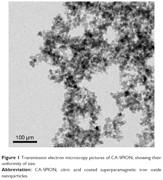 | Figure 1 Transmission electron microscopy pictures of CA-SPION, showing their uniformity of size. |
All samples were prepared in triplicate, and the suspensions were briefly vortexed and centrifuged at 10,016× g for 5 minutes. Then, ~100 μL of supernatant solutions from the sample tube was transferred to a 96-well plate. The absorbance of the supernatant was recorded at 577 nm (corresponding to hemoglobin absorption), with a reference wavelength of 655 nm. The value of the absorbance at 577 nm was correlated with the extent of hemolysis. The result of hemolysis assay and cytotoxicity of SPION and CA-SPION are shown in Figure 2A and 2B, respectively.
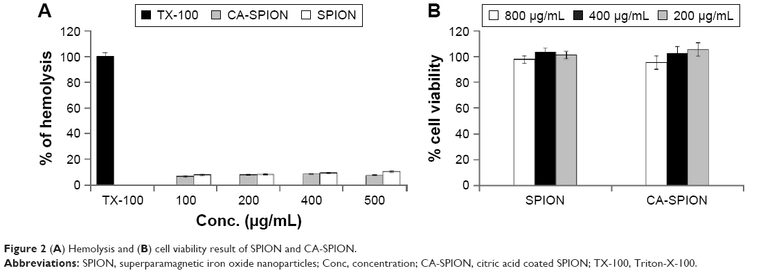 | Figure 2 (A) Hemolysis and (B) cell viability result of SPION and CA-SPION. |
For analyzing the efficacy of magnetically guided drug targeting of cells using iron oxide nanoparticles, Dox-CA-SPION conjugates were added to cells, with and without a bar magnet placed underneath the plates under magnetic guidance.
A total of 12 single-well plates (35 mm) containing A-549 cells (at a confluence of ~60%–70%) were treated with 2 mg/mL of Dox-CA-SPIONs. Magnetic guidance was provided to six plates with nanoparticle-treated cells by placing a bar magnet below them, whereas six remaining plates did not receive magnetic guidance.
After 5 and 15 minutes of incubation, the plates were washed three times with PBS and replenished with 2 mL of fresh media, followed by 2 hours of incubation. After that, the cells were washed with PBS again and lysed by addition of fixed volume of cell lysis reagent (prepared by dissolving 1% mass/vol of surfactant Triton-X-100 in PBS, pH 7.2). After 30 minutes, the lysates are transferred to microcentrifuge tubes, centrifuged to separate the cell debris, and the supernatant was analyzed for intracellular iron content and fluorescence of Dox (λex =490 nm, λem =560 and 590 nm), as shown in Figure 3A and B, respectively. Higher intensity of Dox fluorescence coincides with higher nanoparticle uptake of the cells. The cytotoxic effect of Dox-delivery using CA-SPION under magnetic guidance is shown in Figure 3C.
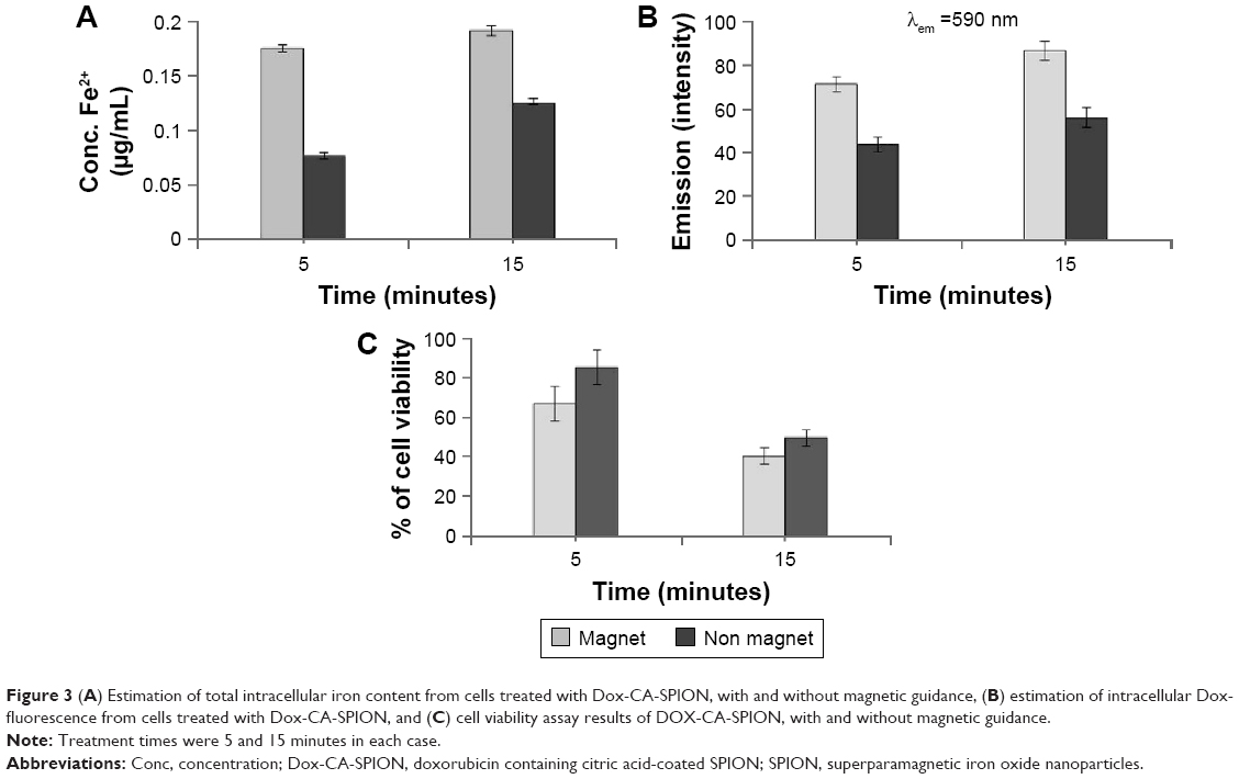 | Figure 3 (A) Estimation of total intracellular iron content from cells treated with Dox-CA-SPION, with and without magnetic guidance, (B) estimation of intracellular Dox-fluorescence from cells treated with Dox-CA-SPION, and (C) cell viability assay results of DOX-CA-SPION, with and without magnetic guidance. |
Conclusion
In this article, we describe the facile synthesis, characterization, and in vitro applications of drug-conjugated CA-SPION. The conjugated drug releases from the nanoparticles in a sustained manner. The cellular uptake of these nanoparticles was found to be substantial, although it can be further enhanced using magnetic guidance. These nanoparticles (drug free) were found to be nontoxic to cells; however, upon drug conjugation, drug-induced toxicity was observed owing to the slow release of drug from the nanoparticles. The therapeutic efficacy of magnetically targeted drug delivery is expected to be more prominent in in vivo studies, where free drugs are known to be largely ineffective.
Acknowledgment
We are grateful to University of Delhi, India, for providing a Research & Development grant to support this study.
Disclosure
The authors report no conflicts of interest in this work.
References
Davis SS. Biomedical applications of nanotechnology – implications for drug targeting and gene therapy. Trends Biotechnol. 1997;15:217–224. | ||
Li K, Shen M, Zheng L, Zhao J, et al. Magnetic resonance imaging of glioma with novel APTS-coated superparamagnetic iron oxide nanoparticles. Nanoscale Res Lett. 2014;9:304. | ||
Sahoo B, Devi KS, Dutta S, et al. Biocompatible mesoporous silica-coated superparamagnetic manganese ferrite nanoparticles for targeted drug delivery and MR imaging applications. J Colloid Interface Sci. 2014;431:31–41. | ||
Prasad PN. Introduction to Biophotonics. New York, NY: Wiley; 2004. | ||
Kumar LC, Hansel CS, Soboyejo W, et al. LHRH-conjugated magnetic iron oxide nanoparticles for detection of breast cancer metastases. Breast Cancer Res Treat. 2006;99:163–176. | ||
Banerjee SS, Chen DH. Multifunctional pH-sensitive magnetic nanoparticles for simultaneous imaging, sensing and targeted intracellular anticancer drug delivery. Nanotechnology. 2008;19(50):505104. | ||
Islam MS, Kurawaki J, Kusumoto Y, et al. Hydrothermal novel synthesis of neck-structured hyperthermia-suitable magnetic (Fe3O4, γ-Fe2O3 and α-Fe2O3) nanoparticles. J Sci Res. 2012;4:97–107. | ||
Kumar P, Anuradha, Roy I. Optically and magnetically doped ormosil nanoparticles for bioimaging: synthesis, characterization, and in vitro studies. RSC Adv. 2014;4(4):16181–16187. | ||
Roy I, Kumar P, Kumar R, et al. Ormosil nanoparticles as a sustained-release drug delivery vehicle. RSC Adv. 2014;4(4):53498–53504. | ||
Gupta AK, Gupta M. Synthesis and surface engineering of iron oxide nanoparticles for biomedical applications. Biomaterials. 2005;26:3995–4021. |
 © 2018 The Author(s). This work is published and licensed by Dove Medical Press Limited. The
full terms of this license are available at https://www.dovepress.com/terms
and incorporate the Creative Commons Attribution
- Non Commercial (unported, 3.0) License.
By accessing the work you hereby accept the Terms. Non-commercial uses of the work are permitted
without any further permission from Dove Medical Press Limited, provided the work is properly
attributed. For permission for commercial use of this work, please see paragraphs 4.2 and 5 of our Terms.
© 2018 The Author(s). This work is published and licensed by Dove Medical Press Limited. The
full terms of this license are available at https://www.dovepress.com/terms
and incorporate the Creative Commons Attribution
- Non Commercial (unported, 3.0) License.
By accessing the work you hereby accept the Terms. Non-commercial uses of the work are permitted
without any further permission from Dove Medical Press Limited, provided the work is properly
attributed. For permission for commercial use of this work, please see paragraphs 4.2 and 5 of our Terms.