
Back to Journals » The Application of Clinical Genetics » Volume 19
Preimplantation Genetic Testing for Cornelia de Lange Syndrome with Low-Level Maternal Gonadal Mosaicism for a Sub-Megabase Deletion in China
Authors Meng L, Wang Y, Zhang J, Zhou R, Wang X  , Qiao F, Zhang Q, Wan C, Jiao S, Hu P, Xu Z
Received 26 February 2026
Accepted for publication 1 July 2026
Published 21 July 2026 Volume 2026:19 603242
DOI https://doi.org/10.2147/TACG.S603242
Checked for plagiarism Yes
Review by Single anonymous peer review
Peer reviewer comments 2
Editor who approved publication: Prof. Dr. Martin Maurer
Lulu Meng,1,* Yan Wang,1,* Junqiang Zhang,2,* Ran Zhou,1 Xingxing Wang,3 Fengchang Qiao,1 Qinxin Zhang,1 Cheng Wan,3 Shujing Jiao,3 Ping Hu,1 Zhengfeng Xu1
1Department of Prenatal Diagnosis, Women’s Hospital of Nanjing Medical University (Nanjing Women and Children’s Healthcare Hospital), Nanjing, Jiangsu, People’s Republic of China; 2State Key Laboratory of Reproductive Medicine, Department of Reproduction Medicine, Women’s Hospital of Nanjing Medical University (Nanjing Women and Children’s Healthcare Hospital), Nanjing, Jiangsu, People’s Republic of China; 3Department of Research and Development, Yikon Genomics Co. Ltd, Suzhou, Jiangsu, People’s Republic of China
*These authors contributed equally to this work
Correspondence: Ping Hu, Department of Prenatal Diagnosis, Women’s Hospital of Nanjing Medical University (Nanjing Women and Children’s Healthcare Hospital), 123 Tianfei Alley, Mochou Road, Nanjing, Jiangsu, 210004, People’s Republic of China, Email [email protected] Zhengfeng Xu, Department of Prenatal Diagnosis, Women’s Hospital of Nanjing Medical University (Nanjing Women and Children’s Healthcare Hospital), 123 Tianfei Alley, Mochou Road, Nanjing, Jiangsu, 210004, People’s Republic of China, Email [email protected]
Purpose: To diagnose and perform preimplantation genetic testing for monogenic disorders (PGT-M) in a Chinese family affected by Cornelia de Lange syndrome type 5, resulting from a microdeletion in Xq13.1q13.2 truncating the HDAC8 gene characterized by low-level gonadal mosaicism.
Patients and Methods: A de novo copy number variation (CNV) was identified through chromosomal microarray analysis and whole exome sequencing in a family with two unsuccessful pregnancies, indicating germline mosaicism. The CNV was validated via real-time quantitative PCR. Whole-genome low-coverage mate-pair sequencing was conducted on female peripheral blood to exclude chromosomal abnormalities. Long-PCR amplified the deleted fragment, utilizing primers designed nearby breakpoints identified through chromosomal microarray analysis. Oxford Nanopore Technology sequencing pinpointed specific breakpoint positions. Droplet-digital PCR (ddPCR) confirmed germline mosaicism in ovarian samples.
Results: A female patient, suspected of harboring a de novo microdeletion at Xq13.1q13.2 exhibiting gonadal mosaicism, along with her husband, participated in this study. The diagnoses of the 172.3 kb microdeletion at Xq13.1q13.2 as low-level gonadal mosaicism was corroborated through nanopore sequencing and ddPCR. We constructed the high-risk haplotype using the affected products of conception as the phasing reference, and performed PGT-M based on SNP haplotype linkage analysis, finally achieving a healthy live birth in February 2023.
Conclusion: Our findings underscore the efficacy of PGT-M employing haplotype linkage analysis for CNVs less than 1 Mb, even within cases involving gonadal mosaicism. We present methodologies to address microdeletions associated with gonadal mosaicism utilizing next-generation sequencing, microarray, nanopore sequencing and ddPCR techniques. Our results advocate for an expansion of PGT-M based on haplotype linkage analysis for families with minor pathogenic CNVs.
Keywords: preimplantation genetic testing for monogenic disease, PGT-M, Cornelia de Lange syndrome, CdLS, low-level gonadal mosaicism, nanopore sequencing, droplet-digital PCR
Introduction
Cornelia de Lange syndrome (CdLS; MIM #122470, 300590, 610759, 300882, 614701) is a clinically heterogeneous developmental disorder characterized by malformations affecting multiple systems. The estimated incidence ranges from 1 in 10,000 to 1 in 30,000 live births;1 however, the actual incidence may be higher due to the presence of mild cases with atypical symptoms that often go undiagnosed. Individuals affected by CdLS typically exhibit dysmorphic facial features, cleft palate, distal limb defects, intrauterine and postnatal growth retardation, as well as severe intellectual disability with a mean IQ of approximately 53.2 Prenatal detection of CdLS manifestations such as fetal skin edema, NT thickening, heart defects occurs in only about 23% of cases,3 which aligns with the experience observed during our family’s second pregnancy.
Cornelia de Lange syndrome 5 (CdLS5, MIM# 300882) is a rare X-linked dominant hereditary disorder caused by mutations in histone deacetylase 8 (HDCA8, MIM *300269), located on chromosome Xq13.1. This genetic variant accounts for approximately 4% of all CdLS cases.4 HDAC8 encodes a class I histone deacetylase, which is a core regulator of the cohesin complex cycle. The cohesin complex is essential for sister chromatid cohesion, accurate chromosome segregation during mitosis/meiosis, DNA damage repair, and long-range transcriptional regulation in embryonic development. HDAC8 specifically catalyzes deacetylation of the SMC3 subunit of cohesin, which is a prerequisite for the release and recycling of cohesin from chromatin after mitosis. Loss of HDAC8 function leads to persistent SMC3 acetylation, impaired cohesin dynamics, widespread dysregulation of developmental genes, and subsequent multi-system malformations characteristic of CdLS.5,6 Notably, hemizygous loss of HDAC8 in male individuals results in complete loss of protein function, which is closely associated with severe developmental defects and even embryonic lethality, consistent with the two consecutive male affected pregnancies in this family. A deficiency in HDAC8 activity leads to an accumulation of acetylated cohesin with reduced affinity for chromatids and subsequently results in abnormal transcriptional regulation.5 The phenotypic variations associated with HDAC8 mutations are notably nonclassical and diverse; however, distinctive features observed alongside typical CdLS characteristics include a large anterior fontanel, a broad or bulbous nasal tip, tooth anomalies, mosaic patches of hyperpigmented skin, orbital hypertelorism as well as generally cheerful dispositions among affected individuals.1 Male patients tend to exhibit more severe manifestations compared to females; conversely, female patients display mildly variable clinical symptoms influenced by patterns of X-inactivation.6
About 100 mutations in the HDAC8 gene have been documented (Human Gene Mutation Database: http://www.hgmd.org). The majority of known disease-causing mutations in HDAC8 are classified as nonsense, missense, splice site variants or CNVs, all of which are predicted to disrupt HDAC8 function. Most of these mutations have been identified as de novo.4,7 To date, there have been few reports concerning microdeletions within the HDAC8 gene. Several cases involving larger CNVs affecting regions of HDAC8 have been reported in individuals with CdLS5, particularly intragenic deletions that span from single to multiple exons. Specifically, exons 1, 1–4, 1–9, 3–4, 5–6, 5–7, 5–10, 7, 8–11 and exon 11 were found to be absent from the HDAC8 gene in a cohort of 13 individuals diagnosed with CdLS5.7–10 This report marks the first instance of an HDAC8 gene deletion leading to male embryo abortion; it suggests that deletion encompassing exon regions from 1 to 9 has a severe developmental defects and even lethal effect on male embryos.
In this case study, we describe a family with products of conception (POC) tissues from one miscarried male fetus and one terminated male fetus exhibiting the same de novo microdeletion at Xq13.1q13.2 that disrupts the HDAC8 gene. Consequently, we hypothesized that the woman is a carrier exhibiting gonadal mosaicism. Long-range PCR and nanopore sequencing were employed on POC to determine specific breakpoint positions accurately. Droplet-digital PCR (ddPCR) was utilized to verify both the existence and proportion of germline mosaicism present in ovarian samples obtained from IVF procedures-including granulosa cells and follicular fluid. Our findings indicate that the extended preimplantation genetic testing for monogenic disorders (PGT-M), based on the haplotype linkage analysis strategies, could be effectively applied to families harboring small pathogenic CNVs in a gonadal mosaic state.
Materials and Methods
Subjects
This couple was enrolled from our hospital clinic. The 29-year-old female patient, gravida 2, para 0, with two unsuccessful pregnancies and no live births, denied any past medical illnesses or regular medication intake. Her 30-year-old spouse was in good general physical condition but suffered from asthenospermia. Her first pregnancy ended in miscarriage at 12+2 weeks’ gestation when she was 27 years old. The chromosomal microarray (CMA) results from both the POC tissue and parental blood samples revealed a de novo pathogenic deletion at Xq13.1q13.2, disrupting the haploinsufficient gene HDAC8. Despite the parents being clinically unaffected and exhibiting no family history associated with CdLS, they sought genetic counseling at the Department of Prenatal Diagnosis of Nanjing Women and Children’s Healthcare Hospital in Jiangsu, China. Subsequently, they opted for natural conception due to the perceived low incidence of germline mosaicism.
During the second pregnancy, fetal nuchal translucency thickening (6.9mm), congenital heart disease and fetal edema—particularly in the head and trunk—were identified at gestational week 13+1. The pregnancy was terminated, and genetic analysis was conducted on the aborted fetus. The fetus was male and identified as carrying a same deletion at Xq13.1q13.2 through CMA and Trio whole-exome sequencing (Trio-WES). Following genetic counseling sessions that took into account their previous two unsuccessful pregnancy outcomes as well as male asthenospermia, the couple decided to pursue assisted reproductive technology at the Department of Reproductive Medicine. The patient’s history and medical process are illustrated in Figure 1. Prior to any interventions, genetic counseling sessions were conducted with the family, during which informed consent was obtained. All procedures and protocols undertaken in this study received approval from the Medical Ethics Committee of Nanjing Women and Children’s Healthcare Hospital.
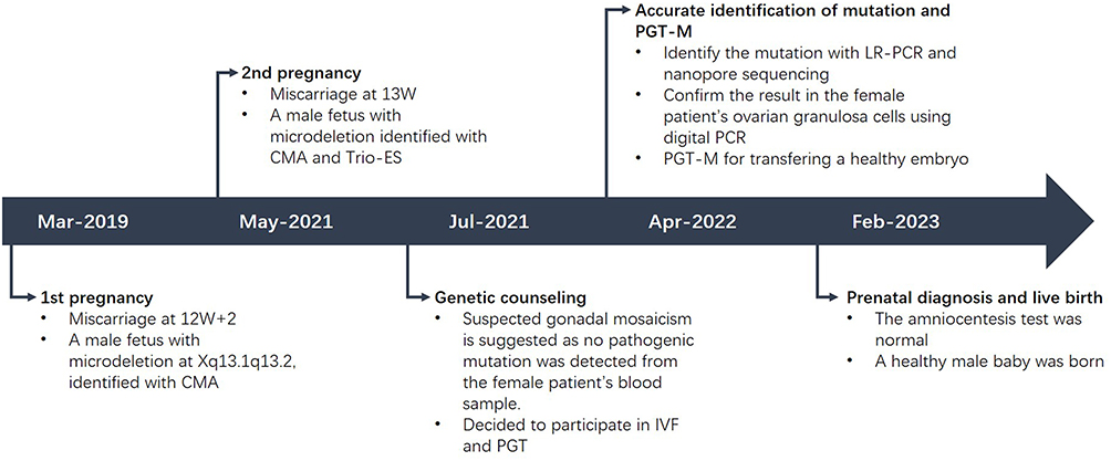 |
Figure 1 History and medical intervention for the patient with gonadal mosaicism of microdeletion at Xq13.1q13.2. |
Sample Collection and Storage
All tissue samples (POC tissues, ovarian granulosa cells) were collected immediately after isolation, snap-frozen in liquid nitrogen, and stored at −80°C until DNA extraction. Peripheral blood samples were stored at 4°C and processed for DNA extraction within 24 h after collection. Follicular fluid samples were centrifuged at 16000×g for 10 min at 4°C immediately after collection, and the supernatant was collected and stored at −80°C until cell-free DNA extraction. Amniotic fluid samples were stored at 4°C and processed within 6 h after amniocentesis. All samples were processed under strict RNase/DNase-free conditions to avoid nucleic acid degradation.
Ethics
Genetic counseling sessions were provided for the family prior to any interventions, all participants signed written informed consent for prenatal diagnosis, PGT testing, participation in this study, and publication of this case report, in full compliance with the Declaration of Helsinki. All procedures and protocols undertaken in this study received approval from the Medical Ethics Committee of Nanjing Women and Children’s Healthcare Hospital.
Genomic DNA Sample Preparation
Genomic DNA (gDNA) from POC tissue samples and peripheral blood was extracted using the whole blood genomic DNA extraction kit based on magnetic bead methodology (M121, Maibo, China); Genomic DNA from 20 mL amniotic fluid samples was extracted utilizing a QIAamp DNA Mini Kit (51306, Qiagen, Germany). Cell-free DNA extracted from follicular fluid samples employed a plasma cell-free DNA extraction kit also based on magnetic bead methodology (M111, Maibo, China).
Chromosomal Microarray Analysis
CMA performs genome-wide detection of chromosomal aneuploidies, CNVs exceeding 100 kb, regions of homozygosity, and mosaicism greater than 30% on POC tissues. It also allows parental origin analysis on parental peripheral blood to distinguish de novo from inherited variants. The Affymetrix CytoScan 750K array (901859, Affymetrix, USA), which comprises approximately 550,000 oligonucleotide probes and 200,000 single nucleotide polymorphism (SNP) probes, was utilized for whole-genome scanning. CNVs were identified at a minimum resolution of 50 kb. Detailed descriptions of the 750K array experiments have been previously reported. The array data was quality-controlled according to the manufacturer’s standard, with median absolute pairwise difference (MAPD) <0.25 and SNP call rate >95% as the qualified threshold for data analysis. Cytogenetic analysis was conducted using the Chromosome Analysis Suite Software (Affymetrix, CA), and CNV coordinates were determined in accordance with the human genome GRCh37/hg19 assembly. The clinical significance of the detected CNVs was evaluated based on the guidelines established by the American College of Medical Genetics and Genomics (ACMG) and the Clinical Genome Resource (ClinGen).
Whole Genome Low-Coverage Mate-Pair Sequencing (WGL-MPS)
WGL-MPS is capable of detecting genome-wide balanced translocations, chromosomal inversions, CNVs larger than 100 Kb abd mosaic states exceeding 30%. To investigate potential cryptic chromosomal abnormalities or mosaic states, WGL-MPS was performed on the blood sample of female participant after the results of CMA. gDNA from the female participant was utilized to construct a non-size-selected mate-pair library followed by subjecting it to 50-bp-end multiplex sequencing using BGISeq-500 technology. After filtering out reads containing sequencing adapters and those identified as low quality, high-quality paired-end reads were aligned against the NCBI human reference genome (hg19) using SOAP2. Only uniquely mapped reads were retained for subsequent analysis, as previously described.
Whole Exome Sequencing
Trio-WES was performed to detect exon-level single-nucleotide variants (SNVs), small insertions and deletions (INDELs) and exonic copy number variations (CNVs) across the whole exome on the couple blood and 2nd POC tissue after WGL-MPS. The Agilent SureSelect XT Library Prep Kit along with the Agilent SureSelect XT Human All Exon V6 kit (Agilent, Santa Clara, CA, USA) were employed to prepare a fragment library and capture gene exons following the manufacturer’s protocols. The captured regions underwent enrichment through PCR amplification and were sequenced on an Illumina Hiseq 2500 platform with a read length of 150 bp. The resultant reads were mapped against GRCh37/hg19 assembly. Only samples with target region coverage >99%, average sequencing depth >100×, and Q30 ratio >85% were included in subsequent variant analysis. Subsequent analysis was carried out as reported previously.
PCR Amplification and Detection of Deletion
A primer validation strategy was designed from the HDAC8 reference sequence (NM_018486.2) at breakpoints informed by WES and CMA results of POC, as illustrated in Figure 2. A Long-PCR method was employed to amplify the deleted fragments, where fragment length dictated the detection methodology. Primers were synthesized by Suzhou Genewiz Biotechnology Co., Ltd., with detailed sequences provided in Table 1. A total volume of 25 μL PCR reaction mix was prepared using LongAmp® Taq 2X premix (M0287; NEB, USA). The reaction conditions included: initial denaturation at 94°C for 1 minute; followed by cycles consisting of denaturation at 94°C for 30s; annealing at 62°C for 30s; extension at 65°C for 10 minutes, repeated over a total of thirty cycles. PCR products were subsequently subjected to electrophoresis on a 0.8% agarose gel at 110 V for 50 minutes followed by visualization and capture using a gel imaging system. The presence of expected specific bands indicates successful amplification.
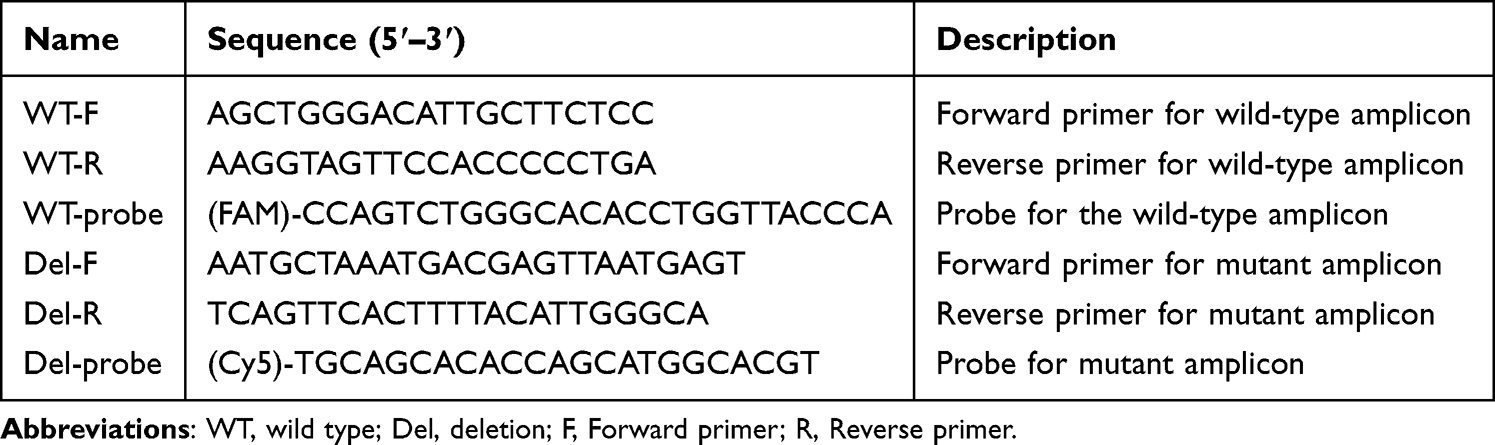 |
Table 1 PCR Primers and Probes Information for the Detection of Microdeletion |
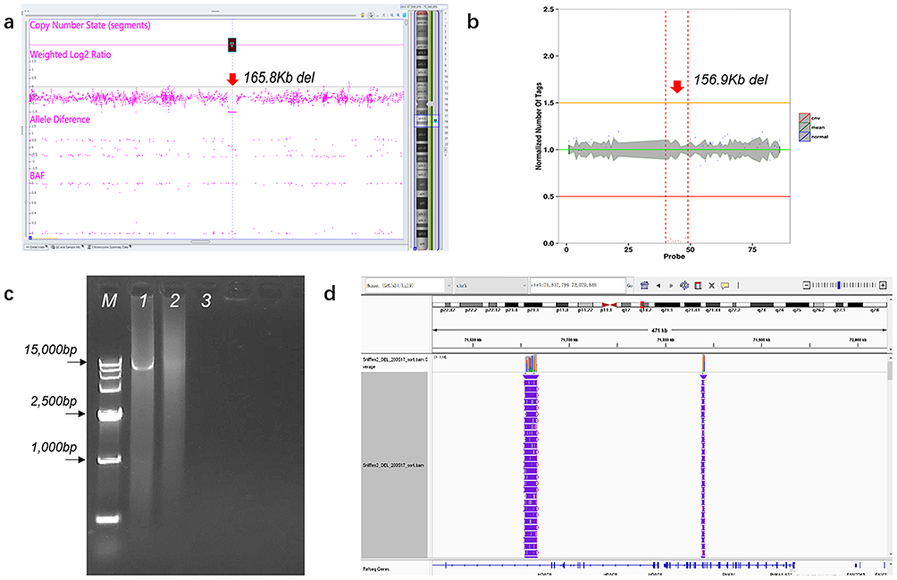 |
Figure 2 Identification of the microdeletion del(X)(q13.1q13.2) in POC tissue samples. (a) CMA result for the 1st POC. The red arrow indicates a 165.8 kb deletion on the Xq13 chromosome, arr Xq13.1q13.2 (71671944×1, 71674803_71840634×0, 71841429×1). (b) WES result for the 2nd POC. The red arrow indicates a 156.9 kb deletion on the Xq13 chromosome, seq del(X)(q13.1q13.2) chrX:g.71681820_71838708del. (c) Agarose gel electrophoresis for the long-range PCR product. The black arrows indicate the fragment sizes of different bands in the DNA ladder, which are 1000 bp, 2500 bp, and 15,000 bp, respectively. M: DL15000 DNA marker, lane 1: PCR product from the POC tissue gDNA, lane 2: PCR product (negative) from the female patient’s blood gDNA; lane 3: Blank control. (d) Nanopore sequencing result for the long-range PCR product of the 2nd POC, visualized by IGV version 2.9.4. It shows a 172.3 kb precise deletion on the Xq13 chromosome, chrX:g.71666527–71838853. |
Third-Generation Sequencing and Analysis of Breakpoint
Compared with CMA, WGL-MPS and short-read WES, Nanopore long-read third-generation sequencing possesses distinctive strengths in CNV breakpoint analysis by achieving base-pair level resolution for precise CNV breakpoint mapping. Following the purification of long-range PCR products from the 2nd POC tissue, a third-generation sequencing library was constructed utilizing the sequencing adapter kit (SQK-LSK110, Oxford Nanopore Technologies, UK). Key steps included employing the Ultra II End Prep module (NEB, E7546, USA) for DNA end repair and dA tailing, followed by ligation of motor protein-tailed adapters using the Quick Ligation Module (NEB, E6056, USA). The purified product represents the third-generation sequencing library. Sequencing was performed with the Oxford Nanopore MinION sequencer. The resulting fastq files were aligned with GRCh37/hg19 using minimap2 software with default parameters; breakpoint positions were validated through Interactive Genomics Viewer software. Only sequencing reads with quality score ≥10 were retained for alignment, and the average sequencing depth of the target region was >3× to ensure breakpoint accuracy.
Droplet-Digital PCR
Based on specific breakpoint positions identified from Oxford Nanopore Technology’s third-generation sequencing data, primers and probes for ddPCR were designed; sequence details are provided in Table 1. Following guidelines outlined in the 2×HQ ddPCR Master Mix for Probe kit (Sniper Co., Ltd. Suzhou, China), the reaction mix consisted of 11 µL of 2×HQ ddPCR Master Mix for Probe, 1 µL each of upstream and downstream primers at a concentration of 10 µmol/L, 0.5 µL of probe at a concentration of 10 µmol/L probe, and 1–5 µL of DNA template. The ddPCR was conducted using the DQ24 Digital PCR System (Sniper Co., Ltd. Suzhou, China). The amplification program included reverse transcription at 65°C for five minutes; enzyme activation at 95°C for fifteen minutes; denaturation at 95°C for twenty seconds; annealing at 60°C for 30 seconds over 40 cycles. After amplification, microdroplet data was analyzed via a droplet reader. Only wells with total valid droplets >18,000 were included in quantitative analysis. The analysis aimed to detect deletion ratios, with POC samples serving as positive controls, female patient’s peripheral blood samples without carrying deletions serving as health controls and buffer solution as negative controls.
Assisted Reproductive Technology Procedure
The couple underwent in vitro fertilization (IVF) at the Department of Reproductive Medicine. The procedure included controlled ovulation, oocytes retrieval, intracytoplasmic sperm injection, embryo culture, trophectoderm biopsy, and cryopreservation through vitrification of the blastocysts, all conducted according to standard protocol. In this IVF cycle, six embryos were cultured to the blastocyst stage; a total of 3–10 trophectoderm (TE) cells from each blastocyst were biopsied on day 5 or 6 and prepared for subsequent whole-genome amplification (WGA).
Whole Genome Amplification
Whole genome amplification (WGA) of the lysed TE cells was performed using multiple annealing and looping-based amplification cycles (MALBAC). This procedure enables the amplification of gDNA from picogram level to nanogram levels suitable for chip experiment. TE cells were transferred into 0.2 µL PCR tubes containing 4.5 µL of lysis buffer (XK-002, Yikon Genomics, China). WGA for each embryo biopsy sample was carried out using a MALBAC WGA kit (XK-028-24, Yikon Genomics, China), following the manufacturer’s standard protocols. Initially, collected cells were lysed in lysis buffer before undergoing MALBAC pre-amplification followed by exponential amplification to yield approximately 2–5 μg of DNA.
Preimplantation Genetic Testing
Purified WGA products from embryo TE biopsies and parental/POC gDNA were utilized for SNP and CNV library preparation via a next-generation sequencing (NGS) library preparation kit (XK-038, Yikon Genomics, China). All procedures adhered strictly to manufacturer’s instructions. Paired-end sequencing was employed with a read length of 150 bp using the MGI T7 platform. The raw data underwent automatic filtering and analysis through ChromGo software (Yikon Genomics, China). Chromosomal aneuploidies and genome-wide SNPs were detected simultaneously using the approach of Mutated Allele Revealed by Sequencing with Aneuploidy and Linkage Analyses (MARSALA).11,12 Embryos exhibiting a mosaic rate greater than 30% were classified as having mosaic aneuploidy, rendering them unsuitable for transfer. Valid reads exceeding 1 Mb with a coefficient of variation less than 0.1 (1000 K bin size) were deemed acceptable for CNV detection. In accordance with the good practice recommendations from the ESHRE PGT Consortium for detecting monogenic disorders, informative SNPs were selected for the haplotyping. High-frequency SNPs located within the 2 Mb regions upstream and downstream of the pathogenic CNV at Xq13.1q13.2, as well as those within the CNV itself, were screened to identify genetic markers necessary for constructing haplotypes. With the X-chromosome segment of affected POC as the phasing reference, concordant maternal segments were defined as “high-risk” haplotypes, and discordant ones aligned with the alternative maternal segments as “low-risk” haplotypes.
Embryo Transfer and Prenatal Diagnosis
The prioritization of embryo transfer was determined based on results from PGT-M, which involved SNP haplotype linkage analysis; preimplantation genetic testing for aneuploidy (PGT-A), which included aneuploidy/CNV analysis; and morphological scores in accordance with Istanbul consensus guidelines. One euploid blastocyst possessing a low-risk haplotype was selected for transfer. Following genetic counseling, this transferable blastocyst underwent frozen‑thawing before being transferred into the uterus. At 18 weeks of gestation, amniotic fluid cells were obtained via amniocentesis to conduct prenatal diagnosis aimed at screening for chromosome aneuploidy and CNVs through CMA.
Results
CMA Results and WGL-MPS Analysis
The CMA results from two POC tissues and their parents revealed a de novo 165.8 kb deletion on the long arm of chromosome X, arr Xq13.1q13.2 (71671944×1, 71674803_71840634×0, 71841429×1), which involves two OMIM genes (HDAC8 exons 1–9 and PHKA1 exons 19–32) and maps to an intergenic region located 47.7 kb from HDAC8 5′UTR (Figure 2a). The sex chromosomes of two POC tissues were both XY. According to the ClinGen Dosage Sensitivity, the HDAC8 gene has a haploinsufficiency score of 3 and a probability of loss-of-function intolerance score of 0.98, indicating that it is intolerant of loss of function mutations. Loss of function due to haploinsufficiency in the HDAC8 gene can lead to CdLS, which is well-documented. The 165.8 kb deletion also involves exons 19–32 of the PHKA1 gene (MIM *306000), which encodes the α subunit of phosphorylase kinase, and loss-of-function variants of PHKA1 cause X-linked glycogen storage disease type IXa (GSD IXa). The CNVs were classified as “Pathogenic” based on ACMG and ClinGen Technical standards.13 Real-time quantitative PCR confirmed the presence of this CNV within the two POC tissues. The result of WGL-MPS conducted on female peripheral blood yielded negative results, thereby excluding the presence of cryptic chromosomal abnormalities or a mosaic state in the female peripheral blood.
WES Results
A trio-WES was conducted for the family during the second pregnancy to investigate potential genetic factors associated with fetal abnormal symptoms in addition to the identified HDAC8 deletion. Exome sequencing achieved an average depth ranging from 159.53× to 172.98×, with qualified reads mapping between approximately 99.89% and 99.97% of the human reference genome sequence. The analysis did not identify any candidate single nucleotide variants or insertion-deletion variants related to this family’s phenotype; however, it did reveal a similar de novo hemizygous deletion measuring approximately 156.9 kb at chromosome Xq13.1q13.2 seq[GRCh37] del(X)(q13.1q13.2)chrX:g.71681820_71838708del encompassing exons 1–9 of the HDAC8 gene as well as exons 21–32 of the PHKA1 gene in samples obtained from the second POC (Figure 2b).
Breakpoint Identification with Long-Range PCR and Nanopore Sequencing
Using genomic DNA (gDNA) from POC samples and blood gDNA as templates, PCR amplification was conducted utilizing the primers listed in Table 1. The results of gel electrophoresis demonstrated that the amplification results with various primers aligned with the anticipated results based on the experimental design (Figure 2c). Notably, the F1R1 primer yielded an amplification product with a length ranging from 10k to 15k, which exceeds the length limitations of Sanger sequencing. Consequently, the purified amplification product was employed for nanopore sequencing instead of insufficient POC samples.
The breakpoint position corresponding to the amplification product generated by the F1R1 primer was validated through nanopore sequencing. Following processing of the raw data obtained post-sequencing, data suitable for genome alignment were generated. The results indicated that while the breakpoint position was close to the chromosome and ES-reported positions, it exhibited enhanced precision down to a single-base resolution (Figure 2d). The precise location of 172.3 kb microdeletion on POC sample was identified as chrX:g.71666527-71838853.
Droplet-Digital PCR
The results from ddPCR demonstrated the presence of deletion-positive droplets in ovarian granulosa cells and follicular fluid samples (Figure 3 and Table 2). In a system volume of 22 μL, copy number concentrations were determined to be 6.32 for granular cell samples, 0.31 for follicular fluid samples, and undetectable levels in blood samples. The positive mutation ratio in ovarian granulosa cells was calculated as approximately 1% (6.32/627.38), while follicular fluid samples was 0.05% (0.31/683.96). This suggests detection of roughly 1% of the deletion segment within the female ovarian granulosa cells, thereby confirming a germinal mosaicism involving this deletion in the female; however, no mosaicism was observed in the female peripheral blood.
 |
Table 2 Detection Results for the Wild-Type and Mutant by Using ddPCR |
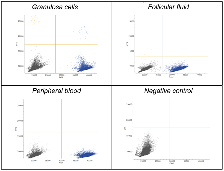 |
Figure 3 Detection of mutation abundance in different samples with ddPCR. The X-axis represents the FAM channel fluorescence intensity, and the Y-axis represents the CY5 channel fluorescence intensity. Each dot represents an individual droplet: black dots indicate droplets without DNA template (both negative), blue dots indicate droplets containing only the wild-type sequence (FAM positive), yellow dots indicate droplets containing only the mutant sequence (CY5 positive), and purple dots indicate double-positive droplets containing both wild-type and mutant sequences. |
PGT Cycle and Follow-Up
In this PGT cycle, six embryos were cultured into blastocysts following intracytoplasmic sperm injection and successfully biopsied. Among the six embryos, two were female and four were male (Table 3). In the NGS-based PGT-A platform, we only reported CNVs larger than 10 Mb and mosaicism ranging from 30% to 70% (specifically those exceeding 30 Mb). The results of PGT-A indicated that four out of the six embryos were euploid (E02, E03, E05, E06). Of the remaining two embryos, one was identified as a mosaic embryo (E01) with a high percentage of mosaic aneuploidy (>40%), while the other embryo (E04) was classified as a carrier with aneuploidy (45, XX, −22).
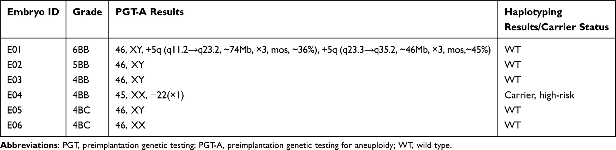 |
Table 3 Summary of the PGT Results for the Patient |
In the haplotype diagram (Figure 4), the blue bar represents the paternal normal haplotype; the dark orange bar denotes the maternal low-risk haplotype; and the light orange bar with a diagonal stripes indicates the maternal high-risk haplotype. Consequently, it is evident that E04 is a high-risk female carrier. E01, E02, E03 and E05 categorized as low-risk males; whereas E06 is classified as a low-risk female. Therefore, the transfer of E01 and E04 was not recommended; genetic counseling was advised prior to transplantation. Four unaffected euploid embryos exhibiting low-risk haplotypes could be transferred in a subsequent frozen-thawed embryo transfer cycle. This family underwent the first frozen-thawed embryo transfer cycle using embryo E03 in April 2022 and achieved clinical pregnancy. PGT-M results showed no allele dropout and were comparable to prenatal diagnosis. An amniocentesis test was performed subsequently; prenatal diagnosis results revealed no chromosomal abnormalities. After completing 40 weeks of gestation, this family welcomed a healthy live baby in February 2023.
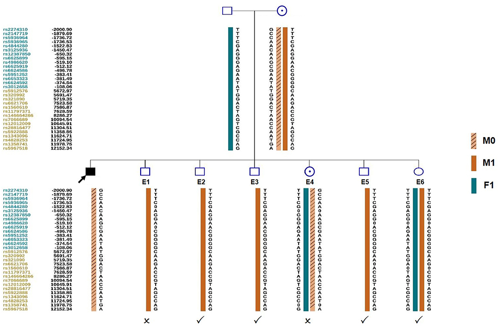 |
Figure 4 SNP-based haplotype linkage analysis of the HDAC8 microdeletion. The use of the pedigree symbols follows the standards of ACMG and ESHG, where squares represent males, circles represent females, a black square with an arrow indicates the male proband, and a circle with a dot indicates the female carrier. The Figure displays only part of the SNP results. RS IDs with blue and yellow represent upstream and downstream SNPs of the microdeletion, respectively. The haplotype linkage used POC tissue as the phasing reference. M0: maternal high-risk chromosome X. M1: maternal low-risk chromosome X. F1: paternal normal chromosome X. ✔: transferable embryo. ×: untransferable embryo. |
Discussion
CdLS is characterized by its clinically heterogeneous presentation affecting multiple systems. CdLS5 represents a rare X-linked dominant hereditary disorder caused by mutations in the HDAC8 gene located at Xq13.1 and accounts for approximately 4% of all CdLS cases.9 As a haploinsufficient gene, nonsense/missense variants along with CNVs are primarily responsible for CdLS5 manifestations. Reports indicate thirteen deletions and one duplication involving the HDAC8 gene.4,7–10 In this study, we identified two male fetuses exhibiting the same deletions in two POC tissues, while no deletions were detected in maternal peripheral blood. This finding suggests the presence of maternal gonadal mosaicism. We successfully determined the CNV breakpoints using long-range PCR and nanopore sequencing on the POC tissue. Additionally, ddPCR was employed to confirm these results and assess the ratio of mosaicism within female gonads. Our findings underscore the clinical significance of quantitatively assessing maternal mosaicism through CNVs by accurately identifying genomic breakpoints. Furthermore, analyzing DNA from ovarian granulosa cells and follicular fluid samples obtained from women suspected of having gonadal mosaicism during routine IVF procedures may serve as an effective source for verifying maternal gonadal mosaicism.
Mosaicism refers to the existence of two or more cell lines with distinct genomic information within an individual, resulting from mutations that occur during early embryonic development.14 Germline mosaicism is characterized by a combination of both normal and mutated gametes due to gonosomal and gonadal factors.14 Patients with germline mosaicism often present phenotypically normal; however, they face a significantly increased risk of recurrently giving birth to affected children. The ddPCR results show approximately 1% deletion-positive signal in ovarian granulosa cells, implying roughly 0.5% of oocytes might carry the deleted Xq13.1 segment. However, 2 out of 2 natural pregnancies of our patient were affected by the same deletion, which suggested the gondal mosaic ratio was extraordinarily underestimated. This severe discrepancy might be associated with tissue heterogeneity, while samples from ovarian granulosa cells and follicular fluid could not fully reflect the in vivo ovary spectrum. Further ovarian biopsy is unfeasible due to potential harm to women. By contrast, sampling of ovarian granulosa cells is non-invasive for women undergoing assisted reproductive treatment. Even with a quite low mosaic proportion, it confirms the genuine existence of gonadal mosaicism. Therefore, it is valuable and should be advocated in clinical utility to collect specimens such as ovarian granulosa cells and follicular fluid to verify pathogenic variants in females suspected of gonadal mosaicism.
The first child of a parent with germline mosaicism is frequently misdiagnosed as having a de novo mutation since germline mosaicism is typically not suspected until another affected child is born. The recurrence risk associated with this condition depends on whether the mosaic mutation resides in the paternal or maternal germlines and on the proportion of germ cells carrying the mutation. To date, only three patients have been reported concerning point mutations in HDAC8 related to mosaicism15,16 along with one case involving a deletion.5 Somatic mosaicism linked to a CNV disrupting HDAC8 has previously been documented once in relation to a CdLS phenotype.17 This report represents the first instance documenting gonadal mosaicism with a deletion affecting the HDAC8 gene. All family members exhibited none of the typical symptoms associated with CdLS. Only our proband experienced two adverse pregnancies, both linked to the same deletion in the HDAC8 gene. This finding underscores the importance of analyzing DNA from POC tissues as a routine molecular genetic diagnostic tool for individuals suffering from recurrent unfavorable pregnancies, even in the absence of a family history of genetic disease.
When germline mosaicism is suspected, it is crucial to consider factors such as disease severity, widely expressed manifestations, and the current lack of cost-effective treatments. In this context, appropriate family planning and prevention through prenatal diagnosis or PGT-M represent two viable strategies for at-risk couples seeking to avoid giving birth to affected fetuses. As an early form of prenatal diagnosis aimed at preventing birth defects, PGT-M can identify embryos carrying low-risk genetic mutations prior to pregnancy. This approach allows couples to be spared from the mental anguish and physical pain associated with terminating affected pregnancies. However, detecting smaller CNVs, such as the 170 kb deletion observed in this particular case, poses challenges at the single-cell level due to NGS-based PGT-A typically having a resolution limit exceeding 5–10 Mb.18 The advent of NGS, which sequences flanking genetic markers like SNPs more efficiently than traditional PCR methods used for short tandem repeat, has revolutionized this field. NGS-based SNP analysis has been successfully employed in PGT for haplotype linkage analysis across multiple monogenic diseases and matching human leukocyte antigens. Consequently, our center’s strategy for implementing PGT in families with inherited CNVs less than 1 Mb involves identifying at-risk chromosomes by detecting SNPs within 2 Mb regions upstream and downstream of pathogenic CNVs while screening for unaffected embryos devoid of these at-risk chromosomes. For cases involving CNVs present in a germline mosaic state, PGT-M primarily relies on haplotyping construction to differentiate high-risk haplotypes—similar segments derived from two pregnancies—from low-risk haplotypes that correspond to other maternal or paternal chromosome segments. This study is the first to report the application of PGT-M for germline mosaicisms associated with HDAC8 deletion.
The haplotype linkage approach is inherently over-conservative in the mosaic setting. It excludes approximately 50% of embryos (hemizygous in males, heterozygous in females) in such X-linked dominant hereditary disorders based on high-risk haplotypes alone, even though only a small fraction of those would actually carry the deletion. Typically, it is not advisable to transfer embryos with a high-risk haplotype without confirming the actual presence of CNVs. However, it may lead to unnecessary discarding of potentially unaffected embryos. With advancements of CNV direct detection using the precise breakpoints in nanopore sequencing and ddPCR, it may be possible to determine whether high-risk embryos genuinely carry CNVs and rescue the majority of “high-risk” embryos that are in fact unaffected. In our study, our patients have only one embryo E04 exhibiting a high-risk haplotype. The reason why we did not directly detect the deletion using the precise breakpoints on embryo E04 by ddPCR was that the length of MALBAC product that our WGA method initially adopted is only about 2 kb, which fell far short of the length of ddPCR product, nearly 10 to 15 kb. Given that we have sufficient available embryos, we discontinued further experimental exploration on the single high-risk haplotype embryo E04. Nevertheless, it inevitably leaves a regret for this research. Accordingly, suitable WGA methods should be chosen for follow-up tests. Nevertheless, for cases characterized by a significant proportion of gonadal mosaicism or those with multiple high-risk embryos, this methodology can assist patients in identifying viable embryos and preserving unaffected ones in principle.
The probability of recurrence is influenced by the extent of gonadal mosaicism, which could be estimated by gonad sampling techniques such as semen analysis and egg retrieval.19 The risks and discomfort associated with egg retrieval surgery, coupled with the limited number of eggs obtainable and the intrinsic value of follicle samples for women, pose challenges for clinical implementation compared to semen sampling—which is generally easier, more convenient, and incurs no additional physical or psychological burden on man. Consequently, there are few clinical reports addressing female gonadal mosaicism. In fact, for couples suspected of maternal gonadal mosaicism who require IVF assistance for conception purposes: during routine egg retrieval procedures millions of granulosa cells from the outer layer surrounding oocytes—as well as follicular fluid—can be collected; these share identical genetic backgrounds suitable for further investigation. Our ovarian sampling method does not impose any additional pain or expense on women, nor does it increases the failure rate of embryo culture. This approach provides a solid foundation for successfully demonstrating gonadal mosaicism in our study and will be utilized to validate the existence of suspected maternal gonadal mosaicism.
In this case, the deletion simultaneously disrupts HDAC8 and PHKA1 genes. HDAC8 haploinsufficiency is the core driver of the CdLS phenotype and male embryonic lethality in this family, as the clinical manifestations of the two affected fetuses are fully consistent with the typical features of CdLS. For female carriers, heterozygous deletion of PHKA1 may lead to mild or subclinical symptoms of GSD IXa due to X-chromosome inactivation. Therefore, our PGT-M strategy simultaneously excluded the transmission of this multi-gene deletion, avoiding the risk of both CdLS and GSD IXa in the offspring.
In summary, several key insights derived from this case are as follows. Firstly, in situations where couples seek consultation regarding potential germline mosaicism, if there is only one affected child in the family and both partners exhibit no variations in peripheral blood, initial consideration should favor the possibility that the child has a de novo mutation. This scenario can be managed through natural conception combined with prenatal diagnosis as a reproductive strategy. However, if there are recurrent instances of the identical gene variations among children with de novo mutations within the family or observed in fetuses, suspicion should be raised regarding the potential for germline mosaicism. In such cases, options may include natural conception accompanied by prenatal diagnosis or employing PGT-M alongside prenatal diagnosis to mitigate the occurrences of allele dropout or recombination.20 Secondly, when suspecting the presence of germline mosaicism, efforts should be made to confirm the diagnosis at a technical level. Nevertheless, due to challenges related to sample specificity and testing complexities—based on collective experiences across multiple centers—it is generally not mandatory to pursue confirmation at this level. For families seeking further validation but facing limitations in obtaining and testing maternal germ cells, priority may be given to assessing sperm for evidence of mosaicism. If there is no mosaicism is detected in the sperm samples, attention can be shift towards preserving granulosa cells, follicular fluid, or other viable sample alternatives during ovum retrieval from the female partner undergoing ovarian stimulation for future testing needs. Thirdly, families with suspicions of germline mosaicism who intend to pursue reproduction through PGT-M should receive comprehensive genetic counseling. This counseling should encompass a full disclosure of various possibilities, including the clinical complexities and costs associated with diagnosing germline mosaicism, comparisons between PGT-M and natural conception accompanied by prenatal diagnosis, considerations regarding embryo utilization rates, as well as the economic and emotional investments required from the patient’s family.
To our knowledge, our study is by far the first to report the application of PGT-M for germline mosaicisms associated with HDAC8 deletion. However, there were some limitations in this research. Firstly, due to a lack of pathological sections or samples from skin, nails, saliva or buccal mucosa, the existence of somatic mosaicism remains questionable. Secondly, the diagnosis of gonadal mosaicism was determined merely by genetic testing on peripheral blood from parents and ovarian granulosa cells from the female participant, without pathological validation in gonadal tissues or abundant clinical manifestation. In addition, these results may not accurately reflect mutations present in other tissues or exact germline mosaic ratio. This underscores the importance of retaining an adequate number and variety of multi-tissue samples when encountering suspected cases of gonadal mosaicism. Lastly, genetic deletions were not verified directly on MDA products of embryos, so embryos without deletions among high-risk ones were not able to be screened, and the number of available embryos could not be increased in this case. However, it is an area of promising application with technical feasibility and suitable WGA choices for direct CNV detection.
Conclusion
Our data demonstrate the PGT-M based on haplotype linkage analysis is effective for CNVs less than 1 Mb even in a gonadal mosaic state. Furthermore, it highlights the significance of parental testing in families affected by CdLS5, as well as the reproductive utility of IVF combined with PGT in families exhibiting low-level parental gonadal mosaicism. By employing various techniques—including NGS, array comparative genomic hybridization, nanopore sequencing, and ddPCR—to precisely define mutations, we illustrate how to address some uncommon genetic mechanisms involved in diagnosing microdeletions within gonadal mosaicism. The broader implementation of this approach in many families with mosaic state will benefit from the technical feasibility of direct confirmation to improve embryo utilization.
Abbreviations
PGT-M, preimplantation genetic testing for monogenic disorders; CdLS5, Cornelia de Lange syndrome type 5; CNV, copy number variation; CMA, chromosomal microarray analysis; WES, whole exome sequencing; ddPCR, droplet-digital PCR; CdLS, Cornelia de Lange syndrome; HDCA8, histone deacetylase 8; gDNA, Genomic DNA; SNP, single nucleotide polymorphism; WGL-MPS, Whole genome low-coverage mate-pair sequencing; IVF, in vitro fertilization; ICSI, intracytoplasmic sperm injection; TE, trophectoderm; WGA, whole-genome amplification; MALBAC, multiple annealing and looping-based amplification cycles; NGS, next-generation sequencing; MARSALA, Mutated Allele Revealed by Sequencing with Aneuploidy and Linkage Analyses; PGT-A, preimplantation genetic testing for aneuploidy.
Data Sharing Statement
All data supporting the findings of this report are available from the corresponding author Ping Hu, e-mail [email protected], upon reasonable request.
Ethics Approval and Informed Consent
Writing and publishing this case report was approved by Nanjing Women and Children’s Healthcare Hospital.
Consent for Publication
Written consent from the patient for case publication of potentially identifying images and clinical details.
Acknowledgments
We would like to express our deepest gratitude to the family who participated in this study for their courage and willingness to share their experiences. We are also grateful for the compassionate care provided by our genetic counseling team. This paper has been uploaded to https://www.researchsquare.com/article/rs-5668001/v1.
Author Contributions
All authors made a significant contribution to the work reported, whether that is in the conception, study design, execution, acquisition of data, analysis and interpretation, or in all these areas; took part in drafting, revising or critically reviewing the article; gave final approval of the version to be published; have agreed on the journal to which the article has been submitted; and agree to be accountable for all aspects of the work.
Funding
This work was supported by the National Key R&D Program of China (2022YFC2703400 to Z.X. and P.H. and 2021YFC1005301 to P.H.), National Natural Science Foundation of China (No. 81971398 and No. 82371862 to P.H), Jiangsu Province Capability Improvement Project through Science, Technology and Education Jiangsu Provincial Medical Key Discipline (ZDXK202211 to Z.X.), Open Project of the Genetic Diseases Key Laboratory of Sichuan Province (2025kflx004 to Y.W.).
Disclosure
The authors report no conflicts of interest in this work.
References
1. Kline AD, Moss JF, Selicorni A, et al. Diagnosis and management of Cornelia de Lange syndrome: first international consensus statement. Nat Rev Genet. 2018;19(10):649–14. doi:10.1038/s41576-018-0031-0
2. Deardorff MA, Noon SE, Krantz ID. Cornelia de Lange Syndrome. In: Adam MP, Bick S, Mirzaa GM, Pagon RA, Wallace SE, Amemiya A, editors. GeneReviews®. Seattle: University of Washington, Seattle; 2005.
3. Avagliano L, Bulfamante GP, Massa V. Cornelia de Lange syndrome: to diagnose or not to diagnose in utero? Birth Defects Res. 2017;109(10):771–777. doi:10.1002/bdr2.1045
4. Lucia-Campos C, Valenzuela I, Latorre-Pellicer A, et al. A novel intragenic duplication in the HDAC8 gene underlying a case of Cornelia de Lange Syndrome. Genes. 2022;13(8):1413. doi:10.3390/genes13081413
5. Helgeson M, Keller-Ramey J, Knight Johnson A, et al. Molecular characterization of HDAC8 deletions in individuals with atypical Cornelia de Lange syndrome. J Hum Genet. 2018;63(3):349–356. doi:10.1038/s10038-017-0387-6
6. Kaur M, Blair J, Devkota B, et al. Genomic analyses in Cornelia de Lange Syndrome and related diagnoses: novel candidate genes, genotype-phenotype correlations and common mechanisms. Am J Med Genet A. 2023;191(8):2113–2131. doi:10.1002/ajmg.a.63247
7. Kaiser FJ, Ansari M, Braunholz D, et al. Loss-of-function HDAC8 mutations cause a phenotypic spectrum of Cornelia de Lange syndrome-like features, ocular hypertelorism, large fontanelle and X-linked inheritance. Hum Mol Genet. 2014;23(11):2888–2900. doi:10.1093/hmg/ddu002
8. Salehi Karlslätt K, Pettersson M, Jäntti N, et al. Rare copy number variants contribute pathogenic alleles in patients with intestinal malrotation. Mol Genet Genomic Med. 2019;7(3):e549. doi:10.1002/mgg3.549
9. Gross AM, Ajay SS, Rajan V, et al. Copy-number variants in clinical genome sequencing: deployment and interpretation for rare and undiagnosed disease. Genet Med. 2019;21(5):1121–1130. doi:10.1038/s41436-018-0295-y
10. Liu C, Li X, Cui J, et al. Analysis of clinical and genetic characteristics in 10 Chinese individuals with Cornelia de Lange syndrome and literature review. Mol Genet Genomic Med. 2020;8(10):e1471. doi:10.1002/mgg3.1471
11. Wu H, Shen X, Huang L, et al. Genotyping single-sperm cells by universal MARSALA enables the acquisition of linkage information for combined pre-implantation genetic diagnosis and genome screening. J Assist Reprod Genet. 2018;35(6):1071–1078. doi:10.1007/s10815-018-1158-9
12. Li S, Chang C, Bai H, et al. A novel and comprehensive whole-genome sequencing-based preimplantation genetic testing approach for different genetic conditions. J Mol Diagn. 2025;27(5):395–404. doi:10.1016/j.jmoldx.2025.02.002
13. Riggs ER, Andersen EF, Cherry AM, et al. Technical standards for the interpretation and reporting of constitutional copy-number variants: a joint consensus recommendation of the American College of Medical Genetics and Genomics (ACMG) and the Clinical Genome Resource (ClinGen). Genet Med. 2020;22(2):245–257. doi:10.1038/s41436-019-0686-8
14. Biesecker LG, Spinner NB. A genomic view of mosaicism and human disease. Nat Rev Genet. 2013;14(5):307–320. doi:10.1038/nrg3424
15. Krawczynska N, Wierzba J, Wasag B. Genetic mosaicism in a group of patients with Cornelia de Lange Syndrome. Front Pediatr. 2019;7:203. doi:10.3389/fped.2019.00203
16. Jezela-Stanek A, Murcia PV, Jurkiewicz D, et al. Novel variant in HDAC8 gene resulting in the severe Cornelia de Lange phenotype. Clin Dysmorphol. 2019;28(3):126–130. doi:10.1097/MCD.0000000000000277
17. Parenti I, Gervasini C, Pozojevic J, et al. Expanding the clinical spectrum of the ‘HDAC8-phenotype’ - implications for molecular diagnostics, counseling and risk prediction. Clin Genet. 2016;89(5):564–573. doi:10.1111/cge.12717
18. Chen HF, Chen M, Ho HN. An overview of the current and emerging platforms for preimplantation genetic testing for aneuploidies (PGT-A) in in vitro fertilization programs. Taiwan J Obstet Gynecol. 2020;59(4):489–495. doi:10.1016/j.tjog.2020.05.004
19. Dai C, Cheng D, Li W, Zeng S, Lu G, Zhang Q. Identification of paternal germline mosaicism by MicroSeq and targeted next-generation sequencing. Mol Genet Genomic Med. 2020;8(9):e1394. doi:10.1002/mgg3.1394
20. Practice Committee and Genetic Counseling Professional Group of the American Society for Reproductive Medicine ASfRM, Washington, D.C. Indications and management of preimplantation genetic testing for monogenic conditions: a committee opinion. Fertil Steril. 2023;120(1):61–71. doi:10.1016/j.fertnstert.2023.03.003
 © 2026 The Author(s). This work is published and licensed by Dove Medical Press Limited. The
full terms of this license are available at https://www.dovepress.com/terms
and incorporate the Creative Commons Attribution
- Non Commercial (unported, 4.0) License.
By accessing the work you hereby accept the Terms. Non-commercial uses of the work are permitted
without any further permission from Dove Medical Press Limited, provided the work is properly
attributed. For permission for commercial use of this work, please see paragraphs 4.2 and 5 of our Terms.
© 2026 The Author(s). This work is published and licensed by Dove Medical Press Limited. The
full terms of this license are available at https://www.dovepress.com/terms
and incorporate the Creative Commons Attribution
- Non Commercial (unported, 4.0) License.
By accessing the work you hereby accept the Terms. Non-commercial uses of the work are permitted
without any further permission from Dove Medical Press Limited, provided the work is properly
attributed. For permission for commercial use of this work, please see paragraphs 4.2 and 5 of our Terms.