Back to Journals » Clinical Ophthalmology » Volume 17
Preclinical Investigation of Ab Interno Goniotomy Using Three Different Techniques
Authors Ammar DA , Porteous E, Kahook MY
Received 8 June 2023
Accepted for publication 17 August 2023
Published 4 September 2023 Volume 2023:17 Pages 2619—2623
DOI https://doi.org/10.2147/OPTH.S424977
Checked for plagiarism Yes
Review by Single anonymous peer review
Peer reviewer comments 2
Editor who approved publication: Dr Scott Fraser
David A Ammar,1 Eric Porteous,2 Malik Y Kahook3
1Research Department, Lions World Vision Institute, Tampa, FL, USA; 2New World Medical, Rancho Cucamonga, CA, USA; 3Department of Ophthalmology, University of Colorado Anschutz Medical Campus, Aurora, CO, USA
Correspondence: Malik Y Kahook, Department of Ophthalmology, University of Colorado Anschutz Medical Campus, Aurora, CO, USA, Tel +1 720 848 2501, Email [email protected]
Purpose: To evaluate incisional or excisional tissue-level effects of ab interno goniotomy techniques on human trabecular meshwork (TM).
Methods: The TM from human cadaveric corneal rim tissue was treated using three devices: (1) Kahook Dual Blade (KDB) GLIDE, (2) iAccess, and (3) SION. Two human corneal rims were used for each of the iAccess and SION devices and one with the KDB GLIDE, with 360 degrees of TM treated in each case. Sections were then prepared for analysis and comparison between devices. Tissue samples underwent standard histologic processing with H&E stain, followed by comparative analyses.
Results: Areas treated with the KDB GLIDE device resulted in nearly complete excision of TM overlying the canal of Schlemm without injury to surrounding tissues. The iAccess device can be used as a focal trephine to create holes or dragged for TM disruption. When used to create holes, iAccess punched through the full thickness of the TM and also disrupted the anterior scleral tissue. It caused some incisional openings through the TM but with significant leaflets remaining and minimal true “hole-punch” effect. When the device tip was dragged, iAccess incised the TM and left debris behind with little, if any, excision of tissue. SION led to both incision and excision of TM with incision predominating over excision.
Conclusion: The various methods evaluated to perform ab interno goniotomy resulted in varying degrees of TM incision or excision. Only the KDB GLIDE device resulted in reliable excision of TM, while the other devices produced incision or minimal excision of tissue with residual leaflets and debris. Use of iAccess resulted in focal disruption of the anterior scleral wall. Because incisional approaches that leave longer residual leaflets may be more prone to fibrosis and closure compared to excisional treatments, clinical correlation will be necessary to better understand the significance of these findings with respect to relative effectiveness of intraocular pressure lowering in eyes with glaucoma.
Keywords: glaucoma, goniotomy, excision, incision, trabecular meshwork
Introduction
Goniotomy was first described by Otto Barkan as incision of the trabecular meshwork (TM) to facilitate the flow of aqueous humor from the anterior chamber to the canal of Schlemm with the goal of reducing intraocular pressure (IOP) in eyes with glaucoma.1 More recently, an array of surgical techniques and instrumentation have been developed to optimize goniotomy outcomes. These include procedures and instruments that perform incisional goniotomy (the microvitreoretinal [MVR] blade, the Trabectome® device (MicroSurgical Technology, Redmond, WA), excisional goniotomy (the Kahook Dual Blade® (KDB®) and KDB GLIDE® [New World Medical, Rancho Cucamonga, CA]) or the TrabEx™/TrabEx+™ device (Microsurgical Technology), as well as, 360° trabeculotomy (gonioscopy-assisted transluminal trabeculotomy [GATT] using a suture, among others.
The effects of each of these procedures on TM tissue are variable. By design, incisional techniques cut through the TM without removing a large amount of tissue, leaving residual leaflets on either side of the incision that can reapproximate and reverse the surgical effect.2–4 Conversely, excisional instruments are intended to remove portions of the TM; ideally, these excised portions are of uniform and adequate size to prevent reapproximation of tissue edges. In two previous preclinical studies, the histologic appearance of the TM following incisional and excisional goniotomy with the instruments listed above has been described. As would be expected, the various techniques produced varying degrees of TM removal; only the KDB and KDB GLIDE devices provided consistent TM excision,2,3 likely related to its dual blade and ramp configuration designed to stretch, elevate, and excise TM (Figure 1A).
 |
Figure 1 Images of the 3 devices with tips enlarged. (A) Kahook Dual Blade (KDB) GLIDE, (B) iAccess Trabecular Trephine, and (C) SION Surgical Instrument. |
Since these prior studies were conducted, additional devices for performing goniotomy have been developed, including the iAccess® Trabecular Trephine (Glaukos, Aliso Viejo, CA) and the SION™ Surgical Instrument (Sight Sciences, Menlo Park, CA). The iAccess features a 30g circular blade trephine for creation of holes in the TM and features a 300-micron deep backstop designed to prevent damage to the canal (Figure 1B).5 The SION features an upper and lower foot that bracket the TM to pull/tear tissue with advancement (Figure 1C).6 In this study, we describe the histologic appearance of the TM and canal of Schlemm following goniotomy with the KDB GLIDE, iAccess, and SION instruments.
Materials and Methods
Prior to study commencement, this preclinical laboratory study was granted exemption by the Colorado Multiple Institutional Review Board for use of human material on the basis that all information was deidentified and devoid of non-public information disclosures.
Five corneoscleral rims were provided by the Lions World Vision Institute (Tampa, FL). Each tissue sample was removed from storage medium, mounted with the TM facing up, and secured with tissue pins. Two samples were used for the iAccess and SION technique and one sample for the KDB GLIDE technique (as histology with KDB GLIDE excisional goniotomy has been reported previously3). In all cases, the full 360 degrees of TM was treated under direct microscopic visualization. The KDB GLIDE was used as per the manufacturer’s instructions7 as follows: the instrument’s blade tip was used to pierce TM and the footplate positioned within the canal of Schlemm and advanced along the canal. Upon completion of the intended excision in one direction, the tip was redirected to excise the trailing TM strip. The iAccess instrument was used as previously described:5 under gentle pressure, multiple full-thickness circles of TM were removed. Also, while not described previously, the tip was placed in one of the holes and dragged along the canal, disrupting TM in an incisional manner. The SION instrument was used as per the manufacturer’s instructions6 as follows: the instrument’s toe was used to puncture the TM and the lower foot seated in the canal. A viewing window in the upper foot permitted visualization of TM to ensure proper placement before advancement along the canal to disrupt TM.
After each procedure, rims were preserved in 4% paraformaldehyde/phosphate-buffered saline overnight at 4° C, then cut into 2–3 mm wide radial pieces and embedded in paraffin with the cut edge of each rim segment facing the front of the block. Two to three 10 µm-thick tissue sections were then cut from randomly selected locations of each sample and stained using Mayer’s hematoxylin and eosin Y (H&E, Richard-Allan Scientific, Kalamazoo, Michigan, USA). Bright-field imaging was obtained using a Nikon Eclipse 80i microscope (Nikon, Melville, New York, USA) fitted with a Nikon D5-Fi1 color camera and a Nikon CFI 10x or 20x/Plan Fluor objective lens. Masked evaluation of the images was undertaken to qualitatively characterize the tissue effects of each technique.
Results
Systematic histological examination of each imaged section was undertaken. Findings were consistent between sections within each corneal rim and, for the iAccess and SION evaluations, between corneal rims as well.
Representative sections of rims undergoing goniotomy with the KDB GLIDE are illustrated in Figure 2. The KDB GLIDE resulted in nearly complete excision of the TM overlying the canal, leaving leaflet remnants ranging from 0 to 50 µm in width. No collateral damage to adjacent structures (outer canal wall, anterior sclera) was seen in any section.
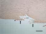
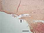
When used as a trephine, the iAccess instrument punched through the full thickness of the TM with incisional rather than excisional openings and residual leaflets ranging from 50 to 150 µm, with accompanying disruption of anterior scleral tissue (Figure 3A). When dragged along the canal, the iAccess incised the TM and pulled it free from the canal but with significant residual debris and little, if any, tissue excision (Figure 3B).
SION goniotomy excises 50–75 µm of central TM in only approximately one-third of the images analyzed, with either simple incision of the TM (or excision of <50 µm) in the other instances (Figure 4). Residual leaflets ranging from 50 to 150 µm in width dominated in areas of excision. No collateral injury to adjacent structures was seen.
Discussion
Elevated IOP in eyes with open-angle glaucoma arises from increased aqueous outflow resistance through the TM.8,9 Goniotomy overcomes this outflow resistance and lowers IOP by restoring aqueous flow through the trabecular outflow pathway. Multiple instruments and techniques have been developed to achieve IOP reduction with goniotomy. Incisional techniques cut through but do not remove TM tissue. Residual tissue leaflets remain on both sides of the incision which can reapproximate to close the opening resulting in surgical failure.2–4 In contrast, excisional techniques are intended to remove regions of the TM, whether in strips or trephined circles. The ideal excision dimension (width for strips, diameter for circles) should be large enough to minimize the risk of reapproximation of any residual tissue leaflets.
In this study, we have evaluated the characteristics of goniotomies created using the KDB GLIDE, iAccess, and SION instruments. Consistent with a similar prior study of histology following goniotomy with the KDB, MVR blade, and Trabectome,2 as well as a study of histology following goniotomy with the KDB GLIDE and MVR blade, and trabeculotomy with a 5–0 prolene suture or TrabEx,3 the present study demonstrated more complete excision of TM with fewer and smaller residual tissue leaflets with the KDB GLIDE compared to the iAccess and SION devices. The iAccess device is intended to create circular 30g full-thickness TM excisions via trephination.5 While openings through the TM were observed after iAccess goniotomy, these did not have the expected “hole-punch” appearance and left significant residual leaflets behind. The SION device, described as bladeless, grasps and pulls/drags TM, tearing it away from the scleral spur and Schwalbe’s line by blunt force.6 Incomplete separation from either the scleral spur or Schwalbe’s line, or both, would be expected to produce a mixed incisional and excisional effect. This was observed in the current study, with incisional effects seen more extensively than excisional effects, suggesting that the device effectively incises TM rather than grasping and dragging to tear the TM from its attachments.
From a safety perspective, an ideal goniotomy device would cause little or no collateral damage to adjacent, non-targeted tissues. In the current study, the iAccess device – when used as a trephine produced significant injury to the anterior wall of the canal of Schlemm. This occurs, despite the device having a 300-micron deep backstop, specifically to prevent injury to tissues deep to the TM.5 No significant injury to neighboring non-target tissues was seen with either the KDB GLIDE or the SION device.
Conclusion
In summary, this study in combination with two prior studies2,3 demonstrates that the various available methods of performing goniotomy produce significant tissue effect variation, with some providing more incision of the TM, others more excision, and some resulting in damage to adjacent structures. Only the KDB GLIDE device consistently excised TM without collateral injury. Clinical correlation is necessary to better characterize the influence of these differential tissue effects on IOP reduction in eyes with glaucoma.
Funding
Research funded by New World Medical.
Disclosure
Dr David A Ammar reports Contract Research from Lions World Vision Institute, during the conduct of the study; Contract Research from Lions World Vision Institute, outside of the submitted work. Mr Eric Porteous is an employee of New World Medical. Dr Malik Kahook reports being a consultant to New World Medical during the conduct of the study. In addition, Dr Malik Kahook has a patent US10,327,947B2 issued to and owned by New World Medical.
References
1. New Eye Surgery Used In Glaucoma. Dr. Otto Barkan’s technique is hailed by specialists at convention here. New York Times; 1936.
2. Seibold LK, Soohoo JR, Ammar DA, Kahook MY. Preclinical investigation of ab interno trabeculectomy using a novel dual-blade device. Am J Ophthalmol. 2013;155(3):524–9 e2. doi:10.1016/j.ajo.2012.09.023
3. Ammar DA, Seibold LK, Kahook MY. Preclinical investigation of goniotomy using four different techniques. Clin Ophthalmol. 2020;14:3519–3525. doi:10.2147/OPTH.S281811
4. Amari Y, Hamanaka T, Futa R. Pathologic investigation failure of trabeculotomy. J Glaucoma. 2015;24(4):316–322. doi:10.1097/IJG.0b013e31829e1d6e
5. iAccess: a novel goniotomy tool for the reduction of intraocular pressure. Available from: https://ophthalmology360.com/glaucoma/iaccess-trabecular-meshwork-trephine-a-novel-goniotomy-tool-for-the-reduction-of-intraocular-pressure/.
6. Sight Sciences. SION Surgical Instrument. Menlo Park, CA: Sight Sciences; 2022.
7. New World Medical. KDB GLIDE Instructions for Use. Rancho Cucamonga, CA: New World Medical; 2021.
8. Grant WM. Experimental aqueous perfusion in enucleated human eyes. Arch Ophthalmol. 1963;69(6):783–801. doi:10.1001/archopht.1963.00960040789022
9. Maepea O, Bill A. Pressures in the juxtacanalicular tissue and Schlemm’s canal in monkeys. Exp Eye Res. 1992;54(6):879–883. doi:10.1016/0014-4835(92)90151-H
 © 2023 The Author(s). This work is published and licensed by Dove Medical Press Limited. The full terms of this license are available at https://www.dovepress.com/terms.php and incorporate the Creative Commons Attribution - Non Commercial (unported, v3.0) License.
By accessing the work you hereby accept the Terms. Non-commercial uses of the work are permitted without any further permission from Dove Medical Press Limited, provided the work is properly attributed. For permission for commercial use of this work, please see paragraphs 4.2 and 5 of our Terms.


