
Back to Journals » Cancer Management and Research » Volume 18


Pre-Infusion Host-Marrow Vulnerability in CD19- and BCMA-Directed CAR T-Cell Therapy: Clonal Hematopoiesis, Hematotoxicity, and Therapy-Related Myeloid Neoplasia
Authors Liu Y, Su G, Li R, Wang S
Received 2 June 2026
Accepted for publication 8 July 2026
Published 18 July 2026 Volume 2026:18 629588
DOI https://doi.org/10.2147/CMAR.S629588
Checked for plagiarism Yes
Review by Single anonymous peer review
Peer reviewer comments 2
Editor who approved publication: Professor Seema Singh
Yong Liu,1 Guiyu Su,2 Ruijin Li,3 Shuang Wang4
1Department of Hematology, the First Hospital of China Medical University, Shenyang, 110001, People’s Republic of China; 2Department of Oncology Medicine, the First Hospital of China Medical University, Shenyang, 110001, People’s Republic of China; 3Emergency Department, the First Hospital of China Medical University, Shenyang, 110001, People’s Republic of China; 4Operation Room, the First Hospital of China Medical University, Shenyang, 110001, People’s Republic of China
Correspondence: Shuang Wang, Operation Room, the First Hospital of China Medical University, No. 155 Nanjing North Street Heping District, Shenyang, Liaoning, People’s Republic of China, Email [email protected]
Abstract: CD19- and B-cell maturation antigen (BCMA)-directed chimeric antigen receptor T-cell (CAR T-cell) therapies have improved outcomes in relapsed or refractory B-cell lymphoid malignancies and multiple myeloma, but late hematologic complications are increasingly relevant as survivorship expands. This narrative review examines how pre-infusion clonal hematopoiesis (CH), clonal hematopoiesis of indeterminate potential (CHIP), clonal cytopenia of undetermined significance (CCUS), marrow reserve, prior genotoxic exposure, inflammatory stress, and disease-platform context shape the risks of prolonged cytopenia and therapy-related myeloid neoplasms (t-MN) after CAR T-cell therapy. Available evidence suggests that high-risk clonal architecture, particularly TP53-mutated or DNA damage response-associated clones, clonal cytopenia, larger or multiple clones, and heavy prior cytotoxic exposure, is more clinically informative than CHIP positivity alone. By contrast, associations between unstratified CH and prolonged cytopenia remain heterogeneous. CD19 lymphoma and BCMA myeloma settings share clonal-selection biology but differ in marrow ecology, treatment history, baseline cytopenia, inflammatory burden, and surveillance windows. For clinical translation, risk assessment should not rely on universal CHIP screening or binary genomic classification. Instead, clonal-risk models, hematotoxicity-risk models, baseline blood counts, inflammatory markers, prior therapy exposure, and disease-platform context should be integrated to identify patients who may benefit from intensified myeloid surveillance, supportive-care planning, and earlier marrow reassessment while preserving access to CAR T-cell therapy.
Keywords: chimeric antigen receptor T-cell therapy, CD19, B-cell maturation antigen, clonal hematopoiesis, clonal hematopoiesis of indeterminate potential, clonal cytopenia of undetermined significance, immune effector cell-associated hematotoxicity, therapy-related myeloid neoplasms, TP53, DNA damage response
Introduction
CD19-directed and B-cell maturation antigen (BCMA)-directed chimeric antigen receptor T-cell (CAR T-cell) therapies have improved outcomes in relapsed or refractory B-cell lymphoid malignancies and multiple myeloma. In large B-cell lymphoma, axicabtagene ciloleucel, tisagenlecleucel, and lisocabtagene maraleucel have produced durable responses in selected patients with historically poor outcomes.1–4 In multiple myeloma, idecabtagene vicleucel and ciltacabtagene autoleucel have produced deep responses in heavily pretreated disease, with randomized studies further expanding the clinical context of BCMA-directed cellular therapy.5–8 As CAR T-cell therapy moves into earlier lines and survivorship expands, delayed hematologic complications are becoming increasingly relevant to treatment planning.
The safety discussion has therefore broadened beyond acute cytokine release syndrome (CRS) and immune effector cell-associated neurotoxicity syndrome (ICANS). Prolonged cytopenia, delayed hematopoietic recovery, infectious vulnerability, impaired immune reconstitution, second primary malignancies, and therapy-related myeloid neoplasms (t-MN) are now important survivorship concerns.9–16 These events may require prolonged supportive care, complicate subsequent therapy, and, in selected patients, indicate evolving myeloid disease. Risk assessment is most useful when it improves counseling, surveillance, and supportive planning without creating inappropriate barriers to CAR T-cell access.
Several adjacent topics have been reviewed, including general CAR T-cell biomarkers, immune effector cell-associated hematotoxicity, and supportive management after BCMA-directed CAR T-cell therapy.10,11,17,18 However, a specific gap remains: how pre-infusion myeloid clonal architecture, clonal cytopenia, inflammatory marrow stress, prior therapy exposure, and disease-platform context should be integrated when assessing host-marrow vulnerability before CD19- or BCMA-directed CAR T-cell therapy.
This narrative review focuses on pre-infusion clonal hematopoiesis (CH), clonal hematopoiesis of indeterminate potential (CHIP), clonal cytopenia of undetermined significance (CCUS), marrow reserve, and therapy-related myeloid neoplasia across CD19 lymphoma and BCMA myeloma settings. A key gap is that these factors are often discussed separately, whereas their combined relevance before CAR T-cell therapy remains insufficiently defined. Cross-platform comparison is useful because CD19 and BCMA CAR T-cell settings share prior genotoxic exposure, DNA damage response-associated clonal selection, inflammatory marrow stress, and impaired hematopoietic reserve, while differing in marrow ecology, treatment history, inflammatory patterns, and surveillance windows.19–21 The objective of this review is to integrate clonal-risk, hematotoxicity-risk, prior therapy exposure, and disease-platform context into a practical framework for interpreting prolonged cytopenia and therapy-related myeloid neoplasia risk while preserving appropriate access to CAR T-cell therapy.
Definitions and Conceptual Boundaries
A clear vocabulary is necessary because CH, CHIP, CCUS, prolonged hematotoxicity, second primary malignancy, and therapy-related myeloid neoplasm are frequently used in overlapping ways.
CH refers to expansion of a hematopoietic clone carrying acquired somatic genetic alterations.22–25 CHIP is conventionally used for individuals with myeloid-malignancy-associated somatic mutations, commonly at a variant allele fraction (VAF) of ≥2%, in the absence of diagnostic evidence of a hematologic malignancy and in the absence of otherwise unexplained persistent cytopenia.26 CHIP comprises biologically distinct states. A low-level isolated DNMT3A clone in an older patient differs substantially from a TP53- or PPM1D-mutated clone in a heavily pretreated lymphoma or myeloma patient. Clone size, mutation number, mutation class, cytopenia, red cell indices, prior cytotoxic exposure, and inflammatory context modify risk interpretation rather than simply confirming CHIP positivity.19,27,28
CCUS occupies a higher-risk myeloid precursor context than CHIP because it combines clonal mutation with otherwise unexplained cytopenia. The presence of cytopenia raises the possibility that clonal hematopoiesis coexists with impaired hematopoietic output or reduced marrow reserve.29 In CAR T-cell candidates, this distinction is clinically important because cytopenia may precede lymphodepletion and may reflect active disease, prior chemotherapy, autologous transplantation, marrow involvement, inflammation, infection, impaired reserve, or an emerging myeloid disorder.10,18 A low-level isolated DNMT3A clone with preserved blood counts therefore represents a different risk state from TP53-mutated clonal cytopenia accompanied by macrocytosis, thrombocytopenia, and heavy prior genotoxic exposure.27
For CAR T-cell risk interpretation, therapy-related clonal cytopenia, referred to here operationally as t-CCUS and overlapping with the t-CC concept, should be analytically separated from incidental CHIP and de novo CCUS. This working category can be considered when three elements coexist: a myeloid-type somatic clone, persistent or otherwise unexplained cytopenia, and prior cytotoxic or genotoxic exposure.21,30 This concept does not constitute a formal diagnosis of therapy-related myeloid neoplasm, but it enriches for a higher-risk clinical context than CHIP alone, especially when cytopenia and prior genotoxic exposure coexist.21 A CAR T-cell candidate with unexplained thrombocytopenia, macrocytosis, prior melphalan or alkylator exposure, and a TP53- or PPM1D-mutated clone should not be interpreted in the same way as a patient with incidental low-VAF DNMT3A CHIP and preserved blood counts.19,31 This working category is therefore useful for CAR T-cell studies because it captures the intersection between pre-existing clonal hematopoiesis, cytopenia, and prior genotoxic selection pressure.19,21,30,31
Prolonged hematotoxicity after CAR T-cell therapy also requires careful definition. Early cytopenia may reflect lymphodepletion, marrow involvement, prior chemotherapy, infection, inflammation, and CAR T-cell-associated immune toxicity. Late or prolonged cytopenia, particularly when persisting beyond day 30 or during day 60 and day 90–100 assessment windows, should be evaluated as a composite endpoint shaped by baseline marrow reserve, inflammatory marrow injury, immune reconstitution, infection, prior therapy, disease involvement, and possible clonal myeloid vulnerability.10,11,18 Detailed supportive management with growth factors, thrombopoietin receptor agonists, transfusion strategies, antimicrobial prophylaxis, or stem-cell boost is outside the scope of this review. Here, prolonged cytopenia and delayed hematopoietic recovery are used as clinical endpoints that may reveal underlying host-marrow vulnerability.
Finally, second primary malignancy (SPM) is broader than secondary myeloid neoplasm. SPM encompasses solid tumors, non-melanoma skin cancers, T-cell and other lymphoid malignancies, and myeloid neoplasms.15 By contrast, therapy-related myeloid neoplasms, including therapy-related myelodysplastic syndromes/neoplasms (MDS) and acute myeloid leukemia (AML), represent a biologically distinct subset shaped by prior genotoxic therapy, pre-existing or therapy-selected myeloid clones, and marrow stress.32 This distinction matters when interpreting post-CAR T-cell safety data. In a systematic review and meta-analysis of 5,517 lymphoma and myeloma patients, Tix et al identified 326 SPMs and reported an overall SPM point estimate of 5.8% (95% CI, 4.7–7.2) after a median follow-up of 21.7 months, but this global estimate combined heterogeneous malignant categories rather than specifically estimating t-MN risk.15 Hamilton et al analyzed a single-center cohort of 724 patients who had received adoptive cellular therapies and emphasized the rarity of second tumors while providing a molecular framework for assessing clonal relationship and vector integration in suspected secondary T-cell lymphoma after CAR T-cell therapy.14 These studies are important for long-term safety surveillance, but global SPM rates should not be treated as surrogates for t-MN risk.15,32 Post-CAR T-cell lymphoma, non-melanoma skin cancer, and TP53-mutated therapy-related MDS arise from different biological and clinical risk architectures.14,32
Pre-Infusion CH/CCUS as a Host-Marrow Vulnerability Signature
Pre-infusion CH/CCUS is most informative when interpreted as part of a host-marrow vulnerability phenotype, not as a binary genomic result. This phenotype integrates aging, prior genotoxic exposure, inflammatory burden, hematopoietic stem-cell competition, cytopenia, and myeloid clonal fitness.19,27,29,33 The overall conceptual relationship among prior therapy exposure, CH/CCUS, inflammatory marrow stress, prolonged cytopenia, and therapy-related myeloid neoplasms is summarized in Figure 1.
 |
Figure 1 Conceptual model of host-marrow vulnerability before and after CAR T-cell therapy. Prior therapy exposure may reduce marrow reserve and select for clonal hematopoiesis/clonal cytopenia states, particularly those involving TP53, PPM1D, or other DNA damage response-associated clones. After lymphodepletion and CAR T-cell infusion, inflammatory marrow stress and regenerative pressure may contribute to prolonged cytopenia and may reveal, select, or amplify vulnerable clones, thereby increasing the risk of therapy-related myeloid neoplasms. |
The genes most often encountered in CH do not carry the same implications. DNMT3A, TET2, and ASXL1 are among the most frequent genes in age-related CH, but isolated low-burden age-related CH, particularly single DNMT3A-mutated CH, generally carries a lower short-term myeloid-risk signal than TP53-mutated, multi-hit, or cytopenic clonal states.22–25,27 TP53, PPM1D, and other DNA damage response-associated mutations more often indicate therapy-selected clonal fitness, particularly after DNA-damaging exposures such as radiation, platinum agents, topoisomerase II inhibitors, and cytotoxic chemotherapy; in transplant-treated lymphoma or myeloma, they should be interpreted within the broader context of high-dose chemotherapy and cumulative genotoxic exposure.19,20,28,31,34,35 TP53-mutated CH is particularly relevant in heavily pretreated hematologic malignancies because TP53-mutated clones can predate therapy-related myeloid neoplasms and undergo therapy-associated selection or expansion.19,20,32,36 Clone metrics also matter. Variant allele frequency, mutation number, high-risk mutation class, cytopenia, platelet count, mean corpuscular volume (MCV), red cell distribution width (RDW), and prior cytotoxic exposure provide more clinically interpretable risk information than CHIP positivity alone.27,29 The biological relevance of TP53- and PPM1D-mutated clones is partly related to DNA damage response signaling. TP53-mutated hematopoietic clones may survive and expand after genotoxic stress because impaired p53-mediated damage sensing and apoptosis can create a selective advantage in heavily treated marrow. PPM1D mutations can similarly attenuate p53-pathway signaling and have been associated with clonal expansion after cytotoxic chemotherapy. These features do not mean that every TP53- or PPM1D-mutated clone is destined to progress to therapy-related myeloid neoplasm, but they explain why DNA damage response-associated CH may be more clinically informative than low-risk age-related CH in CAR T-cell candidates with extensive prior therapy exposure.19,20,31,32,36
CAR T-cell candidates may already carry several of these vulnerability features before lymphodepletion. Patients with relapsed or refractory large B-cell lymphoma may have received anthracycline-based chemoimmunotherapy, salvage platinum-containing therapy, radiation, and bridging regimens. Patients with multiple myeloma may have received prolonged proteasome inhibitor, immunomodulatory drug, anti-CD38 antibody, alkylator exposure, and melphalan-based autologous stem cell transplantation (ASCT). This cumulative treatment history affects marrow reserve, whereas DNA-damaging exposures such as melphalan, alkylators, radiation, platinum agents, and selected cytotoxic therapies are more directly implicated in therapy-selected DNA damage response-associated clones.19,28,31,35 In addition, active malignancy, marrow involvement, chronic inflammation, infection, and limited marrow reserve may contribute to baseline cytopenia or impaired hematopoietic reserve before CAR T-cell infusion.10,18
Recent BCMA CAR T-cell studies in multiple myeloma provide direct clinical examples.33,37 Avigan et al linked CH, inflammatory markers, hematologic toxicity, delayed hematopoietic recovery, and secondary myeloid disease within the same BCMA-directed CAR T-cell framework, and reported expansion of underlying TP53-mutated CH from a median VAF of 3.4% before CAR T-cell therapy to 44.0% at myeloid disease diagnosis.33 Waldschmidt et al further highlighted prior melphalan exposure, TP53 clonal expansion, and post-CAR T-cell myeloid disorders in myeloma, reinforcing the relevance of clonal evolution in a heavily pretreated marrow context.37 These observations do not isolate CAR T-cell therapy as the sole driver of myeloid neoplasia. They instead support a model in which pre-existing high-risk myeloid clonal architecture may be revealed, selected, or amplified within a heavily pretreated, inflamed, and regenerating marrow environment.33,37
This shift moves the clinically relevant unit of interpretation from CHIP positivity alone to the composite marrow phenotype. This phenotype may include persistent cytopenia, thrombocytopenia, macrocytosis, elevated RDW, increased ferritin or C-reactive protein (CRP), prior melphalan or alkylator exposure, multiple somatic mutations, TP53 or PPM1D mutation, high VAF, or a pattern consistent with CCUS.27,29,33,38 These variables can be organized into complementary risk layers represented by the Clonal Hematopoiesis Risk Score (CHRS), the clonal cytopenia risk model proposed by Xie et al, CAR-HEMATOTOX, and disease-platform context, although this layered framework still requires prospective validation as an integrated CAR T-cell risk model.27,29,33,38,39
Pre-Infusion Clonal Architecture in CAR T-Cell Recipients
Several studies have assessed CH/CHIP in CAR T-cell recipients, and their results are informative precisely because they are not uniform.
Miller et al found CHIP in 48% of a 154-patient cohort of non-Hodgkin lymphoma or multiple myeloma patients receiving CAR T-cell therapy, with associations involving complete response and CRS severity, particularly in patients younger than 60 years, but no clear difference in progression-free or overall survival.40 This relatively high prevalence should be interpreted in light of the mixed non-Hodgkin lymphoma and multiple myeloma cohort composition and study-specific sequencing strategy, rather than directly compared with lymphoma-only cohorts using different assay designs or VAF thresholds. Teipel et al detected pre-infusion CHIP in 34% of aggressive B-cell lymphoma patients undergoing CD19-directed CAR T-cell therapy and found no signal of inferior response, CRS/ICANS severity, hematopoietic recovery, or outcome.41 Panagiota et al evaluated CH prevalence and clonal dynamics in anti-CD19 CAR T-cell recipients and found no clear association between CH status and CAR T-related toxicity, treatment response, progression-free survival, or overall survival.42 Together, these studies establish a useful baseline: pre-infusion CH is frequent in CAR T-cell candidates, but CH positivity alone is insufficient as a universal toxicity or outcome biomarker.
Signals linking CH to immune toxicity have also been reported, although they appear endpoint-specific rather than uniform. Saini et al reported a signal linking CH with severe neurotoxicity after axicabtagene ciloleucel in large B-cell lymphoma; however, because a subsequent expression of concern noted sample-labeling problems affecting part of the sequencing dataset, this finding is best treated as a cautionary risk signal rather than definitive evidence.43,44 Goldsmith et al found that pre-CAR T CH was associated with a higher incidence of grade ≥2 CRS in a CD19/BCMA CAR T cohort of non-Hodgkin lymphoma and multiple myeloma, supporting the possibility that CH-associated myeloid inflammatory programs may modify CRS susceptibility.45 This interpretation is biologically plausible because some CH genotypes have been linked to altered myeloid inflammatory programs. Overall, however, the evidence remains heterogeneous: CH has not shown a consistent relationship with CRS, ICANS, cytopenia, response, progression-free survival, or overall survival across studies. This heterogeneity is plausibly explained by differences in cohort size, disease platform, CAR construct, prior therapy exposure, sequencing approach, VAF threshold, follow-up duration, and endpoint definition.
The BCMA myeloma setting has a distinct marrow-risk context. Gustine et al evaluated CH in multiple myeloma patients receiving BCMA CAR T-cell therapy and linked CH to prolonged cytopenia or delayed hematopoietic recovery, while finding no consistent association with response, CRS, ICANS, progression-free survival, or overall survival.46 Avigan et al provided a more integrated BCMA-specific signal by linking baseline inflammation, CH, elevated ferritin, delayed hematologic recovery, and secondary myeloid disease, with TP53-mutated CH emerging as a particularly relevant high-risk feature.33 Waldschmidt et al further identified post-CAR T-cell myeloid clonality in myeloma and highlighted prior melphalan exposure, pre-existing CH, and TP53 expansion as key elements of post-CAR T myeloid disorder biology.37 These studies suggest that CH becomes clinically interpretable only when disease platform, prior therapy exposure, baseline cytopenia, inflammatory state, and mutation class are considered together.
Pre-infusion CH is therefore context-dependent: frequent enough to warrant attention, but too nonspecific to guide risk interpretation in isolation. Its interpretive value increases when combined with cytopenia, inflammatory markers, prior genotoxic exposure, high-risk clonal architecture, and the CD19 lymphoma or BCMA myeloma platform context. Although outside the CD19 and BCMA disease settings, the CD30.CAR-T study by Kapadia et al is relevant because longitudinal sampling showed preferential expansion of initially small CH clones after CD30.CAR-T administration, while pre-treatment CH was not associated with survival outcomes or inflammatory toxicities in that cohort.47 This observation supports the hypothesis that conditioning, inflammatory stress, and post-infusion marrow regeneration may reshape clonal hematopoietic dynamics.
Inflammatory Marrow Stress, Clonal Selection, and Delayed Hematopoietic Recovery
Prolonged hematotoxicity after CAR T-cell therapy is not adequately explained by lymphodepletion-related marrow suppression alone. It is increasingly viewed as an immune effector cell-associated hematotoxicity phenotype shaped by baseline hematopoietic reserve, inflammatory toxicity, and post-infusion marrow stress.10,11 In this model, lymphodepletion contributes to early cytopenia, whereas baseline cytopenia, prior therapy, marrow involvement, bridging intensity, infection, CRS/ICANS-associated inflammation, and CH/CCUS may modify the trajectory of count recovery. Mechanistically, CAR T-cell-associated marrow injury may be mediated through overlapping inflammatory and regenerative pathways rather than by direct lymphodepletion alone. IFN-γ-rich T-cell infiltration can suppress hematopoietic progenitor function and alter the marrow niche, whereas IL-6 and other cytokine signals may amplify systemic inflammation, endothelial activation, myeloid-cell activation, and stress hematopoiesis during CRS and immune effector cell-associated hematotoxicity. In this setting, hematopoietic recovery occurs under selective pressure. Clones with stress-adapted fitness, particularly those involving DNA damage response biology, may gain relative advantage during repeated cycles of cytotoxic injury, inflammation, and regeneration. These mechanisms provide a biologically plausible bridge between pre-infusion CH/CCUS and post-CAR T-cell outcomes, but they should be interpreted as interacting pathways rather than as evidence that CH alone causes delayed recovery.10,11,33,48–50
Strati et al identified an inflammatory marrow component in CD19 CAR T-cell recipients, linking prolonged cytopenia to bone marrow infiltration by clonally expanded IFNγ-expressing CD8 T cells. This finding supports an IFNγ-rich, T-cell-infiltrated marrow microenvironment as a contributor to impaired hematopoietic recovery after CD19 CAR T-cell therapy.48
Palacios-Berraquero et al extended this inflammatory marrow framework to BCMA CAR T-cell therapy in multiple myeloma. Long-term cytopenia after BCMA CAR T-cell therapy was associated with baseline cytopenia and peak inflammatory markers, and experimental analyses suggested that paracrine signals from activated BCMA CAR T-cells can impair hematopoietic progenitor maturation and differentiation.49 This observation is particularly relevant in myeloma, where the marrow is both the disease site and the compartment of hematopoietic regeneration.
Ben Khelil et al provided translational evidence that CAR T-cell mediated bone marrow inflammation can drive hematotoxicity and favor clonal hematopoiesis.50 These data support a host-marrow vulnerability model in which inflammatory marrow stress and regenerative pressure after CAR T-cell infusion may reshape clonal competition, particularly when susceptible hematopoietic clones are already present or emerge during recovery.
Avigan et al provided a clinically anchored BCMA example of this interaction, linking CH, baseline inflammation, delayed hematopoietic recovery, and secondary myeloid disease within the same BCMA-treated myeloma cohort.33 This finding indicates that clonal risk and inflammatory risk are clinically interrelated rather than separable domains. Elevated ferritin and CH may therefore capture complementary dimensions of marrow vulnerability, particularly in patients with TP53-mutated CH.
This framework should not reduce delayed recovery to CH alone. Frenking et al showed that bridging intensity is associated with impaired hematopoietic recovery after BCMA CAR T-cell therapy for multiple myeloma,51 indicating that treatment intensity and cumulative marrow stress can independently shape post-infusion recovery. Duffy-null-associated delayed neutrophil recovery further illustrates that post-CAR T neutrophil kinetics can be influenced by host neutrophil biology and should not automatically be interpreted as pathologic marrow failure.52 Important confounding factors must therefore be considered before attributing prolonged cytopenia to CH/CCUS or clonal evolution. Active marrow involvement by lymphoma or myeloma, residual or progressive disease, recent bridging therapy, lymphodepletion intensity, antiviral or antimicrobial drugs, viral infections including CMV or EBV reactivation, autoimmune cytopenias, immune-mediated hemophagocytic or inflammatory syndromes, marrow fibrosis, nutritional deficiency, and prior stem-cell reserve can all contribute to delayed count recovery. In practice, persistent cytopenia after CAR T-cell therapy should be interpreted only after integrating marrow morphology, disease status, infection assessment, drug exposure, inflammatory markers, and clonal data. This distinction is essential because CH/CCUS may coexist with these conditions without being the dominant cause of hematotoxicity.10,18,51
Overall, prolonged hematotoxicity is best interpreted as a multidimensional recovery phenotype rather than as a CH-defined event. CH/CCUS may identify one component of vulnerable marrow biology, but the clinical phenotype reflects the combined effects of hematopoietic reserve, inflammatory cytokine signaling, bridging intensity, marrow disease burden, infection or viral reactivation, drug exposure, immune-mediated cytopenias, marrow fibrosis, host neutrophil background, and regenerative pressure. Accordingly, the most defensible interpretation is not that CH directly explains post-CAR T cytopenia, but that selected clonal states may interact with inflammatory and regenerative stress in a susceptible marrow environment.
Therapy-Related Myeloid Neoplasms After CAR T-Cell Therapy
Across available studies, the link between high-risk myeloid clonal architecture and post-CAR T-cell myeloid neoplasia appears more reproducible than the association between CH positivity alone and nonspecific prolonged cytopenia. This interpretation rests on repeated directionally concordant clinical and biologic observations, although it has not yet been tested in a formal quantitative comparison. The strongest signals involve TP53-mutated or DNA damage response-associated clones, prior genotoxic exposure, impaired baseline hematopoietic reserve, and post-infusion clonal evolution.16,33,37
Post-CAR T-Cell Myeloid Neoplasms as a Late Survivorship Risk
Early descriptions of therapy-related myeloid neoplasms after CAR T-cell therapy for non-Hodgkin lymphoma helped move post-CAR T-cell myeloid neoplasia beyond isolated case awareness. In a single-institutional cohort of 189 patients treated with commercial CAR T-cell therapy for relapsed or refractory non-Hodgkin lymphoma, Alkhateeb et al identified 10 cases of t-MN and reported 1- and 2-year cumulative incidences of 5% and 11% among patients with complete follow-up, framing t-MN as a late complication requiring systematic survivorship attention.53
The broader second-malignancy literature is relevant but should be interpreted carefully. Hamilton et al reported on second tumors and T-cell lymphoma after CAR T-cell therapy in a large institutional adoptive cellular therapy cohort, emphasizing that second tumors are uncommon and that suspected T-cell malignancies require clonal and vector-integration assessment.14 This NEJM report informs long-term CAR T-cell safety surveillance, but it addresses the broader SPM landscape rather than replacing t-MN-specific studies.14
Gurney et al performed a key analysis of myeloid neoplasms following CAR T-cell therapy. They reported cumulative incidence estimates of post-CAR T-cell myeloid neoplasm of 4%, 6%, and 9% at 1, 2, and 3 years, respectively.16 Importantly, baseline samples were available in only a limited subset; among 11 cases with baseline material, 7, or 64%, had detectable CH before CAR T-cell therapy, and paired sample analysis established clonal relatedness in 7 of 10 evaluable cases, although in three cases the baseline clone was below the usual clinical reporting threshold, with a VAF of <2%.16 Although based on a small baseline-sample subset, this pattern provides clinically relevant biologic support for pre-existing clonal vulnerability. Older age, lower hemoglobin, lower platelet count, and higher CAR-HEMATOTOX score were also associated with risk, reinforcing that clinical marrow reserve and clonal architecture should be interpreted together. Galli et al subsequently provided an early CD19 CAR T-cell application of CHRS for post-CAR T-cell t-MN prediction, suggesting that CHRS may be clinically relevant in this setting while still requiring validation in larger cohorts.54
These observations argue against attributing t-MN risk to CAR T-cell infusion alone. Patients who develop post-CAR T-cell myeloid neoplasms have usually received multiple prior cytotoxic regimens. CAR T-cell therapy may reveal or potentially amplify vulnerable hematopoiesis, but prior genotoxic therapy and pre-existing clonal lesions remain central to the risk landscape.16,37 This distinction is important for risk communication and for future model development.
CD19 CAR T-Cell Therapy in Lymphoma
Gazeau et al reported a multicenter French analysis of 539 patients with B-cell lymphoma treated with CD19-directed CAR T-cell therapy.55 Their study identified therapy-related myeloid neoplasms as severe late complications in long-term responders, with pre-lymphodepletion MCV and ICANS grade emerging as key risk factors. Among t-MN cases with available pre-CAR T-cell next-generation sequencing (NGS), 85.7% had pre-existing mutations, with TP53 among the most frequent mutations.55 These data support a model in which many post-CAR T-cell t-MN cases arise on a background of pre-existing clonal hematopoiesis, rather than being generated de novo after infusion.55
Farina et al provided complementary real-world evidence from the ClonHema program, collecting secondary myeloid neoplasm cases after commercial CD19 CAR T-cell therapy across Italian centers within a framework designed to assess CH before infusion and at prolonged cytopenia or secondary myeloid neoplasm onset. Its main value is that it links real-world CAR T-cell practice with planned longitudinal clonal assessment; as an initial report, it is best interpreted as programmatic and signal-generating evidence rather than as a definitive estimate of baseline clonal risk.56
Sillito et al analyzed clonal evolution and secondary myeloid neoplasia following CAR T-cell therapy.57 In their Haematologica report, 10 post-CAR T-cell myeloid malignancies were observed among 403 NHL CAR T-cell recipients, corresponding to approximately 2.5%.57 Although the event count remains small, the study adds support to a clonal-evolution model and reinforces the need for marrow and molecular reassessment when cytopenia persists or evolves after CAR T-cell therapy.57
BCMA CAR T-Cell Therapy in Multiple Myeloma
The BCMA myeloma setting has distinct biology. Myeloma is a marrow-resident malignancy. Many patients have prior autologous transplantation, melphalan exposure, multiple lines of therapy, prolonged immune dysfunction, and baseline cytopenia.35 These factors may create a stronger background for therapy-selected CH and delayed marrow recovery.37
Avigan et al studied CH and inflammation in 213 patients receiving BCMA-directed CAR T-cell therapy and reported secondary myeloid disease within a broader hematotoxicity framework, including high-grade MDS requiring therapy in 5% of patients and expansion of underlying TP53-mutated CH among patients who developed high-grade MDS.33 Their findings support the idea that TP53-mutated CH and inflammatory marrow stress may interact in high-risk patients. Waldschmidt et al further linked prior melphalan exposure, TP53 expansion, and post-CAR T-cell myeloid disorders in myeloma, identifying post-CAR T-cell myeloid clonality in 12 of 179 patients, or 6.7%, including AML, MDS, and CCUS.37 These data are consistent with transplant-era myeloma evidence showing that CH is common and clinically relevant in patients undergoing ASCT, while Waldschmidt et al provide more direct post-CAR T-cell evidence linking melphalan-associated mutational signatures to myeloid clonal evolution.35,37
These data are better read as evidence that BCMA CAR T-cell therapy is often delivered into a marrow environment already shaped by myeloma, melphalan, ASCT, prolonged therapy, inflammation, and clonal hematopoiesis, rather than as evidence of intrinsic leukemogenicity. In that setting, CAR T-cell-associated inflammation and regenerative stress may reveal, select, or amplify vulnerable myeloid clones.33,37
The available estimates indicate a cross-platform late myeloid signal, but endpoint heterogeneity prevents crude incidence ranking. Gurney et al reported post-CAR T-cell myeloid neoplasm cumulative incidence up to 9% at 3 years across CAR T-cell recipients.16 In CD19 lymphoma, Gazeau et al reported 4.5% at 2 years and 10% at 4 years among long-term responders,55 while Sillito and et al observed 10 post-CAR T-cell myeloid malignancies among 403 NHL recipients, corresponding to approximately 2.5%.57 In BCMA myeloma, Waldschmidt et al identified post-CAR T-cell myeloid clonality in 12 of 179 patients, or 6.7%, including AML, MDS, and CCUS, in a context strongly shaped by melphalan exposure and TP53 expansion.37 These values should not be read as directly comparable incidence estimates because they differ by endpoint definition, cohort selection, competing-risk structure, sequencing intensity, baseline sampling, disease platform, and follow-up duration. The wider 2-year estimate reported by Alkhateeb et al compared with the 2-year estimate from Gurney et al likely reflects differences in single-institutional versus broader cohort structure, denominators based on patients with complete follow-up versus full analytic cohorts, sequencing intensity, and competing-risk handling, rather than a clear biological discrepancy. Representative clinical evidence linking CH/CCUS, hematotoxicity, inflammatory marrow stress, and therapy-related myeloid neoplasms after CAR T-cell therapy is summarized in Table 1.
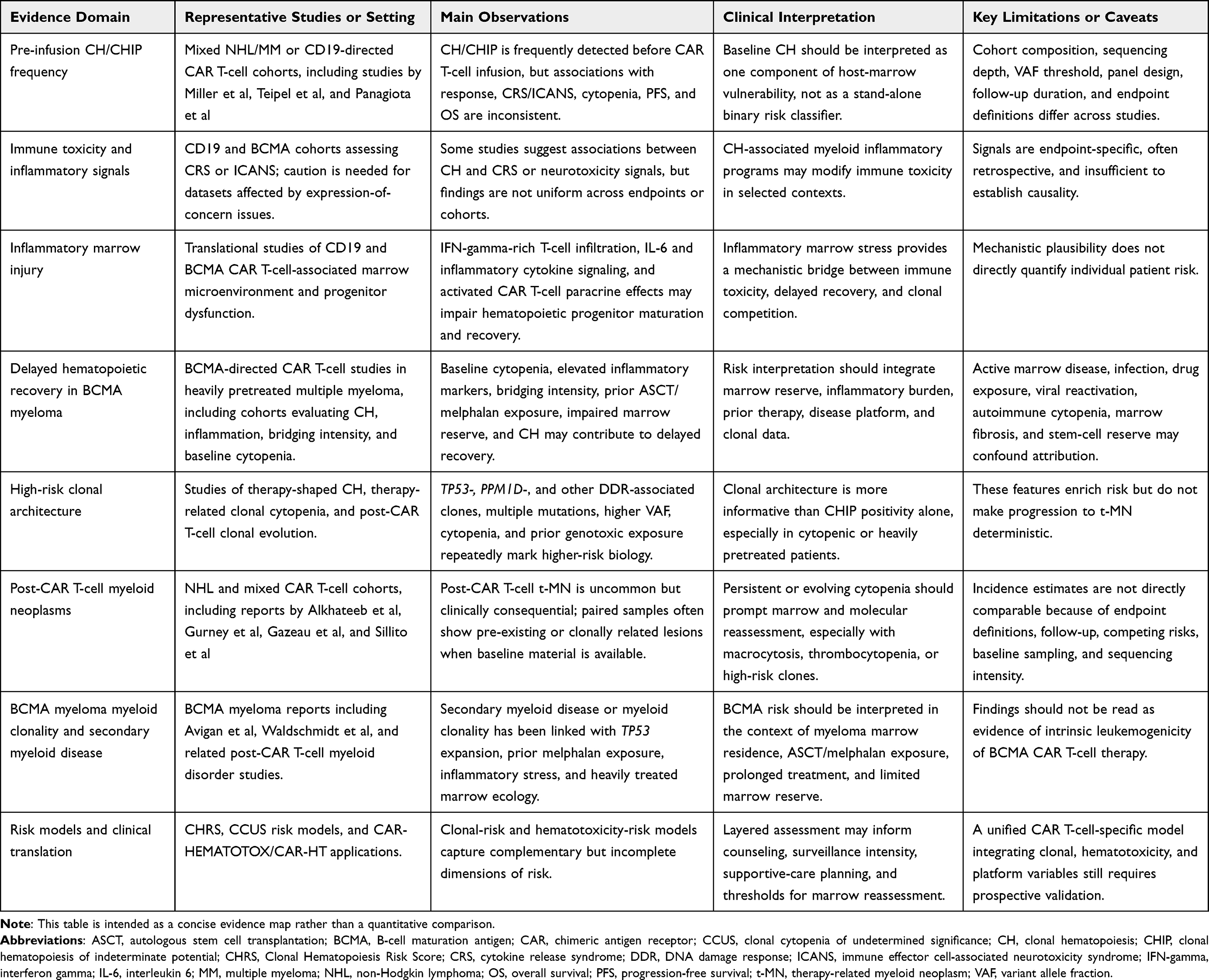 |
Table 1 Representative Evidence Informing CH/CCUS, Hematotoxicity, and Post-CAR T-Cell Myeloid Risk |
Disease-Platform Divergence: CD19 Lymphoma versus BCMA Myeloma
A cross-platform analysis is useful because CD19 lymphoma and BCMA myeloma CAR T-cell settings share several clonal-risk elements, including older age, prior genotoxic therapy, baseline cytopenia, inflammatory stress, impaired marrow reserve, and TP53- or DNA damage response-associated CH. However, these shared elements operate within different disease and treatment ecosystems. The comparison is therefore more informative when framed as risk architecture rather than as a crude ranking of platform-specific leukemogenicity.16,33,37,55 A schematic comparison of these platform-specific risk architectures is shown in Figure 2.
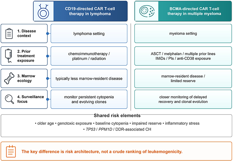 |
Figure 2 CD19 versus BCMA CAR T-cell platforms: different risk architectures. This figure compares CD19-directed CAR T-cell therapy in lymphoma and BCMA-directed CAR T-cell therapy in multiple myeloma across disease context, prior treatment exposure, marrow ecology, and surveillance focus. The figure emphasizes that the key distinction is risk architecture rather than a crude ranking of leukemogenicity, while also highlighting shared risk elements such as older age, prior genotoxic exposure, baseline cytopenia, impaired reserve, inflammatory stress, and TP53/PPM1D or other DNA damage response-associated clonal hematopoiesis. |
In the CD19 lymphoma platform, marrow vulnerability is often shaped by prior chemoimmunotherapy, salvage platinum-containing therapy, radiation in selected patients, bridging therapy, and the survivorship window created by durable lymphoma control. In this context, late myeloid risk is most interpretable when baseline blood indices and clonal data are integrated with post-infusion clinical events. Gurney et al identified older age, lower hemoglobin, lower platelet count, and CAR-HEMATOTOX score as relevant clinical risk features, while baseline CH was documented in a subset of post-CAR T-cell myeloid neoplasm cases with available paired material. Gazeau et al further showed that, among long-term B-cell lymphoma responders after CD19-directed CAR T-cell therapy, higher pre-lymphodepletion MCV and ICANS grade were associated with t-MN risk, and most t-MN cases with available pre-CAR T-cell NGS had pre-existing mutations, with TP53 among the most frequent mutations.16,55 Farina and Sillito add complementary CD19-focused evidence: the former links real-world secondary myeloid neoplasm collection with longitudinal CH assessment, whereas the latter reinforces a clonal-evolution model for post-CAR T myeloid malignancies.56,57
The BCMA myeloma platform has a different center of gravity. Myeloma is marrow-resident, and many candidates enter CAR T-cell therapy after prolonged plasma cell-directed treatment, baseline cytopenia, prior ASCT, high-dose melphalan exposure, inflammatory burden, and constrained stem-cell reserve. Avigan et al linked CH, elevated ferritin or inflammatory state, delayed hematologic recovery, and secondary myeloid disease after BCMA-directed CAR T-cell therapy, including expansion of TP53-mutated CH in patients who developed high-grade MDS. Waldschmidt et al similarly identified post-CAR T-cell myeloid clonality in myeloma and connected this signal with prior melphalan exposure, TP53 expansion, and melphalan-associated mutational signatures.33,37 In this platform, practical hematotoxicity issues such as delayed neutrophil or platelet recovery, transfusion burden, cytopenia-directed interventions, CAR-HT assessment, and availability of stored stem cells become part of the same risk architecture rather than separate supportive-care details.18,46
These platform differences shape surveillance. In CD19 lymphoma, persistent or evolving cytopenia after early recovery should prompt closer myeloid reassessment, particularly when macrocytosis, platelet decline, high CAR-HEMATOTOX score, or pre-existing high-risk mutations are present. In BCMA myeloma, the same cytopenic phenotype must be interpreted against active or prior marrow disease, prior ASCT and melphalan exposure, inflammatory burden, delayed neutrophil or platelet recovery, infection, transfusion burden, and stem-cell availability. Baseline myeloid NGS is therefore most clinically interpretable in selected patients with unexplained cytopenia, macrocytosis, thrombocytopenia, rising RDW, prior genotoxic exposure, TP53- or PPM1D-mutated CH, or abnormal marrow findings; current evidence does not support universal CHIP screening as a routine pre-lymphodepletion requirement for all CAR T-cell candidates.18,38,39,46
The more useful comparison is therefore not product-level leukemogenicity, but how each disease-platform context may select, reveal, or amplify vulnerable myeloid clones under inflammatory and regenerative stress. Incidence estimates vary with follow-up duration, competing risks, sequencing intensity, baseline marrow evaluation, prior therapy exposure, disease platform, and event definition. A risk-architecture comparison is therefore more defensible than a crude incidence ranking.16,37,55,57
Reconciling Clonal-Risk and Hematotoxicity-Risk Models Before CAR T-Cell Therapy
A clinically useful approach is to reconcile clonal-risk and hematotoxicity-risk assessment in a disease-platform-specific manner, rather than treating myeloid sequencing and clinical scores as competing tools. This approach supports both a risk-adapted surveillance pathway for CAR T-cell candidates with known or suspected CH/CCUS risk (Figure 3) and a complementary three-layer interpretive framework integrating clonal-risk, hematotoxicity-risk, and disease-platform context (Figure 4).
 |
Figure 3 Risk-adapted surveillance for CAR T-cell candidates with known or suspected CH/CCUS risk. This figure outlines a staged surveillance pathway beginning with pre-infusion assessment, followed by identification of high-risk features that may warrant hematology-guided myeloid work-up, and post-infusion follow-up with marrow reassessment when persistent or evolving abnormalities are present. The figure emphasizes that CH/CCUS should guide surveillance and follow-up intensity rather than automatically restrict access to CAR T-cell therapy. |
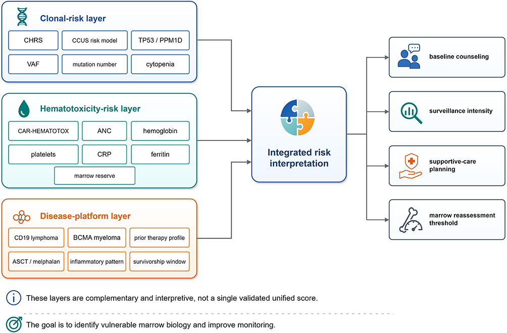 |
Figure 4 Integrating clonal-risk, hematotoxicity-risk, and disease-platform context in CAR T-cell practice. This figure illustrates a three-layer interpretive framework integrating clonal-risk variables, hematotoxicity-risk variables, and disease-platform context into a unified clinical interpretation that may inform baseline counseling, surveillance intensity, supportive-care planning, and the threshold for marrow reassessment. These layers are complementary and interpretive, not a single validated unified score. |
CHRS and clonal cytopenia risk modeling provide complementary clonal-risk frameworks. CHRS was developed to estimate myeloid malignancy risk in individuals with CHIP or CCUS, incorporating mutation class, VAF, cytopenia, age, RDW, MCV, and the presence of a single DNMT3A mutation.27 Galli et al subsequently applied CHRS in a CD19-directed CAR T-cell cohort and reported that therapy-related myeloid neoplasms occurred only among patients with intermediate-high baseline CHRS, providing a preliminary application signal rather than a definitive CAR T-specific validation.54 Xie et al recently proposed a clonal cytopenia risk model for CCUS, referred to here as the CCUS risk model, which may be conceptually closer to CAR T-cell candidates with baseline cytopenia, thrombocytopenia, multiple myeloid-type mutations, or splicing mutations.29 These models should not yet be treated as CAR T-cell-specific algorithms, but they support a clinically important distinction between low-risk incidental CH and higher-risk clonal cytopenia or high-risk myeloid architecture.
CAR-HEMATOTOX and related CAR-HT applications provide hematotoxicity-risk frameworks. These models use baseline absolute neutrophil count (ANC), hemoglobin, platelet count, CRP, and ferritin to estimate the risk of CAR T-cell-related hematologic toxicity and delayed recovery.38,39 Their clinical practicality lies in the use of routinely available variables, including in BCMA-directed supportive-care settings.18 However, they do not capture clonal architecture and were not designed as molecular models of myeloid transformation.
Avigan et al illustrate how these layers may converge clinically. In a BCMA CAR T-cell myeloma cohort, CH in the setting of elevated ferritin or inflammation was associated with delayed hematologic recovery, and secondary myeloid disease was linked to expansion of underlying TP53-mutated CH.33 Although this remains short of a validated combined model, it shows that clonal-risk and hematotoxicity-risk dimensions can converge in clinically observable recovery and late myeloid outcomes. Gurney et al provided complementary evidence that clinical marrow-reserve variables were associated with post-CAR T-cell myeloid neoplasm risk; baseline CH and clonal relatedness were observed in a limited subset with available paired samples, supporting biologic plausibility rather than a fully integrated predictive model.16
A working framework includes three partially overlapping layers. The clonal-risk layer includes CHRS, the CCUS risk model, TP53, PPM1D, DNA damage response-associated mutations, VAF, mutation number, and CCUS or therapy-related clonal cytopenia. The hematotoxicity-risk layer includes CAR-HEMATOTOX or CAR-HT variables, baseline ANC, hemoglobin, platelets, CRP, ferritin, marrow reserve, inflammatory burden, and bridging intensity. The disease-platform layer includes CD19 lymphoma versus BCMA myeloma context, prior therapy profile, marrow disease background, inflammatory toxicity pattern, follow-up window, and expected late complication spectrum.
These layers are interpretive rather than algorithmic, and their overlap requires integrated clinical judgment. A patient with low-level isolated DNMT3A CHIP, preserved counts, low inflammatory markers, and limited prior genotoxic exposure should be interpreted differently from a patient with cytopenia, macrocytosis, prior melphalan exposure, elevated ferritin, TP53-mutated CH, and delayed recovery after BCMA CAR T-cell therapy. Clinically, the aim is to construct an individualized risk interpretation that informs baseline counseling, surveillance intensity, and supportive-care planning, rather than to assign a single universal score.
Risk-Adapted Myeloid Assessment and Surveillance Before and After CAR T-Cell Therapy
Using these three layers, risk-adapted assessment can be organized as a staged clinical pathway. Recent BCMA-directed CAR T-cell hematotoxicity management work indicates that universal CHIP screening before lymphodepletion is not supported at present and that targeted myeloid evaluation is most appropriate for prolonged and unexplained cytopenias.18 On that basis, a platform-specific surveillance approach can map clinical decisions to clonal-risk, hematotoxicity-risk, and disease-platform layers rather than applying a uniform testing strategy across all CAR T-cell recipients. As shown in Figure 3, this risk-adapted pathway begins with routine pre-infusion assessment, identifies high-risk features that may justify hematology-guided myeloid work-up, and links post-infusion abnormalities to marrow reassessment and molecular evaluation when clinically indicated. A practical summary of the proposed risk-assessment layers, their clinical uses, and important caveats is provided in Table 2.
 |
Table 2 Practical Layered Framework for CH/CCUS-Informed Risk Assessment and Surveillance in CAR T-Cell Candidates |
Before CAR T-cell therapy, assessment should first use routine clinical, laboratory, and treatment-history variables. The hematotoxicity-risk and disease-platform layers include age, prior therapy burden, prior ASCT or melphalan exposure when applicable, other alkylator or radiation exposure, baseline ANC, hemoglobin, platelet count, CRP, ferritin, marrow involvement, prior prolonged cytopenia, and planned bridging intensity.38,39 MCV and RDW provide additional clonal-risk context; together, these routine variables do not substitute for myeloid sequencing, but help identify patients in whom sequencing results are more likely to be clinically interpretable.
A risk-adapted approach is therefore more clinically defensible than universal pre-lymphodepletion CHIP testing. The clonal-risk layer becomes clinically relevant in selected candidates with persistent unexplained cytopenia, macrocytosis, rising RDW, thrombocytopenia, prior ASCT or melphalan exposure, multiple genotoxic exposures, or suspicion for CCUS or therapy-related clonal cytopenia, where myeloid NGS, marrow morphology, conventional cytogenetics, and fluorescence in situ hybridization (FISH) may clarify baseline risk and provide a comparator for post-infusion reassessment.27,29 The purpose is risk communication, baseline documentation, supportive-care planning, and longitudinal interpretation if cytopenia persists or evolves after infusion, while preserving appropriate access to CAR T-cell therapy.
After CAR T-cell therapy, surveillance should integrate all three layers across early, intermediate, and late phases. During the first month, cytopenia is often multifactorial and may reflect lymphodepletion, inflammatory toxicity, marrow disease, infection, or early immune effector cell-associated toxicity. Around day 30 and during day 90–100 follow-up, persistent severe cytopenia, transfusion dependence, recurrent infection, platelet decline, macrocytosis, rising RDW, abnormal peripheral-blood morphology, or recovery that deviates from the expected clinical trajectory should trigger reassessment. Bone marrow morphology, flow cytometry, cytogenetics, FISH, and myeloid NGS may be warranted when cytopenia is prolonged, unexplained, progressive, or discordant with expected recovery.10
The t-CCUS or t-CC concept is useful as an operational category, rather than a CAR T-cell-specific diagnostic label, in this setting. Persistent cytopenia with a myeloid clone after prior DNA-damaging therapy represents more than a “CHIP-positive” state. This pattern may represent therapy-related clonal cytopenia, a higher-risk precursor context than incidental CHIP, and should support closer longitudinal monitoring rather than follow-up appropriate for incidental CHIP alone.21,30 TP53 or PPM1D mutations, multiple mutations, high VAF, macrocytosis, thrombocytopenia, and prior melphalan or alkylator exposure should further increase concern.19,31
Risk assessment should preserve access to CAR T-cell therapy while improving the quality of counseling and surveillance. CH/CCUS is better used to guide surveillance than to gate access. Reported post-CAR T-cell myeloid neoplasms are clinically consequential but still occur in a minority of treated patients,16 supporting informed consent, individualized surveillance intensity, early recognition of evolving myeloid disease, and prospective study design rather than categorical exclusion.
Future Directions
Several Priorities Deserve Prospective Study
First, prospective cohorts should determine whether CH/CCUS adds independent predictive information for prolonged hematotoxicity beyond baseline marrow reserve and time-dependent post-infusion events. Future models should account for age, baseline cytopenia, marrow involvement, bridging intensity, lymphodepletion, infection, CRS, ICANS, CRP, ferritin, and other markers of hematopoietic vulnerability, while moving beyond CHIP-positive versus CHIP-negative comparisons to evaluate clonal-risk variables, hematotoxicity-risk variables, and disease-platform context within the same models.33,38
Second, longitudinal tracking should prioritize TP53-mutated clones and should analyze PPM1D or other DNA damage response-associated clones where detected.20,31,33 Sampling before lymphodepletion, during peak inflammatory toxicity, around day 30, around day 100, and during long-term follow-up would help distinguish clonal selection, clinical unmasking, and parallel evolution under prior genotoxic and post-infusion inflammatory stress.33,37
Third, CD19 lymphoma and BCMA myeloma should be modeled separately during derivation and early validation before pooled analyses are attempted. These platforms differ in disease biology, marrow ecology, prior therapy, inflammatory pattern, competing-risk structure, and survivorship window; a combined model may obscure disease-specific signals.37,55
Fourth, CHRS, the CCUS risk model, and CAR-HEMATOTOX should be evaluated as complementary rather than interchangeable risk dimensions.27,29,38,39 Prospective CAR T-cell cohorts should test whether combined clinical and molecular assessment improves prediction of prolonged cytopenia, infection, transfusion burden, need for marrow reassessment, secondary myeloid neoplasm, and survival.
Fifth, the clinical utility, workflow feasibility, and resource implications of baseline myeloid NGS need prospective definition in CAR T-cell candidates. Current evidence does not support universal CHIP screening before lymphodepletion.18 Selected testing in patients with unexplained cytopenia, macrocytosis, thrombocytopenia, prior genotoxic exposure, or other high-risk features may be more rational, but selection criteria require validation.
Finally, risk assessment should evolve toward testable intervention strategies. If high-risk CH/CCUS identifies vulnerable marrow biology, future studies should evaluate whether risk-adapted bridging intensity,51 pre-emptive stem-cell planning, infection prophylaxis, earlier marrow reassessment, or individualized supportive care can reduce clinically meaningful outcomes such as severe prolonged cytopenia, infection, transfusion burden, and secondary myeloid neoplasm.
Conclusions
Pre-infusion CH/CCUS functions best as a host-marrow vulnerability marker that refines CAR T-cell risk assessment without determining eligibility. Its clinical value lies in connecting clonal-risk and hematotoxicity-risk models and identifying patients who may warrant intensified myeloid surveillance as CAR T-cell survivorship expands. At present, risk assessment is strongest when it integrates cytopenia, mutation class, clone size, prior genotoxic exposure, inflammatory state, marrow reserve, and disease platform, instead of relying on universal CHIP screening or binary genomic classification. Across CD19 lymphoma and BCMA myeloma, TP53-mutated or DNA damage response-associated clonal cytopenia appears to carry the clearest link with therapy-related myeloid neoplasms, whereas the relationship between CH and prolonged cytopenia remains more heterogeneous. Prospective studies should now test whether this layered risk architecture improves surveillance, supportive planning, and early recognition of evolving myeloid disease while preserving access to potentially life-extending CAR T-cell therapy.
Abbreviations
AML, acute myeloid leukemia; ANC, absolute neutrophil count; ASCT, autologous stem cell transplantation; BCMA, B-cell maturation antigen; CAR-HT, CAR-HEMATOTOX; CAR T-cell, chimeric antigen receptor T-cell; CCUS, clonal cytopenia of undetermined significance; CH, clonal hematopoiesis; CHIP, clonal hematopoiesis of indeterminate potential; CHRS, Clonal Hematopoiesis Risk Score; CRP, C-reactive protein; CRS, cytokine release syndrome; FISH, fluorescence in situ hybridization; ICANS, immune effector cell-associated neurotoxicity syndrome; MCV, mean corpuscular volume; MDS, myelodysplastic syndromes/neoplasms; NGS, next-generation sequencing; RDW, red cell distribution width; SPM, second primary malignancy; t-MN, therapy-related myeloid neoplasms; VAF, variant allele fraction.
Data Sharing Statement
No new datasets were generated or analyzed for this review.
Author Contributions
All authors made a significant contribution to the work reported, whether that is in the conception, study design, execution, acquisition of data, analysis and interpretation, or in all these areas; took part in drafting, revising or critically reviewing the article; gave final approval of the version to be published; have agreed on the journal to which the article has been submitted; and agree to be accountable for all aspects of the work.
Funding
No specific funding was received for this work.
Disclosure
The author declares no competing interests.
References
1. Neelapu SS, Locke FL, Bartlett NL, et al. Axicabtagene ciloleucel CAR T-Cell therapy in refractory large B-Cell lymphoma. N Engl J Med. 2017;377:2531–18. doi:10.1056/NEJMoa1707447
2. Schuster SJ, Bishop MR, Tam CS, et al. Tisagenlecleucel in adult relapsed or refractory diffuse Large B-Cell lymphoma. N Engl J Med. 2019;380:45–56. doi:10.1056/NEJMoa1804980
3. Abramson JS, Palomba ML, Gordon LI, et al. Lisocabtagene maraleucel for patients with relapsed or refractory large B-cell lymphomas (TRANSCEND NHL 001): a multicentre seamless design study. Lancet. 2020;396:839–852. doi:10.1016/s0140-6736(20)31366-0
4. Westin JR, Oluwole OO, Kersten MJ, et al. Survival with axicabtagene ciloleucel in Large B-Cell lymphoma. N Engl J Med. 2023;389:148–157. doi:10.1056/NEJMoa2301665
5. Munshi NC, Anderson LD, Shah N, et al. Idecabtagene vicleucel in relapsed and refractory multiple myeloma. N Engl J Med. 2021;384:705–716. doi:10.1056/NEJMoa2024850
6. Berdeja JG, Madduri D, Usmani SZ, et al. Ciltacabtagene autoleucel, a B-cell maturation antigen-directed chimeric antigen receptor T-cell therapy in patients with relapsed or refractory multiple myeloma (CARTITUDE-1): a phase 1b/2 open-label study. Lancet. 2021;398(10297):314–324. doi:10.1016/s0140-6736(21)00933-8
7. Rodriguez-Otero P, Ailawadhi S, Arnulf B, et al. Ide-cel or standard regimens in relapsed and refractory multiple myeloma. N Engl J Med. 2023;388:1002–1014. doi:10.1056/NEJMoa2213614
8. San-Miguel J, Dhakal B, Yong K, et al. Cilta-cel or standard care in lenalidomide-refractory multiple myeloma. N Engl J Med. 2023;389:335–347. doi:10.1056/NEJMoa2303379
9. Fried S, Avigdor A, Bielorai B, et al. Early and late hematologic toxicity following CD19 CAR-T cells. Bone Marrow Transplant. 2019;54:1643–1650. doi:10.1038/s41409-019-0487-3
10. Rejeski K, Jain MD, Shah NN, Perales MA, Subklewe M. Immune effector cell-associated haematotoxicity after CAR T-cell therapy: from mechanism to management. Lancet Haematol. 2024;11:e459–e470. doi:10.1016/s2352-3026(24)00077-2
11. Rejeski K, Subklewe M, Aljurf M, et al. Immune effector cell-associated hematotoxicity: EHA/EBMT consensus grading and best practice recommendations. Blood. 2023;142:865–877. doi:10.1182/blood.2023020578
12. Logue JM, Zucchetti E, Bachmeier CA, et al. Immune reconstitution and associated infections following axicabtagene ciloleucel in relapsed or refractory large B-cell lymphoma. Haematologica. 2021;106:978–986. doi:10.3324/haematol.2019.238634
13. Cordeiro A, Bezerra ED, Hirayama AV, et al. Late events after treatment with CD19-targeted chimeric antigen receptor modified T cells. Biol Blood Marrow Transplant. 2020;26:26–33. doi:10.1016/j.bbmt.2019.08.003
14. Hamilton MP, Sugio T, Noordenbos T, et al. Risk of second tumors and T-Cell lymphoma after CAR T-Cell therapy. N Engl J Med. 2024;390:2047–2060. doi:10.1056/NEJMoa2401361
15. Tix T, Alhomoud M, Shouval R, et al. Second primary malignancies after CAR T-Cell therapy: a systematic review and meta-analysis of 5,517 lymphoma and myeloma patients. Clin Cancer Res. 2024;30:4690–4700. doi:10.1158/1078-0432.Ccr-24-1798
16. Gurney M, Baranwal A, Rosenthal A, et al. Features and factors associated with myeloid neoplasms after chimeric antigen receptor T-Cell therapy. JAMA Oncol. 2024;10:532–535. doi:10.1001/jamaoncol.2023.7182
17. Wang Y, Cheng W, Kang K, Niu T, Zhao A, Wu Y. Biomarker-empowered precision navigation of CAR-T cell therapy. Mol Cancer. 2026. doi:10.1186/s12943-026-02647-0
18. Cook J, Gupta S, Abdallah N, et al. Management of hematological toxicities after BCMA-directed CAR-T cell therapy. Blood Cancer J. 2026;16. doi:10.1038/s41408-026-01455-5
19. Bolton KL, Ptashkin RN, Gao T, et al. Cancer therapy shapes the fitness landscape of clonal hematopoiesis. Nat Genet. 2020;52:1219–1226. doi:10.1038/s41588-020-00710-0
20. Wong TN, Ramsingh G, Young AL, et al. Role of TP53 mutations in the origin and evolution of therapy-related acute myeloid leukaemia. Nature. 2015;518:552–555. doi:10.1038/nature13968
21. Shah MV, Mangaonkar AA, Begna KH, et al. Therapy-related clonal cytopenia as a precursor to therapy-related myeloid neoplasms. Blood Cancer J. 2022;12(106). doi:10.1038/s41408-022-00703-8
22. Genovese G, Kähler AK, Handsaker RE, et al. Clonal hematopoiesis and blood-cancer risk inferred from blood DNA sequence. N Engl J Med. 2014;371:2477–2487. doi:10.1056/NEJMoa1409405
23. Jaiswal S, Fontanillas P, Flannick J, et al. Age-related clonal hematopoiesis associated with adverse outcomes. N Engl J Med. 2014;371:2488–2498. doi:10.1056/NEJMoa1408617
24. Xie M, Lu C, Wang J, et al. Age-related mutations associated with clonal hematopoietic expansion and malignancies. Nat Med. 2014;20:1472–1478. doi:10.1038/nm.3733
25. Jaiswal S, Ebert BL. Clonal hematopoiesis in human aging and disease. Science. 2019;366. doi:10.1126/science.aan4673
26. Steensma DP, Bejar R, Jaiswal S, et al. Clonal hematopoiesis of indeterminate potential and its distinction from myelodysplastic syndromes. Blood. 2015;126:9–16. doi:10.1182/blood-2015-03-631747
27. Weeks LD, Niroula A, Neuberg D, et al. Prediction of risk for myeloid malignancy in clonal hematopoiesis. NEJM Evid. 2023;2. doi:10.1056/evidoa2200310
28. Coombs CC, Zehir A, Devlin SM, et al. Therapy-related clonal hematopoiesis in patients with non-hematologic cancers is common and associated with adverse clinical outcomes. Cell Stem Cell. 2017;21:374–382.e374. doi:10.1016/j.stem.2017.07.010
29. Xie Z, Komrokji R, Al Ali N, et al. Risk prediction for clonal cytopenia: multicenter real-world evidence. Blood. 2024;144:2033–2044. doi:10.1182/blood.2024024756
30. Li M, Baranwal A, Gurney M, et al. The impact of cytotoxic therapy on the risk of progression and death in clonal cytopenia(s) of undetermined significance. Blood Adv. 2024;8:3130–3139. doi:10.1182/bloodadvances.2023012357
31. Hsu JI, Dayaram T, Tovy A, et al. PPM1D mutations drive clonal hematopoiesis in response to cytotoxic chemotherapy. Cell Stem Cell. 2018;23(5):700–713.e706. doi:10.1016/j.stem.2018.10.004
32. McNerney ME, Godley LA, Le Beau MM. Therapy-related myeloid neoplasms: when genetics and environment collide. Nat Rev Cancer. 2017;17:513–527. doi:10.1038/nrc.2017.60
33. Avigan ZM, Catlett J, Bodnar S, et al. Clonal hematopoiesis and inflammation predict hematologic toxicity and secondary myeloid malignancies after B-cell maturation antigen-directed chimeric antigen receptor T-cell therapy. Clin Cancer Res. 2025;31:4333–4344. doi:10.1158/1078-0432.Ccr-25-1587
34. Gibson CJ, Lindsley RC, Tchekmedyian V, et al. Clonal hematopoiesis associated with adverse outcomes after autologous stem-cell transplantation for lymphoma. J Clin Oncol. 2017;35:1598–1605. doi:10.1200/jco.2016.71.6712
35. Mouhieddine TH, Sperling AS, Redd R, et al. Clonal hematopoiesis is associated with adverse outcomes in multiple myeloma patients undergoing transplant. Nat Commun. 2020;11:2996. doi:10.1038/s41467-020-16805-5
36. Takahashi K, Wang F, Kantarjian H, et al. Preleukaemic clonal haemopoiesis and risk of therapy-related myeloid neoplasms: a case-control study. Lancet Oncol. 2017;18:100–111. doi:10.1016/s1470-2045(16)30626-x
37. Waldschmidt J, Fandrei D, Hecker JS, et al. Flare of clonal hematopoiesis, TP53 expansion and prior melphalan drive post-CAR-T myeloid disorders in multiple myeloma. Leukemia. 2026;40:1054–1057. doi:10.1038/s41375-026-02941-2
38. Rejeski K, Perez A, Sesques P, et al. CAR-HEMATOTOX: a model for CAR T-cell-related hematologic toxicity in relapsed/refractory large B-cell lymphoma. Blood. 2021;138:2499–2513. doi:10.1182/blood.2020010543
39. Rejeski K, Hansen DK, Bansal R, et al. The CAR-HEMATOTOX score as a prognostic model of toxicity and response in patients receiving BCMA-directed CAR-T for relapsed/refractory multiple myeloma. J Hematol Oncol. 2023;16:88. doi:10.1186/s13045-023-01465-x
40. Miller PG, Sperling AS, Brea EJ, et al. Clonal hematopoiesis in patients receiving chimeric antigen receptor T-cell therapy. Blood Adv. 2021;5:2982–2986. doi:10.1182/bloodadvances.2021004554
41. Teipel R, Kroschinsky F, Kramer M, et al. Prevalence and variation of CHIP in patients with aggressive lymphomas undergoing CD19-directed CAR T-cell treatment. Blood Adv. 2022;6:1941–1946. doi:10.1182/bloodadvances.2021005747
42. Panagiota V, Kerschbaum JF, Penack O, et al. Clinical implications and dynamics of clonal hematopoiesis in Anti-CD19 CAR T-cell treated patients. Hemasphere. 2023;7(10):e957. doi:10.1097/hs9.0000000000000957
43. Saini NY, Swoboda DM, Greenbaum U, et al. Clonal hematopoiesis is associated with increased risk of severe neurotoxicity in axicabtagene ciloleucel therapy of large B-Cell lymphoma. Blood Cancer Discov. 2022;3:385–393. doi:10.1158/2643-3230.Bcd-21-0177
44. Saini NY, Swoboda DM, Greenbaum U, et al. Expression of concern: clonal hematopoiesis is associated with increased risk of severe neurotoxicity in axicabtagene ciloleucel therapy of large B-Cell lymphoma. Blood Cancer Discov. 2023;4:246. doi:10.1158/2643-3230.Bcd-23-0040
45. Goldsmith SR, Shouse G, Wong FL, et al. Clonal hematopoiesis is associated with severe cytokine release syndrome in patients treated with chimeric antigen receptor T-Cell (CART) therapy. Transplant Cell Ther. 2024;30:
46. Gustine JN, Branagan AR, Cirstea DD, et al. Impact of clonal hematopoiesis on clinical outcomes to BCMA CAR-T in multiple myeloma. Blood Adv. 2025;9:3026–3030. doi:10.1182/bloodadvances.2025015981
47. Kapadia CD, Rosas G, Thakkar SG, et al. Incipient clonal hematopoiesis is accelerated following CD30.CAR-T therapy. Cytotherapy. 2024;26:261–265. doi:10.1016/j.jcyt.2023.11.013
48. Strati P, Li X, Deng Q, et al. Prolonged cytopenia following CD19 CAR T cell therapy is linked with bone marrow infiltration of clonally expanded IFNγ-expressing CD8 T cells. Cell Rep Med. 2023;4:101158. doi:10.1016/j.xcrm.2023.101158
49. Palacios-Berraquero ML, Rodriguez-Marquez P, Calleja-Cervantes ME, et al. Molecular mechanisms promoting long-term cytopenia after BCMA CAR-T therapy in multiple myeloma. Blood Adv. 2024;8:5479–5492. doi:10.1182/bloodadvances.2023012522
50. Ben Khelil M, Arbab A, Srikanthan J, et al. CAR T cell-mediated bone marrow inflammation causes hematotoxicity and favors clonal hematopoiesis. Sci Transl Med. 2025;17:eadu9790. doi:10.1126/scitranslmed.adu9790
51. Frenking JH, Zhou X, Rejeski K, et al. Bridging intensity is associated with impaired hematopoietic recovery after BCMA CAR-T therapy for multiple myeloma. Blood Adv. 2025;9:4151–4166. doi:10.1182/bloodadvances.2024015732
52. Avigan ZM, Bodnar S, Pan D, et al. Delayed neutrophil recovery following BCMA CAR-T therapy in Duffy-null myeloma does not impact severe infections or survival. Blood Adv. 2025;9:202–206. doi:10.1182/bloodadvances.2024014255
53. Alkhateeb HB, Mohty R, Greipp P, et al. Therapy-related myeloid neoplasms following chimeric antigen receptor T-cell therapy for Non-Hodgkin Lymphoma. Blood Cancer J. 2022;12:113. doi:10.1038/s41408-022-00707-4
54. Galli E, Rossi M, Pansini I, et al. Predicting therapy-related myeloid neoplasms after CAR-T with the Clonal Haematopoiesis Risk Score (CHRS). Br J Haematol. 2025;206:372–374. doi:10.1111/bjh.19905
55. Gazeau N, Beauvais D, Tilmont R, et al. Myeloid neoplasms after CD19-directed CAR T cells therapy in long-term B-cell lymphoma responders, a rising risk over time? Leukemia. 2025;39:1714–1722. doi:10.1038/s41375-025-02605-7
56. Farina M, Bernardi S, Malagola M, et al. Real-world collection of secondary myeloid neoplasms after CD19 CAR-T cell therapy: first report of the ClonHema study. Bone Marrow Transplant. 2025;60:702–704. doi:10.1038/s41409-025-02529-x
57. Sillito F, Rampotas A, Cheok K, et al. Clonal evolution and the risk of secondary myeloid neoplasia following chimeric antigen receptor T-cell therapy. Haematologica. 2025;110:2812–2818. doi:10.3324/haematol.2025.287341
 © 2026 The Author(s). This work is published and licensed by Dove Medical Press Limited. The
full terms of this license are available at https://www.dovepress.com/terms
and incorporate the Creative Commons Attribution
- Non Commercial (unported, 4.0) License.
By accessing the work you hereby accept the Terms. Non-commercial uses of the work are permitted
without any further permission from Dove Medical Press Limited, provided the work is properly
attributed. For permission for commercial use of this work, please see paragraphs 4.2 and 5 of our Terms.
© 2026 The Author(s). This work is published and licensed by Dove Medical Press Limited. The
full terms of this license are available at https://www.dovepress.com/terms
and incorporate the Creative Commons Attribution
- Non Commercial (unported, 4.0) License.
By accessing the work you hereby accept the Terms. Non-commercial uses of the work are permitted
without any further permission from Dove Medical Press Limited, provided the work is properly
attributed. For permission for commercial use of this work, please see paragraphs 4.2 and 5 of our Terms.