Back to Journals » Journal of Inflammation Research » Volume 18
Potato Peel Extract Reduces MMP-9 Expression During Oral Mucosal Wound Healing in Rats
Authors Sufiawati I ![]() , Finita N, Hidayat W
, Finita N, Hidayat W ![]() , Yuslianti ER
, Yuslianti ER
Received 7 July 2025
Accepted for publication 7 November 2025
Published 19 November 2025 Volume 2025:18 Pages 16261—16272
DOI https://doi.org/10.2147/JIR.S552188
Checked for plagiarism Yes
Review by Single anonymous peer review
Peer reviewer comments 4
Editor who approved publication: Professor Ning Quan
Irna Sufiawati,1,* Nur Finita,2,* Wahyu Hidayat,1,3 Euis Reni Yuslianti4
1Department of Oral Medicine, Faculty of Dentistry, Universitas Padjadjaran, Bandung, West Java, Indonesia; 2Undergraduate Program, Faculty of Dentistry, Universitas Padjadjaran, Bandung, West Java, Indonesia; 3Doctoral Program, Faculty of Dentistry, Universitas Padjadjaran, Bandung, West Java, Indonesia; 4Department of Oral Biology, Faculty of Dentistry, Universitas Jenderal Achmad Yani, Bandung, West Java, Indonesia
*These authors contributed equally to this work
Correspondence: Irna Sufiawati, Department of Oral Medicine, Faculty of Dentistry, Universitas Padjadjaran, Jl. Sekeloa Selatan No. 1, Bandung, West Java, 40132, Indonesia, Email [email protected]
Purpose: This study aimed to analyze the differences in matrix metalloproteinase-9 (MMP-9) expression during oral mucosal wound healing in Wistar rats after administration of 4% and 6% potato peel extract gel compared to 0.1% triamcinolone acetonide.
Material and Methods: A true experimental design was employed involving 48 Wistar rats, randomly assigned to four groups (n = 12 per group), consisting of placebo gel, 0.1% triamcinolone acetonide, 4% potato peel extract gel, and 6% potato peel extract gel. Excisional oral mucosal wounds were created using a punch biopsy instrument. For each group, three rats were euthanized and analyzed at each time point (days 0, 1, 3, 7, and 14) to assess MMP-9 expression. Histopathological evaluation was performed using hematoxylin-eosin staining, and immunohistochemistry was used for MMP-9 analysis. Data were analyzed using one-way ANOVA followed by post hoc t-tests, with significance set at p < 0.05.
Results: Both 4% and 6% potato peel extract gel groups showed reduced MMP-9 expression compared to placebo and 0.1% triamcinolone acetonide across all time points (p < 0.05). Post hoc analysis showed that on day 14, the 4% extract group did not differ significantly from the 6% extract group (p = 0.584) or from day 7 groups (p > 0.05), but showed significant differences compared to day 0 and the 0.1% triamcinolone acetonide group on day 7 (p < 0.05), indicating a distinct treatment effect at day 14. Among the two concentrations, the 4% extract group showed a lower mean MMP-9 expression compared to the 6% group.
Conclusion: Granola potato peel ethanol extract gel at a concentration of 4% was the most effective in reducing MMP-9 expression compared to the 6% extract and 0.1% triamcinolone acetonide during oral mucosal wound healing, highlighting its potential as a natural alternative to corticosteroids and contributing to innovation in pharmaceutical research.
Keywords: healing process, mucosal wound, Granola potato peel extract, MMP-9, pharmaceutical research
Introduction
The oral mucosa is composed of stratified squamous epithelium, a basement membrane, lamina propria, and submucosa, which collectively serve to protect against pathogenic invasion and mechanical stress.1 Nevertheless, the oral mucosa remains vulnerable to injury, including lesions or ulcers, which are frequently associated with trauma, dysfunction, or infectious processes.2,3 Wound healing progresses through four distinct phases: hemostasis, inflammation, proliferation, and remodeling, each involving the coordinated activity of cells, cytokines, growth factors, and enzymes, including matrix metalloproteinases (MMP).1–4
Matrix metalloproteinase-9 plays a crucial role in extracellular matrix (ECM) degradation, keratinocyte migration, and the production of pro-inflammatory chemokines. However, excessive levels of MMP-9 may impair the healing process and contribute to the transition of acute wounds into chronic wounds. Peng et al reported that high MMP-9 expression is correlated with impaired wound healing in the context of infected wounds.4–7
Oral mucosal ulcers are typically treated with anti-inflammatory agents, including non-steroidal anti-inflammatory drugs (NSAIDs) and corticosteroids. Commonly prescribed NSAIDs include ibuprofen and naproxen, while corticosteroids include systemic corticosteroids (SAIDs) such as prednisone and dexamethasone, and topical corticosteroids such as triamcinolone acetonide. However, these therapies are associated with adverse effects such as hypertension, gastric ulceration, insulin resistance, osteoporosis, dermal thinning, and increased susceptibility to oral candidiasis with prolonged use.8–12 Therefore, natural substances such as potato peel extract (Solanum tuberosum L.) represent a promising alternative. Potato peel contains flavonoids, anthocyanins, and other bioactive compounds that possess anti-inflammatory properties and can accelerate the healing of oral mucosal wounds.13–15
Our previous studies demonstrated that 4% and 6% potato peel extract gel effectively accelerated oral mucosal wound healing in Wistar rats, with efficacy comparable to that of 0.1% triamcinolone acetonide, as evidenced by wound length measurements.15 Moreover, both concentrations exhibited anti-inflammatory effects similar to 0.1% triamcinolone acetonide and even promoted superior healing outcomes, as indicated by increased fibroblast proliferation, collagen deposition, and blood vessel formation.16
Despite these promising findings, the specific role of potato peel extract in modulating MMP-9 expression during oral mucosal wound healing remains unclear, representing a knowledge gap this study aims to address. Furthermore, limited studies have directly compared potato peel extract with corticosteroids in oral mucosal wound models, highlighting the novelty of this work. Previous studies have explored the wound-healing potential of other natural products in comparison to corticosteroids in oral mucosal models. Mhapuskar et al reported that honey was more effective than 0.1% triamcinolone acetonide in reducing pain and ulcer size in recurrent minor aphthous stomatitis.17 Ferreira et al emphasized the potential of natural products in preventing and treating oral mucositis, while noting the limited direct comparisons with standard topical corticosteroids. These prior findings underscore the relevance of investigating potato peel extract as a natural alternative, addressing the current knowledge gap regarding its effect on MMP-9 expression during oral mucosal wound healing and highlighting the novelty of our study.18
We therefore hypothesize that 4% and 6% Granola potato peel ethanol extract gels reduce MMP-9 expression more effectively than 0.1% triamcinolone acetonide during oral mucosal wound healing in Wistar rats, linking the rationale directly to the study objectives. Accordingly, this study aimed to further investigate and analyze MMP-9 expression during the oral mucosal wound healing process in Wistar rats following administration of 4% and 6% Granola variety potato peel extract gel, providing focused insight into its anti-inflammatory and wound healing potential in comparison to a well-established topical corticosteroid.
Materials and Methods
Ethical Approval
This research employed a true experimental design and was approved by the Health Research Ethics Committee of Universitas Padjadjaran (Approval No. 1076/UN6.KEP/EC/2024) and conducted in accordance with the Declaration of Helsinki. All experimental procedures involving animals were conducted in accordance with the Animal Research: Reporting of In Vivo Experiments (ARRIVE) guidelines to ensure adherence to ethical standards and proper reporting.
Extract Gel Preparation
Granola variety potato peels (Solanum tuberosum L.) used in this study were obtained from Pangalengan District, West Java, and botanically identified by Heri Santoso, S.Si, at the Generasi Biologi Indonesia Laboratory. A voucher specimen (No. GBI-08.159/Genbinesia/I/2023) has been deposited in Herbarium Generasi Biologi Indonesia, Bandung, Indonesia.
The extraction process began with thoroughly washing 100 kg of potatoes, followed by peeling to obtain potato peels with a thickness of 1–2 mm. The peels were oven-dried at 40–42 °C for 24 hours and then coarsely ground to obtain dried fragments. Extraction was performed using the maceration method with 96% ethanol at a 1:10 ratio for 72 hours at room temperature, with stirring during the first two hours of each solvent replacement. The maceration process was carried out in a sealed container, with the solvent replaced every 24 hours. Extract was subsequently concentrated using a rotary evaporator (75 rpm, condenser temperature of 16 °C) for 2 hours at 45–50 °C, and further processed in a water bath at 60 °C for 48 hours.
Extract Gel Formula/Stability Study of the Gel Formula
The process continued with the preparation of the potato peel extract gel, starting with the thickening of the ethanolic potato peel extract in a water bath at 60°C for 36 hours. The materials were measured based on the formulation outlined in Table 1. The gel base was prepared by dispersing Carbomer 940 in distilled water, followed by the addition of methylparaben and propylparaben until a homogeneous mixture was achieved. The extract was dissolved in glycerin, combined with the gel base, and thoroughly mixed to form a homogeneous gel. The gel base was optimized for homogeneity, pH, consistency (viscosity), and spreadability. Visual inspection ensured uniform distribution of components. The pH was measured to confirm compatibility with oral tissues. Viscosity was adjusted for appropriate consistency using a rotational viscometer, and spreadability was assessed with a glass plate method to ensure ease of application. These evaluations guided the selection of the final gel concentration used in the study.
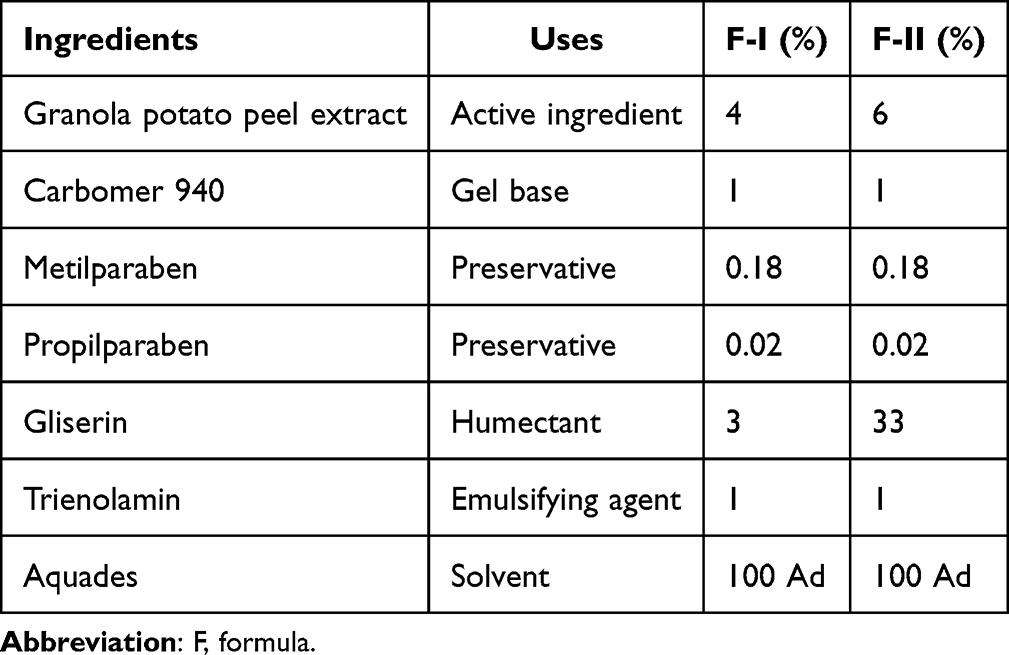 |
Table 1 Formulation of Granola Variety Potato Peel Extract Gel |
Experimental Animals
The study subjects were male white rats (Rattus norvegicus) of the Wistar strain, weighing approximately 150–250 grams and aged 6–8 weeks. Rats were housed for 7 days for an adaptation period prior to the initiation of treatment. Throughout the adaptation period, rats were provided with a standard pellet diet containing low fiber (5%), protein (20%), and fat (5–10%), along with ad libitum access to water. Cages and drinking water containers were routinely cleaned to maintain hygiene. Rats were divided into four groups: Group 1 as the negative control group (placebo gel), Group 2 as the positive control group (0.1% triamcinolone acetonide), Group 3 as the first treatment group (4% potato peel extract gel), and Group 4 as the second treatment group (6% potato peel extract gel). The minimum sample size per group was determined using Federer’s formula, with an additional 10% included to account for potential animal mortality, resulting in a total of 48 rats. Each group comprised 12 rats, with oral mucosal specimen collection performed on days 0, 1, 3, 7, and 14. For each group, three animals were euthanized and analyzed at each observation day (0, 1, 3, 7, and 14), ensuring adequate statistical power and transparency regarding sample size per analysis. The 14-day observation period was selected to encompass the inflammatory, proliferative, and early remodeling phases of oral mucosal wound healing.
Experimental Animals to Induce Ulcers
Before the induction of oral ulceration, all 48 rats were intraperitoneally anesthetized using ketamine (45 mg/kg) and xylazine (0.35 mg/kg). The palatal mucosa of the rats was sterilized using a 10 mm diameter cotton swab soaked in a 0.2% chlorhexidine gluconate mouthwash solution. Oral ulcers were induced at the median region of the hard palate using a disposable punch biopsy instrument (Mentok Co., Ltd., India) with a diameter of 4 mm and an excision depth of approximately ±2 mm. Surrounding mucosa and periosteum were excised using sharp surgical instruments. Following the completion of the punch biopsy procedure, the wound site was irrigated with 0.9% NaCl solution using a blunt-tipped syringe, then gently dried with sterile gauze while applying mild pressure to achieve hemostasis. Once hemostasis was achieved, the assigned topical agent (1 mL) was applied to each group of rats using a blunt-cannula syringe, ensuring no direct contact with the wound surface.
Histopathological Analysis
On days 0, 1, 3, 7, and 14, euthanasia was performed on the rats via CO2 inhalation under the supervision of qualified personnel. After euthanasia, the experimental animals were observed for over 60 seconds to confirm the absence of respiration, cardiac activity, and any muscle movement before proceeding with tissue collection. Tissue sampling was carried out by surgically excising the ulcer-induced lesion using a No.11 scalpel. After tissue collection was completed, the rat carcasses were disinfected with 70% alcohol and subsequently buried directly in the ground, without plastic covering, at a depth of 40 cm to promote rapid decomposition.
Oral mucosal tissues were fixed in 4% neutral-buffered formalin for 24 hours and processed into paraffin blocks (4 µm thick). Sections were deparaffinized in xylene, rehydrated in graded alcohol series, and underwent antigen retrieval using 10 mM citrate buffer (pH 6) at 100 °C for 10–15 minutes. Endogenous peroxidase activity was inhibited using hydrogen peroxide for 10 minutes, followed by immersion of the sections in blocking solution for 60–90 minutes. Subsequently, the tissue sections were stained with hematoxylin-eosin (HE).
For immunohistochemical analysis, sections were incubated with primary anti-MMP-9 antibody (rabbit polyclonal, Novus Biologicals, NBP1-57940, at 1:250 dilution), followed by a horseradish peroxidase (HRP)-conjugated secondary antibody (goat anti-rabbit IgG-HRP, Scytek Laboratories, at 1:500 dilution). Positive and negative controls were included in each staining batch to validate the specificity of the staining. The reaction was visualized using DAB substrate for 10 minutes and counterstained with hematoxylin for 1 minute.
MMP-9 expression was assessed using ImageJ software. Five microscopic fields per slide were randomly selected for analysis, and all measurements were performed at 400× magnification. Expression levels were quantified based on the positive staining area, and optical density (OD) values were determined, with background correction applied to each image to eliminate non-specific staining. All measurements were independently performed by two observers blinded to group allocation to ensure objectivity and reproducibility. Data are presented as mean ± SD for comparison across groups.
Statistical Analyses
Data were analyzed using Excel MegaStat software version 10.6 and SPSS version 26. Normality was assessed using the Shapiro–Wilk test and homogeneity with Levene’s test at α = 0.05. One-way ANOVA was used to assess group differences, followed by the Tukey post hoc test for pairwise comparisons.
Results
Histopathological Findings of MMP-9 Expression
Observational analysis was conducted to assess and quantify MMP-9 expression in the palatal mucosa of Wistar rats, as shown in Figure 1. MMP-9 expression within the tissue was identified using immunohistochemical (IHC) staining and visualized as brown-stained areas. On day 0, MMP-9 expression levels appeared comparable across all groups, with no distinct differences observed. However, brown-stained regions were more prominent in the placebo gel and 0.1% triamcinolone acetonide groups compared to the 4% and 6% potato peel extract gel groups.
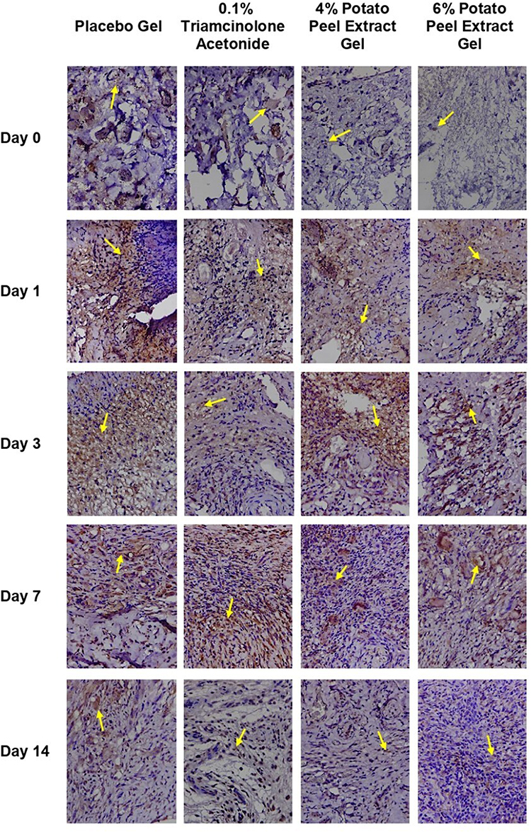 |
Figure 1 Histopathological features of MMP-9 expression in ulcerated palatal mucosa of Wistar rats, visualized by immunohistochemical (IHC) staining at 400× magnification. The yellow arrow indicates MMP-9, highlighting areas of extracellular matrix degradation (brown staining). |
On day 1, MMP-9 expression was evident in all groups. In the 0.1% triamcinolone acetonide and placebo gel groups, brown-stained regions were extensively distributed, while in the 4% and 6% extract gel groups, the staining appeared less intense and more localized. On day 3, the placebo gel, 4%, and 6% extract gel groups exhibited an increase in MMP-9 expression compared to the previous day, whereas a reduction was observed in the 0.1% triamcinolone acetonide group. On day 7, MMP-9 expression declined in the placebo, 4%, and 6% extract gel groups, while the 0.1% triamcinolone acetonide group showed a renewed increase in expression. By day 14, a general decrease in MMP-9 expression was observed across all groups. Brown-stained areas remained clearly visible in the placebo group, whereas in the 0.1% triamcinolone acetonide, 4%, and 6% extract gel groups, brown staining was no longer distinctly observed.
Quantitative Analysis of MMP-9 Expression
The collected data were first analyzed using the Shapiro–Wilk test to assess data normality. Table 2 presents the mean and standard deviation (SD) of MMP-9 expression in each treatment group. One-way analysis of variance (ANOVA) revealed a statistically significant difference among the groups (p < 0.0001), indicating that the treatments produced differential effects on MMP-9 expression levels. Table 3 summarizes the mean percentage change in MMP-9 expression for each group compared with the placebo group on days 1, 3, 7, and 14, illustrating the dynamic progression of MMP-9 expression throughout the wound healing process.
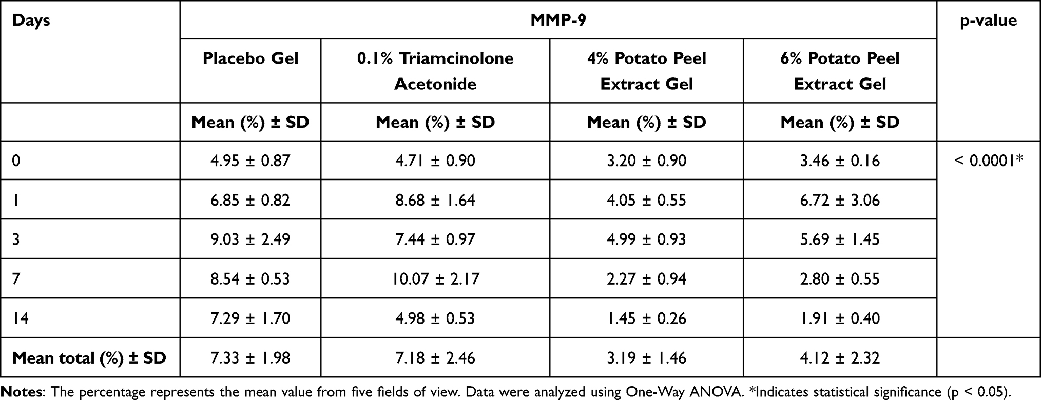 |
Table 2 MMP-9 Expression (%) in Oral Mucosal Wounds Across Different Treatment Groups |
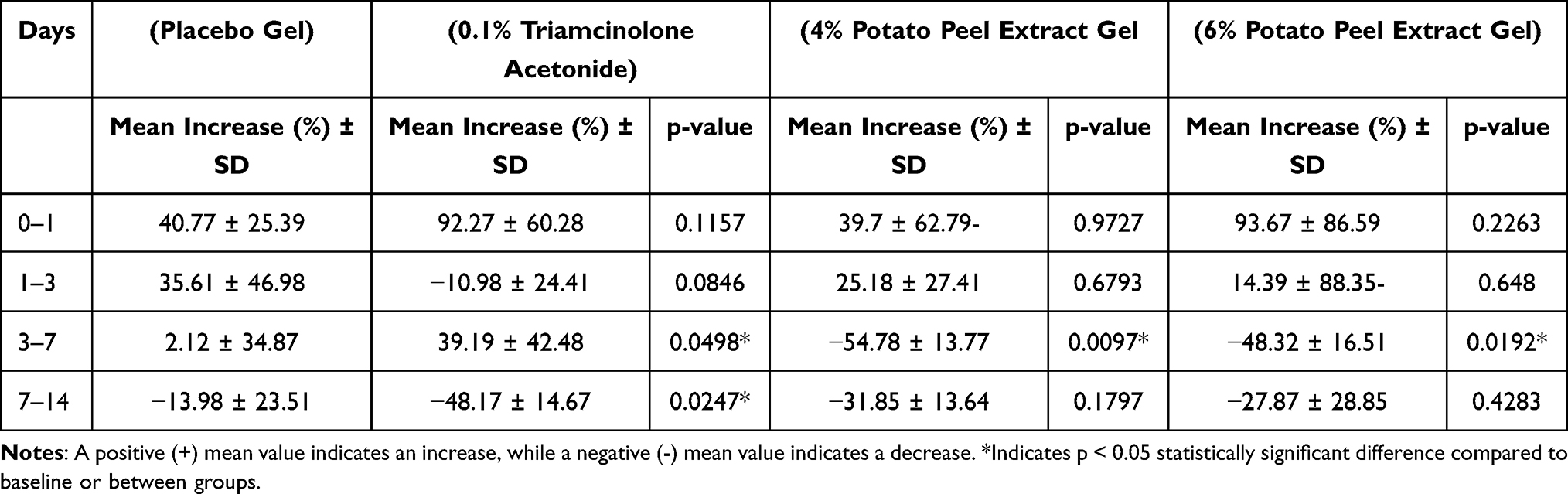 |
Table 3 Mean Percentage Change in MMP-9 Expression Relative to Placebo |
Post Hoc Statistical Comparisons Between Groups
The results from the post hoc analysis are summarized in Table 4. The values presented are p-values from pairwise statistical comparisons among the treatment groups. For example, in the 4% potato peel extract gel group on day 14, comparison with the 6% potato peel extract gel group on the same day yielded a p-value of 0.5840 (p > 0.05), indicating no significant difference between the two treatments. Similarly, comparisons between the 4% potato peel extract gel group on day 14 and both the 4% potato peel extract gel group on day 7 and the 6% potato peel extract gel group on day 7 also produced non-significant results (p > 0.05). In contrast, the 4% potato peel extract gel group on day 14 showed statistically significant differences compared to the 4% potato peel extract gel group on day 0 and the 0.1% triamcinolone acetonide group on day 7 (p < 0.05), indicating that the treatment effect at day 14 was distinct from these groups.
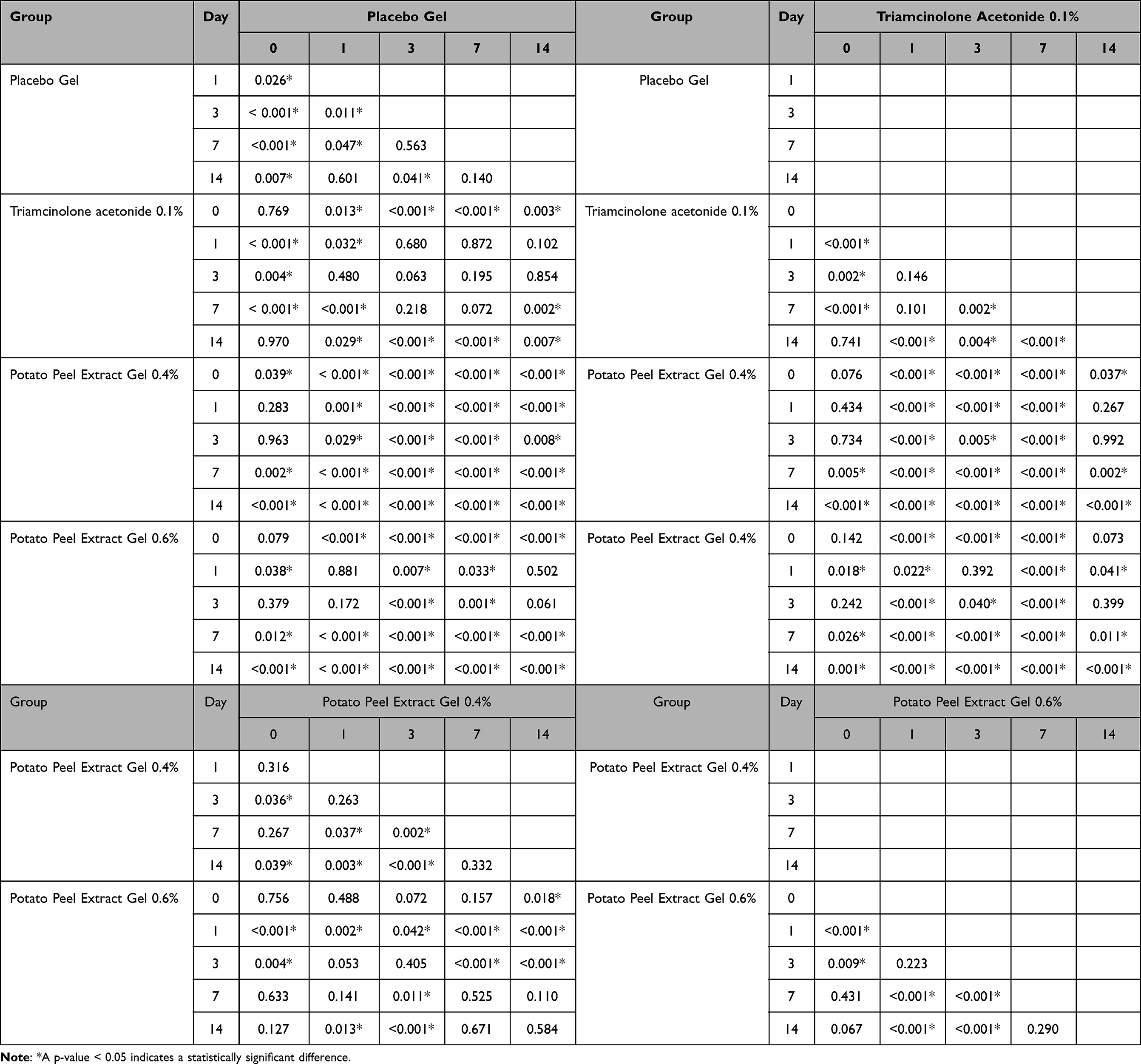 |
Table 4 Post-Hoc Analysis of MMP-9 Expression Differences Among the Treatment Groups |
Novelty of the Findings
The novelty of our results lies in demonstrating that Granola potato peel extract gel at 4% and 6% concentrations can effectively modulate MMP-9 expression and promote oral mucosal wound healing. Unlike previous studies that focused only on flavonoid content or general antioxidant activity, this study shows a direct comparison with 0.1% triamcinolone acetonide and placebo, highlighting the potential of a natural extract as an alternative to steroid treatment.
Discussion
Potatoes contain various bioactive compounds such as phenolic compounds, glycoalkaloids, polysaccharides, proteins and amino acids, as well as vitamins and minerals. Approximately 50% of these phenolic compounds are concentrated in the peel and surrounding tissues.19–21 Our prior study confirmed the presence of flavonoids, tannins, saponins, triterpenoids, and alkaloids in potato peel.15 Flavonoids act as antioxidants by reducing reactive species through mechanisms such as inhibition of nitric oxide synthase and xanthine oxidase synthase and modulation of ion channels, thereby decreasing cellular oxidative damage and promoting wound healing. They also limit the oxidation of low-density lipoprotein (LDL) caused by peroxynitrite, a product of nitric oxide (NO) and superoxide ions produced by activated macrophages. Moreover, flavonoids exhibit anti-inflammatory activity by reducing neutrophil infiltration, modulating macrophage activity, and inhibiting key inflammatory mediators such as NF-κB, activator protein-1 (AP-1), interleukin-1β (IL-1β), tumor necrosis factor-alpha (TNF-α), IL-6, IL-8, and COX-2,14,20 which is comparable to the mechanism of action of 0.1% triamcinolone acetonide.12 In addition, flavonoids and other bioactive compounds in potato peel, such as anthocyanins, tannins, and terpenoids, exhibit antibacterial effects by disrupting bacterial membranes, inhibiting enzymatic activity, and interfering with nucleic acid formation and protein transport, thereby supporting tissue regeneration and preventing infection during wound healing.13
Potato peel extract contains bioactive compounds such as flavonoids, tannins, saponins, and triterpenoids, which reduce inflammatory cell infiltration, exert antioxidant and antibacterial effects, and modulate MMP-9 expression. These effects collectively accelerate wound healing, as evidenced by the reduction in MMP-9 over time in the extract-treated groups compared to the placebo and triamcinolone acetonide groups. In our previous study, lower concentrations (2% and 4%) were tested for anti-inflammatory activity. For this study, 4% and 6% concentrations were chosen to optimize efficacy while minimizing potential local irritation or toxicity. The 4% dose proved slightly more effective than 6%, likely due to better tissue compatibility and optimal availability of bioactive compounds, suggesting an optimal concentration range rather than a simple dose-response effect.
We have confirmed that the bioactive compounds present in potato peel are capable of reducing inflammatory cell infiltration in mucosal wounds of Wistar rats. The study demonstrated that groups treated with 4% and 6% ethanol extract potato peel gel exhibited higher inflammatory cell counts on days 1 and 3, which subsequently decreased by day 7 compared to the 0.1% triamcinolone acetonide and placebo gel groups, indicating a faster progression through the inflammatory phase.16 In addition to its anti-inflammatory effects, potato peel extract also exerts antioxidant and antibacterial activities. Previous reports, such as the study by Gebrechristos et al, have shown that potato peel extract inhibits common pathogenic bacteria and exhibits substantial radical-scavenging activity, supporting its role in wound healing.23 These combined properties of antioxidant, antibacterial, and anti-inflammatory activity give potato peel extract an advantage over 0.1% triamcinolone acetonide, despite the gel formulation being more prone to loss or dissolution in saliva. The uniform coverage and local bioavailability of bioactive compounds likely compensate for this, modulating MMP-9 expression and promoting tissue regeneration effectively. The gel base used in this study was a neutral formulation optimized for stability and spreadability, and was designed to be pharmacologically inert, thus not contributing to ulcer healing on its own.
MMP-9 is a zinc-dependent enzyme crucial for wound healing, facilitating the degradation of damaged extracellular matrix (ECM), recruitment of inflammatory cells, and tissue remodeling. During the inflammatory phase, it helps clear damaged proteins and prepare the wound margins for new ECM formation. However, excessive or prolonged MMP-9 activity, as seen in chronic wounds, can degrade essential growth factors and impair healing.24,25 The time-dependent changes in MMP-9 expression observed in this study correspond well with the inflammatory, proliferative, and early remodeling phases of wound healing, reinforcing its mechanistic relevance.
In this study, the inflammatory phase observed on days 1 and 3 showed the highest MMP-9 expression in the placebo group, 6.85% on day 1 and 9.03% on day 3. This was followed by the 4% extract group (4.05% and 4.99%) and the 6% extract group (6.72% and 5.69%). Interestingly, on day 1, MMP-9 expression in the positive control (0.1% triamcinolone acetonide) and 6% extract groups was higher (92% and 93%, respectively) compared to the other groups (40%). This early peak is likely due to the acute inflammatory response, with rapid recruitment of neutrophils and macrophages to degrade damaged extracellular matrix and initiate tissue repair. The transient elevation reflects a strong but controlled early inflammatory response, which subsequently decreases as the wound progresses into the proliferative phase by day 7.
These findings align with those of Jindatanmanusan et al, who reported that high MMP-9 levels are indicative of active inflammation, since the enzyme is secreted by neutrophils and macrophages, the primary cells involved in the inflammatory response.26 LeBert et al also emphasized that elevated MMP-9 levels contribute to the regulation of leukocyte infiltration at sites of inflammation, whereas decreased MMP-9 levels may impede leukocyte recruitment in both acute inflammatory responses and chronic tissue injury.27
By day 7, a decrease in MMP-9 expression was observed across all groups, marking the transition from the inflammatory phase to the proliferative phase. Specifically, MMP-9 expression was 8.54% in the placebo group, 2.27% in the 4% extract group, 2.80% in the 6% extract group, and 2.10% in the positive control group (0.1% triamcinolone acetonide). This low level in the positive control reflects the anti-inflammatory effect of triamcinolone, which effectively suppresses inflammatory cell infiltration and MMP-9 production, facilitating progression toward tissue repair. This reduction reflects decreased inflammatory activity and progression toward tissue repair. Persistent overexpression of MMP-9 at this stage may impair wound healing by degrading ECM, altering cytokine profiles, and depleting essential growth factors.23
By day 14, further reductions in MMP-9 expression were seen in all groups. This decline indicates the progression into the remodeling phase, supporting LeBert’s findings, which stated that MMP-9 plays a role in collagen restructuring during the final stage of the wound healing process.27 Although clinical closure of superficial oral ulcers may occur by day 7, low-level MMP-9 activity can persist into day 14 as part of collagen remodeling and extracellular matrix reorganization during the final stage of healing. Interestingly, the 4% potato peel extract gel appeared slightly more effective than the 6% dose. This greater effectiveness may be attributed to an optimal balance between bioactive compound availability and tissue compatibility at the 4% concentration, whereas higher concentrations could cause local irritation or saturation of biological pathways, limiting efficacy and modulation of MMP-9 expression.
We acknowledge that 0.1% triamcinolone acetonide is a potent anti-inflammatory agent whose vasoconstrictive properties may temporarily reduce MMP-9 expression. However, the multi-target effects of the potato peel extract, encompassing anti-inflammatory, antioxidant, and antibacterial activities, provide a broader and sustained modulation of MMP-9 and tissue regeneration over time. From clinical perspective, these results suggest that Granola potato peel extract gel could potentially serve as a plant-based topical agent for managing oral mucosal wounds, offering an alternative to corticosteroids where long-term use is limited by adverse effects. Further preclinical studies, including dose optimization, long-term safety evaluation, and eventual clinical trials, are needed to confirm its applicability in humans.
It should be noted that the MMP-9 level in the positive control (0.1% triamcinolone acetonide) group remained higher than in the extract-treated groups on day 14. This can be explained by the difference in mechanisms. Triamcinolone primarily suppresses inflammatory cell activity but does not directly provide antioxidant or antibacterial effects, whereas potato peel extract contains multiple bioactive compounds (flavonoids, tannins, saponins, triterpenoids) that synergistically modulate MMP-9 expression, reduce oxidative stress, and support tissue regeneration. Additionally, the gel formulation of the extract enhances local bioavailability, resulting in more sustained regulation of MMP-9 during the later healing phase.
In this study, the main objective was to evaluate the modulation of MMP-9 expression and wound healing in ulcerated tissue. While a normal group would show baseline histological features of intact oral mucosa, the histology of normal oral mucosa is well-documented in the literature, and our results were interpreted relative to these standard features. Future studies could include a normal control group for direct comparison.
This study has certain limitations. The absence of a normal control group and the restriction to only two concentrations (4% and 6%) of potato peel extract limit the scope of the findings, in addition to the relatively short 14-day observation period and the focus on a single biomarker (MMP-9) without evaluating other inflammatory or angiogenic mediators. Future studies should include a normal control group, evaluate other inflammatory or angiogenic mediators, assess higher or intermediate doses, explore different formulations, and extend the observation period to confirm safety, optimize efficacy, and compare results directly with intact mucosa.
Taken together, these results provide evidence that Granola potato peel extract gels can modulate MMP-9 expression and support oral mucosal wound healing, highlighting their potential as a natural alternative to corticosteroid therapy and their relevance within the broader context of natural-based therapies.
Conclusion
This study demonstrated that Granola potato peel ethanol extract gel at 4% and 6% concentrations, particularly 4%, was more effective than 0.1% triamcinolone acetonide in suppressing MMP-9 expression during oral mucosal wound healing. By combining anti-inflammatory, antioxidant, and antibacterial effects, the extract provides broader and more sustained modulation of MMP-9 and tissue regeneration compared to triamcinolone acetonide. The slightly greater effectiveness of the 4% concentration may reflect an optimal balance between bioactive compound availability and tissue compatibility. These findings underscore the potential clinical relevance of MMP-9 modulation in accelerating oral wound healing and support the development of plant-based alternatives to corticosteroids. As these are pre-clinical findings in an animal model, further studies, including dose optimization, long-term safety evaluation, and clinical trials, are needed to confirm efficacy and safety in humans.
Acknowledgments
The authors would like to express their sincere gratitude to the technical staff for their valuable assistance during the data collection process.
Funding
The authors gratefully acknowledge the financial support from the Internal Research Grant of Universitas Padjadjaran (Grant No. 3135/UN6.F/HK.07/2025).
Disclosure
The authors report no conflicts of interest in this work.
References
1. Toma AI, Fuller JM, Willett NJ, Goudy SL. Oral wound healing models and emerging regenerative therapies. Transl Res. 2021;236:17–34. doi:10.1016/j.trsl.2021.06.003
2. Babu NA, Malathi LK, Kasthuri M, Jimson S. Ulcerative lesions of the oral cavity - An overview. Biomed Pharmacol J. 2017;10(1):401–405. doi:10.13005/bpj/1122
3. Fitzpatrick SG, Cohen DM, Clark AN. Ulcerated lesions of the oral mucosa: clinical and histologic review. Head Neck Pathol. 2019;13(1):91–102. doi:10.1007/s12105-018-0981-8
4. Kandhwal M, Behl T, Singh S, et al. Role of matrix metalloproteinase in wound healing. Am J Transl Res. 2022;14(7):4391–4405.
5. Cabral-Pacheco GA, Garza-Veloz I, Castruita-De la Rosa C, et al. The roles of matrix metalloproteinases and their inhibitors in human diseases. Int J Mol Sci. 2020;21(24):9739. doi:10.3390/ijms21249739
6. Caley MP, Martins VLC, O’Toole EA. Metalloproteinases and wound healing. Adv Wound Care (New Rochelle). 2015;4(4):225–234. doi:10.1089/wound.2014.0581
7. Peng Z, Nguyen TT, Song W, et al. Selective MMP-9 inhibitor (R)-ND-336 alone or in combination with linezolid accelerates wound healing in infected diabetic mice. ACS Pharmacol Transl Sci. 2021;4(1):107–117. doi:10.1021/acsptsci.0c00104
8. Bindu S, Mazumder S, Bandyopadhyay U. Non-steroidal anti-inflammatory drugs (NSAIDs) and organ damage: a current perspective. Biochem Pharmacol. 2020;180:114147. doi:10.1016/j.bcp.2020.114147
9. Ericson-Neilsen W, Kaye AD. Steroids: pharmacology, complications, and practice delivery issues. Ochsner J. 2014;14(2):203–207.
10. Spence JD, Grosser T, Fitzgerald GA. Acetaminophen, nonsteroidal anti-inflammatory drugs, and hypertension. Hypertension. 2022;79(9): 1922–1926. doi: 10.1161/HYPERTENSIONAHA.122.19315.
11. Tai FWD, McAlindon ME. Non-steroidal anti-inflammatory drugs and the gastrointestinal tract. Clin Med. 2021;21(2):131–134. doi:10.7861/clinmed.2021-0039
12. Sidhu G, Preuss CV. Triamcinolone. [Updated 2024 Feb 28]. In: StatPearls [Internet]. Treasure Island (FL): StatPearls Publishing; 2025 Jan-. Available from: https://www.ncbi.nlm.nih.gov/books/NBK544309/
13. Krisnadita A, Lestari ES, Setyawan A, Antari AL. Antibacterial effectiveness test of potato peel ethanol extract (Solanum tuberosum L.) against Lactobacillus acidophilus: an in vitro study. Majalah Obat Tradisional. 2023;28(2):86–92. doi:10.22146/mot.81476
14. Al-Khayri JM, Ravikumar Sahana G, Nagella P, Joseph BV, Alessa FM, Al-Mssallem MQ. Flavonoids as potential anti-inflammatory: a review. Molecules. 2022;27(9):2901. doi:10.3390/molecules27092901
15. Tiarasanti F, Sufiawati I, Amalia E, Sari KI, Zubaedah C, Takarini V. The effects of potato (Solanum tuberosum L. vs. Granola; Solanaceae) peel extract gel on gingival wound healing in wistar rats. J Exp Pharmacol. 2024;16:25–35. doi:10.2147/JEP.S443355
16. Santoso AW, Amalia E, Sari KKI, Takarini V, Sufiawati I. Histopathological evaluation of wound healing and anti-inflammatory effects of Granola potato peel ethanol extract in rat oral mucosa. J Exp Pharmacol. 2024;16:377–395. doi:10.2147/JEP.S487373
17. Mhapuskar AA, Thakare S, Kale IP, Karmarkar P, Hiremutt DR, Jadhav A. Comparison of effects of honey and 0.1% triamcinolone acetonide in the management of recurrent aphthous stomatitis - A randomised, single-blind study. J Indian Acad Oral Med Radiol. 2022;34(3):276–280. doi:10.4103/jiaomr.jiaomr_157_22
18. Ferreira AS, Macedo C, Silva AM, Delerue-Matos C, Costa P, Rodrigues F. Natural products for the prevention and treatment of oral mucositis—a review. Int J Mol Sci. 2022;23(8):4385. doi:10.3390/ijms23084385
19. Rodríguez-Martínez B, Gullón B, Yáñez R. Identification and recovery of valuable bioactive compounds from potato peels: a comprehensive review. Antioxidants. 2021;10(10):1630. doi:10.3390/antiox10101630
20. Jimenez-Champi D, Romero-Orejon FL, Moran-Reyes A, Muñoz AM, Ramos-Escudero F. Bioactive compounds in potato peels, extraction methods, and their applications in the food industry: a review. CyTA - J Food. 2023;21(1):418–432. doi:10.1080/19476337.2023.2213746
21. Sampaio SL, Petropoulos SA, Alexopoulos A, et al. Potato peels as sources of functional compounds for the food industry: a review. Trends Food Sci Technol. 2020;103:118–129. doi:10.1016/j.tifs.2020.07.015
22. Zulkefli N, Che Zahari CNM, Sayuti NH, et al. Flavonoids as potential wound-healing molecules: emphasis on pathways perspective. Int J Mol Sci. 2023;24(5):4607. doi:10.3390/ijms24054607
23. Gebrechristos HY, Ma X, Xiao F, et al. Potato peel extracts as an antimicrobial and potential antioxidant in active edible film. Food Sci Nutr. 2020;8(12):6338–6345. doi:10.1002/fsn3.1119
24. Fu K, Zheng X, Chen Y, et al. Role of matrix metalloproteinases in diabetic foot ulcers: potential therapeutic targets. Front Pharmacol. 2022;13:1050630. doi:10.3389/fphar.2022.1050630
25. Sabino F, Auf Dem Keller U. Matrix metalloproteinases in impaired wound healing. Metalloproteinases in Medicine. 2015;2:1–8. doi:10.2147/MNM.S68420
26. Jindatanmanusan P, Luanraksa S, Boonsiri T, Nimmanon T, Arnutti P. Wound fluid matrix metalloproteinase-9 as a potential predictive marker for the poor healing outcome in diabetic foot ulcers. Patholog Res Int. 2018;16:1–5. doi:10.1155/2018/1631325
27. LeBert DC, Squirrell JM, Rindy J, et al. Matrix metalloproteinase 9 modulates collagen matrices and wound repair. Development. 2015;142(12):2136–2146. doi:10.1242/dev.121160
 © 2025 The Author(s). This work is published and licensed by Dove Medical Press Limited. The
full terms of this license are available at https://www.dovepress.com/terms
and incorporate the Creative Commons Attribution
- Non Commercial (unported, 4.0) License.
By accessing the work you hereby accept the Terms. Non-commercial uses of the work are permitted
without any further permission from Dove Medical Press Limited, provided the work is properly
attributed. For permission for commercial use of this work, please see paragraphs 4.2 and 5 of our Terms.
© 2025 The Author(s). This work is published and licensed by Dove Medical Press Limited. The
full terms of this license are available at https://www.dovepress.com/terms
and incorporate the Creative Commons Attribution
- Non Commercial (unported, 4.0) License.
By accessing the work you hereby accept the Terms. Non-commercial uses of the work are permitted
without any further permission from Dove Medical Press Limited, provided the work is properly
attributed. For permission for commercial use of this work, please see paragraphs 4.2 and 5 of our Terms.