Back to Journals » Journal of Experimental Pharmacology » Volume 13
Phytochemical Screening and in-vitro Evaluation of Antibacterial Activities of Echinops amplexicaulis, Ruta chalepensis and Salix subserrata Against Selected Pathogenic Bacterial Strains in West Shewa Zone, Ethiopia
Authors Marami LM ![]() , Dilba GM
, Dilba GM ![]() , Babele DA
, Babele DA ![]() , Sarba EJ
, Sarba EJ ![]() , Gizaw A
, Gizaw A ![]() , Bune WM
, Bune WM ![]() , Bayu MD, Admasu P
, Bayu MD, Admasu P ![]() , Mekbeb A, Tadesse M, Abdisa K, Bayisa D
, Mekbeb A, Tadesse M, Abdisa K, Bayisa D
Received 9 February 2021
Accepted for publication 22 April 2021
Published 18 May 2021 Volume 2021:13 Pages 511—520
DOI https://doi.org/10.2147/JEP.S305936
Checked for plagiarism Yes
Review by Single anonymous peer review
Peer reviewer comments 2
Editor who approved publication: Prof. Dr. Héctor Mora-Montes
Lencho Megersa Marami,1 Getachew Mulatu Dilba,1 Dagmawit Atalel Babele,1 Edilu Jorga Sarba,2 Askale Gizaw,1 Wakuma Mitiku Bune,1 Morka Dandecha Bayu,1 Petros Admasu,2 Abraham Mekbeb,2 Miressa Tadesse,3 Kebede Abdisa,2 Dejene Bayisa4
1Ambo University, College of Agriculture and Veterinary Science, Department of Veterinary Laboratory Technology, Ambo, Oromia, Ethiopia; 2Ambo University, College of Agriculture and Veterinary Science, Department of Veterinary Science, Ambo, Oromia, Ethiopia; 3Ambo University, College of Computational and Natural Science, Department of Chemistry, Ambo, Oromia, Ethiopia; 4Jeldu Woreda, Livestock and Fishery Development Office, Gojo, Oromia, Ethiopia
Correspondence: Getachew Mulatu Dilba
Ambo University, College of Agriculture and Veterinary Science, Department of Veterinary Laboratory Technology, P.O. Box 19, Ambo, Oromia, Ethiopia
Tel +251-912717217
Email [email protected]
Background: Although traditional healers in Ethiopia have a long history of using medicinal plants to treat diseases in animals and humans, studies on the antibacterial activities and potential bioactive ingredients of most medicinal plants have been insufficient. Therefore, this study aimed to evaluate the in-vitro antibacterial activities and to screen phytochemical constituents of selected medicinal plants against reference bacterial strains.
Methods: The fresh and healthy roots of Echinops amplexicaulis, fruits of Ruta chalepensis, and leaves of Salix subserrata were collected from West Shewa Zone, Ethiopia. Agar well diffusion and agar dilution methods were used to evaluate antibacterial activities and minimum inhibitory concentrations (MIC). All the crude plant extracts were tested against Staphylococcus aureus, Streptococcus pneumoniae, Escherichia coli and Pseudomonas aeruginosa at concentrations of 100, 50, and 25 mg/mL in each triplet (3x). MIC of crude extracts ranging from 1.5625 to 12.50 mg/mL was applied to all bacterial strains. The positive control was ciprofloxacin disk (5 μg) and the negative control was 5% dimethyl sulfoxide. The presence of secondary metabolites of each crude extract was screened. The group means comparisons were done using one-way ANOVA and results were presented as mean ± standard deviation.
Results: Although all selected plant extracts had shown antibacterial activities, methanol extracts had a greater zone of inhibition against all reference bacterial strains when compared to petroleum ether extracts. The growth of P. aeruginosa was inhibited at a minimum concentration of both methanol and petroleum extracts (1.5625 mg/mL) when compared to the remaining bacterial strains. Phytochemical screening showed that saponins and alkaloids were found in all crude plant extracts, while phytosterol was meager.
Conclusion: This study revealed that all tested plants had significant secondary metabolites and antibacterial activities against reference bacterial strains.
Keywords: antibacterial activity, crude plant extract, ethnomedicine, methanol, petroleum ether
Introduction
Ethnomedicine is a holistic study of indigenous knowledge of traditional medicine, socio-cultural experience, and the environment as it relates to health care and animal husbandry.1 Ethnomedicinal practice has been developed through trial and error, which later on is supported by actual experimentation.2 Also, ethnoveterinary medicine is often undertaken as part of a community-based approach that serves to improve animal health and provide basic veterinary services, most commonly in rural areas.3 Since plants are rich in phytochemicals (secondary metabolites), they are naturally occurring substances and provide health benefits. Alkaloids, flavonoids, tannins, phenols, saponin, steroids, glycosides, and terpenes are some of the plant’s major secondary metabolites4 that have antioxidant, anti-inflammatory, anti-cancer, and anti-microbial properties.5 According to the WHO reports, at least 80% of people in developing countries depend largely on indigenous practices for the control and treatment of various diseases affecting both humans and animals.6 In most developing countries where there is a shortage of animal health facilities and veterinarians, ethnoveterinary medicine provides alternative treatments for animal diseases that are locally available and usually cheaper than the standard treatments because livestock holders can prepare and use homemade remedies with least cost.7,8
Echinops amplexicaulis (“Kosorruu Harree” in Afan Oromo) belongs to the family Asteraceae and its species are found in Eastern and Southern Europe, Tropical North and East Africa, and Asia.9 Traditional medicine practitioners in Uganda used the entire root of this plant to treat HIV/AIDS and related conditions.10 This plant is also used to treat ulcerative lymphangitis and hepatitis.9 The frequently described application is to treat symptoms like inflammation, pain, and fever, and ailments related to the respiratory tract, including cough and sore throat.11 Members of the genus have been used as an aphrodisiac,12 facilitation of expulsion of retained placenta and delivery,13,14 as an abortifacient,15 treatment of uterus tumor,16 and leucorrhoea.17 E. bannaticus Rochel ex Schrad, E. cornigerus D.C., and E. polyceras Boiss are the species of Echinops that have been used in the management of kidney stones.18–20
Ruta chalepensis belongs to the Rutaceae family, which is distributed in temperate and tropical countries.21 The decoction of R. chalepensis is used as a medicinal remedy against the evil eye and for “spiritual cleansings”, whereas the infusions of its fresh leaves are widely used as a treatment for gastric disorders, headache, and rheumatism. It is also reported for its diuretic, anti-inflammatory, and anti-spasmodic properties.21 This plant has also been used as a mosquito repellent, an antidote to snake and scorpion venom poisoning, and as a poultice for bites and stings.22
Salix subserrata (Alaltuu in Afan Oromo) is a deciduous bush or small tree 2–10 m tall that grows along streams throughout Africa. The leaf furnishes a laxative for human and veterinary medicine. Roots are used in medicines that help cure stomach pains, fever, and headaches. Crushed leaves of S. subserrata are also reported to be used in treating patients with rabies after mixing with milk.23
Escherichia coli is a Gram-negative bacillus found in the large intestine and excreted naturally through the feces and urinary tracts. It is one of the most frequent causes of many common bacterial diseases including cholecystitis, bacteremia, cholangitis, urinary tract infections and travelers’ diarrhea, and other clinical infections, such as neonatal meningitis and pneumonia.24
Pseudomonas aeruginosa is also a gram-negative, rod-shaped, and mono flagellated microorganism. It has a pearlescent manifestation and a grape or tortilla. This organism grows well at 25–37°C and its ability to grow at 42°C distinguishes it from other Pseudomonas species.25
Staphylococcus aureus is a gram-positive coccus that colonizes the nasal mucosa and skin of healthy individuals. The organism can cause a wide range of diseases ranging from skin or soft tissue infections to systemic and fatal diseases.26 S. aureus possesses a specific virulence factor called coagulase, which plays a significant role in biofilm formation during S. aureus infection. Coagulase binds to host prothrombin and forms active staphylothrombin complexes that convert soluble monomeric fibrinogen into self-polymerizing insoluble fibrin and activates a coagulation cascade.26
Streptococcus pneumoniae is a gram-positive facultative anaerobe, currently one of the most considerable pathogens worldwide. It is a major cause of bacterial pneumonia, meningitis, and sepsis.27 It is also the principal cause of potentially life-threatening community-acquired disease and is associated with a predictable global mortality rate which is of the same order of magnitude as that of tuberculosis, and subsequent investigation has identified a unique mechanism of penicillin resistance in this bacterium.28
Antimicrobial resistance is spreading at an alarming rate, making treating bacterial infections in animals and humans more difficult. Even modern medicines have failed to treat those resistant bacteria since they are developing resistant behavior.29 Therefore, it is logical to search for alternative methods to manage infectious diseases. Ethnomedicinal practices are believed to be one of the potential bases for the development of safe and effective treatments. Ethiopia has a long history of a traditional health care system, but studies on traditional medicinal plants (TMP) have been limited in comparison to the country’s multiethnic, cultural, and flora diversity.30 Also, the use of medicinal plants to treat infections is an old practice in large parts of Ethiopia to solve health problems for livestock and humans.31–36 However, most of these experiences are not documented and supported by scientific experiments. Among medicinal plants, leaves of S. subserrata, fruits of R. chalepensis, and roots of E. amplexicaulis were frequently used for the treatments of disease-causing bacterial strains like S. aureus, E. coli, S. pneumoniae, and P. aeruginosa. Therefore, the objective of the current study was to evaluate the in-vitro antibacterial activities and to screen major phytochemical constituents of selected medicinal plants against reference bacterial strains.
Materials and Methods
Description of Plant Collection Areas
Plants were collected from the Ejere, Toke Kutaye, and Dendi districts of West Shewa, Oromia Regional State, Ethiopia from December 2019 to January 2020. Ejere woreda is in the central part of Ethiopia with longitude and latitude of 9.03° N 38.40° E and 9°2ʹN 38°24ʹ with an elevation of about 2360 MASL (meters above sea level). While Toke Kutaye district is located 162 km west of the capital city, Addis Ababa and the area lies within central Ethiopia with altitudes ranging from 1580 to 3194 MASL. It receives an annual rainfall of 800–100 mm and has an annual temperature range of 10–29 °C. Dendi is located 79 km west of Addis Ababa, and 35 km east of Ambo. The area lies within the central country with altitudes ranging from 2000 to 3288 MASL. It receives an annual rainfall of 750–1170 mm and has an annual temperature range of 9.3°C-23.8°C. The traditional knowledge and practice of ethnomedicine areas are well known. Moreover, the area is rich in different natural resources. For instance, “Chilimo” forest is the main source of the medicinal plant for local practitioners.37 All the study areas’ soil types ranged from loam to clay, and the subsurface horizons were clayey, with low percentage base saturation, soil pH, and exchangeable cations.38
Study Design
The phytochemical screening test and in-vitro antibacterial evaluation were done through a randomized experimental design. All experimental tests were done with triplicates alongside the positive and negative controls.
Plant Collection and Preparation
Fresh and healthy leaves of S. subserrata, roots of E. amplexicaulis, and fruits of R. chalepensis were collected after being identified by a botanist from the department of plant science at Ambo University. The collected plant parts were authenticated (herbarium) in the Plant Science Laboratory of Ambo University Guder Mamo Mezemir Campus, cleaned using sterilized distilled water, cut into smaller sizes of about 1–2 cm long, and dried under shade at room temperature for 15 (fifteen) days. Then, it was ground by using a conventional wooden-made pestle and mortar, pounded using an electric grinder into a fine powder, and finally kept in a refrigerator (4 °C) until used.36,39
Plant Extraction
Although a standardized extraction protocol has not been developed for herbal extracts, 20–95% of the solvents (polar or/and non-polar) substances are frequently used by the herbal medicine industry to prepare plant crude extracts.40 All three medicinal plants: E. amplexicaulis, R. chalepensis, and S. subserrata were extracted with methanol (99.8%) and petroleum ether (99.5%) based on the information from the traditional claim and previous studies. Two hundred grams of air-dried powdered plant materials were placed in a flat-bottom flask filled with 1000 mL of extracting solvents and macerated for 24 hrs over a rotary shaker at 121 rpm. The suspension was filtered with Whatman №. 1 paper. The resulting filtrate was then concentrated under reduced pressure in a rotary evaporator. The gummy residue was further dried, followed by a water bath and oven at 45 °C and 42 °C, respectively, until the solvent was removed. After the solvent was evaporated, the remaining crude extracts were diluted with 10 mL of sterile distilled water and kept in an airtight bottle in the refrigerator until the experiment was carried out.36
Preliminary Phytochemical Test
According to the concept of Pandey and Tripathi,41 each crude extract was tested for the presence of secondary metabolites that might be playing a significant role in the antibacterial activities of medicinal plants. Accordingly, each plant was tested for the presence of alkaloids, saponins, phenols, tannins, flavonoids, and phytosterols.
Test for alkaloids: 0.25 gm of the crude extract was added to five drops of HCl and then it was filtered and finally the filtrate was mixed with Wagner’s reagents to form a Brown or precipitate which indicates the presence of an alkaloid.
Test for saponin: 0.25 gm of crude plant extract was mixed with 20 mL distilled water and shaken in a graduate cylinder for 15 minutes to form a foam with a 1 cm layer indicating the presence of saponin.
Test for phenol: 0.25 gm of the crude extract was added to 4 drops of FeCl3 for the formation of a blue-black color which indicates the presence of phenols and this test is considered as a ferric chloride test.
Test for tannin: 0.25 gm of the crude extract was added to 1% gelatin solution containing NaCl for the formation of a precipitate, which in turn indicates the presence of tannin in the crude extract.
Test for flavonoid: Using an alkaline reagent test, a few drops of NaOH solution were added to the crude extract for the formation of an intense yellow color that becomes colorless and the addition of 10 drops of 1% hydrochloric acid showed the presence of flavonoids.
Test for phytosterol: The Szarkowski test was conducted by adding a few drops of chloroform to 0.25gm of crude extract and then filtered. The filtrate was then mixed with a few drops of concentrated H2SO4, shacked, and left to stand for a few minutes to obtain a golden yellow color, indicating the presence of phytosterols in the crude extract.
Source of Bacterial Strains
Reference bacterial strains, namely S. aureus (ATCC 25923), S. pneumoniae (ATCC 49619), E. coli (ATCC 25922), and P. aeruginosa (ATCC 27853) were utilized to evaluate antibacterial activities of crude plant extracts. These four American-type cell cultures (ATCC) were aseptically collected from the Ethiopian Public Health Institute (EPHI), Addis Ababa, and transported under the cold chain.
In-vitro Antibacterial Activity Test
The bacterial strains were cultured on blood agar plates at 37 °C for 18–24; then, pure colonies were transferred to the nutrient agar plate. Four to five bacteria colonies were inoculated into the nutrient broth and incubated for 2–6 hrs. Each cultured isolate was compared with 0.5 McFarland turbidity standards. Sterile physiological saline solution was used to standardize the turbidity of the isolates.
Agar-Well Diffusion Method
After inoculation of the bacteria with sterile swabs on the surface of Mueller Hinton agar plates, it was left to dry at room temperature. Four holes were made on the surface of the Mueller Hinton agar plate at equal distances from each other using a 6 mm diameter cork borer. The holes were then filled with the test sample (crude plant extracts) at a concentration of 100, 50, and 25 mg/mL, and negative control (5% DMSO). An antibiotic disk, ciprofloxacin (5 μg), was placed on the surface of the agar plate as a positive control.42 The experiment was repeated three times (3x). The plates were then left at room temperature for about an hour to favor diffusion and incubated at 37°c for 24 hrs. After 24 hrs. of incubation, the antibacterial activity was evaluated by measuring the diameter of the zone of inhibition including the hole.43
Minimum Inhibitory Concentration (MIC): Agar Dilution Method
The minimum inhibitory concentration (MIC) of all crude extracts was evaluated against S. aureus, P. aeruginosa, E. coli, and S. pneumoniae. A 5% Dimethyl sulfoxide (DMSO) was used to dilute crude plant extracts. Then, after serial dilution, the crude extract (2 mL) was mixed with molten Mueller Hinton agar (18 mL) and poured on sterilized Petri dishes. The plate was inoculated with the standardized (0.5 McFarland standard) bacterial inoculum and incubated at 37°C for 24 hrs. The result of bacterial inhibition was judged by comparison with growth in positive and negative controls.44
Data Analysis
Data were entered into Microsoft Excel and exported to STATA version 14 (1985–2015 Stata Corp) for statistical analysis. One-way ANOVA was performed to compare group means. The results for zones of inhibition of antibacterial activities and MIC were summarized in the form of means ± standard deviation. If secondary metabolites were present, slightly present, and absent, the phytochemical screening result was classified as “++”, “+”, and “-”, respectively.
Results
Phytochemical Screening of Crude Plant Extracts
The results of phytochemical screening of both methanol and petroleum ether plant extracts (E. amplexicaulis, R. chalepensis, and S. subserrata) were summarized (Table 1). The methanol extracts of R. chalepensis consisted of all tested secondary metabolites. Saponins and alkaloids were the most abundant secondary metabolites found in all crude extracts, while phytosterol was the least abundant.
 |
Table 1 Results of Preliminary Phytochemical Screening of Selected Medicinal Plants |
Antibacterial Activities of the Crude Extracts: Agar-Well Diffusion Method
The results of a zone of inhibition (mm, diameter) created by antibacterial activities of crude methanol and petroleum ether extracts of S. subserrata (Leaf), R. chalepensis (Fruit), and E. amplexicaulis plants were measured and summarized in Table 2. The concentration of these crude extracts was classified into 100, 50, and 25 mg/mL in three replicates and subjected to S. aureus, P. aeruginosa, E. coli, and S. pneumoniae. The methanol and petroleum ether plant extracts had shown a zone of inhibition against the selected bacterial strains and had a statistical significance difference between and within-group replicates at p <0.05. However, petroleum ether extract of S. subserrata against S. aureus and P. aeruginosa and methanol extracts of R. chalepensis against S. aureus were not statistically significant. All crude methanol plant extracts had better antibacterial activity when compared with petroleum ether extracts.
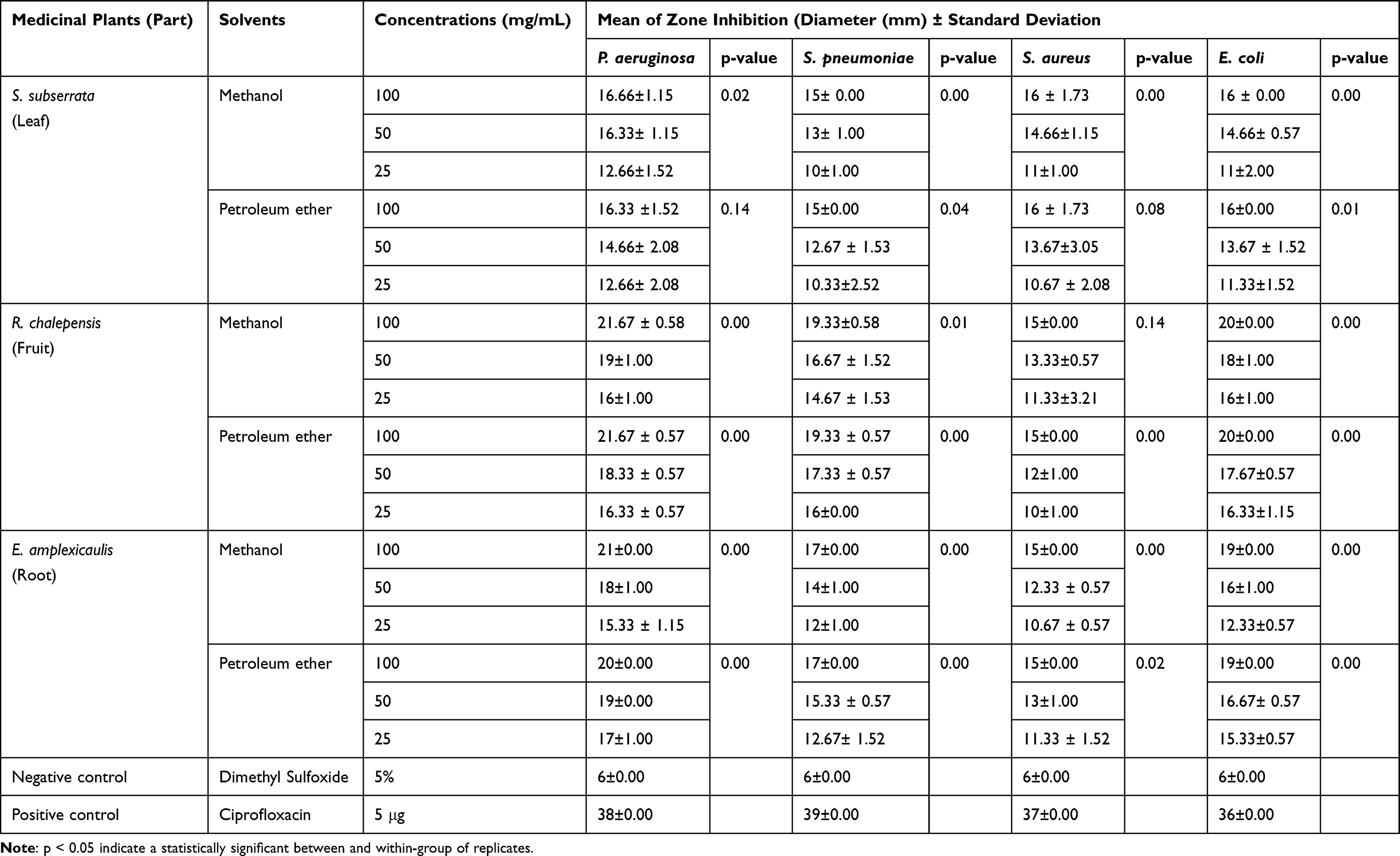 |
Table 2 The Antibacterial Activity of Methanol and Petroleum Ether Extracts of Three Medicinal Plants (Mean of Zone Inhibition of Three Replicates (Diameter in mm) ± Standard Deviation) |
Minimum Inhibitory Concentration (MIC): Agar Dilution Method
The MIC test was done using a serial dilution of crude plant extract using 5% DMSO. For extracts showing a diameter greater than or equal to 6 mm of the growth inhibition zone at 25 mg/mL, MIC was calculated. The methanol and petroleum ether crude extracts were applied to all reference bacterial strains with concentrations ranging from 1.5625 mg/mL to 12.5 mg/mL and the results are shown in Table 3. Compared to the remaining bacterial strains, P. aeruginosa had better antibacterial activities at a lower concentration. P. aeruginosa and E. coli were inhibited at 1.5625 mg/mL concentration by both crude plant extracts.
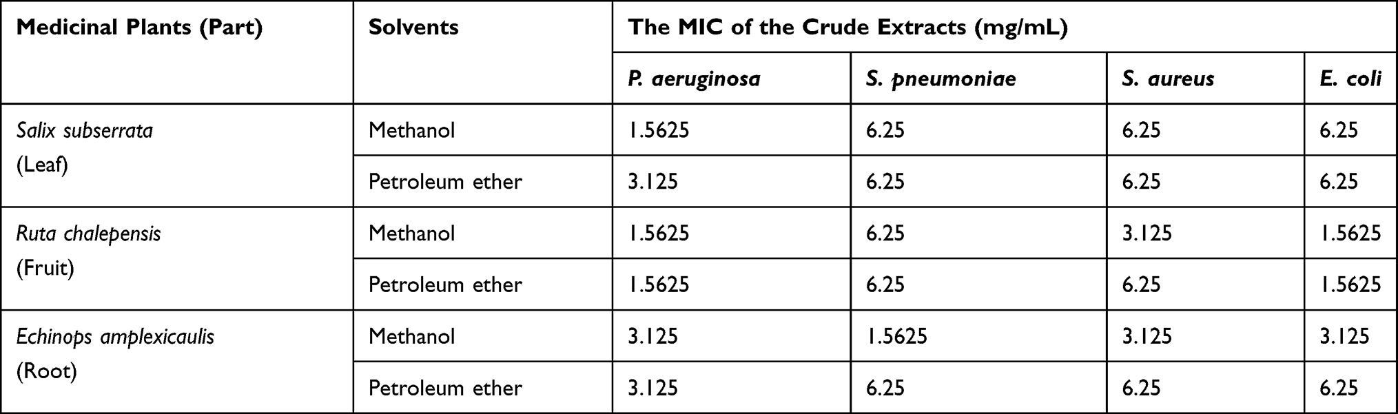 |
Table 3 Minimum Inhibitory Concentrations of Crude Extracts of Selected Medicinal Plants Against Selected Bacterial Strains (mg/ml) |
Discussion
The current study revealed that alkaloids, tannins, saponins, flavonoids, and phytosterols were the secondary metabolites found in the crude extract of E. amplexicaulis, R. chalepensis, and S. subserrata. The methanol extracts of E. amplexicaulis and S. subserrata contain most of the secondary metabolites. This is in line with a previous study by Najem et al,45 who reported saponins, alkaloids, tannins, and flavonoids found in methanol extracts of E. amplexicaulis, but phytosterols and phenols were absent. Kevin et al9 found small amounts of tannins and saponins in E. amplexicaulis crude extract but no flavonoids, in contrast to the current study. The difference might be due to soil content, geographical area, seasons of plant collection, plant parts, and growth stage of the plants. According to Copp,46 secondary metabolites of the terpenoids’ family had antibacterial activities, which may be related to their lipophilic nature and thus able to penetrate the bacterial cell wall. In general, the antibacterial activities of plant extracts may be associated with the presence of important secondary metabolites, which play a significant role in the treatment and control of various bacterial diseases, and thus these medicinal plants are used as alternative medicines.
A study by Emam et al47 indicated that the ethanol extract (80%) of the R. chalepensis leaves had alkaloids that are suggested to exhibit antifungal activity against the three phytopathogenic fungi R. solani, S. rolfsii, and F. solani when tested with the disk diffusion technique. Also, the secondary metabolites, namely alkaloids, flavonoids, and tannin of R. chalepensis leaves crude ethanolic extract, had anti-acetylcholinesterase inhibition and antioxidant activities in the gastrointestinal tract.48,49
The current experimental study showed that all three medicinal plants had remarkable antibacterial activities against all tested bacteria. This finding supports the traditional use in the treatment of bacterial infections in both humans and livestock. In the current study, petroleum ether and methanol extracts of R. chalepensis had a considerable antibacterial activity against the use of gram-positive and gram-negative bacterial species. Similarly, Amdouni et al50 used gas chromatography to identify the chemical composition of R. chalepensis leaves, stems, and roots grown under salt stress, and the extract of these plants had antibacterial activity against eight bacteria (Salmonella All, Salmonella K, Escherichia coli 45AG, Escherichia coli 45AI, Staphylococcus aureus 9402, Staphylococcus aureus 02B145, Listeria 477 and Pseudomonas aeruginosa ATCC 10145).
The methanol and petroleum ether extracts of R. chalepensis in the current study showed a higher zone of inhibition (21.67 ± 0.5) at 100 mg/mL concentration, which is contrary to the report of Seid and Ayisha51 that states the leaf extract of R. chalepensis showed the least zone of inhibition (2.49 mm). This is because a higher amount of secondary metabolites is found in the fruit than in the leaf of the indicated plant Krayni et al;52 in this study, the fruit part was used but in the previous study it was the leaf.
The antibacterial evaluation of the three plants results in various sizes of inhibition zones with both solvents against both the Gram-positive and Gram-negative bacteria. In particular, methanol extracts relatively showed a greater zone of inhibition (antibacterial activities) compared to the petroleum ether extracts. Seid and Ayisha51 were also reported the same finding in Ethiopia. This is associated with the extracting efficiency of methanol to liberate most of the biologically active phytochemical compounds like flavonoids, tannins, and carotenoids, which agrees with the ideas of Bakari et al53 and Felhi et al.54 On the other hand, in this study, Gram-positive bacterial strains were relatively less sensitive to both the petroleum ether and methanol extracts, which is in agreement with the results of Fentahun et al.30
The MIC determination test revealed that a different minimum concentration of the crude extract was gained in three of the plants that could inhibit the growth of the reference bacteria. The least MIC value was recorded for methanol extract of R. chalepensis against P. aeruginosa and E. coli. On the contrary, the higher concentration of crude extracts was inhibiting the growth of G-positive bacteria, S. pneumoniae. The MIC value is in line with the antibacterial activities of this plant on the same bacteria. This could be logically explained that R. chalepensis has a better antibacterial activity on G-negative bacteria than G-positives, at least those which are tested in this study. On the other hand, the MIC values of both the methanol and petroleum ether extracts of S. subserrata were 6.25 mg/mL against all the tested organisms except for P. aeruginosa in which the MIC value was 1.5625 mg/mL and 3.125 mg/mL for methanol and petroleum ether extracts, respectively. This also indicates that most of the potential phytochemicals are better extracted with methanol than petroleum ether. Overall, P. aeruginosa is highly susceptible to plant crude extracts even with a small dose.
Despite the important finding, in-vivo evaluation of antibacterial activities of the selected plants, toxicity studies, and quantification of secondary metabolites were not conducted in the current study due to a lack of laboratory animals and facilities.
Conclusion
The current study revealed that all tested plants had important active phytochemical constituents and antibacterial activities against reference bacterial strains. Methanol extracts of S. subserrata (leaves), R. chalepensis (fruits), and E. amplexicaulis (roots) had a greater zone of inhibition against all reference bacterial strains when compared to petroleum ether extracts. The growth of P. aeruginosa was inhibited at a minimum concentration of both methanol and petroleum extracts when compared to the remaining bacterial strains. Saponins and alkaloids were the abundant phytochemicals found in methanol and petroleum ether extracts of S. subserrata (Leaf), R. chalepensis (Fruit), and E. amplexicaulis (Roots), while phytosterol was meager. In addition to the current finding, more research on in-vivo antibacterial activity evaluation, toxicity studies, and quantitative analysis of secondary metabolites of crude extracts using advanced techniques is encouraged.
Data Sharing Statement
All supplementary data used in the present study are available from the corresponding author and first author upon reasonable request.
Acknowledgments
This work was financed by Ambo University. The authors deeply acknowledge Ambo University on various occasions during the duration of the study. We would also like to thank those traditional healers found in the study area who assisted us during the plant collection.
Author Contributions
All authors made a significant contribution to the overall research activities either in the conception of the study, execution, acquisition of data, analysis, and interpretation or in all these areas. Moreover, all authors took part in revising or critically reviewing the article; gave final approval for the version to be published; have agreed on the journal to which the article has been submitted, and agreed to be accountable for all aspects of the work.
Disclosure
The funder had no role in the study design, data collection, management, and analysis, decision to publish, or preparation of the manuscript.
The authors report no conflicts of interest in this work.
References
1. Eshetu GR, Dejene TA, Telila LB, Bekele DF. Ethnoveterinary medicinal plants: preparation and application methods by traditional healers in selected districts of southern Ethiopia. Vet World. 2015;8(5):674–684. doi:10.14202/vetworld.2015.674-684
2. Bekele A, Musa A. Ethnoveterinary practice in Chiro District, western Hararge, Ethiopia. Pharmacologyonline. 2009;1:128–139.
3. Oyda S. Review on traditional ethno-veterinary medicine and medicinal plants used by indigenous people in Ethiopia: practice and application system. Int J Res. 2017. doi:10.5281/zenodo.884998
4. De Silva GO, Abeysundara AT, Aponso MMW. Extraction methods, qualitative and quantitative techniques for screening of phytochemicals from plants. Am J Essent Oil. 2017;5(2):29–32.
5. Majekodunmi SO. Review of extraction of medicinal plants for pharmaceutical research. Merit Res J Med. 2015;3:521–527.
6. Django S, Nuwanyakpa M, Toyang NJ, Wanyama J. Ethnoveterinary Medicine a Practical Approach to the Treatment of Cattle Diseases in Sub-Saharan Africa. The Netherland: Agromisa Foundation; 2007:1–78.
7. McCorkle CM. Back to the future: lessons from ethnoveterinary research, development, and extension for studying and applying local knowledge. Agr Hum Val. 1995;12(2):52–80. doi:10.1007/BF02217297
8. Tekle Y. Medicinal plants in the ethno veterinary practices of Bensa woreda, Southern Ethiopia. OALib Journal. 2015;2(01):1. doi:10.4236/oalib.1101258
9. Kevin K, John K, Carolyn N, Derrick S, Lubega A. In vitro anti-tuberculosis activity of total crude extract of Echinops amplexicaulis against multi-drug resistant Mycobacterium tuberculosis. J Health Sci. 2018;6:296–303. doi:10.17265/2328-7136/2018.04.008
10. Lamorde M, Tabuti JR, Obua C, et al. Medicinal plants used by traditional medicine practitioners for the treatment of HIV/AIDS and related conditions in Uganda. J Ethnopharmacol. 2010;130(1):43–53. doi:10.1016/j.jep.2010.04.004
11. Ghasemi PA, Momeni M, Bahmani M. Ethnobotanical study of medicinal plants used by Kurd tribe in Dehloran and Abdanan districts, Ilam Province, Iran. Afr J Tradit Complement Altern Med. 2013;10(2):368–385. doi:10.4314/ajtcam.v10i2.24
12. Hamayun M, Khan SA, Sohn EY, Lee I-J. Folk medicinal knowledge and conservation status of some economically valued medicinal plants of District Swat, Pakistan. Lyonia. 2006;11(2):101–113.
13. Okello J, Ssegawa P. Medicinal plants used by communities of Ngai Subcounty, Apac District, northern Uganda. Afr J Ecol. 2007;45:76–83. doi:10.1111/j.1365-2028.2007.00742.x
14. Qureshi R, Bhatti GR, Memon RA. Ethnomedicinal uses of herbs from northern part of Nara desert, Pakistan. Pak J Bot. 2010;42(2):839–851.
15. Abouri M, El Mousadik A, Msanda F, Boubaker H, Saadi B, Cherifi K. An ethnobotanical survey of medicinal plants used in the Tata Province, Morocco. Int J Med Plants Res. 2012;1(7):99–123.
16. Abderrahim O, Martin GJ, Abdelaziz A. Botanical identification and ethno-medicinal uses of some underground part of medicinal plants collected and traded in Marrakech region. J Med Plants Res. 2013;7(29):2165–2169. doi:10.5897/JMPR11.1597
17. Wagh VV, Jain AK. Status of ethnobotanical invasive plants in western Madhya Pradesh, India. S Afr J Bot. 2018;114:171–180. doi:10.1016/j.sajb.2017.11.008
18. Mustafa B, Hajdari A, Krasniqi F, et al. Medical ethnobotany of the Albanian Alps in Kosovo. J Ethnobiol Ethnomed. 2012;8(1):6. doi:10.1186/1746-4269-8-6
19. Nawash O, Shudiefat M, Al-Tabini R, Al-Khalidi K. Ethnobotanical study of medicinal plants commonly used by local bedouins in the badia region of Jordan. J Ethnopharmacol. 2013;148(3):921–925. doi:10.1016/j.jep.2013.05.044
20. Kumar H, Khajuria AK, Bisht N. Traditional phytoremedies used to treat urolithiasis in Pauri (Garhwal) Uttarakhand India. J Pharmacogn Phytochem. 2018;7(3):2941–2944.
21. Boudjema K, Guerdouba A, Composition HL, Analysis P. Antimicrobial and anti-inflammatory activities of the essential oils obtained from Ruta chalepensis. L growing wild in northern of Algeria. J Chem Soc Pak. 2018;40(6):1054.
22. Ali A, Demirci B, Kiyan HT, et al. Biting deterrence, repellency, and larvicidal activity of Ruta chalepensis (Sapindales: rutaceae) essential oil and its major individual constituents against mosquitoes. J Med Entomol. 2013;50(6):1267–1274. doi:10.1603/me12177
23. Hussain H, Badawy A, Elshazly A, et al. Chemical constituents and antimicrobial activity of Salix Subserrata. Rec Nat Prod. 2011;5(2):133.
24. Singh RK, Chang HW, Yan D, et al. Influence of diet on the gut microbiome and implications for human health. J Transl Med. 2017;15(1):73. doi:10.1186/s12967-017-1175-y
25. Wu W, Jin Y, Bai F, Jin S. Pseudomonas aeruginosa. In: Molecular Medical Microbiology. Elsevier; 2015:753–767.
26. Taylor TA, Unakal CG. Staphylococcus Aureus. StatPearls [Internet]. Treasure Island, FL: StatPearls Publishing; 2020.
27. Weiser JN, Ferreira DM, Paton JC. Streptococcus pneumoniae: transmission, colonization and invasion. Nat Rev Microbiol. 2018;16(6):355–367. doi:10.1038/s41579-018-0001-8
28. Blevins LK, Wren JT, Holbrook BC, et al. Coinfection with Streptococcus pneumoniae negatively modulates the size and composition of the ongoing influenza-specific CD8(+) T cell response. J Immunol. 2014;193(10):5076–5087. doi:10.4049/jimmunol.1400529
29. Laxminarayan R, Duse A, Wattal C, et al. Antibiotic resistance-the need for global solutions. Lancet Infect Dis. 2013;13(12):1057–1098. doi:10.1016/S1473-3099(13)70318-9
30. Fentahun M, Ayele Y, Amsalu N, Alemayehu A, Amsalu G. Antibacterial evaluation and phytochemical analysis of selected medicinal plants against some pathogenic enteric bacteria in Gozamin District, Ethiopia. J Pharmacovigil. 2017;5(5):1–6. doi:10.4172/2329-6887.1000244
31. Redda YT, Kebede E, Cruz C, Gugsa G, Awol N, Mengeste B. Potential antibacterial activity of crude extracts from Aloe vera, Zingiber officinale and Vinca major medicinal plants. Intl J. 2014;5(3):202–207.
32. Giday M, Asfaw Z, Woldu Z. Medicinal plants of the Meinit ethnic group of Ethiopia: an ethnobotanical study. J Ethnopharmacol. 2009;124(3):513–521. doi:10.1016/j.jep.2009.05.009
33. Regassa R. Assessment of indigenous knowledge of medicinal plant practice and mode of service delivery in Hawassa city, southern Ethiopia. J Med Plants Res. 2013;7(9):517–535. doi:10.5897/JMPR012.1126
34. Abera B. Medicinal plants used in traditional medicine by Oromo people, Ghimbi District, Southwest Ethiopia. J Ethnobiol Ethnomed. 2014;10(1):1–15. doi:10.1186/1746-4269-10-40
35. Tamene S. Ethnobotanical study of indigenous knowledge on medicinal plant uses and threatening factors around the Malga District, Southern Ethiopia. Int J Biodivers Conserv. 2020;12(3):215–226. doi:10.5897/IJBC2020.1416
36. Mulatu G. Antibacterial activities of Calpurnia aurea against selected animal pathogenic bacterial strains. Adv Pharmacol Pharm Sci. 2020;2020:8840468. doi:10.1155/2020/8840468
37. DLFDO. Dendi districts livestock and fishery development office, the annual report. Dendi, Ethiopia; 2017.
38. Damene S, Assen M, Esayas A. Characteristics and classification of the soils of tenocha-wenchacher micro-catchment, South-west Shewa, Ethiopia. Ethiop J Nat Res. 2007.
39. Sulaiman SF, Sajak AAB, Ooi KL, Seow EM. Effect of solvents in extracting polyphenols and antioxidants of selected raw vegetables. J Food Compos Anal. 2011;24(4–5):506–515. doi:10.1016/j.jfca.2011.01.020
40. Zhang QW, Lin LG, Ye WC. Techniques for extraction and isolation of natural products: a comprehensive review. Chin Med. 2018;13:20. doi:10.1186/s13020-018-0177-x
41. Pandey A, Tripathi S. Concept of standardization, extraction and pre phytochemical screening strategies for herbal drug. J Pharmacogn Phytochem. 2014;2(5).
42. Romha G, Admasu B, Hiwot Gebrekidan T, Aleme H, Gebru G. Antibacterial activities of five medicinal plants in Ethiopia against some human and animal pathogens. Evid-Based Compl Alt. 2018;2018:1–10. doi:10.1155/2018/2950758
43. Tambekar D, Dahikar S. Antibacterial activity of some Indian Ayurvedic preparations against enteric bacterial pathogens. J Adv Pharm Technol Res. 2011;2(1):24. doi:10.4103/2231-4040.79801
44. Wayne P. Performance standards for antimicrobial susceptibility testing; twenty-second informational supplement. CLSI; 2012.
45. Najem M, Bachiri L, Bouiamrine E, Ibijbijen J, Nassiri L. Ruta chalepensis (l.): phytochemical study and bioinsecticidal effect against Tribolium castaneum (herbst.). Int J Herb Med. 2019;7(4):1–5.
46. Copp BR. Antimycobacterial natural products. Nat Prod Rep. 2003;20(6):535–557. doi:10.1039/b212154a
47. Emam A, Eweis M, Elbadry M. A new furoquinoline alkaloid with antifungal activity from the leaves of Ruta chalepensis L. Drug Discov Ther. 2010;4(6):399–404.
48. Khadhri A, Bouali I, Belkhir S, et al. In vitro digestion, antioxidant and antiacetylcholinesterase activities of two species of Ruta: ruta chalepensis and Ruta montana. Pharm Biol. 2017;55(1):101–107. doi:10.1080/13880209.2016.1230634
49. Loizzo MR, Falco T, Bonesi M, Sicari V, Tundis R, Bruno M. Ruta chalepensis L. (Rutaceae) leaf extract: chemical composition, antioxidant and hypoglicaemic activities. Nat Prod Res. 2018;32(5):521–528. doi:10.1080/14786419.2017.1326491
50. Amdouni T, Abdallah SB, Msilini N, et al. Effect of salt stress on the antimicrobial activity of Ruta chalepensis essential oils. Acta Physiol Plant. 2016;38(6):147. doi:10.1007/s11738-016-2167-x
51. Seid M, Ayisha A. Extraction and phytochemical determination of some selected traditional medicinal plants for antimicrobial susceptibility test, in Adama, Ethiopia. Int J Sci Eng Technol. 2015;3(5):1290–1297. doi:10.2348/ijset09151290
52. Krayni H, Fakhfakh N, Kossentini M, Zouari S. Fruits of Ruta chalepensis L. (Rutaceae) as a Source of 2-undecanone. J Essent Oil-Bear Plants. 2018;21(3):789–795. doi:10.1080/0972060X.2018.1473054
53. Bakari S, Daoud A, Felhi S, Smaoui S, Gharsallah N, Kadri A. Proximate analysis, mineral composition, phytochemical contents, antioxidant and antimicrobial activities and GC-MS investigation of various solvent extracts of cactus cladode. Food Sci Technol. 2017;37(2):286–293. doi:10.1590/1678-457x.20116
54. Felhi S, Daoud A, Hajlaoui H, Mnafgui K, Gharsallah N, Kadri A. Solvent extraction effects on phytochemical constituents profiles, antioxidant and antimicrobial activities and functional group analysis of Ecballium elaterium seeds and peels fruits. Food Sci Technol. 2017;37(3):483–492. doi:10.1590/1678-457x.23516
 © 2021 The Author(s). This work is published and licensed by Dove Medical Press Limited. The
full terms of this license are available at https://www.dovepress.com/terms
and incorporate the Creative Commons Attribution
- Non Commercial (unported, 3.0) License.
By accessing the work you hereby accept the Terms. Non-commercial uses of the work are permitted
without any further permission from Dove Medical Press Limited, provided the work is properly
attributed. For permission for commercial use of this work, please see paragraphs 4.2 and 5 of our Terms.
© 2021 The Author(s). This work is published and licensed by Dove Medical Press Limited. The
full terms of this license are available at https://www.dovepress.com/terms
and incorporate the Creative Commons Attribution
- Non Commercial (unported, 3.0) License.
By accessing the work you hereby accept the Terms. Non-commercial uses of the work are permitted
without any further permission from Dove Medical Press Limited, provided the work is properly
attributed. For permission for commercial use of this work, please see paragraphs 4.2 and 5 of our Terms.