Back to Journals » Drug Design, Development and Therapy » Volume 18
Phytochemical Characterization and Synergistic Antibacterial Effects of Colebrookea Oppositifolia Essential Oil as Adjuvants to Modern Antibiotics in Combating Drug Resistance
Authors Shang Z, Sharma V, Kumar T, Dev K, Patil S ![]()
Received 16 August 2024
Accepted for publication 5 October 2024
Published 15 October 2024 Volume 2024:18 Pages 4601—4614
DOI https://doi.org/10.2147/DDDT.S489517
Checked for plagiarism Yes
Review by Single anonymous peer review
Peer reviewer comments 2
Editor who approved publication: Professor Anastasios Lymperopoulos
Zifang Shang,1,* Vipasha Sharma,2,* Tarun Kumar,3 Kamal Dev,4,5 Sandip Patil6
1Guangdong Engineering Technological Research Center of Clinical Molecular Diagnosis and Antibody Drugs, Meizhou Academy of Medical Sciences, Meizhou People’s Hospital (Huangtang Hospital), Meizhou, 514031, People’s Republic of China; 2Department of Biotechnology, University Institute of Biotechnology, Chandigarh University, Mohali, Punjab, India; 3Mkelly Biotech Pvt Ltd., Mohali, Punjab, India; 4Faculty of Applied Sciences and Biotechnology, Shoolini University of Biotechnology and Management Sciences, Solan, Himachal Pradesh, India; 5Department of Pharmacology and Toxicology, Wright State University, Dayton, OH, USA; 6Department of Haematology and Oncology, Shenzhen Children’s Hospital, Shenzhen, People’s Republic of China
*These authors contributed equally to this work
Correspondence: Sandip Patil; Vipasha Sharma, Email [email protected]; [email protected]
Background: The global threat of multi-drug-resistant bacteria has severely limited the options available for effective antibiotics. This study focuses on the antimicrobial activity and phytochemical characterization of C. oppositifolia extracts, aiming to identify novel plant-based therapeutic agents.
Methods: C. oppositifolia specimens-leaves and inflorescence. Specimens were cleaned, sterilized, dried, and ground into a fine powder. Extracts were obtained using methanol and petroleum ether via a Soxhlet apparatus, followed by fractionation with chloroform, n-butanol, and ethyl acetate. Volatile oil was extracted through hydro distillation using a Clevenger apparatus. Phytochemical analysis was conducted to identify bioactive compounds. Biophysical techniques, including UV-visible spectrophotometry, TLC, HPLC, GC-MS, FTIR, and NMR, were employed for characterization. Antimicrobial activity was tested against S. aureus ATCC25922 and E. coli ATCC25922 using agar well and disc diffusion methods, and synergistic effects were assessed with erythromycin and amoxicillin.
Results: Methanol extract exhibited bacteriostatic activity with inhibition zones of 13.0 ± 0.2 mm for both S. aureus and E. coli. Petroleum ether, chloroform, n-butanol, and ethyl acetate fractions showed varying inhibition zones. Erythromycin demonstrated bactericidal activity, which was enhanced synergistically when combined with methanol extract and volatile oil, increasing inhibition zones against S. aureus. Phytochemical analysis identified phenols, flavonoids, tannins, coumarins, alkaloids, terpenoids, saponins, and glycosides. FTIR analysis revealed functional groups such as amines, aldehydes, nitriles, alkenes, and sulfones. GC-MS identified 24 compounds, with α-pinene, caryophyllene, and carene as major components. NMR spectra indicated no complex formation between oils and antibiotics, suggesting the compounds act as synergists.
Conclusion: The C. oppositifolia extracts possess significant antimicrobial activity and synergistic potential, particularly against S. aureus. The presence of various bioactive compounds suggests a promising role in developing new plant-based therapeutics.
Keywords: C. oppositifolia, antimicrobial activity, phytochemical characterization, multidrug-resistant pathogens, synergistic effects
Introduction
Emergence of multi-drug-resistant (MDR) bacteria poses a global health concern, significantly limiting the current antibiotic therapies.1 This problem required an urgent need for novel therapeutic strategies to combat antibiotic-resistant pathogens. In recent years, natural products, particularly plant-derived essential oils, have garnered considerable attention for their potential to enhance antibiotic efficacy and offer alternative or complementary solutions to traditional antibiotics.2 C. oppositifolia, commonly known as Indian Squirrel Tail, is a medicinal plant belonging to the Lamiaceae family. Traditionally used in various indigenous medical systems.3 This plant has been recognized for its therapeutic properties, including antimicrobial, anti-inflammatory, and antioxidant activities.4 Despite its long history of medicinal use, scientific investigations into the plant’s essential oils and their potential synergistic effects with antibiotics are relatively sparse.5 Essential oils are complex mixtures of volatile compounds, typically extracted from various parts of plants, including leaves, flowers, and stems.6 These oils have been widely studied for their antimicrobial properties against a range of bacterial, fungal, and viral pathogens.7 The antimicrobial activity of essential oils is primarily attributed to their high content of bioactive compounds such as terpenes, terpenoids, phenolics, and aldehydes. These compounds can disrupt bacterial cell membranes, interfere with enzyme activity, and inhibit biofilm formation, making essential oils potent antimicrobial agents. Specifically, terpenes can cause cell membrane destabilization, leading to increased permeability and cell lysis, while phenolic compounds can inhibit key enzymatic processes in bacterial metabolism.8,9 The objective of this study was to investigate the synergistic antibacterial activity of essential oils extracted from the leaves and inflorescences of C. oppositifolia in combination with conventional antibiotics against Staphylococcus aureus (S. aureus) and Escherichia coli (E. coli). The rationale behind this study stems from the hypothesis that combining essential oils with antibiotics could enhance their antimicrobial efficacy, potentially reducing the required dosage of antibiotics and mitigating the development of resistance as the study published.10,11 The antimicrobial potential of the extracted oils was assessed using the disc diffusion method, a standard technique for evaluating the efficacy of antimicrobial agents. The minimum inhibitory concentration (MIC) of the essential oils was determined to quantify their antimicrobial potency. To explore the synergistic effects of the essential oils with antibiotics, the checkerboard assay was employed, and the fractional inhibitory concentration (FIC) index was calculated. This assay allows for the identification of potential synergistic interactions between two antimicrobial agents, providing valuable insights into the combinatorial effects of essential oils and antibiotics. Further characterization of the essential oils was conducted using a suite of analytical techniques, enabling a comprehensive analysis of the chemical composition of the essential oils, and identifying key bioactive compounds responsible for their antimicrobial activity. By demonstrating significant synergistic effects with conventional antibiotics, these essential oils offer a promising avenue for developing new strategies to combat MDR pathogens. Further research is warranted to explore the safety, efficacy, and mechanisms of action of these essential oils in clinical settings, paving the way for their potential integration into modern antimicrobial therapy.
Methods
Plant Material and Extract Preparation
The C. oppositifolia was collected from two locations in Himachal Pradesh, India: Solan (altitude 1350 m, temperature 20–30°C, humidity 55–68%) and Shimla (altitude 2202 m, temperature 12–25°C, humidity 62–80%). Plant specimens- leaves were collected in sterile plastic bags. The plant identification was confirmed by botanist Bhupinder Thakur at Dr. Y.S. Parmar University of Horticulture and Forestry, Nauni, Himachal Pradesh. A voucher specimen (GB-HFU-22) has been deposited in the university’s herbarium. There was no special approval required for this plant study according to university regulations. The collected specimens were cleaned with water and surface sterilized using 1% hydrogen peroxide (H2O2), followed by rinsing with autoclaved distilled water to remove any residual H2O2 as we well-established protocol in our laboratory. The surface-sterilized plant material was then dried in an oven at 40°C until completely dried. The dried plant material was ground into a fine powder using an electronic mixer grinder.
Solvent Extraction
Five grams (5 g) of the fine powder was subjected to extraction using a Soxhlet apparatus and the protocol was followed previously.12 Two different solvents, methanol and petroleum ether, were used for the extraction process: The powdered plant material was extracted with methanol and petroleum ether. The methanolic extract was evaporated using a rotary evaporator at 40°C to remove the solvent. The residue was collected and stored at −20°C until further use. The methanolic extract was further fractionated to isolate different phytochemical components: The methanolic extract was dissolved in lukewarm water to make a suspension. The mixture was transferred into a separating funnel, and 100 mL of chloroform was added. The funnel was gently shaken, and the chloroform fraction was collected. This step was repeated five times, collecting the chloroform fractions each time. The same procedure was repeated using n-butanol and ethyl acetate to obtain different solvent fractions from the methanolic extract. The solvents may affect the solubility and stability of the bioactive compounds in the essential oils, which can alter their antimicrobial potency. Furthermore, certain solvents can exhibit intrinsic antibacterial properties, potentially affecting the overall assessment of antimicrobial activity when combined with antibiotics.
Extraction of Volatile Oil
Fresh specimens of leaves and inflorescence of C. oppositifolia were subjected to hydrodistillation using a Clevenger apparatus.13 Two hundred grams (200 g) of chopped specimens were placed in a round-bottom flask, and 400 mL of distilled water was added. The mixture was subjected to 3 hours of hydrodistillation. The organic phase was dried over sodium sulphate, and filtered, and the solvent was evaporated to dryness. The volatile oil was stored at 4°C until analysis.
Phytochemical Tests
Fresh stock solutions of each extract were prepared at a concentration of 100 mg/mL by dissolving 1 g of extract in 10 mL of distilled water and shaking vigorously. For glycoside testing, 100 mg of extract was used. The procedures followed were described previously14,15 (Table 1).
 |
Table 1 Phytochemical Tests and Procedures Performed for Specimen-Leaves of C. Oppositifolia |
Analysis of Phytochemicals and Their Derivatives by Biophysical Techniques
The sequential fractionation of methanolic leaf extract of C. oppositifolia was performed using chloroform, n-butanol, and ethyl acetate as solvents. UV-visible spectrophotometry was conducted using a Systronics double-beam UV spectrophotometer to analyse spectral changes in drug-phyto-compound combinations, with absorbance measured in the 200–800 nm range. Thin Layer Chromatography (TLC) was employed for the separation and purity determination of compounds, using a toluene acetate mobile phase and visualizing with vanillin dye. High-Performance Liquid Chromatography (HPLC) was performed with a C18 column, using acetonitrile and water as the mobile phases,16 and flow rates optimized for separation. Gas Chromatography-Mass Spectrometry (GC-MS) was utilized to observe the mass spectra of volatile oil constituents with a Varian cp sil 8 capillary column.17 Fourier Transform Infrared Spectroscopy (FTIR) was conducted with an Agilent Cary 630 Series spectrometer to identify functional groups in the samples. Nuclear Magnetic Resonance (NMR) spectroscopy was carried out using a Bruker Avance II 400 NMR spectrophotometer for structural elucidation of samples,18 using CDCl₃ for erythromycin and DMSO d6 for amoxicillin.
Antimicrobial and Synergistic Assay
Antimicrobial activity was tested against S. aureus ATCC 25923 and E. coli ATCC25922 using both the agar well diffusion method and the disc diffusion method, as described by Perez et al. For the agar diffusion method, the Inoculum of each bacterium (2×10⁸ cells from 12–14-hour cultures) was mixed with 0.7% Muller Hinton (M.H) agar (soft agar), which was overlaid on M.H agar plate. Wells were punched into the agar using an 8 mm cork borer, and 5 µg, 10µg, 20µg, 30µg, and 40 µg of plant extract including solvent extract methanol and petroleum ether in addition to chloroform fractions each time. The same procedure was repeated using n-butanol and ethyl acetate was loaded into each well. Plates were incubated at 37°C for 12 hours, and the zone of inhibition was measured in mm.19 For the disc diffusion assay, discs made from Whatman filter paper no. 1 were autoclaved and loaded with plant extracts. The zones of inhibition (in mm) were measured for three independent experiments. Antibacterial activity was tested against S. aureus and E. coli. Erythromycin (30 µg) served as a positive control. The amount of extract used for antibacterial assays, alone and in combination with erythromycin, was 5 µg, 10µg, 20µg, 30µg, 40 µg. Ethanol was used as the solvent control. Synergistic activity was assessed using erythromycin (30µg) and amoxicillin (30 µg) discs alone and in combination with 40µg of plant extract. Synergy was indicated by an increased zone of inhibition compared to individual extracts or antibiotics. Well, no. 1: Solvent alone (control), Well no. 2: Crude plant extract, well no. 3: Drug alone Well no. 4: Combination of plant extract (1) and antibiotics. To determine whether the extracts/antibiotics exhibited bactericidal or bacteriostatic activity, cells from the zone of clearance were streaked onto nutrient agar plates and incubated at 37°C for 24 hours. Growth indicated bacteriostatic activity, while no growth indicated bactericidal activity.
Results
The identified plant specimen leaf was successfully sterilized and processed without contamination. The dried leaf was ground into a fine powder to facilitate efficient extraction.
Extraction
A total of Five grams of powder extracted was used for ethanolic extract. The yield of the ethanolic extract was found to be 12% (0.6 g from 5 g of dried leaf powder) while the yield of the volatile oil was found to be 0.8% (1.6 g from 200 g of fresh leaves). The resulting volatile oil was stored at 4°C until analysis.
Antimicrobial Activity and Synergistic Effects of C. Oppositifolia Extracts
The antimicrobial activity of C. oppositifolia extracts was evaluated against S. aureus and E. coli using both the agar well diffusion method and the disc diffusion method. The results, including the zones of inhibition (in mm) are summarized in Table 2. The methanol extract showed bacteriostatic activity with inhibition zones of 13.0 ± 0.2 mm for S. aureus and 13.0 ± 0.2 mm for E. coli, consistent in both agar well and disc diffusion methods. Petroleum ether, chloroform, n-butanol, and ethyl acetate fractions also exhibited bacteriostatic effects with varying inhibition zones ranging from 9.0 to 12.0 mm. Erythromycin demonstrated bactericidal activity against both strains, maintaining zones of inhibition at 14.0 ± 0.3 mm. Combining erythromycin with the methanol extract and essential oil enhanced its bactericidal effect synergistically against S. aureus. showing increased inhibition zones of 18 ± 0.1mm (Figure 1). Amoxicillin alone displayed bactericidal effects similar to erythromycin, but its combination with the methanol extract did not show a synergistic effect.
 |
Table 2 Antimicrobial and Synergistic Assay for C. Oppositifolia Extracts Using Agar Well Diffusion and Disc Diffusion Methods |
 |
Figure 1 Synergistic Antimicrobial Activity of Erythromycin Combined with Methanol Extract and Essential Oil Against S. aureus and Amoxicillin for E. coli. (A and D) (Ethyl acetate fraction), (B and E) (n- n-butanol fraction) and (C and F) (chloroform fraction). Note: c- Indicates solvent alone as control; 1- erythromycin (30µg); 2- amoxicillin (30 µg); 3- fraction alone; 4- The combination of erythromycin and fraction; 5- The combination of amoxicillin. |
Phytochemical Compounds
The phytochemical analysis of the extracts revealed the presence of various bioactive compounds. Phenolic compounds were detected, which are known for their antioxidant properties. Flavonoids, known for their anti-inflammatory and antimicrobial activities, were also present. Tannins, which have astringent properties, were found in the extracts. Coumarins, which possess anticoagulant, anti-inflammatory, and antimicrobial properties, were detected as well. The presence of alkaloids, known for their pharmacological effects, was confirmed. Terpenoids, which are known for their diverse therapeutic properties including antimicrobial and anti-inflammatory activities, were present in the extracts. Saponins, which have immunomodulatory and antimicrobial properties, were also detected. Glycosides, which can have various therapeutic effects including cardiac activity, were found in the extracts. However, sterols, which are important components of cell membranes and have various health benefits, were absent in the leaves (Supplementary Table 1).
Phytochemicals Characterisation
FTIR Spectra Analysis of Ethyl Acetate Fractions of Methanolic Leaf Extract of C. Oppositifolia
The FTIR spectra analysis of the ethyl acetate fractions of the methanolic leaf extract with standard n- n-butanol fraction revealed the presence of several functional groups at various wavelengths (Supplementary Figure 1). An OH group was detected at 3342 cm−1, an aromatic group at 1607 cm−1, sulfones, sulfonyl chloride, sulfates, and sulphonamide at 1272 cm−1, and sulfoxide at 1074 cm−1.
Essential Oil Characterisation
FTIR Analysis
The FTIR analysis of the volatile oils extracted from the leaves and inflorescence revealed the presence of various functional groups. The leaf spectrum displayed several key peaks. At 3547 cm⁻¹ and 3499 cm⁻¹, N-H stretches indicated the presence of amines and amides. A peak at 2933 cm⁻¹ suggested the presence of a CH stretch associated with aldehydes. The presence of nitrile (C≡N) groups was confirmed by a peak at 2259 cm⁻¹. Additionally, a peak at 1640 cm⁻¹ was attributed to the C=C stretch characteristic of alkenes. An aromatic peak was identified at 1451 cm⁻¹, while a peak at 1380 cm⁻¹ corresponded to S=O stretches, which could be attributed to sulfones, sulfonyl chloride, sulfates, and sulfonamides. For the inflorescence, Peaks at 2921 cm⁻¹ and 2884 cm⁻¹ were identified as C-H stretches associated with aldehydes. A C-H bend indicative of alkanes was observed at 1451 cm⁻¹. Peaks at 1380 cm⁻¹ and 1220 cm⁻¹ suggested the presence of S=O stretches, indicating sulfones, sulfonyl chloride, sulfates, and sulfonamides. A peak at 1086 cm⁻¹ indicated the presence of amines, C-O stretches (associated with alcohols, esters, or ethers), or C-X stretches (fluorides). Lastly, a peak at 989 cm⁻¹ corresponded to out-of-plane bends characteristic of alkenes. The major difference in the functional groups of both specimens is indicated by the start (*) (Figure 2A–C). These findings highlight the complexity and diversity of the chemical composition of C. oppositifolia volatile oils, providing valuable insights into their potential chemical behaviour and applications.
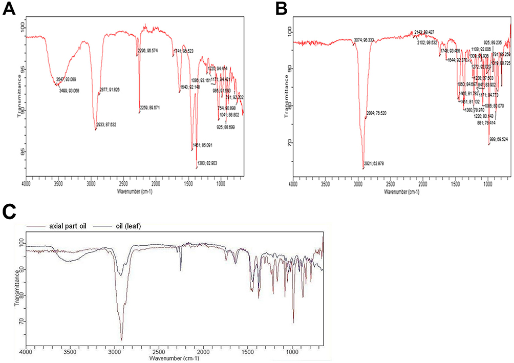 |
Figure 2 FTIR Spectra Analysis of Ethyl Acetate Fractions of Methanolic Leaf Extract of C. oppositifolia. (A) Leaf Extract Spectrum. (B) Inflorescence Extract Spectrum. (C) Comparison of Functional Groups Present in Leaf and Inflorescence Extracts. Note: Detection of functional groups including OH at 3342 cm⁻¹, aromatic groups at 1607 cm⁻¹, sulfones, sulfonyl chloride, sulfates, and sulphonamide at 1272 cm⁻¹, and sulfoxide at 1074 cm⁻¹. |
Thin Layer Chromatography
TLC of the volatile oil of C. oppositifolia (leaf and inflorescence) revealed significant differences in the concentration of various compounds present. As shown in Figure 3A and B, the compounds in lanes 1, 3, 5, 8, and 10 exhibited lower concentrations in the leaf extract compared to the inflorescence oil, with the inflorescence displaying more intense bands indicative of higher concentrations. TLC effectively separates compounds based on their polarity and molecular weight; thus, more polar compounds migrate shorter distances and appear as distinct bands on the chromatogram. For instance, the presence of terpenoids in the inflorescence resulted in stronger bands compared to phenolic compounds found in the leaf, underscoring the chemical richness of the inflorescence oil. This method not only highlights the diversity of phytochemicals but also suggests potential therapeutic applications based on the identified compounds’ concentrations.
 |
Figure 3 Thin Layer Chromatography (TLC) of Volatile Oil of C. oppositifolia (Leaf and Inflorescence). (A): TLC profile of leaf volatile oil showing differences in compound concentration; (B): TLC profile of inflorescence volatile oil showing differences in compound concentration. |
GC/MS Analysis
The GC/MS analysis of the volatile oil from the leaves of C. oppositifolia (inflorescence not performed) revealed the presence of 24 compounds in various concentrations (Table 3). The compounds present in the highest amounts in the whole oil were α-pinene (23.85), caryophyllene (11.312), and carene (13.642).
 |
Table 3 Compounds Present in the Volatile Oil of Leaves in C. Oppositifolia Through GC/MS Spectra |
FTIR Spectra of Volatile Oil (Leaf and Inflorescence) and Combinations with Antibiotics
The overlay of FTIR spectra of volatile oils (leaves and inflorescence) (Figure 4A and B) with antibiotics-amoxicillin and erythromycin respectively (Figure 4C and D) showed significant changes, including the disappearance and shift of peaks. Notably, in the combination of leaf oil and amoxicillin, a peak shift was observed at 1599 cm⁻¹ and 1406 cm⁻¹, as shown by the star in Figure 4E. For the combination of leaf oil and erythromycin, the peak at 2259 cm⁻¹ disappeared, and a shift was observed at 1715 cm⁻¹ in the erythromycin spectrum, as illustrated in Figure 4F. These observations suggest that the combination of volatile oils and antibiotics leads to functional group changes, which could potentially enhance the potency of these traditional antibiotics.
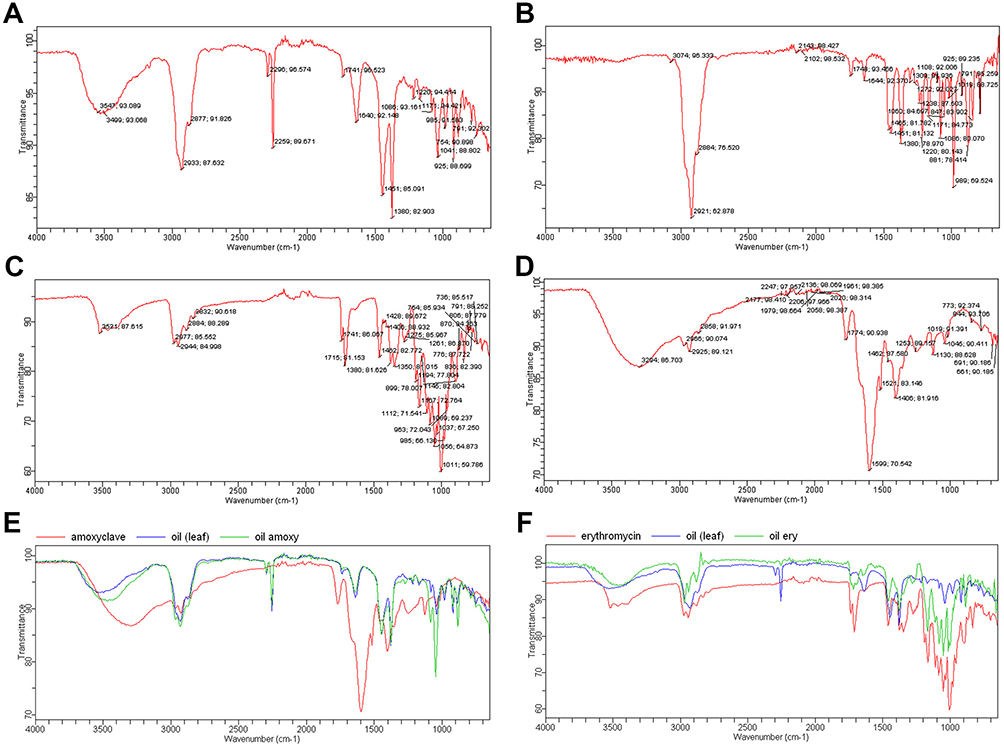 |
Figure 4 FTIR Spectra of Volatile Oils (Leaves and Inflorescence) and Their Combinations with Antibiotics. (A): FTIR spectrum of leaf volatile oil with key peaks including N-H stretches at 3547 cm⁻¹ and 3499 cm⁻¹, CH stretch at 2933 cm⁻¹, nitrile group at 2259 cm⁻¹, C=C stretch at 1640 cm⁻¹, aromatic peak at 1451 cm⁻¹, and S=O stretches at 1380 cm⁻¹; (B): FTIR spectrum of inflorescence volatile oil with key peaks including C-H stretches at 2921 cm⁻¹ and 2884 cm⁻¹, C-H Bend at 1451 cm⁻¹, S=O stretches at 1380 cm⁻¹ and 1220 cm⁻¹, and amines or C-O stretches at 1086 cm⁻¹; (C): FTIR spectrum of leaf volatile oil combined with amoxicillin showing significant changes including peak shifts; (D): FTIR spectrum of leaf volatile oil combined with erythromycin showing significant changes including disappearance of peaks; (E): Combination of leaf oil and amoxicillin showing peak shift at 1599 cm⁻¹ and 1406 cm⁻¹; (F): Combination of leaf oil and erythromycin showing disappearance of the peak at 2259 cm⁻¹ and a shift at 1715 cm⁻¹. |
UV Spectrophotometer Spectra of Volatile Oil (Leaf and Inflorescence) Combinations with Antibiotics
The overlays of UV-visible spectra for the combinations of oil (inflorescence and leaf) with erythromycin and amoxicillin did not show any changes in the peaks. When comparing the results with those of the oil and drug alone, as shown in Figure 5A–D, no significant differences were observed. This indicates that no complex formation occurred in these combinations.
 |
Figure 5 UV-visible spectra of Volatile Oil (Leaf and Inflorescence) Combinations with Erythromycin and Amoxicillin. (A): UV-visible spectrum of leaf volatile oil combined with erythromycin showing no changes in peaks; (B): UV-visible spectrum of inflorescence volatile oil combined with erythromycin showing no changes in peaks; (C): UV-visible spectrum of leaf volatile oil combined with amoxicillin showing no changes in peaks; (D): UV-visible spectrum of inflorescence volatile oil combined with amoxicillin showing no changes in peaks. |
NMR Spectra of Combinations of Oil (Leaf and Inflorescence) and Antibiotics
The NMR spectra of pure compounds (erythromycin and amoxicillin) and their mixtures with inflorescence oil and leaf oil are shown as peaks. The 1H NMR of amoxicillin (400 MHz, DMSO, δ with TMS=0) showed peaks at 1.08 (s, 6H), 2.11 (s, 6H), 2.23 (s, 3H), 1.23 (s, 2H), 1.24 (s, 1H), 1.56 (s, 1H), 3.03 (s, 1H), and 3.00 (s, 1H). The 1H NMR of erythromycin (400 MHz, CDCl3, δ with TMS=0) displayed peaks at 1.05 (s, 6H), 1.00 (s, 6H), 1.17 (s, 3H), 1.99 (s, 2H), 2.29 (s, 1H), 3.39 (s, 1H), 4.35 (s, 1H), 3.24 (s, 1H), 1.21 (s, 6H), 2.60 (s, 6H), 7.16 (s, 3H), 2.07 (s, 2H), 4.26 (s, 1H), 4.17 (s, 1H), 2.29 (s, 1H), 5.48 (s, 1H), 11.96 (s, 1H), 17.16 (s, 1H), and 4.97 (s, 1H). The 1H NMR of volatile oil (leaves) (400 MHz, CDCl3, δ with TMS=0) revealed peaks at 0.09 (s, 6H), 0.39 (s, 6H), 0.10 (s, 3H), 0.17 (s, 2H), 0.19 (s, 1H), 0.07 (s, 1H), 4.35 (s, 1H), 1.00 (s, 1H), 0.82 (s, 6H), 3.03 (s, 6H), 1.16 (s, 3H), 2.57 (s, 2H), 2.29 (s, 1H), 2.22 (s, 1H), 2.13 (s, 1H), 1.06 (s, 1H), 1.30 (s, 1H), 2.47 (s, 1H), 1.11 (s, 1H), 1.22 (s, 1H), and 0.18 (s, 1H). The 1H NMR of volatile oil (inflorescence) (400 MHz, CDCl3, δ with TMS=0) showed peaks at 0.66 (s, 6H), 0.24 (s, 6H), 1.10 (s, 3H), 0.67 (s, 2H), 1.00 (s, 1H), 0.95 (s, 1H), 1.15 (s, 1H), 1.07 (s, 1H), 6.97 (s, 6H), 9.54 (s, 6H), 3.20 (s, 3H), 10.59 (s, 2H), 4.45 (s, 1H), 6.24 (s, 1H), 3.40 (s, 1H), 15.51 (s, 1H), 2.48 (s, 1H), 4.75 (s, 1H), 1.45 (s, 1H), 4.19 (s, 1H), 4.33 (s, 1H), 0.79 (s, 1H), and 3.11 (s, 1H). The results suggested no complex formation was observed; all the independent peaks (Supplementary Figure 2) of drugs and oils remained at the same positions as shown by their independent spectra. From the spectral analysis, it could be concluded that no complex formation took place, and the compounds behaved only as synergists.
Discussion
C. oppositifolia, a plant with significant historical and medicinal value, has been utilized for its therapeutic properties in various traditional medicinal systems.20 The plant is known for its rich phytochemical composition, which includes compounds such as phenols, flavonoids, tannins, coumarins, alkaloids, terpenoids, saponins, and glycosides.21 These bioactive compounds contribute to the plant’s antioxidant, anti-inflammatory, antimicrobial, and pharmacological properties, making it a potent candidate for developing new therapeutic agents.22,23 The rise of multidrug-resistant (MDR) bacteria has become a major global health threat. MDR pathogens are responsible for increasing mortality rates and complicating treatment protocols, leading to longer hospital stays and higher medical costs.24 According to the World Health Organization (WHO), antibiotic resistance is one of the biggest threats to global health, food security, and development today. This underscores the urgent need to discover new antibiotics and alternative treatments to combat resistant strains.25 Plants like C. oppositifolia, with their diverse and potent bioactive compounds, offer a promising avenue for developing new antimicrobial agents. In our study, the extraction of C. oppositifolia yielded 12% ethanolic extract and 0.8% essential oil. The relatively low yield, especially for the volatile oil, could be attributed to several factors including the extraction method used, the specific part of the plant processed, and the environmental conditions during plant growth. These yields are somewhat lower compared to other studies on similar plants. For instance, Dhanani et al reported a 13.2% yield from ethanolic extraction of a related species, and Pandey et al obtained a 10% yield from the volatile oil of another medicinal plant.26,27 Optimizing extraction parameters, such as solvent polarity, extraction time, and temperature, could enhance the efficiency and yield of bioactive compounds from C. oppositifolia. The lower yields in our study suggest that optimizing extraction parameters, such as solvent polarity, extraction time, and temperature, could potentially enhance the efficiency and yield of bioactive compounds from C. oppositifolia. Despite the lower yields, the potent antimicrobial activity observed in our extracts highlights the plant’s therapeutic potential. The antimicrobial activity of C. oppositifolia extracts against S. aureus ATCC25923 and E. coli ATCC25922 was evaluated using both the agar well diffusion and disc diffusion methods. The methanol extract demonstrated consistent bacteriostatic activity with inhibition zones of 13.0 ± 0.2 mm for both bacteria. This is comparable to the findings of Nigussie et al and his coworkers, who reported inhibition zones of 14 mm using methanol extracts from a similar plant.28 Additionally, petroleum ether, chloroform, n-butanol, and ethyl acetate fractions exhibited varying bacteriostatic effects, with inhibition zones ranging from 9.0 to 12.0 mm. These results align with studies by Besieged and Baek, who observed similar antimicrobial activities in various plant fractions.29 The synergistic effect of combining erythromycin with the methanol extract and essential oil of C. oppositifolia significantly enhanced the bactericidal effect against S. aureus, increasing the inhibition zone to 18 ± 0.1 mm. This suggests that the extract may enhance the efficacy of traditional antibiotics, potentially through a mechanism that alters bacterial cell membrane permeability or inhibits efflux pumps. Literature supports similar synergistic interactions, highlighting combinations of plant extracts and antibiotics against MDR bacteria. Such a synergistic effect is novel and has not been widely reported in previous studies. The selection of erythromycin and amoxicillin was based on their well-documented efficacy against S. aureus ATCC25923 and E. coli ATCC25922, respectively. This targeted approach is innovative and highlights the potential of combining plant extracts with traditional antibiotics to combat MDR bacteria effectively. Our phytochemical analysis revealed the presence of various bioactive compounds including phenols, flavonoids, tannins, coumarins, alkaloids, terpenoids, saponins, and glycosides. Each of these compounds is known for its unique therapeutic properties. For example, phenols and flavonoids are recognized for their antioxidant and anti-inflammatory activities, while alkaloids and terpenoids exhibit significant antimicrobial and pharmacological effects.30 The presence of these compounds in C. oppositifolia corroborates its traditional use in medicine and suggests a broad spectrum of therapeutic applications. The FTIR spectra analysis of the ethyl acetate fractions of the methanolic leaf extract revealed the presence of several functional groups, including OH, aromatic, sulfonyl, and sulfoxide groups. Notably, the FTIR spectra of the volatile oils from the leaves and inflorescence displayed key peaks indicative of various functional groups. The shifting peaks in FTIR spectra when combined with antibiotics suggest interactions that may enhance antimicrobial potency without forming new chemical entities.For instance, N-H stretches at 3547 cm⁻¹ and 3499 cm⁻¹ indicated the presence of amines and amides, while a peak at 2933 cm⁻¹ suggested the presence of a CH stretch associated with aldehydes. The presence of nitrile (C≡N) groups was confirmed by a peak at 2259 cm⁻¹. The shifting of peaks in the FTIR spectra, such as the shift observed at 1599 cm⁻¹ and 1406 cm⁻¹ in the combination of leaf oil and amoxicillin, indicates potential interactions between the volatile oils and antibiotics, which could enhance the potency of these traditional antibiotics. The UV-visible spectra overlay for the combinations of volatile oil (inflorescence and leaf) with erythromycin and amoxicillin did not show any significant changes in the peaks. This indicates that no complex formation occurred in these combinations, suggesting that the oils and antibiotics act synergistically without forming new chemical entities. The NMR spectra of the pure compounds (erythromycin and amoxicillin) and their mixtures with inflorescence and leaf oils showed no significant changes, with all independent peaks remaining at the same positions. This further supports the conclusion that no complex formation took place, and the compounds functioned as synergists. The consistent NMR peaks suggest that the bioactive compounds in C. oppositifolia enhance the efficacy of the antibiotics without altering their chemical structure. To fully realize the therapeutic potential of C. oppositifolia, further research should focus on purifying the identified bioactive compounds. This will allow for a more detailed investigation into their individual and synergistic effects. Additionally, understanding the molecular mechanisms underlying the observed antimicrobial synergy with antibiotics is crucial for developing new therapeutic formulations. Expanding the scope of antimicrobial testing to include a wider range of resistant bacterial strains will provide a more comprehensive understanding of the plant’s antimicrobial potential. Furthermore, evaluating the cytotoxicity of these compounds will be essential to ensure their safety for therapeutic use. The development of new formulations combining purified bioactive compounds from C. oppositifolia with traditional antibiotics could lead to novel treatments for combating MDR bacteria. Such formulations could be particularly valuable in clinical settings where traditional antibiotics are becoming less effective.
Conclusion
Our study demonstrated the significant therapeutic potential of C. oppositifolia, highlighting its antimicrobial activity against S. aureus and E. coli with inhibition zones of 13.0 ± 0.2 mm. Despite the relatively low extraction yields of 12% for the ethanolic extract and 0.8% for the essential oil, the extracts exhibited potent effects when combined with traditional antibiotics. Notably, the synergistic interaction with erythromycin resulted in an enhanced inhibition zone of 18 ± 0.1 mm against S. aureus, underscoring the effectiveness of this combination. The rich phytochemical profile, characterized by FTIR, UV, and NMR analyses, provides a strong foundation for future research. By focusing on purifying the identified bioactive compounds and exploring their synergistic mechanisms, we can develop new and effective treatments to combat the growing threat of multidrug-resistant bacteria.
Data Sharing Statement
The original data presented in the study are included in the article and its Supplementary Materials. Further inquiries can be directed to the corresponding authors.
Author Contributions
All authors made a significant contribution to the work reported, whether that is in the conception, study design, execution, acquisition of data, analysis and interpretation, or in all these areas; took part in drafting, revising or critically reviewing the article; gave final approval of the version to be published; have agreed on the journal to which the article has been submitted; and agree to be accountable for all aspects of the work.
Funding
This work was supported by the Scientific Research Cultivation Project of Meizhou People’s Hospital (no. PY-C2023042), the High-level Talent Scientific Research Startup Fund Project of Meizhou People’s Hospital (no. KYQD202303).
Disclosure
The authors declare that the research was conducted in the absence of any commercial or financial relationships that could be construed as a potential conflict of interest.
References
1. Muteeb G, Rehman MT, Shahwan M, Aatif M. Origin of antibiotics and antibiotic resistance, and their impacts on drug development: a narrative review. Pharmaceuticals. 2023;16(11):1615. doi:10.3390/ph16111615
2. Abdallah EM, Alhatlani BY, de Paula Menezes R, Martins CHG. Back to nature: medicinal plants as promising sources for antibacterial drugs in the post-antibiotic era. Plants. 2023;12(17):3077. doi:10.3390/plants12173077
3. Uritu CM, Mihai CT, Stanciu GD, et al. Medicinal plants of the family Lamiaceae in pain therapy: a review. Pain Res Manag. 2018;2018:7801543. doi:10.1155/2018/7801543
4. Peron G, Hošek J, Prasad Phuyal G, Raj Kandel D, Adhikari R, Dall’Acqua S. Comprehensive characterization of secondary metabolites from colebrookea oppositifolia (Smith) leaves from Nepal and assessment of cytotoxic effect and anti-Nf-κb and ap-1 activities in vitro. Int J Mol Sci. 2020;21(14):4897. doi:10.3390/ijms21144897
5. Yap PSX, Yiap BC, Ping HC, Lim SHE. Essential oils, a new horizon in combating bacterial antibiotic resistance. Open Microbiol J. 2014;8(1):6–14. doi:10.2174/1874285801408010006
6. Manzoor A, Yousuf B, Pandith JA, Ahmad S. Plant-derived active substances incorporated as antioxidant, antibacterial or antifungal components in coatings/films for food packaging applications. Food Bioscience. 2023;53:102717. doi:10.1016/j.fbio.2023.102717
7. Drioiche A, Baammi S, Zibouh K, et al. A study of the synergistic effects of essential oils from Origanum compactum and Origanum elongatum with commercial antibiotics against highly prioritized multidrug-resistant bacteria for the World Health Organization. Metabolites. 2024;14(4):210. doi:10.3390/metabo14040210
8. Aljaafari MN, AlAli AO, Baqais L, et al. An overview of the potential therapeutic applications of essential oils. Molecules. 2021;26(3):628. doi:10.3390/molecules26030628
9. Yammine J. Essential oils and their active components applied as: free, encapsulated and in hurdle technology to fight microbial contaminations. A review. Heliyon. 2022;8. doi:10.1016/j.heliyon.2022.e12472
10. Dhanda G, Acharya Y, Haldar J. Antibiotic adjuvants: a versatile approach to combat antibiotic resistance. ACS Omega. 2023;8(12):10757–10783. doi:10.1021/acsomega.3c00312
11. Lupia C, Castagna F, Bava R, et al. Use of essential oils to counteract the phenomena of antimicrobial resistance in livestock species. Antibiotics. 2024;13(2):163. doi:10.3390/antibiotics13020163
12. Zhang QW, Lin LG, Ye WC. Techniques for extraction and isolation of natural products: a comprehensive review. Chin Med. 2018;13(1):20. doi:10.1186/s13020-018-0177-x
13. Fagbemi KO, Aina DA, Olajuyigbe OO. Soxhlet Extraction versus Hydrodistillation Using the Clevenger Apparatus: a Comparative Study on the Extraction of a Volatile Compound from Tamarindus indica Seeds. ScientificWorldJournal. 2021;2021:5961586. doi:10.1155/2021/5961586
14. Usman H, Abdulrahman F, Usman A. Qualitative phytochemical screening and in vitro antimicrobial effects of methanol stem bark extract of ficus thonningii (Moraceae). Afr J Tradit Complement Altern Med. 2009;6(3):289–295. doi:10.4314/ajtcam.v6i3.57178
15. Phytochemical Methods A Guide to Modern Techniques of Plant Analysis. Available from: https://link.springer.com/book/9780412572609.
16. Pyka A. Detection Progress of Selected Drugs in TLC. Biomed Res Int. 2014;2014:732078. doi:10.1155/2014/732078
17. Hossain MA, Al-Hdhrami SS, Weli AM, Al-Riyami Q, Al-Sabahi JN. Isolation, fractionation and identification of chemical constituents from the leaves crude extracts of Mentha piperita l grown in sultanate of Oman. Asian Pac J Trop Biomed. 2014;4(Suppl 1):S368–S372. doi:10.12980/APJTB.4.2014C1051
18. Mateus T, Almeida I, Costa A, et al. Fourier-transform infrared spectroscopy as a discriminatory tool for myotonic dystrophy type 1 metabolism: a pilot study. Int J Environ Res Public Health. 2021;18(7):3800. doi:10.3390/ijerph18073800
19. Manandhar S, Luitel S, Dahal RK. In vitro antimicrobial activity of some medicinal plants against human pathogenic bacteria. J Trop Med. 2019;2019:1895340. doi:10.1155/2019/1895340
20. Ambu G, Chaudhary RP, Mariotti M, Cornara L. Traditional uses of medicinal plants by ethnic people in the Kavrepalanchok District, Central Nepal. Plants. 2020;9(6):759. doi:10.3390/plants9060759
21. Riaz M, Khalid R, Afzal M, et al. Phytobioactive compounds as therapeutic agents for human diseases: a review. Food Sci Nutr. 2023;11(6):2500–2529. doi:10.1002/fsn3.3308
22. Atanasov AG, Zotchev SB, Dirsch VM, Supuran CT, Supuran CT. Natural products in drug discovery: advances and opportunities. Nat Rev Drug Discov. 2021;20(3):200–216. doi:10.1038/s41573-020-00114-z
23. Chiș A, Noubissi PA, Pop OL, et al. Bioactive compounds in moringa oleifera: mechanisms of action, focus on their anti-inflammatory properties. Plants. 2024;13(1):20. doi:10.3390/plants13010020
24. Patil S, Pai L, Chen X, et al. Genomic characterisation of multi-drug resistant Escherichia coli and Klebsiella pneumoniae co-harbouring mcr-1 and mcr-3 genes on a single plasmid from paediatric clinical cases. J Global Antimicrob Resist. 2023;34:134–140. doi:10.1016/j.jgar.2023.07.012
25. Global antimicrobial resistance and use surveillance system (GLASS) report: 2022. Available from: https://www.who.int/publications/i/item/9789240062702.
26. Dhanani T, Shah S, Gajbhiye NA, Kumar S. Effect of extraction methods on yield, phytochemical constituents and antioxidant activity of Withania somnifera. Arabian J Chem 2017;10:S1193–S1199. doi:10.1016/j.arabjc.2013.02.015
27. Pandey VK, Tripathi A, Srivastava S, et al. Exploiting the bioactive properties of essential oils and their potential applications in food industry. Food Sci Biotechnol. 2023;32(7):885–902. doi:10.1007/s10068-023-01287-0
28. Nigussie D, Davey G, Legesse BA, Fekadu A, Makonnen E. Antibacterial activity of methanol extracts of the leaves of three medicinal plants against selected bacteria isolated from wounds of lymphoedema patients. BMC Complement Med Ther. 2021;21(1):2. doi:10.1186/s12906-020-03183-0
29. Basavegowda N, Baek KH. Combination strategies of different antimicrobials: an efficient and alternative tool for pathogen inactivation. Biomedicines. 2022;10(9):2219. doi:10.3390/biomedicines10092219
30. Mousavi L, Salleh RM, Murugaiyah V. Phytochemical and bioactive compounds identification of Ocimum tenuiflorum leaves of methanol extract and its fraction with an anti-diabetic potential. Int J Food Prop. 2018;21(1):2390–2399. doi:10.1080/10942912.2018.1508161
 © 2024 The Author(s). This work is published and licensed by Dove Medical Press Limited. The
full terms of this license are available at https://www.dovepress.com/terms
and incorporate the Creative Commons Attribution
- Non Commercial (unported, 3.0) License.
By accessing the work you hereby accept the Terms. Non-commercial uses of the work are permitted
without any further permission from Dove Medical Press Limited, provided the work is properly
attributed. For permission for commercial use of this work, please see paragraphs 4.2 and 5 of our Terms.
© 2024 The Author(s). This work is published and licensed by Dove Medical Press Limited. The
full terms of this license are available at https://www.dovepress.com/terms
and incorporate the Creative Commons Attribution
- Non Commercial (unported, 3.0) License.
By accessing the work you hereby accept the Terms. Non-commercial uses of the work are permitted
without any further permission from Dove Medical Press Limited, provided the work is properly
attributed. For permission for commercial use of this work, please see paragraphs 4.2 and 5 of our Terms.