
Back to Journals » Drug Design, Development and Therapy » Volume 20
Phytochemical and Anti-Ischemic Stroke Properties from the Vitex L. Genus
Received 29 November 2025
Accepted for publication 31 January 2026
Published 11 February 2026 Volume 2026:20 585338
DOI https://doi.org/10.2147/DDDT.S585338
Checked for plagiarism Yes
Review by Single anonymous peer review
Peer reviewer comments 2
Editor who approved publication: Professor Anastasios Lymperopoulos
Chenqiong Xie,1 Jinjin Wu,1 Ping Huang2
1Department of Traditional Chinese Medicine Pharmacy, The Third Affiliated Hospital of Zhejiang Chinese Medical University, Hangzhou, People’s Republic of China; 2Department of Clinical Pharmacy, Hangzhou TCM Hospital Affiliated to Zhejiang Chinese Medical University, Hangzhou, People’s Republic of China
Correspondence: Ping Huang, Department of Clinical Pharmacy, Hangzhou TCM Hospital Affiliated to Zhejiang Chinese Medical University, No. 453, Tiyuchang Road, Hangzhou, 310007, People’s Republic of China, Tel +86 18268212670, Email [email protected]
Introduction: The genus Vitex L. (Verbenaceae) comprises ~250 species globally, with long-standing ethnopharmacological value. Notably, only Traditional Chinese Medicine (TCM) explicitly applies Vitex negundo for ischemic stroke (documented in classic Materia medica), distinguishing it from other regional uses (eg, menstrual disorders, malaria) and providing a unique basis for anti-stroke research.
Materials and Methods: Through systematic searches of English and Chinese databases such as Web of Science, Pubmed, CNKI, and Wanfang Data. This review systematically summarizes the natural constituents of Vitex L. their anti-ischemic stroke efficacy, and underlying mechanisms, emphasizing the uniqueness of Vitex-specific components and guiding preclinical optimization and clinical translation.
Results: Over 200 constituents were identified, with flavonoids (vitexin, isovitexin, casticin), terpenoids (vitexilactone, rotundifuran), and phenols as core active components. High-evidence compounds (validated by both in vitro and in vivo experiments) such as vitexin (10– 50 mg/kg) reduced rat MCAO infarct volume by 30– 40% via blocking NMDA receptor-mediated Ca2⁺ overload. Mechanistically, components target neurons, glia, and vascular endothelial cells, regulating both classic pathways (Nrf2, NF-κB, PI3K/Akt) and frontier mechanisms (ferroptosis, pyroptosis, epigenetic regulation). Synergistic effects of multi-component mixtures and optimized extraction/synthesis address low-content challenges.
Conclusion: Vitex L. exhibits significant anti-ischemic stroke potential, with unique components and multi-pathway regulation as core advantages. Future research should focus on multi-center validation, synergistic mechanism exploration, and clinical trials of high-evidence components to advance translation.
Keywords: Vitex L., ischemic stroke, natural chemical constituents, neuroprotection, blood-brain barrier, flavonoid
Introduction
Natural products, including traditional Chinese medicine (TCM), have a long history of clinical application. They are increasingly recognized for their curative effects on various physiological conditions and diseases, such as cancer, cardiovascular disease, diabetes, lung damage, kidney disease, and neurodegenerative disease, as well as obesity and aging. Vitex L. genus contains approximately 250 species that distributed from tropical to temperate regions worldwide.1 The primary distribution regions in Asia encompass the Indian subcontinent, Southeast Asia (including Vietnam, Laos, and Cambodia), and southern China (provinces south of the Yangtze River, extending north to the Qinling Mountains–Huaihe River line).2 Additionally, the species is found in tropical Africa, ranging from West Africa to East Africa, as well as in Madagascar, where 42 species have been identified, 41 of which are endemic, such as Vitex lowryi3 and Vitex betsiliensis. In Australia, along the eastern coast and in the northern regions, seven species are present, including Vitex glabrata and Vitex lignum-vitae.4 In the Americas, Vitex gaumeri is distributed from southern Mexico to Nicaragua, thriving in humid tropical forests.5 A limited presence is noted in Bolivia and Brazil in South America, with species such as Vitex trifolia var. subtrisecta.6 In North America, the Mediterranean species Vitex agnus-castus (commonly known as the chaste tree), has become naturalized in Florida and Texas, where it adapts to arid limestone soils. Along the Mediterranean coast, Vitex agnus-castus is indigenous, with a distribution spanning from Greece and Italy to Turkey.7 This species exhibits remarkable drought tolerance and typically grows on rocky slopes. In China, the genus Vitex comprises 14 species, including 7 varieties and 3 forms. Most of species are distributed south of the Yangtze River, with a small number occurring in the northwest, north, and northeast of China (Figure 1).
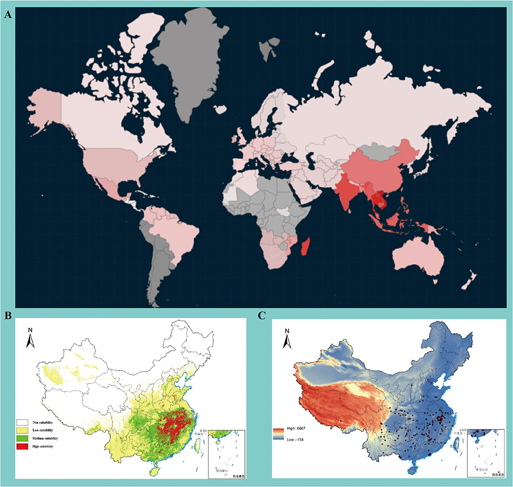 |
Figure 1 Distribution Characteristics of Vitex L. Plants. (A) Distribution quantity of Vitex L. plants in various regions of the world (the redder the color, the greater the quantity; gray represents areas with no data); (B) Suitable growth areas of Vitex L. plants in China (white represents unsuitable growth areas, yellow represents low-suitable growth areas, green represents moderately suitable growth areas, and red represents highly suitable growth areas); (C) Distribution of main harvesting and medicinal areas of Vitex L. plants in China (indicated by red dots in the figure). |
To date, extensive research has been carried out on Vitex L. species globally, with in-depth investigations particularly focusing on the phytochemical profiles and pharmacological activities of representative species, including Vitex trifolia L., Vitex negundo L., and Vitex agnus-castus L. These plants are known to be rich in diverse bioactive compounds, predominantly flavonoids, lignans, and terpenoids. Pharmacological investigations have demonstrated that several of these compounds possess multiple biological activities, including anti-ischemic stroke, anti-inflammatory, anti-tumor, and antioxidant properties, rendering them a prominent research focus.8 Notably, the medicinal potential of Vitex negundo L. var. cannabifolia (Sieb. et Zucc). Hand.-Mazz. (a Verbenaceae plant) has been the most thoroughly investigated, with a focus on its roots and leaves. However, research on the pharmacology and phytochemistry of the type species, Vitex negundo L., remains relatively limited, with most studies concentrating on a small number of monomeric compounds with well-defined pharmacological effects, such as vitexin and casticin.9,10 These monomers have been validated to exert protective effects against ischemic-reperfusion injury and anti-inflammatory activities, with distinct dose-dependent effects observed in preclinical models. The present review aims to systematically summarize the natural bioactive constituents of the genus Vitex L. and their potential therapeutic efficacy, in the treatment of ischemic stroke.
Materials and Methods
Through systematic searches of English and Chinese databases such as Web of Science, Pubmed, CNKI, and Wanfang Data. This review systematically summarizes the natural constituents of Vitex L., their anti-ischemic stroke efficacy, and underlying mechanisms, emphasizing the uniqueness of Vitex-specific components and guiding preclinical optimization and clinical translation.
Results
Traditional Medicinal Values of the Genus Vitex L. Plants
Members of the genus Vitex L. exhibit widespread medicinal use worldwide (Table 1). Notably, only TCM explicitly associates Vitex species with the treatment of cerebrovascular diseases, providing a unique ethnopharmacological foundation for investigating their anti-ischemic stroke potential. In Indian Ayurvedic medicine, fruit extracts of Nirgundi (Vitex negundo) are utilized for the management of menstrual irregularities and dysmenorrhea.11 Traditionally, its “hormone-regulating” effect is thought to be associated with the suppression of prolactin secretion. Additionally, topical application of leaf juice for the treatment of skin infections has been documented. In Indian folk medicine, root decoctions are also employed as anthelmintics, while seeds combined with dried ginger and milk are utilized as an aphrodisiac. In Japan and Vietnam, Vitex trifolia is commonly used for the treatment of wind-heat headaches, conjunctival hyperemia, and ocular pain; its leaf extracts are employed to alleviate coughs and colds.12
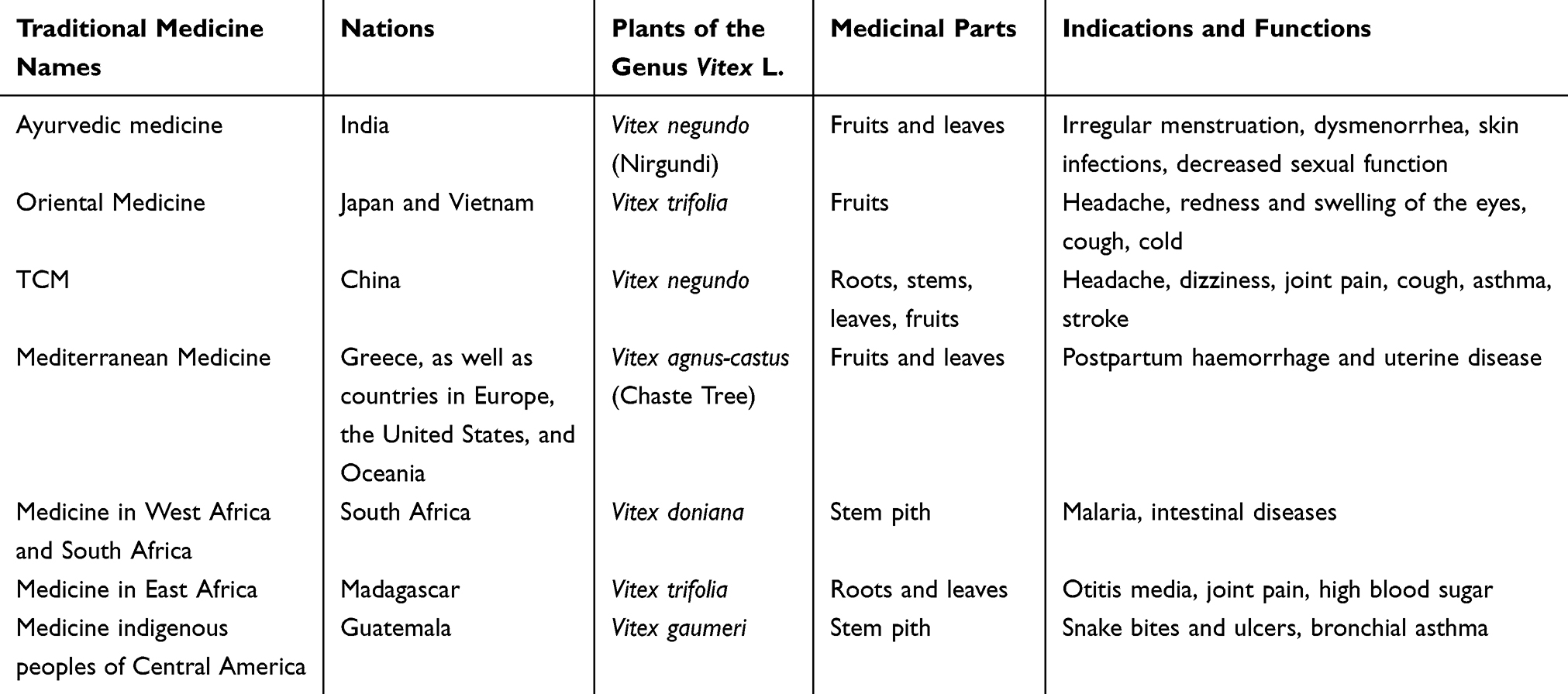 |
Table 1 Traditional Medicinal Use of Genus Vitex L. in the World |
In China, Vitex negundo has a long history of medicinal use, with its earliest documentation in Supplementary Records of Famous Physicians, where it was categorized as a “top-grade” medicinal herb.13 Following stir-frying, it serves as a medicinal component to dispel wind-heat and alleviate headaches and dizziness, as documented in Shennong’s Classic of Materia Medica. Collected Annotations on the Classic of Materia Medica notes: “Vitex grows in fields; their fruits are harvested in August and September and dried in the shade.” Supplements to Materia Medica describes its efficacy as “alleviating wind-damp arthralgia, and muscle-bone contracture.”
In Europe, the use of Vitex agnus-castus (Chaste Tree) is documented in ancient Greek texts, which were employed for postpartum hemostasis and uterine disorders. During the Middle Ages, it was known as “Monk’s Pepper” in monasteries, where it was believed to suppress sexual desire; additionally, fruit decoctions were used in sitz baths for the management of uterine inflammation.14 Furthermore, following the introduction of this species to Australia and North American nations, it has became a key raw material for natural medicinal products.15 In Australia, it is utilized as a dietary supplement to relieve symptoms of premenstrual syndrome (PMS); its standardized extract (containing 550 μg of agnuside per tablet) modulates the menstrual cycle by regulating the luteinizing hormone/follicle-stimulating hormone (LH/FSH) balance. Following its naturalization in Florida and Texas (USA), it is employed as an herbal remedy to regulate hormonal balance and alleviate symptoms related to polycystic ovary syndrome (PCOS).16
In West Africa and South Africa, stem bark extracts of Vitex doniana (Black Plum) are utilized as an antimalarial agent and for the treatment of intestinal disorders, whereas its leaf juice is employed to alleviate diarrhea.17 In East Africa, Vitex trifolia leaf juice is instilled into the ears canal for the treatment of otitis media, and root decoctions are employed to relieve pain associated with rheumatoid arthritis. Its primary medicinal indications are similar to those in East Asia; additionally, its fruit extracts have been shown to reduce blood glucose levels in diabetic mice.
In the Philippines, Vitex negundo (locally referred to as Lagundi) is utilized as a traditional topical remedy: leaf paste is applied for the treatment of snakebites and ulcers. Furthermore, the Philippine Department of Science and Technology has officially recognized it as an herbal medicine for the treatment of coughs and asthma; its tablets and syrups have undergone clinical validation, demonstrating the ability to reduce mucus viscosity and relieve bronchospasm.18 There are also reports indicating that indigenous populations in Guatemala (Central America) utilize decoctions of Vitex gaumeri bark to manage malarial fever.
The most compelling evidence has shown that the inhibition of inflammation and oxidative stress constitutes a crucial molecular mechanism through which natural products exert therapeutic effects on various diseases, including cancer, cardiovascular disease, non-alcoholic fatty liver disease, chronic kidney disease, diabetes mellitus, inflammatory bowel disease, autoimmune disease, degenerative disease, and benign prostatic hyperplasia.19–23 Members of the genus Vitex L. possess potent anti-inflammatory activity, and thus most of their traditional medicinal applications are centered on inflammation-related conditions. Notably, only in TCM, Vitex negundo is also employed for the treatment of cardiovascular and cerebrovascular diseases.24 For instance, Great Dictionary of Chinese Materia Medica records that Vitex negundo can be utilized as an herbal remedy for stroke, and numerous modern studies have been progressively conducted to explore this application.25
The Main Active Ingredients of Vitex L. in the Treatment of Ischemic Stroke
As a type of natural herbal medicine, plants of the genus Vitex contain a complex and diverse range of chemical components, and exert multiple functions such as anti-inflammation, anti-oxidation, resistance to ischemia-reperfusion injury, and cardiovascular protection. Systematic literature reviews have identified over 200 compounds in Vitex plants, covering simple phenols, organic acids, phenylpropanoids, lignans, flavonoids/flavonoid glycosides, terpenoids/saponins, steroids, and a small number of alkaloids. Based on the research results of compounds in the treatment of ischemic stroke, we define “high-level evidence” with reference to the Oxford Centre for Evidence-Based Medicine (OCEBM) evidence grading standards: compounds supported by at least 2 independent in vivo experiments (n≥6 per group, ≥3 repetitions) and 2 independent in vitro experiments (≥2 cell models, reasonable concentration gradients) are classified as high-level evidence; compounds validated only by either in vitro or in vivo experiments (≥1 independent study with rigorous design) are regarded as potential anti-stroke compounds (Figure 2).
 |
Figure 2 Compounds with Experimental Evidence and Potential for Treating Ischemic Stroke in the Genus Vitex. Compounds within the red boxes are those supported by both in vitro and in vivo experiments; compounds within the yellow boxes are those validated only by either in vitro or in vivo experiments, which have potential for treating ischemic stroke. |
Research on the Treatment of Ischemic Stroke with Simple Phenols and Organic Acids
A total of 23 simple phenols and organic acid components have been identified in medicinal plants of the genus Vitex (Table 2). Current evidence highlights protocatechuic acid, vanillic acid, and ferulic acid, which exhibit neuroprotective potential and capacity to improve neurological function in vitro cell models and in vivo animal models. In the rat middle cerebral artery occlusion (MCAO) model, intraperitoneal pretreatment with (25 mg/kg) for 7 days reduced cerebral infarct volume by 28.6% and significantly improved the modified Neurological Severity Score (mNSS).26 Meanwhile, protocatechuic acid (5–20 μM) protected PC12 cells against Oxygen-Glucose Deprivation (OGD)-induced injury, increasing cell survival rate by more than 30% and improving the maintenance rate of mitochondrial membrane potential.27 In the rat bilateral common carotid artery occlusion/reperfusion (BCCAO/R) model, oral pretreatment with vanillic acid (10–50 mg/kg) for 2 weeks reduced cerebral infarct volume by 25–30% and mitigated anxiety-like behaviors.28 Additionally, vanillic acid (10–50 μM) protected human umbilical vein endothelial cells (HUVECs) against H2O2-induced damage, enhancing the recovery of vascular endothelial barrier function by 40%.29 In the rat MCAO model, intraperitoneal injection of ferulic acid (100 mg/kg) reduced cerebral infarct volume by 40% and improved the National Institutes of Health Stroke Scale (NIHSS) score.30 Simultaneously, ferulic acid (1–10 μM) enhanced the resistance of SH-SY5Y cells to oxygen-glucose deprivation/reperfusion (OGD/R)-induced injury, increasing cell survival rate by more than 50%.31
 |
Table 2 Relevant Information on Molecules in the Genus Vitex |
Limited evidence indicates that syringaldehyde, citronellal, p-hydroxybenzyl alcohol, and 3,4-dihydroxybenzoic acid possess potential for treatment of ischemic stroke in vivo animal studies or in vitro cell experiments. Syringaldehyde administration significantly reduced the amyloid plaques in the hippocampus of APPswe/PS1dE9 (APP/PS1) transgenic mice, promoted neuronal repair, and enhanced cognitive function, yet its efficacy has not been validated in the MCAO model.42 Administration of citronellal and p-hydroxybenzyl alcohol reduced cerebral infarct volume and mitigated the severity of brain injury in the MCAO model;43,44 however, studies investigating their mechanism of action in neuronal cells are scarce. As an oxidation product of p-hydroxybenzoic acid, 3,4-dihydroxybenzoic acid exhibits potent free radical scavenging activity (IC50 = 12.5 μM). Studies have demonstrated that 5–20 μM of this compound can enhance the resistance of HUVECs to H2O2-induced damage and improve vascular endothelial barrier function.45 However, direct evidence from stroke models is currently lacking, and further research on blood-brain barrier (BBB) permeability is warranted.
Among the aforementioned compounds, protocatechuic acid holds promise as a candidate for clinical translation. Multicenter animal experiments are recommended to be performed, and its synergistic effects in combination with thrombolytic agents should be explored. For ferulic acid, validation of its long-term safety and elucidation of its regulatory mechanisms on lipid metabolism via metabolomics are needed. Development of a nanolipidic delivery system for citronellal derivatives represents a viable optimization strategy, with an emphasis on assessing their BBB penetration efficiency. However, given the relatively low content of these compounds in Vitex medicinal plants, future applications in stroke treatment should prioritize the chemical synthesis of these compounds or their derivatives over simple extraction.
Research on the Treatment of Ischemic Stroke with Phenylpropanoid
A total of 7 phenylpropanoid components have been identified in the medicinal plants belonging to the genus Vitex (Table 2). Current evidence highlights 3,5-di-O-caffeoylquinic acid, which exhibits potential for neuroprotection and function improvement in animal models and cell models. Studies have demonstrated that pretreatment of SH-SY5Y cells with 3,5-di-O-caffeoylquinic acid (10–40 μM) can counteract damage induced by Aβ1-42 or NMDA;46 meanwhile, oral administration of this compound (6.7 mg/kg/d) to Senescence Accelerated Mouse-Prone 8 (SAMP8) mice enhanced cognitive function and diminished oxidative stress markers in the brain.47 Additionally, the compounds 5,7-dihydroxychromone, 3,4-dihydroxyphenyllactic acid methyl ester, and 2-methoxy-4-(3-methoxy-1-propenyl)-phenol have all exhibited potential for stroke treatment in in vitro experiments. Among these, 5,7-dihydroxychromone and 3,4-dihydroxyphenyllactic acid methyl ester can mitigate H2O2-induced damage to vascular smooth muscle cells to some extent,48,49 while 2-methoxy-4-(3-methoxy-1-propenyl)-phenol has been shown to reduce dopamine depletion in the mouse striatum and improve motor function.50
In particular, 3,5-di-O-caffeoylquinic acid holds promise as a candidate for further mechanistic investigations, with an emphasis on exploring its BBB protective and anti-thrombotic effects in the MCAO model. 5,7-dihydroxy chromone necessitates additional long-term safety data and evaluation of its translational potential as an Nrf2 activator. However, given the relatively low content of this compound in medicinal Vitex plants, its extraction for practical application currently poses a significant challenge.
Research on the Treatment of Ischemic Stroke with Lignans
Lignans are major components and pharmacologically active ingredients in natural products, including TCM.51–54 A total of 44 lignan components have been identified in the medicinal plants of the genus Vitex (Table 2). Current evidence highlights apigenin, atractylenolide I, and (+)-sesamin, all of which have demonstrated efficacy in stroke treatment in both in vitro and in vivo models. Apigenin exhibits promising potential for the treatment of myocardial hypertrophy, insulin resistance and renal fibrosis.55–57 In the rat MCAO model, intraperitoneal injection of apigenin (0–50 mg/kg) reduced infarct volume by 30–40% and significantly improved the mNSS.58 Additionally, apigenin (10–20 μM) enhanced the resistance of SH-SY5Y cells to H2O2-induced or OGD-induced damage, increasing cell survival rate by more than 50%.59 Intraperitoneal injection of atractylenolide I (1–10 mg/kg) also reduced cerebral infarct volume by 25–30% and mitigated cerebral edema in the MCAO model of C57BL/6 mice.60 Meanwhile, atractylenolide I (0.01–1 μM) could counteract lipopolysaccharide (LPS)-induced inflammation in BV2 microglia, reducing the release of nitric oxide (NO) and Interleukin-6 (IL-6) by more than 60%.61 Oral administration of (+)-sesamin (30 mg/kg) reduced cerebral infarct volume by approximately 50% and improved neurological function in the mouse MCAO model.62 Moreover, 4-hydroxysesamin (10–20 μM) could protect HT22 neurons against OGD-induced damage, reducing the cellular apoptosis rate by 40%.63
In addition, three other compounds have exhibited potential for stroke treatment. Methyl rosmarinate (10–50 μM) improved vascular endothelial barrier function against H2O2-induced damage; however, direct evidence from stroke models is lacking.64 Nevertheless, rosmarinic acid has been demonstrated to be effective in the rat myocardial ischemia model (10 mg/kg, intravenous injection).65 3,4,5-tricaffeoylquinic acid (5–20 μM) protected U87MG cells against OGD-induced damage, restoring adenosine triphosphate (ATP) levels to 80% of the normal value.66 Although direct in vivo experimental evidence is lacking, chlorogenic acid enhanced cognitive function in the SAMP8 mouse model, suggesting that this derivative may also exert certain therapeutic effects.67 Furthermore, neoandrographolide has also been reported to exert efficacy in the rat MCAO model.
Apigenin holds promise as a candidate for clinical translation. Multicenter, large-sample animal studies are recommended to explore its synergistic effects in combination thrombolytic drugs. Atractylenolide I requires validation of its long-term safety and the elucidation of its regulatory mechanisms on lipid metabolism via metabolomics. The derivative 4-HS of (+)-sesamin exhibits enhanced activity and represents a viable optimization strategy, with an emphasis on assessing its BBB penetration efficiency. Moreover, when these monomers are utilized for stroke treatment, Vitex plants can be considered potential raw materials for extraction.
Research on the Treatment of Ischemic Stroke with Flavonoid and Flavonoid Glycoside
Flavonoids are among the major components with in the plant kingdom, characterized by the most diverse structures and high abundance.68–71 Flavonoids exert extensive pharmacological activities against various diseases.72–76 A total of 31 flavonoid and flavonoid glycoside components have been identified in medicinal plants of the genus Vitex (Table 2). Among these, luteolin, quercetin, isorhamnetin, kaempferol, and vitexin have demonstrated efficacy in stroke treatment in both in vitro and in vivo models. In the rat MCAO model, intraperitoneal injection of luteolin (10–50 mg/kg) reduced cerebral infarct volume by 30–40% and significantly improved the mNSS.77 Additionally, luteolin (10–20 μM) protected SH-SY5Y cells against damage induced by H2O2 or OGD, increasing cell survival rate by more than 50%.78 In the mouse MCAO model, oral administration of quercetin (30 mg/kg) reduced infarct volume by approximately 50% and enhanced neurological function.79 Meanwhile, quercetin (10–20 μM) protected HT22 neurons against OGD-induced damage, reducing the cellular apoptosis rate.80 Intraperitoneal injection of kaempferol (5–20 mg/kg) reduced cerebral infarct volume by 20–30% and improved neurological function scores in the rat MCAO model.81 Furthermore, kaempferol (5–10 μM) protected PC12 cells against 6-Hydroxydopamine (6-OHDA)-induced damage, increasing cell survival rate by more than 30%.82 In the MCAO model using C57BL/6 mice, intraperitoneal injection of isorhamnetin (1–10 mg/kg) reduced cerebral infarct volume by 20–30% and improved neurological function scores.83 Additionally, isorhamnetin (5–10 μM) mitigated LPS-induced inflammation in BV2 microglia, reducing the release of NO and IL-6 by more than 60%. Hesperidin is regarded as a potential candidate for stroke treatment. In cardiomyocytes, hesperidin (10–50 μM) can mitigate H2O2-induced damage.84 Direct evidence from stroke animal model is lacking; however, Hesperetin has been shown to be effective in a rat myocardial ischemia model (5 mg/kg, intravenous injection).
Research on luteolin and quercetin has already covered multiple signaling pathway mechanisms; however, the complexity of ischemic stroke pathology (such as BBB disruption and excitotoxicity) necessitates more comprehensive model validation. Meanwhile, given widespread distribution of these active components across various plant species, Vitex is not recommended as the extraction source. Future research could further focus on the genus-specific components for the ischemic stroke treatment. Vitexin, casticin, and isovitexin have garnered increasing attention in recent years due to their anti-stroke activity and are widely distributed in Vitex species. Vitexin (10–50 μM) protected HUVECs against H2O2-induced damage and enhanced vascular endothelial barrier function.85 Notably, apigenin has been demonstrated effective in a rat myocardial ischemia model (10 mg/kg via intravenous injection),86 and additional experiments on vitexin are warranted in the MCAO model. Casticin has been shown to potentially inhibit the JAK2/STAT3 pathway, reducing the release of TNF-α and IL-6 from microglia, thereby exerting efficacy in neuroinflammation animal models.79 Isovitexin is also deemed beneficial for microglia recovery.87 However, due to the limitations of current research, these Vitex-specific flavonoids still require extensive studies to validate their therapeutic efficacy in stroke, while they also holding considerable potential for clinical translation.
Research on the Treatment of Ischemic Stroke with Terpenoid and Saponin
Terpenoids and saponins are among the most abundant components in natural products and exhibit diverse pharmacological activities.88–91 A total of 73 terpenoid and saponin components have been identified in plants belonging to the genus Vitex (Table 2). Among these, aucubin, oleanolic acid, and betulinic acid have been demonstrated to possess potential for stroke treatment in both in vitro and in vivo models. In the gerbil global cerebral ischemia model, intraperitoneal injection of aucubin (10 mg/kg) increased the neuronal survival rate in the hippocampal CA1 region by 40% and significantly improved neurological function scores.92 Additionally, aucubin (1–10 μM) protected BV2 microglia against LPS-induced inflammation, reducing the release of NO and IL-6 by more than 60%.93 In the rat MCAO model, intraperitoneal injection of oleanolic acid (20–50 mg/kg) reduced cerebral infarct volume by 25–30% and mitigated cerebral edema.94 Meanwhile, oleanolic acid (5–20 μM) protected PC12 cells against 6-Hydroxydopamine (6-OHDA)-induced damage, increasing the cellular survival rate by more than 30%.95 Intraperitoneal injection of betulinic acid (10–30 mg/kg) reduced cerebral infarct volume by approximately 40% and significantly improved neurological function scores in the mouse MCAO model.96 Furthermore, betulinic acid (5–10 μM) protected HT22 neurons against OGD-induced damage, reducing the cellular apoptosis rate by 40%.97
In addition, rotundifuran (10–50 μM) protected human umbilical vein endothelial cells (HUVECs) against H2O2-induced damage and improved vascular endothelial barrier function.98 Although direct evidence from stroke models is lacking, extract of Vitex rotundifolia has been demonstrated to be effective in a rat myocardial ischemia model (10 mg/kg, intravenous injection). Moreover, maslinic acid (5–20 μM) protected U87MG cells against OGD-induced damage, restoring ATP levels to 80% of the normal value.99 These two compounds are regarded as potential candidates for stroke treatment, and further validation in animal models is warranted.
Aucubin holds promise as a candidate for clinical translation. Multicenter, large-sample animal studies are recommended to explore its synergistic effects in combination with thrombolytic drugs. The long-term safety of oleanolic acid requires validation, and its regulatory mechanisms underlying lipid metabolism should be elucidated via metabolomics. 3-O-acetylbetulinic acid, a derivative of betulinic acid, exhibits enhanced activity and represents a viable optimization strategy, with an emphasis on assessing its BBB penetration efficiency.100 Vitexilactone is another Vitex-specific component that possesses antioxidant properties. However, direct evidence supporting its efficacy in ischemic stroke is currently lacking.
Research on the Treatment of Ischemic Stroke with Steroid
A total of 12 steroid compounds have been identified in plants belonging to the genus Vitex (Table 2). Among these, progesterone, testosterone, and β-sitosterol have been demonstrated to exert broad therapeutic efficacy in stroke in both in vitro and in vivo models. In the rat MCAO model, intraperitoneal injection of Progesterone (10–50 mg/kg) reduced cerebral infarct volume by 30–40% and significantly improved the mNSS.101 Additionally, progesterone (1–10 μM) protected SH-SY5Y cells against OGD-induced damage, increasing cell survival rate by more than 50%.102 Preclinical data indicate that it exhibits a favorable safety profile without significant adverse effects. In the castrated rat MCAO model via subcutaneous implantation of testosterone (10 mg/kg), accelerated recovery of neurological function and reduced astrocyte activation were observed.103 Meanwhile, testosterone (0.1–1 μM) HT22 neurons against H2O2-induced damage, improving the maintenance of mitochondrial membrane potential by 30%.104 In the rat intracranial aneurysm model, oral administration of β-sitosterol (20–50 mg/kg) reduced aneurysm volume by 25–30%.105 Furthermore, β-sitosterol (5–20 μM) protected BV2 microglia against LPS-induced inflammation, reducing the release of NO by more than 60%.106
In addition, in the mouse MCAO model via intraperitoneal injection of stigmasterol (20–80 mg/kg), a 20–30% reduction in cerebral infarct volume was observed.107 Ergosterol peroxide (10–50 μM) protected HUVECs against H2O2-induced damage and enhanced vascular endothelial barrier function.108 Although direct evidence from stroke models is lacking, ganoderma lucidum spore oil extract has been demonstrated to exert efficacy in the rat myocardial ischemia model.109 These two compounds are considered potential candidates for stroke treatment. Currently, these compounds are primarily synthesized or extracted from other plant species. They play a crucial role in the application of Vitex plants for the ischemic stroke treatment, but their specificity is relatively low.
Research on the Treatment of Ischemic Stroke with Other Classes Compounds
Additionally, 10 compounds of other classes have been identified from plants belonging to the genus Vitex. Among these, cineole, caffeic acid, and panaxydol have been demonstrated to exhibit therapeutic efficacy in stroke both in vitro and in vivo models. In the rat MCAO model, intraperitoneal injection of cineole (10–50 mg/kg) reduced cerebral infarct volume and significantly improved the mNSS.110 Meanwhile, cineole (5–20 μM) helped HT22 neurons against OGD-induced damage, improving the maintenance of mitochondrial membrane.111 In the rat Permanent Middle Cerebral Artery Occlusion (PMCAO) model, oral administration of caffeic acid (2 mg/kg) reduced cerebral infarct volume and improved neurological function scores, with the therapeutic time window extendable to 2 hours post-ischemia.112 Caffeic acid (1–10 μM) protected SK-N-SH cells against OGD/R-induced damage, increasing cell survival rate by more than 50%. Panaxydol (5–20 μM) was found to protect PC12 cells against 6-OHDA-induced damage, increasing the cellular survival rate by more than 30%;113 and it also exerted therapeutic effects in cerebral ischemia animal models.
Furthermore, isofraxidin (10–50 μM) mitigated LPS-induced stimulation in BV2 microglia, reducing NO release by more than 60%.114 Oral administration of pyrogallol (1–10 mg/kg) for 7 days to mice resulted in no significant toxicity and enhanced the activity of antioxidant enzymes in brain tissue. However, given the relatively low content of these additional compounds in the Vitex genus plants, their application value as extracted drugs for ischemic stroke treatment is relatively limited.
Main Mechanisms of Medicinal Plants of the Genus Vitex in Treating Ischemic Stroke
Medicinal plants of the genus Vitex harbor diverse active components with therapeutic efficacy in ischemic stroke (Tables 3 and 4). Regarding composition and specificity, flavonoids, including vitexin, isovitexin, casticin, vitexilactone, and rotundifuran, are the most abundant and highly specific active components in medicinal Vitex plants, and thus are presumably the key contributors to the therapeutic effects of these plants. Furthermore, steroids and organic acids may also play crucial roles in stroke treatment. Accumulating evidence have demonstrated that these components can target three core cell populations (neurons, glial cells, and vascular endothelial cells) and regulate key processes during the pathological progression of ischemic stroke, including oxidative stress, inflammatory response, cell apoptosis, and BBB disruption (Figure 3).
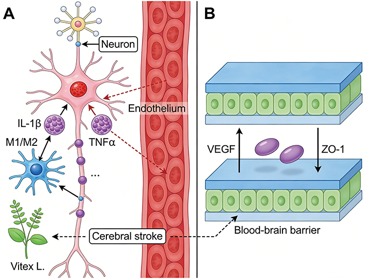 |
Figure 3 Schematic diagram of the mechanism of action of Vitex plants in the treatment of ischemic stroke. (A) Active components of Vitex plants act on neuronal cells and glial cells; (B) Active components of Vitex plants act on vascular endothelial cells and BBB. |
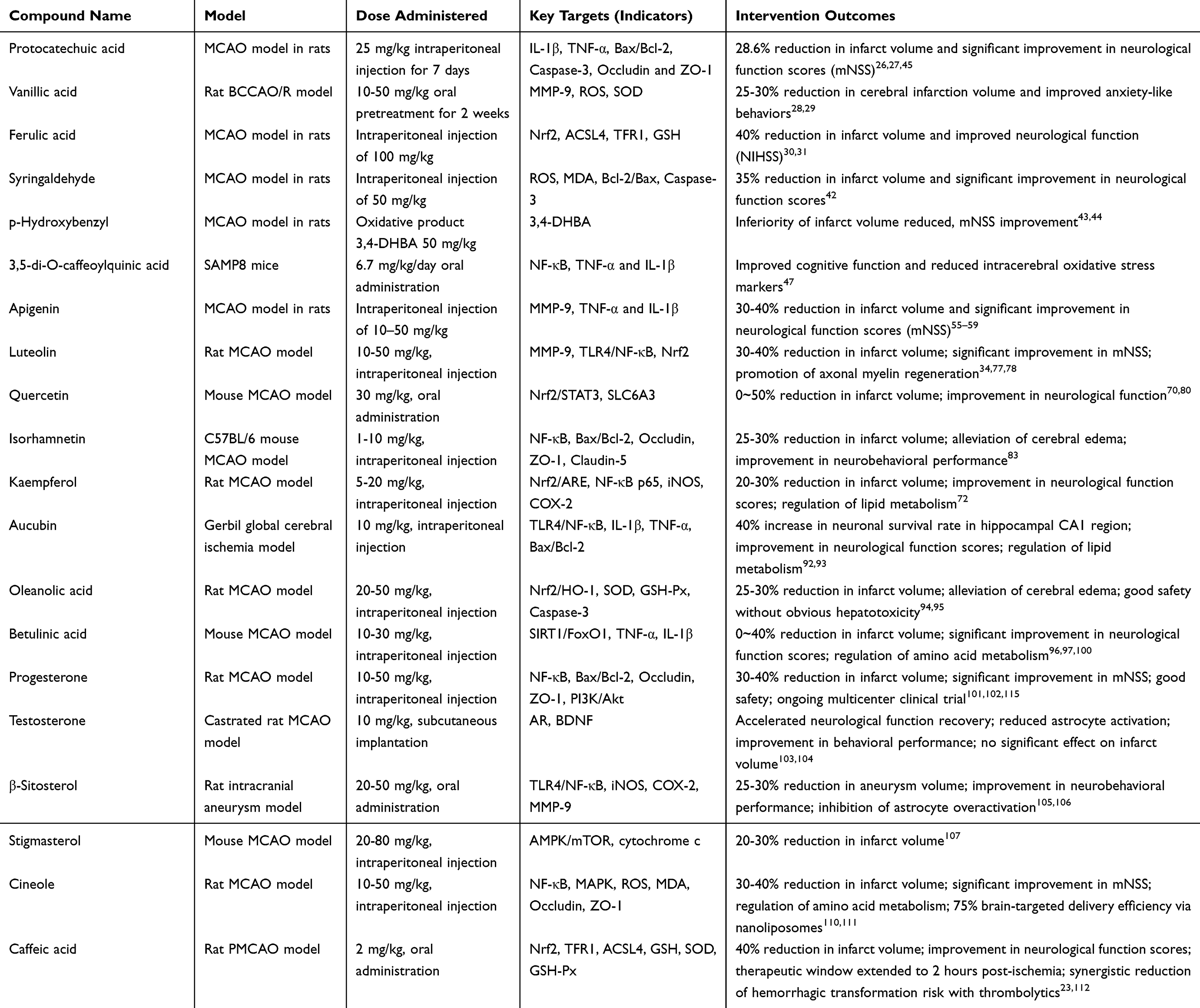 |
Table 3 Experimental Study in vivo on Active Components of Vitex L. Genus for Ischemic Stroke Treatment |
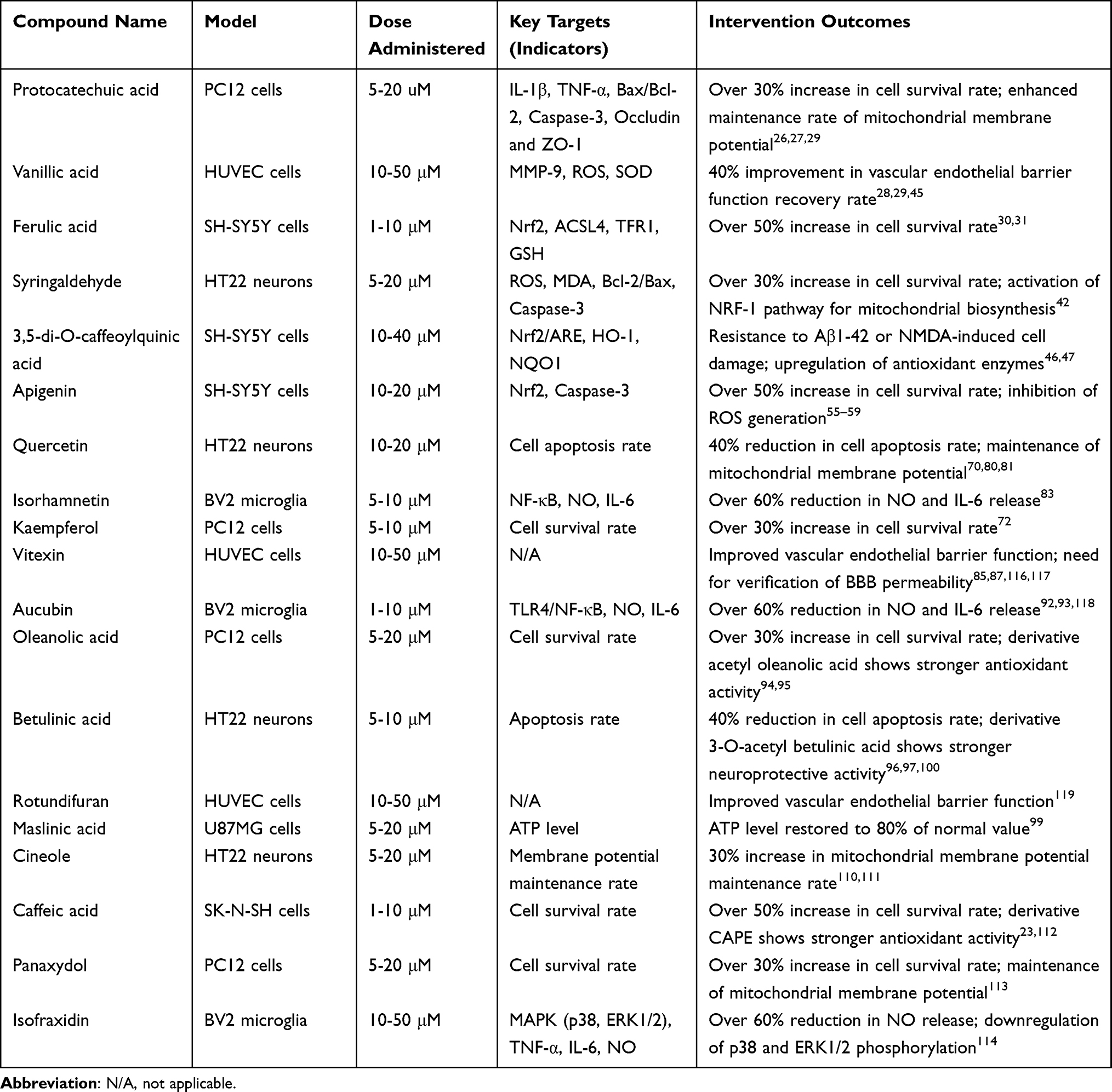 |
Table 4 Experimental Study in vitro on Active Components of Vitex L. Genus for Ischemic Stroke Treatment |
Neuronal Protection Mechanisms of Active Components from the Genus Vitex
Active components from the genus Vitex directly act on neurons to block the cerebral ischemia-induced “oxidative stress-excitotoxicity-apoptosis” cascade, while preserving neuronal metabolism and synaptic function (Figure 4). Accumulating evidence has demonstrated that vitexin and isovitexin activate the Nrf2/ARE pathway for scavenging reactive oxygen species (ROS).116,120 These components can bind to Kelch-like ECH-associated protein 1 (Keap1), thereby inhibiting the Keap1-Nrf2 interaction, and promoting the dissociation of Nrf2 from Keap1 as well as its nuclear translocation. Upon binding of Nrf2 to the antioxidant response element (ARE), it upregulates the transcriptional expression of antioxidant enzymes, including heme oxygenase-1 (HO-1), NAD(P)H: quinone oxidoreductase 1 (NQO1), superoxide dismutase (SOD), and glutathione peroxidase (GSH-Px). Additionally, studies have further confirmed that these flavonoids can directly scavenge excessive ·OH and ·O2− generated during ischemia-reperfusion (I/R), decrease the levels of malondialdehyde (MDA), and attenuate neuronal membranes lipid peroxidation.117
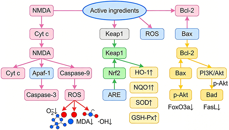 |
Figure 4 Vitex plants protective mechanism against neuronal damage in ischemic stroke: Molecular pathway illustration diagram.↑ Arrow indicates that the target gene or protein is upregulated under the regulation of the limited component; ↓ Arrow indicates that the target gene or protein is downregulated under the regulation of the limited component. |
Meanwhile, these active components also participate in suppressing the mitochondrial-mediated apoptotic pathway. Casticin preserves the stability of mitochondrial membrane potential (ΔΨm), inhibits the opening of mitochondrial permeability transition pores (mPTP), and reduces the release of cytochrome c (Cyt c) from the mitochondrial matrix to the cytoplasm.121 This further inhibits apoptosome formation via the binding of Cyt c to apoptotic protease activating factor-1 (Apaf-1), thereby blocking the cascading activation of Caspase-9 and Caspase-3. Aucubin upregulates the expression of the anti-apoptotic protein Bcl-2, downregulates that of the pro-apoptotic protein Bax, decreases the Bax/Bcl-2 ratio, and suppresses mitochondrial membrane rupture as well as the initiation of apoptotic signals.118 Progesterone activates phosphatidylinositol 3-kinase (PI3K), thereby promoting the phosphorylation of Akt. Phosphorylated Akt (p-Akt) further phosphorylates its downstream target Bad, thereby inactivating Bad and inducing its dissociation from mitochondria.115 Simultaneously, p-Akt inhibits the nuclear translocation of forkhead box transcription factor 3a (FoxO3a), thereby downregulating the expression of apoptosis-related genes.
Furthermore, emerging studies have indicated that these active components can inhibit excitotoxicity and attenuate NMDA receptor-mediated Ca2⁺ overload. Vitexin directly binds to N-methyl-D-aspartate (NMDA) receptors on the neuronal plasma membrane (molecular docking shows a binding energy of approximately −6.8 kcal/mol), thereby competitively inhibiting glutamate binding to NMDA receptors and reducing ion channels opening following receptor activation. By blocking NMDA receptor-mediated Ca2⁺ influx, vitexin prevents cytoplasmic Ca2⁺ overload, attenuates the activation of Ca2⁺-dependent proteases, suppresses the degradation of neuronal cytoskeletal proteins, and concomitantly inhibits Ca2⁺-induced mitochondrial dysfunction.
Regarding the effective components of Vitex species in inhibiting ferroptosis and regulating pyroptosis, some studies have indicated that vitexin downregulates ACSL4 and TFR1 expression in OGD/R-induced SH-SY5Y cells, this reduces lipid peroxidation (with MDA levels decreased by 40%) and elevates GPX4 activity,122,123 while protocatechuic acid chelates iron ions (with a binding constant of 1.2×10⁴ M−1) to prevent ferroptosis-related neuronal death. Casticin suppresses pyroptosis by inhibiting NLRP3 inflammasome activation, which reduces Caspase-1 cleavage and GSDMD-N release in neurons and thereby attenuates pyroptotic cell lysis.124 In terms of epigenetic regulation, apigenin upregulates miR-107 expression to target Bcl-2-associated agonist of cell death (BAD) and inhibit apoptosis, and ferulic acid enhances histone H3 acetylation in neuronal nuclei to promote the transcription of antioxidant genes.125
Mechanisms of Glial Cell Regulation of Active Components from the Genus Vitex
Glial cells (microglia, astrocytes, and oligodendrocytes) are critical mediators of the inflammatory response and neural repair during ischemic stroke. Active components from the genus Vitex exert precise regulation on the functional states of these glial cells, attenuating detrimental responses and potentiating protective effects (Figure 5). Specifically, these active components can promote the polarization of microglia from the pro-inflammatory M1 phenotype to the anti-inflammatory M2 phenotype.
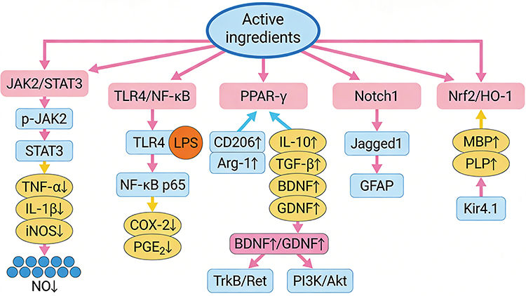 |
Figure 5 Vitex plants protective mechanism against ischemic stroke in neural glial cells: Molecular mechanism diagram.↑ Arrow indicates that the target gene or protein is upregulated under the regulation of the limited component; ↓ Arrow indicates that the target gene or protein is downregulated under the regulation of the limited component. |
Inhibition of M1 Phenotype Activation and Pro-Inflammatory Cytokine Release
Casticin inhibits the Janus kinase 2/signal transducer and activator of transcription 3 (JAK2/STAT3) pathway, attenuating the phosphorylation of JAK2 (p-JAK2) and nuclear translocation of STAT3.126 This further downregulates the transcriptional and expression of pro-inflammatory mediators, including tumor necrosis factor-α (TNF-α), interleukin-1β (IL-1β), and inducible nitric oxide synthase (iNOS), thereby mitigating the toxicity of NO (nitric oxide); as excessive NO reacts with ·O2− to form ONOO−, a highly toxic reactive nitrogen species. Meanwhile, vitexin inhibits the TLR4/NF-κB pathway, abrogating the LPS-induced inflammatory cascade triggered by Toll-like receptor 4 (TLR4) activation.127 This not only reduces the nuclear translocation of the NF-κB p65 subunit but also suppresses the expression of cyclooxygenase-2 (COX-2) and the production of prostaglandin E2 (PGE2). Vitexin activates peroxisome proliferator-activated receptor γ (PPAR-γ), promoting microglia expression of M2 phenotype markers (CD206, Arg-1) along with the secretion of anti-inflammatory cytokines, including interleukin-10 (IL-10) and transforming growth factor-β (TGF-β).126,128 This restricts the propagation of local inflammation and enhances the survival of injured neurons.
Regulation of Astrocytes: Inhibition of Glial Scar Formation and Enhancement of Nutritional Support
First, active components derived from Vitex species inhibit excessive astrocyte activation and glial scar formation. Vitexin suppresses the Notch1 signaling pathway, attenuating the interaction between the Notch1 receptor and its ligand Jagged1, as well as the nuclear translocation of the Notch intracellular domain (NICD).129 This further inhibits astrocyte proliferation and the expression of glial fibrillary acidic protein (GFAP), precluding overactivated astrocytes from secreting collagen fibers to form glial scars and alleviating the physical barrier that impairs axon regeneration. Second, these components augment neurotrophic factor secretion and metabolic support. Under ischemic stress, vitexilactone induces astrocytes to upregulate the expression and secretion of brain-derived neurotrophic factor (BDNF) and glial cell line-derived neurotrophic factor (GDNF).130 Upon binding to TrkB and Ret receptors on the neuronal plasma membrane, BDNF and GDNF activate the PI3K/Akt pathway, thereby suppressing neuronal apoptosis. Additionally, astrocytes upregulate glutamate transporter 1 (GLT-1), facilitating the clearance of excessive extracellular glutamate and mitigating excitotoxicity.131 Active components from Vitex species also exert beneficial effects on oligodendrocytes by preserving myelin integrity and promoting remyelination. Vitexin activates the Nrf2/HO-1 signaling pathway, mitigating ischemia-induced oxidative stress in oligodendrocytes and sustaining their viability.122 Moreover, Vitex-derived components upregulate the expression of myelin-associated proteins, including myelin basic protein (MBP) and proteolipid protein (PLP).132 They also activate the Kir4.1 potassium channel in oligodendrocyte precursor cells (NG2 glia), facilitating the differentiation of NG2 glia into mature oligodendrocytes, restoring ischemia-impaired myelin structures, and preserving axonal conduction integrity.
Regarding the frontier mechanisms of active ingredients in Vitex plants inhibiting ferroptosis and regulating astrocyte function, some studies have indicated that isovitexin reduces LPS-induced NLRP3/caspase-1/GSDMD pathway activation in BV2 cells, decreasing the release of pro-inflammatory cytokines (TNF-α, IL-1) by more than 50%.87
Repair Mechanisms of Vascular Endothelial Cells and BBB of Active Components from the Genus Vitex
Ischemic stroke induces vascular endothelial cell injury, BBB disruption, and cerebral microcirculation dysfunction. Active components from the genus Vitex enhance cerebral blood supply and tissue perfusion by preserving the function of vascular endothelial cells, repairing the BBB, and facilitating angiogenesis (Figure 6). Vitexin activates the PI3K/Akt signaling pathway to induce vascular endothelial cells to upregulate expression of tight junction proteins,133 including Occludin, zonula occludens-1 (ZO-1), and Claudin-5, thereby augmenting intercellular junction integrity.134 Meanwhile, these components suppress the expression and enzymatic activity of matrix metalloproteinase-9 (MMP-9), attenuate the MMP-9-mediated degradation of tight junction proteins, reduce BBB permeability, and mitigate the leakage of macromolecular substances (Evans Blue, EB) into the brain parenchyma.135 Casticin maintains the structural integrity of the BBB by upregulating Bcl-2 and downregulating Bax,136 thereby suppressing vascular endothelial cell apoptosis.
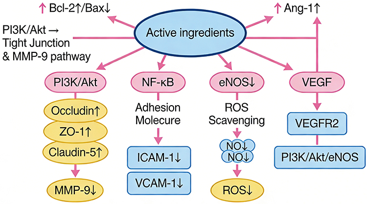 |
Figure 6 Vitex plants protective mechanism against vascular endothelial damage in ischemic stroke: Molecular mechanism diagram.↑ Arrow indicates that the target gene or protein is upregulated under the regulation of the limited component; ↓ Arrow indicates that the target gene or protein is downregulated under the regulation of the limited component. |
Vitex-derived active components suppress the NF-κB pathway to downregulate the expression of intercellular adhesion molecule-1 (ICAM-1) and vascular cell adhesion molecule-1 (VCAM-1) in vascular endothelial cells.137 This abrogates the adhesion of peripheral leukocytes (eg, neutrophils, monocytes) to vascular endothelial cells and their transmigration cross the BBB, thereby attenuating inflammatory cell infiltration and cerebral parenchymal inflammatory injury. Vitexin activates endothelial nitric oxide synthase (eNOS) to promote the NO production,138 which dilates blood vessels, inhibits platelet aggregation, scavenges ROSe, and mitigates oxidative injury to vascular endothelial cells.
Vitexin upregulates the expression of vascular endothelial growth factor (VEGF). Upon binding of VEGF to VEGF receptor 2 (VEGFR2) on the vascular endothelial cell surface, it activates the PI3K/Akt/eNOS signaling pathway, facilitating the proliferation, migration, and tube formation of endothelial cells.139 Additionally, vitexin upregulates the expression of angiopoietin-1 (Ang-1) to augment the stability of newly formed blood vessels and inhibit vascular leakage.140 By promoting angiogenesis and vasodilation, vitexin increases blood perfusion in the ischemic region, shortens the duration of cerebral ischemia-hypoxia, supplies sufficient oxygen and nutrients to neurons and glial cells, and suppresses the progression of ischemic injury.
Regarding the effective components of Vitex species in inhibiting ferroptosis and regulating pyroptosis, some studies have pointed out that vanillic acid confers ferroptosis resistance in endothelial cells by upregulating SLC7A11 expression,141 this maintains GSH levels and inhibits H2O2-induced endothelial ferroptosis, while vitexin mediates epigenetic regulation of angiogenesis, it promotes histone acetylation in the VEGF promoter region by inhibiting HDAC1, thereby enhancing VEGF transcription and angiogenesis.142,143
Research Value and Development Prospects of Unique Components in Vitex L
Vitex L. harbors specific components with structural uniqueness and targeted anti-ischemic stroke activity, which are the core basis for its therapeutic advantages and research value.
Vitexilactone
As a diterpenoid lactone unique to Vitex (eg, Vitex trifolia), vitexilactone exhibits potent antioxidant activity (IC50 = 15.3 μM for DPPH scavenging) and neuroprotective effects. Preclinical studies show it promotes astrocyte secretion of BDNF/GDNF via activating the PI3K/Akt pathway, may inhibiting neuronal apoptosis.144 Its unique structure (C22H34O5) enables specific binding to Keap1 (molecular docking binding energy = −7.2 kcal/mol), enhancing Nrf2 nuclear translocation more efficiently than common flavonoids. Future directions include verifying its inhibition of ferroptosis via regulating GPX4 expression and optimizing synthesis to improve yield.
Rotundifuran
This diterpenoid (C22H34O4) is enriched in Vitex rotundifolia, with demonstrated vascular protective effects. Rotundifuran (10–50 μM) improves HUVEC barrier function by 35% via upregulating Occludin/ZO-1.119,145 Unlike non-specific vascular protectants, it specifically targets VEGFR2 to promote angiogenesis without inducing abnormal vascular proliferation.119 Its mechanism involves inhibiting MMP-9 activity and reducing BBB permeability, making it a potential candidate for BBB repair.98
Casticin
A flavonoid unique to Vitex (Vitex negundo, Vitex agnus-castus), casticin inhibits microglial M1 polarization via targeting JAK2/STAT3 (p-JAK2 reduction by 60%) and NLRP3 inflammasome activation (IL-1β release reduction by 55%).10 It also blocks neuronal excitotoxicity by binding to NMDA receptors (binding energy = −6.9 kcal/mol), synergizing with vitexin to enhance neuroprotection.79 Clinical translation potential is supported by its good safety profile (LD50 = 150 mg/kg in mice) and potential for oral formulation.121
These unique components, together with other common active ingredients, form a “multi-component, multi-target” therapeutic network, highlighting the irreplaceable value of Vitex L. in anti-ischemic stroke research.
Discussion
Scientific Evidence and Mechanistic Prospects for Vitex in Ischemic Stroke Treatment
Accumulating preclinical studies provide compelling evidence for the efficacy of Vitex L. species against ischemic stroke. In vitro models (SH-SY5Y, BV2, HUVEC cells) and in vivo models (rat/mouse MCAO, BCCAO/R) have confirmed that Vitex-derived components reduce cerebral infarct volume, enhance neurological function (mNSS/NIHSS), and attenuate key pathological insults (oxidative stress, inflammation, BBB disruption). Mechanistically, the “multi-cellular target, multi-pathway regulation” characteristic is well-documented: these components target neurons to block the “oxidative stress-excitotoxicity-apoptosis” cascade (via Nrf2/ARE, PI3K/Akt, and NMDA receptor inhibition), regulate glial cells to balance inflammation and tissue repair (via PPAR-γ, Notch1, and Kir4.1 pathways), and protect vascular endothelial cells to restore BBB integrity and cerebral perfusion (via PI3K/Akt-tight junctions signaling and VEGF/Ang-1-mediated angiogenesis).
Current preclinical evidence confirms Vitex L.’s anti-ischemic stroke efficacy, with a mechanistic network spanning both classic and frontier pathways. Future efforts should include validating ferroptosis and pyroptosis regulation, this could involve focusing on vitexin’s role in the ACSL4/TFR1 axis and casticin’s impact on the NLRP3 inflammasome via molecular dynamics simulation, decoding intercellular communication by exploring how astrocyte-derived lncRNA MALAT1 coordinates with neuronal miR-107 to modulate synaptic remodeling, and clarifying epigenetic mechanisms through investigating component-mediated histone modifications (eg, acetylation) and non-coding RNA interactions within ischemic brain tissue.
However, mechanistic understanding of these effects remains incomplete. Future research directions should focus on: (1) Exploring underinvestigated pathological pathways, such as ferroptosis (ie, whether vitexin regulates ACSL4/TFR1) and autophagy (the role of betulinic acid in SIRT1/FoxO1-mediated autophagy balance);146 (2) Investigating epigenetic regulation, including the crosstalk between Vitex-derived components and non-coding RNAs (miR-107, lncRNA MALAT1) or histone modifications (acetylation) in ischemic neurons;147 (3) Elucidating intercellular communication mechanisms, such as how astrocyte-derived BDNF/GDNF crosstalk with neuronal TrkB/Ret receptors to promote synaptic remodeling, and how microglial M2 polarization modulates oligodendrocyte differentiation.148 These investigations will further expand the mechanistic network underlying the anti-ischemic stroke effects of Vitex L. species.
Active Components of Vitex and Progress in Extraction/Application
Vitex L. species harbor diverse active components with distinct anti-ischemic stroke potential, which can be classified based on evidence strength and application potential: High-evidence components, flavonoids (vitexin, isovitexin, casticin, luteolin), are the most specific and potent, with vitexin and isovitexin exhibiting potent BBB-protective effects and NMDA receptor inhibitory activity; terpenoids (oleanolic acid, betulinic acid, aucubin) exert robust anti-apoptotic and anti-inflammatory effects; simple phenols (protocatechuic acid, ferulic acid) possess prominent antioxidant activity but low abundance in Vitex species. Potential components, phenylpropanoids (3,5-di-O-caffeoylquinic acid) and steroids (progesterone, β-sitosterol), demonstrate efficacy in single experimental models but necessitate further validation in stroke-specific models.
Although some key active components (eg, protocatechuic acid) have low abundance in Vitex plants, optimized extraction techniques (ultrasonic-assisted extraction), chemical synthesis, and nanodelivery systems (eg, vitexin microcapsules) have effectively overcome this limitation. Moreover, the unique components of Vitex (eg, vitexilactone) and the synergistic effects of multi-component mixtures highlight the irreplaceable value of Vitex-centered research.
Limitations of This Review and Future Improvements
This review comprehensively collates existing research on Vitex in ischemic stroke treatment, but several limitations should be noted: First, most studies rely on single-cell lines or single animal models (Sprague-Dawley rats, C57BL/6 mice), lacking multi-center, large-sample animal experiments to verify reproducibility. For example, although syringaldehyde reduces amyloid plaques in APP/PS1 mice, its efficacy in MCAO models (the gold standard for ischemic stroke) has not been validated, leading to uncertain translational value.149 Second, while core pathways (Nrf2, NF-κB) are well-studied, molecular dynamics verification of component-target binding (the interaction between vitexin and NMDA receptors at the atomic level) is scarce, and the cross-talk between pathways (how PI3K/Akt interacts with NF-κB to regulate endothelial cell apoptosis) is not clarified. Third, many components (progesterone, β-sitosterol) are widely distributed in other plants, and Vitex-specific components (vitexilactone, rotundifuran) have limited research. Additionally, only progesterone has entered early clinical trials, while other high-evidence components (apigenin, oleanolic acid) lack human safety and efficacy data, hindering clinical translation. Fourth, Vitex extracts contain multiple active components, but current studies focus on monomers rather than exploring how flavonoids, terpenoids, and phenols synergistically regulate pathology. This limits the development of holistic herbal therapies based on Vitex plants.
Single-cell sequencing technology holds great promise for deciphering the cell-type-specific regulatory effects of diverse active components from Vitex L. on ischemic brain tissue, enabling precise identification of subtype-specific responses of core cell populations (eg, microglia, astrocytes, and vascular endothelial cells) and clarification of their distinct functional alterations under the intervention of Vitex-derived compounds. This technology can further uncover the synergistic regulatory mechanisms of multi-component mixtures from Vitex L., such as the crosstalk between flavonoids and terpenoids in modulating inflammatory cascades, ferroptosis, and metabolic pathways across different cell subtypes, while identifying hub genes and key signaling axes analogous to the Spp1-mediated regulation revealed in KBA-Z-GS studies.150 Future research should address these limitations by conducting multi-model validation, integrating multi-omics techniques (proteomics, metabolomics) to decode mechanisms, developing Vitex-specific component screening platforms, and designing Phase I/II clinical trials for promising candidates. These efforts will promote the transformation of Vitex plants from traditional medicine to evidence-based anti-ischemic stroke therapies.
Conclusion
Vitex L. exhibits significant anti-ischemic stroke potential, with unique components and multi-pathway regulation as core advantages. Future research should focus on multi-center validation, synergistic mechanism exploration, and clinical trials of high-evidence components to advance translation.
Abbreviations
Akt, Protein Kinase B; Ang-1, Angiopoietin-1; Apaf-1, Apoptotic Protease Activating Factor-1; ARE, Antioxidant Response Element; Bax, Bcl-2-Associated X Protein; BBB, Blood-Brain Barrier; BCCAO/R, Bilateral Common Carotid Artery Occlusion/Reperfusion; Bcl-2, B-Cell Lymphoma 2; BDNF, Brain-Derived Neurotrophic Factor; COX-2, Cyclooxygenase-2; Cyt c, Cytochrome c; EB, Evans Blue; eNOS, Endothelial Nitric Oxide Synthase; GDNF, Glial Cell Line-Derived Neurotrophic Factor; GFAP, Glial Fibrillary Acidic Protein; GLT-1, Glutamate Transporter 1; GSH-Px, Glutathione Peroxidase; HO-1, Heme Oxygenase-1; ICAM-1, Intercellular Adhesion Molecule-1; IL, Interleukin; iNOS, Inducible Nitric Oxide Synthase; JAK2/STAT3, Janus Kinase 2/Signal Transducer and Activator of Transcription 3; Keap1, Kelch-Like ECH-Associated Protein 1; LPS, Lipopolysaccharide; MBP, Myelin Basic Protein; MCAO, Middle Cerebral Artery Occlusion; MDA, Malondialdehyde; MMP-9, Matrix Metalloproteinase-9; mNSS, Modified Neurological Severity Score; mPTP, Mitochondrial Permeability Transition Pore; NF-κB, Nuclear Factor-κB; NIHSS, National Institutes of Health Stroke Scale; NQO1, NAD(P)H: Quinone Oxidoreductase 1; Nrf2, Nuclear Factor Erythroid 2-Related Factor 2; OGD, Oxygen-Glucose Deprivation; OGD/R, Oxygen-Glucose Deprivation/Reperfusion; PGE2, Prostaglandin E2; PI3K, Phosphatidylinositol 3-Kinase; PLP, Proteolipid Protein; PMCAO, Permanent Middle Cerebral Artery Occlusion; PPAR-γ, Peroxisome Proliferator-Activated Receptorγ; ROS, Reactive Oxygen Species; SOD, Superoxide Dismutase; SAMP8, Senescence Accelerated Mouse-Prone 8; TLR4, Toll-Like Receptor 4; TNF-α, Tumor Necrosis Factor-α; VCAM-1, Vascular Cell Adhesion Molecule-1; VEGF, Vascular Endothelial Growth Factor; VEGFR2, Vascular Endothelial Growth Factor Receptor 2; ΔΨm, Mitochondrial Membrane Potential.
Author Contributions
All authors made a significant contribution to the work reported, whether that is in the conception, study design, execution, acquisition of data, analysis and interpretation, or in all these areas; took part in drafting, revising or critically reviewing the article; gave final approval of the version to be published; have agreed on the journal to which the article has been submitted; and agree to be accountable for all aspects of the work.
Chengqiong Xie and Jinjin Wu drafted and edited the manuscript, Ping Huang wrote manuscript. All data were generated in-house, and no paper mill was used. All authors agree to be accountable for all aspects of work ensuring integrity and accuracy.
Funding
This work was supported by Wu Jinjin National Inheritance Workshop for Veteran TCM Pharmacists Construction Project (Grant No. Guo Zhong Yi Yao Ren Jiao Han [2024] 255), Zhejiang Province Traditional Chinese Medicine Science and Technology Program (2025ZF051), Zhejiang Province Key Discipline Construction Project of Traditional Chinese Medicine (Clinical Chinese Pharmacy) (2024-XK-56).
Disclosure
The authors declare no conflict of interests.
References
1. Sirotkin AV. Effects, Mechanisms of Action and Application of Vitex agnus-castus for Improvement of Health and Female Reproduction. Phytother Res. 2025;39(3):1484–28. doi:10.1002/ptr.8438
2. Yan CX, Wei YW, Li H, et al. Vitex rotundifolia L. f. and Vitex trifolia L.: a review on their traditional medicine, phytochemistry, pharmacology. J Ethnopharmacol. 2023;308:116273. doi:10.1016/j.jep.2023.116273
3. Puglia LT, Lowry J, Tamagno G. Vitex agnus castus effects on hyperprolactinaemia. Front Endocrinol. 2023;14:1269781. doi:10.3389/fendo.2023.1269781
4. Teixeira da Silva JA, Kher MM, Nataraj M. Biotechnological advances in Vitex species, and future perspectives. J Genet Eng Biotechnol. 2016;14(2):335–348. doi:10.1016/j.jgeb.2016.09.004
5. Peraza-Sánchez SR, Cen-Pacheco F, Noh-Chimal A, et al. Leishmanicidal evaluation of extracts from native plants of the Yucatan peninsula. Fitoterapia. 2007;78(4):315–318. doi:10.1016/j.fitote.2007.03.013
6. Mottaghipisheh J, Kamali M, Doustimotlagh AH, et al. A comprehensive review of ethnomedicinal approaches, phytochemical analysis, and pharmacological potential of Vitex trifolia L. Front Pharmacol. 2024;15:1322083. doi:10.3389/fphar.2024.1322083
7. Soleimany Z, Siadat F, Farhadi M, Mirshaby ZS, Sanadgol Z, Eyni H. Improvement of ovarian function in a premature ovarian failure mouse model using Vitex agnus-castus extract. JBRA Assist Reprod. 2025;29(1):117–126. doi:10.5935/1518-0557.20240101
8. Zheng CJ, Li HQ, Ren SC, et al. Phytochemical and pharmacological profile of Vitex negundo. Phytother Res. 2015;29(5):633–647. doi:10.1002/ptr.5303
9. Chen C, Zhang Z, Du B, Lv C. Vitexin alleviates cerebral ischemia/reperfusion injury by regulating mitophagy via the SIRT1/PINK1/Parkin pathway. Brain Res Bull. 2025;227:111404. doi:10.1016/j.brainresbull.2025.111404
10. Huang D, Zhou J, Li W, Zhang L, Wang X, Liu Q. Casticin protected against neuronal injury and inhibited the TLR4/NF-κB pathway after middle cerebral artery occlusion in rats. Pharmacol Res Perspect. 2021;9(2):e00752. doi:10.1002/prp2.752
11. Shivarajan VV, Balachandran I. Botanical notes on the identity of certain herbs used in ayurvedic medicines in Kerala.ii. Anc Sci Life. 1985;4(4):217–219.
12. Kiuchi F, Matsuo K, Ito M, Qui TK, Honda G. New norditerpenoids with trypanocidal activity from Vitex trifolia. Chem Pharm Bull. 2004;52(12):1492–1494. doi:10.1248/cpb.52.1492
13. Ding GY, Hu P. Research progress on the chemical constituents and biological activities of Vitex negundo. J Shenyang Med College. 2020;22(02):162–4+73.
14. Ntalli NG, Ferrari F, Giannakou I, Menkissoglu-Spiroudi U. Phytochemistry and nematicidal activity of the essential oils from 8 Greek Lamiaceae aromatic plants and 13 terpene components. J Agric Food Chem. 2010;58(13):7856–7863. doi:10.1021/jf100797m
15. Webster DE, He Y, Chen SN, Pauli GF, Farnsworth NR, Wang ZJ. Opioidergic mechanisms underlying the actions of Vitex agnus-castus L. Biochem Pharmacol. 2011;81(1):170–177. doi:10.1016/j.bcp.2010.09.013
16. Kar TK, Sil S, Ghosh A, Barman A, Chattopadhyay S. Mitigation of letrozole induced polycystic ovarian syndrome associated inflammatory response and endocrinal dysfunction by Vitex negundo seeds. J Ovarian Res. 2024;17(1):76. doi:10.1186/s13048-024-01378-4
17. Bunu MI, Ndinteh DT, Macdonald JR, et al. Ecdysteroids from the stem bark of Vitex doniana Sweet (Lamiaceae; ex. Verbenaceae): a geographically variable African Medicinal Species. Antibiotics. 2021;10(8):937. doi:10.3390/antibiotics10080937
18. Gentallan RP, Sengun S, Bartolome MCB, et al. The Vitextrifolia complex (Lamiaceae) in the Philippines. PhytoKeys. 2024;248:1–40. doi:10.3897/phytokeys.248.120387
19. Pan X, Jiang S, Zhang X, et al. Recent strategies in target identification of natural products: exploring applications in chronic inflammation and beyond. Br J Pharmacol. 2025;182(20):4841–4860. doi:10.1111/bph.17356
20. Feng HY, Wang YQ, Yang J, Miao H, Zhao YY, Li X. Anthraquinones from Rheum officinale ameliorate renal fibrosis in acute kidney injury and chronic kidney disease. Drug Des Devel Ther. 2025;19:5739–5760. doi:10.2147/DDDT.S521265
21. Boaru DL, Fraile-Martinez O, De Leon-Oliva D, et al. Harnessing the anti-inflammatory properties of polyphenols in the treatment of inflammatory bowel disease. Int J Biol Sci. 2024;20(14):5608–5672. doi:10.7150/ijbs.98107
22. Ma D, Yang J, Zhao Z, et al. Research progress on the mechanism of Chinese herbal compounds treating benign prostatic hyperplasia by regulating inflammatory response. Integrat Med Nephrol Androl. 2024;11(3).
23. Wang YN, Wu X, Shan QY, et al. Acteoside-containing caffeic acid is bioactive functional group of antifibrotic effect by suppressing inflammation via inhibiting AHR nuclear translocation in chronic kidney disease. Acta Pharmacol Sin. 2025;46:2975–2988. doi:10.1038/s41401-025-01598-4
24. Song H, Xu D, Yu Q. The effect of isovitexin on neuronal damage induced by OGD/R by modulating the cAMP/PKA signaling pathway. Stroke and Nervous Diseases. 2023;30(6):557–62+76.
25. Alimohamadi R, Fatemi I, Naderi S, Hakimizadeh E, Rahmani MR, Allahtavakoli M. Protective effects of Vitex agnus-castus in ovariectomy mice following permanent middle cerebral artery occlusion. Iran J Basic Med Sci. 2019;22(9):1097–1101. doi:10.22038/ijbms.2019.31692.7625
26. Kale S, Sarode LP, Kharat A, et al. Protocatechuic acid prevents early hour ischemic reperfusion brain damage by restoring imbalance of neuronal cell death and survival proteins. J Stroke Cerebrovasc Dis. 2021;30(2):105507. doi:10.1016/j.jstrokecerebrovasdis.2020.105507
27. Kaewmool C, Kongtawelert P, Phitak T, Pothacharoen P, Udomruk S. Protocatechuic acid inhibits inflammatory responses in LPS-activated BV2 microglia via regulating SIRT1/NF-κB pathway contributed to the suppression of microglial activation-induced PC12 cell apoptosis. J Neuroimmunol. 2020;341:577164. doi:10.1016/j.jneuroim.2020.577164
28. Khoshnam SE, Farbood Y, Fathi Moghaddam H, Sarkaki A, Badavi M, Khorsandi L. Vanillic acid attenuates cerebral hyperemia, blood-brain barrier disruption and anxiety-like behaviors in rats following transient bilateral common carotid occlusion and reperfusion. Metab Brain Dis. 2018;33(3):785–793. doi:10.1007/s11011-018-0187-5
29. Festa J, Hussain A, Al-Hareth Z, Bailey SJ, Singh H, Da Boit M. Phenolic metabolites protocatechuic acid and vanillic acid improve nitric oxide bioavailability via the Akt-eNOS pathway in response to TNF-α induced oxidative stress and inflammation in endothelial cells. Metabolites. 2024;14(11):613. doi:10.3390/metabo14110613
30. Cheng CY, Kao ST, Lee YC. Ferulic acid exerts anti-apoptotic effects against ischemic injury by activating HSP70/Bcl-2- and HSP70/Autophagy-mediated signaling after permanent focal cerebral ischemia in rats. Am J Chin Med. 2019;47(1):39–61. doi:10.1142/S0192415X19500034
31. Verma H, Yadav A, Gangwar P, et al. A cross-sectional in vitro study on the synergistic neuroprotective effects of phytochemicals ferulic acid and Ginkgolide B against amyloid beta-induced oxidative stress and modulation of multifunctional enzyme APE1/Ref-1 in human neuroblastoma SH-SY5Y cells. Cell Biochem Biophys. 2025;83(4):4731–4748. doi:10.1007/s12013-025-01799-y
32. Hoberg E, Meier B, Sticher O. Quantitative high performance liquid chromatographic analysis of diterpenoids in agni-casti fructus. Planta Med. 2000;66(4):352–355. doi:10.1055/s-2000-8535
33. Krishna V, Sharma S, Pareck R, Singh P. Terpenoid constituents from some indigenous plants. J Indian Chem Society. 2002;79:550–552.
34. Ko W, Kang T, Lee S, Kim Y, Lee B. Effects of luteolin on the inhibition of proliferation and induction of apoptosis in human myeloid leukaemia cells. Phytotherapy Res. 2002;16(3):295–298. doi:10.1002/ptr.871
35. Kawazoe K, A Y, Y T. Aryl naphthalenes norlignans from Vitex rotundifolia. Phytochemistry. 1999;52:1657–1659. doi:10.1016/S0031-9422(99)00405-7
36. Leitão S, Fonseca E, Santos T. Essential oils from two brazilian Vitex species. Acta Hortic. 1999;500:89–92. doi:10.17660/ActaHortic.1999.500.11
37. Azhar-Ul-Haq M, Khan M, Anwar-Ul-Haq K, Ahmad A, Choudhary M. Tyrosinase inhibitory lignans from the methanol extract of the roots of Vitex negundo Linn. and their structure-activity relationship. Phytomedicine. 2006;13(4):255–260. doi:10.1016/j.phymed.2004.09.001
38. Ono M, Sawamura H, Ito Y, Mizuki K, Nohara T. Diterpenoids from the fruits of Vitex trifolia. Phytochemistry. 2000;55(8):873–877. doi:10.1016/S0031-9422(00)00214-4
39. Suksamrarn A, Kumpun S, Kirtikara K, Yingyongnarongkul B, Suksamrarn S. Iridoids with anti-inflammatory activity from Vitex peduncularis. Planta medica. 2002;68(1):72–73. doi:10.1055/s-2002-20048
40. Thuy T, Sung T, Adam G. Study on chemical constituents of Vitex leptobotrys II. Chalcones and alkaloid. Tap Chi Hoa Hoc. 2000;38(2):1–7.
41. Gu J, Li X, Yu H. Establishment of an acute inflammatory model of Xylene-induced mouse ear edema. J Hunan Univ Chin Med. 2016;36(05):32–35.
42. Chen R, Gao H, Zhu T, et al. Syringaldehyde ameliorates cognitive dysfunction in APP/PS1 mice by stabilizing the NLRP3 pathway. Mol Neurobiol. 2025;62(12):15844–15858. doi:10.1007/s12035-025-05227-3
43. Vamshi G, DSNBK P, Sampath A, et al. Possible cerebroprotective effect of citronellal: molecular docking, MD simulation and in vivo investigations. J Biomol Struct Dyn. 2024;42(3):1208–1219. doi:10.1080/07391102.2023.2220025
44. Xu C, Feng J, Sun H, et al. Pharmacokinetics of 4-Hydroxybenzaldehyde in normal and cerebral ischemia-reperfusion injury rats based on microdialysis technique. Eur J Drug Metab Pharmacokinet. 2024;49(1):23–32. doi:10.1007/s13318-023-00863-3
45. Festa J, Hussain A, Singh H, Da Boit M. Cyanidin-3-glucoside phenolic metabolites, protocatechuic acid and vanillic acid, attenuate the adhesion of monocytes to endothelial cells in response to TNF-α by targeting NF-κB and Nrf2 pathways. Eur J Nutr. 2025;64(5):208. doi:10.1007/s00394-025-03725-7
46. Kim SS, Park RY, Jeon HJ, Kwon YS, Chun W. Neuroprotective effects of 3,5-dicaffeoylquinic acid on hydrogen peroxide-induced cell death in SH-SY5Y cells. Phytother Res. 2005;19(3):243–245. doi:10.1002/ptr.1652
47. Han J, Miyamae Y, Shigemori H, Isoda H. Neuroprotective effect of 3,5-di-O-caffeoylquinic acid on SH-SY5Y cells and senescence-accelerated-prone mice 8 through the up-regulation of phosphoglycerate kinase-1. Neuroscience. 2010;169(3):1039–1045. doi:10.1016/j.neuroscience.2010.05.049
48. Kim DW, Lee KT, Kwon J, Lee HJ, Lee D, Mar W. Neuroprotection against 6-OHDA-induced oxidative stress and apoptosis in SH-SY5Y cells by 5,7-Dihydroxychromone: activation of the Nrf2/ARE pathway. Life Sci. 2015;130:25–30. doi:10.1016/j.lfs.2015.02.026
49. Bai M, Cui N, Liao Y, et al. Astrocytes and microglia-targeted Danshensu liposomes enhance the therapeutic effects on cerebral ischemia-reperfusion injury. J Control Release. 2023;364:473–489. doi:10.1016/j.jconrel.2023.11.002
50. Zhang P, Yang XW. Studies on chemical constituents in roots and rhizomes of Notopterygium incisum. China J Chin Materia Medica. 2008;33(24):2918–2921.
51. Tułacz Z, Włodarczyk M. Comprehensive review of isoflavones and lignans in the prophylaxis and treatment of breast cancer. J Am Nutr Assoc. 2025;1–18.
52. Xiao HH, Mok DK, Yao XS, Wong MS. Lignans from Sambucus williamsii protect bone via microbiome. Curr Osteoporos Rep. 2024;22(6):497–501. doi:10.1007/s11914-024-00883-1
53. Ehambarampillai D, Wan MLY. A comprehensive review of Schisandra chinensis lignans: pharmacokinetics, pharmacological mechanisms, and future prospects in disease prevention and treatment. Chin Med. 2025;20(1):47. doi:10.1186/s13020-025-01096-z
54. Cao G, Miao H, Wang YN, et al. Intrarenal 1-methoxypyrene, an aryl hydrocarbon receptor agonist, mediates progressive tubulointerstitial fibrosis in mice. Acta Pharmacol Sin. 2022;43(11):2929–2945. doi:10.1038/s41401-022-00914-6
55. Yan N, Wang X, Xu Z, Zhong L, Yang J. Apigenin attenuates transverse aortic constriction-induced myocardial hypertrophy: the key role of miR-185-5p/SREBP2-mediated autophagy. Drug Des Devel Ther. 2024;18:3841–3851. doi:10.2147/DDDT.S464004
56. Hsu MC, Chen CH, Wang MC, et al. Apigenin targets fetuin-A to ameliorate obesity-induced insulin resistance. Int J Biol Sci. 2024;20(5):1563–1577. doi:10.7150/ijbs.91695
57. Wang YN, Li XJ, Wang WF, Zou L, Miao H, Zhao YY. Geniposidic acid attenuates chronic tubulointerstitial nephropathy through regulation of the NF‐ƙB/Nrf2 pathway via aryl hydrocarbon receptor signaling. Phytother Res. 2024;38(11):5441–5457. doi:10.1002/ptr.8324
58. Pang Q, Zhao Y, Chen X, Zhao K, Zhai Q, Tu F. Apigenin protects the brain against ischemia/reperfusion injury via Caveolin-1/VEGF in vitro and in vivo. Oxid Med Cell Longev. 2018;2018:7017204. doi:10.1155/2018/7017204
59. Gao AX, Xia TC, Lin LS, Dong TT, Tsim KW. The neurotrophic activities of brain-derived neurotrophic factor are potentiated by binding with apigenin, a common flavone in vegetables, in stimulating the receptor signaling. CNS Neurosci Ther. 2023;29(10):2787–2799. doi:10.1111/cns.14230
60. Li H, Yu W, Yang Y, et al. Combination of Atractylenolide I, Atractylenolide III, and Paeoniflorin promotes angiogenesis and improves neurological recovery in a mouse model of ischemic stroke. Chin Med. 2024;19(1):3. doi:10.1186/s13020-023-00872-z
61. Jeong YH, Li W, Go Y, Oh YC. Atractylodis rhizoma alba attenuates neuroinflammation in BV2 microglia upon LPS stimulation by inducing HO-1 activity and inhibiting NF-κB and MAPK. Int J Mol Sci. 2019;20(16):4015. doi:10.3390/ijms20164015
62. Ahmad S, Elsherbiny NM, Haque R, et al. Sesamin attenuates neurotoxicity in mouse model of ischemic brain stroke. Neurotoxicology. 2014;45:100–110. doi:10.1016/j.neuro.2014.10.002
63. Wang L, Qu Z, Sun Q, Mao Z, Si P, Wang W. 4-Hydroxysesamin, a modified natural compound, attenuates neuronal apoptosis after ischemic stroke via inhibiting MAPK pathway. Neuropsychiatr Dis Treat. 2024;20:523–533. doi:10.2147/NDT.S444760
64. Avasthi AS, Bhatnagar M, Sarkar N, Kitchlu S, Ghosal S. Bioassay guided screening, optimization and characterization of antioxidant compounds from high altitude wild edible plants of Ladakh. J Food Sci Technol. 2016;53(8):3244–3252. doi:10.1007/s13197-016-2300-2
65. Verma H, Bhattacharjee A, Shivavedi N, Nayak PK. Evaluation of rosmarinic acid against myocardial infarction in maternally separated rats. Naunyn Schmiedebergs Arch Pharmacol. 2022;395(10):1189–1207. doi:10.1007/s00210-022-02273-9
66. Skała E, Toma M, Kowalczyk T, Śliwiński T, Sitarek P. Rhaponticum carthamoides transformed root extract inhibits human glioma cells viability, induces double strand DNA damage, H2A.X phosphorylation, and PARP1 cleavage. Cytotechnology. 2018;70(6):1585–1594. doi:10.1007/s10616-018-0251-3
67. Li Y, Ren X, Lio C, et al. A chlorogenic acid-phospholipid complex ameliorates post-myocardial infarction inflammatory response mediated by mitochondrial reactive oxygen species in SAMP8 mice. Pharmacol Res. 2018;130:110–122. doi:10.1016/j.phrs.2018.01.006
68. Xia Y, Liang C, Luo H, Zhang Y. Therapeutic potential of flavonoids and flavonoid-rich compounds in irritable bowel syndrome. Drug Des Devel Ther. 2025;19:4895–4910. doi:10.2147/DDDT.S515004
69. Wang Y, Huang M, Zhou X, Li H, Ma X, Sun C. Potential of natural flavonoids to target breast cancer angiogenesis (review). Br J Pharmacol. 2025;182(10):2235–2258. doi:10.1111/bph.16275
70. Huang M, Liu X, Ren Y, et al. Quercetin: a flavonoid with potential for treating acute lung injury. Drug Des Devel Ther. 2024;18:5709–5728. doi:10.2147/DDDT.S499037
71. Shang W, Li XH, Zeng LH, et al. Mechanistic insights into flavonoid subclasses as cardioprotective agents against doxorubicin-induced cardiotoxicity: a comprehensive review. Drug Des Devel Ther. 2025;19:5553–5596. doi:10.2147/DDDT.S535517
72. Gautheron G, Péraldi-Roux S, Vaillé J, et al. The flavonoid resokaempferol improves insulin secretion from healthy and dysfunctional pancreatic β-cells. Br J Pharmacol. 2025;182(1):52–68. doi:10.1111/bph.17304
73. Cai J, Tan X, Hu Q, et al. Flavonoids and gastric cancer therapy: from signaling pathway to therapeutic significance. Drug Des Devel Ther. 2024;18:3233–3253. doi:10.2147/DDDT.S466470
74. Miao H, Cao G, Wu XQ, et al. Identification of endogenous 1-aminopyrene as a novel mediator of progressive chronic kidney disease via aryl hydrocarbon receptor activation. Br J Pharmacol. 2020;177(15):3415–3435. doi:10.1111/bph.15062
75. Niu YJ, Xia CJ, Ai X, et al. Sequential activation of ERα-AMPKα signaling by the flavonoid baicalin down-regulates viral HNF-dependent HBV replication. Acta Pharmacol Sin. 2025;46(3):653–661. doi:10.1038/s41401-024-01408-3
76. Li XJ, Wang YN, Wang WF, Nie X, Miao H, Zhao YY. Barleriside A, an aryl hydrocarbon receptor antagonist, ameliorates podocyte injury through inhibiting oxidative stress and inflammation. Front Pharmacol. 2024;15:1386604. doi:10.3389/fphar.2024.1386604
77. Li L, Pan G, Fan R, et al. Luteolin alleviates inflammation and autophagy of hippocampus induced by cerebral ischemia/reperfusion by activating PPAR gamma in rats. BMC Complement Med Ther. 2022;22(1):176. doi:10.1186/s12906-022-03652-8
78. Zhu L, Bi W, Lu D, Zhang C, Shu X, Lu D. Luteolin inhibits SH-SY5Y cell apoptosis through suppression of the nuclear transcription factor-κB, mitogen-activated protein kinase and protein kinase B pathways in lipopolysaccharide-stimulated cocultured BV2 cells. Exp Ther Med. 2014;7(5):1065–1070. doi:10.3892/etm.2014.1564
79. Yang X, Liu Z, Xu X, He M, Xiong H, Liu L. Casticin induces apoptosis and cytoprotective autophagy while inhibiting stemness involving Akt/mTOR and JAK2/STAT3 pathways in glioblastoma. Phytother Res. 2024;38(1):305–320. doi:10.1002/ptr.8048
80. Cai H, Zhao MY, Wang CZ, et al. Nrf2/STAT3-mediated activation of SLC6A3 underlies the neuroprotective effect of quercetin in ischemic stroke. Phytomedicine. 2025;145:157061. doi:10.1016/j.phymed.2025.157061
81. Dabeek WM, Marra MV. Dietary quercetin and kaempferol: bioavailability and potential cardiovascular-related bioactivity in humans. Nutrients. 2019;11(10):2288. doi:10.3390/nu11102288
82. Almarfadi OM, Siddiqui NA, Shahat AA, et al. Isolation of a novel isoprenylated phenolic compound and neuroprotective evaluation of Dodonaea viscosa extract against cerebral ischaemia-reperfusion injury in rats. Saudi Pharm J. 2024;32(1):101898. doi:10.1016/j.jsps.2023.101898
83. Kim SY, Jin CY, Kim CH, et al. Isorhamnetin alleviates lipopolysaccharide-induced inflammatory responses in BV2 microglia by inactivating NF-κB, blocking the TLR4 pathway and reducing ROS generation. Int J Mol Med. 2019;43(2):682–692. doi:10.3892/ijmm.2018.3993
84. He S, Wang X, Zhong Y, et al. Hesperetin post-treatment prevents rat cardiomyocytes from hypoxia/reoxygenation injury in vitro via activating PI3K/Akt signaling pathway. Biomed Pharmacother. 2017;91:1106–1112. doi:10.1016/j.biopha.2017.05.003
85. Li W, Deng Z, Xiao S, et al. Protective effect of vitexin against high fat-induced vascular endothelial inflammation through inhibiting trimethylamine N-oxide-mediated RNA m6A modification. Food Funct. 2024;15(13):6988–7002. doi:10.1039/D3FO04743A
86. Tian C, Yu B, Liu Y, Diao Z, Wang Y, Zhou J. Apigenin attenuates myocardial ischemia-reperfusion injury through miR-448/SIRT1 axis. Iran J Basic Med Sci. 2025;28(5):602–611. doi:10.22038/ijbms.2025.80172.17365
87. Liu B, Huang B, Hu G, et al. Isovitexin-mediated regulation of microglial polarization in lipopolysaccharide-induced neuroinflammation via activation of the CaMKKβ/AMPK-PGC-1α signaling axis. Front Immunol. 2019;10:2650. doi:10.3389/fimmu.2019.02650
88. Guo ZY, Wu X, Zhang SJ, Yang JH, Miao H, Zhao YY. Poria cocos: traditional uses, triterpenoid components and their renoprotective pharmacology. Acta Pharmacol Sin. 2025;46(4):836–851. doi:10.1038/s41401-024-01404-7
89. Du Y, Li J, Ye M, et al. Hyperuricemia-induced acute kidney injury in the context of chronic kidney disease: a case report. Integr Med Nephrol Androl. 2023;10(4):e00008. doi:10.1097/IMNA-D-23-00008
90. Lu D, Huang L, Weng C. Unveiling the novel anti-tumor potential of digitonin, a steroidal saponin, in gastric cancer: a network pharmacology and experimental validation study. Drug Des Devel Ther. 2025;19:2653–2666. doi:10.2147/DDDT.S504671
91. Wang M, Yuan C, Wu Z, et al. Paris saponin VII reverses resistance to PARP inhibitors by regulating ovarian cancer tumor angiogenesis and glycolysis through the RORα/ECM1/VEGFR2 signaling axis. Int J Biol Sci. 2024;20(7):2454–2475. doi:10.7150/ijbs.91861
92. Park JH, Lee TK, Kim DW, et al. Neuroprotective effects of Aucubin against cerebral ischemia and ischemia injury through the inhibition of the TLR4/NF-κB inflammatory signaling pathway in gerbils. Int J Mol Sci. 2024;25(6).
93. Wang C, Cui X, Dong Z, et al. Attenuated memory impairment and neuroinflammation in Alzheimer’s disease by aucubin via the inhibition of ERK-FOS axis. Int Immunopharmacol. 2024;126:111312. doi:10.1016/j.intimp.2023.111312
94. Sapkota A, Choi JW. Oleanolic acid provides neuroprotection against ischemic stroke through the inhibition of microglial activation and NLRP3 inflammasome activation. Biomol Ther. 2022;30(1):55–63. doi:10.4062/biomolther.2021.154
95. Msibi ZNP, Mabandla MV. Oleanolic acid mitigates 6-Hydroxydopamine neurotoxicity by attenuating intracellular ROS in PC12 cells and striatal microglial activation in rat brains. Front Physiol. 2019;10:1059. doi:10.3389/fphys.2019.01059
96. Zhao Y, Shi X, Wang J, Mang J, Xu Z. Betulinic acid ameliorates cerebral injury in middle cerebral artery occlusion rats through regulating autophagy. ACS Chem Neurosci. 2021;12(15):2829–2837. doi:10.1021/acschemneuro.1c00198
97. Jiao S, Zhu H, He P, Teng J. Betulinic acid protects against cerebral ischemia/reperfusion injury by activating the PI3K/Akt signaling pathway. Biomed Pharmacother. 2016;84:1533–1537. doi:10.1016/j.biopha.2016.11.028
98. Hajdú Z, Hohmann J, Forgo P, et al. Diterpenoids and flavonoids from the fruits of Vitex agnus-castus and antioxidant activity of the fruit extracts and their constituents. Phytother Res. 2007;21(4):391–394. doi:10.1002/ptr.2021
99. Phang SW, Ooi BK, Ahemad N, Yap WH. Maslinic acid suppresses macrophage foam cells formation: regulation of monocyte recruitment and macrophage lipids homeostasis. Vascul Pharmacol. 2020;128-129:106675. doi:10.1016/j.vph.2020.106675
100. Hoenke S, Heise NV, Kahnt M, Deigner HP, Csuk R. Betulinic acid derived amides are highly cytotoxic, apoptotic and selective. Eur J Med Chem. 2020;207:112815. doi:10.1016/j.ejmech.2020.112815
101. Dhote V, Mandloi AS, Singour PK, Kawadkar M, Ganeshpurkar A, Jadhav MP. Neuroprotective effects of combined trimetazidine and progesterone on cerebral reperfusion injury. Curr Res Pharmacol Drug Discov. 2022;3:100108. doi:10.1016/j.crphar.2022.100108
102. Castelnovo LF, Thomas P. Progesterone exerts a neuroprotective action in a Parkinson’s disease human cell model through membrane progesterone receptor α (mPRα/PAQR7). Front Endocrinol. 2023;14:1125962. doi:10.3389/fendo.2023.1125962
103. Fanaei H, Karimian SM, Sadeghipour HR, et al. Testosterone enhances functional recovery after stroke through promotion of antioxidant defenses, BDNF levels and neurogenesis in male rats. Brain Res. 2014;1558:74–83. doi:10.1016/j.brainres.2014.02.028
104. Zhang Y, Chen M, Chen H, et al. Testosterone reduces hippocampal synaptic damage in an androgen receptor-independent manner. J Endocrinol. 2024;260(2). doi:10.1530/JOE-23-0114
105. Tang X, Yan T, Wang S, et al. Treatment with β-sitosterol ameliorates the effects of cerebral ischemia/reperfusion injury by suppressing cholesterol overload, endoplasmic reticulum stress, and apoptosis. Neural Regen Res. 2024;19(3):642–649. doi:10.4103/1673-5374.380904
106. Sun Y, Gao L, Hou W, Wu J. β-Sitosterol alleviates inflammatory response via inhibiting the activation of ERK/p38 and NF-κB pathways in LPS-exposed BV2 cells. Biomed Res Int. 2020;2020:7532306. doi:10.1155/2020/7532306
107. Li S, Xu F, Yu L, Yu Q, Yu N, Fu J. Stigmasterol protects human brain microvessel endothelial cells against ischemia-reperfusion injury through suppressing EPHA2 phosphorylation. Chin J Nat Med. 2023;21(2):127–135. doi:10.1016/S1875-5364(23)60390-5
108. Noh HJ, Yang HH, Kim GS, et al. Chemical constituents of Hericium erinaceum associated with the inhibitory activity against cellular senescence in human umbilical vascular endothelial cells. J Enzyme Inhib Med Chem. 2015;30(6):934–940. doi:10.3109/14756366.2014.995181
109. Chen LW, Wang YQ, Wei LC, Shi M, Chan YS. Chinese herbs and herbal extracts for neuroprotection of dopaminergic neurons and potential therapeutic treatment of Parkinson’s disease. CNS Neurol Disord Drug Targets. 2007;6(4):273–281. doi:10.2174/187152707781387288
110. Meng C, Zeng W, Lv J, et al. 1,8-cineole ameliorates ischaemic brain damage via TRPC6/CREB pathways in rats. J Pharm Pharmacol. 2021;73(7):979–985. doi:10.1093/jpp/rgab035
111. Liu Z, Wang J, Jin X, et al. 1,8-cineole alleviates OGD/R-induced oxidative damage and restores mitochondrial function by promoting the Nrf2 pathway. Biol Pharm Bull. 2023;46(10):1371–1384. doi:10.1248/bpb.b23-00154
112. Li XN, Shang NY, Kang YY, et al. Caffeic acid alleviates cerebral ischemic injury in rats by resisting ferroptosis via Nrf2 signaling pathway. Acta Pharmacol Sin. 2024;45(2):248–267. doi:10.1038/s41401-023-01177-5
113. Li WP, Ma K, Jiang XY, et al. Molecular mechanism of panaxydol on promoting axonal growth in PC12 cells. Neural Regen Res. 2018;13(11):1927–1936. doi:10.4103/1673-5374.239439
114. Wang TA, Li SY, Fann LY, et al. Neuroprotective potential of isofraxidin: alleviating parkinsonian symptoms, inflammation and microglial activation. J Cent Nerv Syst Dis. 2025;17:11795735241312661. doi:10.1177/11795735241312661
115. Ishrat T, Sayeed I, Atif F, Hua F, Stein DG. Progesterone is neuroprotective against ischemic brain injury through its effects on the phosphoinositide 3-kinase/protein kinase B signaling pathway. Neuroscience. 2012;210:442–450. doi:10.1016/j.neuroscience.2012.03.008
116. Li XS, Tang XY, Su W, Li X. Vitexin protects melanocytes from oxidative stress via activating MAPK-Nrf2/ARE pathway. Immunopharmacol Immunotoxicol. 2020;42(6):594–603. doi:10.1080/08923973.2020.1835952
117. Tao M, Li R, Xu T, et al. Vitexin and isovitexin delayed ageing and enhanced stress-resistance through the activation of the SKN-1/Nrf2 signaling pathway. Int J Food Sci Nutr. 2023;74(6):685–694. doi:10.1080/09637486.2023.2243055
118. Xue HY, Niu DY, Gao GZ, Lin QY, Jin LJ, Xu YP. Aucubin modulates Bcl-2 family proteins expression and inhibits caspases cascade in H2O2-induced PC12 cells. Mol Biol Rep. 2011;38(5):3561–3567. doi:10.1007/s11033-010-0466-7
119. Kang MJ, Moon DO, Park JY, et al. Rotundifuran Induces ferroptotic cell death and mitochondria permeability transition in lung cancer cells. Biomedicines. 2024;12(3):576. doi:10.3390/biomedicines12030576
120. Lin CM, Chen CT, Lee HH, Lin JK. Prevention of cellular ROS damage by isovitexin and related flavonoids. Planta Med. 2002;68(4):365–367. doi:10.1055/s-2002-26753
121. Qiao Z, Cheng Y, Liu S, Ma Z, Li S, Zhang W. Casticin inhibits esophageal cancer cell proliferation and promotes apoptosis by regulating mitochondrial apoptotic and JNK signaling pathways. Naunyn Schmiedebergs Arch Pharmacol. 2019;392(2):177–187. doi:10.1007/s00210-018-1574-5
122. Guo L, Shi L. Vitexin improves cerebral ischemia‑reperfusion injury by attenuating oxidative injury and ferroptosis via Keap1/Nrf2/HO-1signaling. Neurochem Res. 2023;48(3):980–995. doi:10.1007/s11064-022-03829-0
123. Song J, Wang H, Sheng J, et al. Vitexin attenuates chronic kidney disease by inhibiting renal tubular epithelial cell ferroptosis via NRF2 activation. Mol Med. 2023;29(1):147. doi:10.1186/s10020-023-00735-1
124. Li X, Mei W, Huang Z, et al. Casticin suppresses monoiodoacetic acid-induced knee osteoarthritis through inhibiting HIF-1α/NLRP3 inflammasome signaling. Int Immunopharmacol. 2020;86:106745. doi:10.1016/j.intimp.2020.106745
125. Wu J, Zhang D, Liu H, et al. Neuroprotective effects of apigenin on retinal ganglion cells in ischemia/reperfusion: modulating mitochondrial dynamics in in vivo and in vitro models. J Transl Med. 2024;22(1):447. doi:10.1186/s12967-024-05260-1
126. Kundu A, Ghosh P, Bishayi B. Vitexin along with verapamil downregulates efflux pump P-glycoprotein in macrophages and potentiate M1 to M2 switching via TLR4-NF-κB-TNFR2 pathway in lipopolysaccharide treated mice. Immunobiology. 2024;229(1):152767. doi:10.1016/j.imbio.2023.152767
127. Kang I, Choi S, Ha TJ, et al. Effects of Mung Bean (Vigna radiata L.) ethanol extracts decrease proinflammatory cytokine-induced lipogenesis in the KK-Ay diabese mouse model. J Med Food. 2015;18(8):841–849. doi:10.1089/jmf.2014.3364
128. Gire D, Acharya J, Malik S, Inamdar S, Ghaskadbi S. Molecular mechanism of anti-adipogenic effect of vitexin in differentiating hMSCs. Phytother Res. 2021;35(11):6462–6471. doi:10.1002/ptr.7300
129. Fei Y, Ge M, Bian Q. Improvement effects of vitexin on asthmatic model mice based on Notch signaling pathway. China Pharmacy. 2024;35(15):1849–1854.
130. Nishina A, Itagaki M, Sato D, et al. The rosiglitazone-like effects of Vitexilactone, a constituent from Vitex trifolia L. in 3T3-L1 preadipocytes. Molecules. 2017;22(11):2030. doi:10.3390/molecules22112030
131. Pajarillo E, Rizor A, Lee J, Aschner M, Lee E. The role of astrocytic glutamate transporters GLT-1 and GLAST in neurological disorders: potential targets for neurotherapeutics. Neuropharmacology. 2019;161:107559. doi:10.1016/j.neuropharm.2019.03.002
132. Rasool R, Ullah I, Shahid S, et al. In vivo assessment of the ameliorative impact of some medicinal plant extracts on lipopolysaccharide-induced multiple sclerosis in Wistar rats. Molecules. 2022;27(5):1608. doi:10.3390/molecules27051608
133. Liu Y, Zhu S, Liu J, et al. Vitexin regulates angiogenesis and osteogenesis in ovariectomy-induced osteoporosis of rats via the VDR/PI3K/AKT/eNOS signaling pathway. J Agric Food Chem. 2023;71(1):546–556. doi:10.1021/acs.jafc.2c07005
134. Zhang J, Liang F, Chen Z, et al. Vitexin protects against dextran sodium sulfate-induced colitis in mice and its potential mechanisms. J Agric Food Chem. 2022;70(38):12041–12054. doi:10.1021/acs.jafc.2c05177
135. Cui YH, Zhang XQ, Wang ND, Zheng MD, Yan J. Vitexin protects against ischemia/reperfusion-induced brain endothelial permeability. Eur J Pharmacol. 2019;853:210–219. doi:10.1016/j.ejphar.2019.03.015
136. Liu E, Kuang Y, He W, Xing X, Gu J. Casticin induces human glioma cell death through apoptosis and mitotic arrest. Cell Physiol Biochem. 2013;31(6):805–814. doi:10.1159/000350098
137. Koh DJ, Ahn HS, Chung HS, et al. Inhibitory effects of casticin on migration of eosinophil and expression of chemokines and adhesion molecules in A549 lung epithelial cells via NF-κB inactivation. J Ethnopharmacol. 2011;136(3):399–405. doi:10.1016/j.jep.2011.01.014
138. Min JW, Hu JJ, He M, et al. Vitexin reduces hypoxia-ischemia neonatal brain injury by the inhibition of HIF-1alpha in a rat pup model. Neuropharmacology. 2015;99:38–50. doi:10.1016/j.neuropharm.2015.07.007
139. Nyamweya BM, Rukshala D, De Silva R, Premawansa S, Fernando N, Handunnetti S. Aqueous leaf extract of Vitex negundo modulates M1-M2 phenotypic switch and functional changes in human macrophages in an in vitro model of hypertension. J Ayurveda Integr Med. 2025;16(3):101148. doi:10.1016/j.jaim.2025.101148
140. Zhang J, Shi Y, Wu W. Analysis of flavonoids components in Lophatherum gracile Brongn. and their protective effects on cardiomyocytes injury induced by AngII. Cereals Oils. 2025;38(01):131–138.
141. Wang C, Zhou J, Wang S, et al. Guanxining injection alleviates fibrosis in heart failure mice and regulates SLC7A11/GPX4 axis. J Ethnopharmacol. 2023;310:116367. doi:10.1016/j.jep.2023.116367
142. Lyu Z, Cao J, Wang J, Lian H. Protective effect of vitexin reduces sevoflurane-induced neuronal apoptosis through HIF-1α, VEGF and p38 MAPK signaling pathway in vitro and in newborn rats. Exp Ther Med. 2018;15(3):3117–3123. doi:10.3892/etm.2018.5758
143. Zheng L, Huang J, Su Y, Wang F, Kong H, Xin H. Vitexin ameliorates preeclampsia phenotypes by inhibiting TFPI-2 and HIF-1α/VEGF in a l-NAME induced rat model. Drug Dev Res. 2019;80(8):1120–1127. doi:10.1002/ddr.21596
144. Deniz GY, Laloglu E, Altun S, Yiğit N, Gezer A. Antioxidant and anti-apoptotic effects of vitexilactone on cisplatin-induced nephrotoxicity in rats. Biotech Histochem. 2020;95(5):381–388. doi:10.1080/10520295.2019.1703220
145. Kuo WT, Odenwald MA, Turner JR, Zuo L. Tight junction proteins occludin and ZO-1 as regulators of epithelial proliferation and survival. Ann N Y Acad Sci. 2022;1514(1):21–33. doi:10.1111/nyas.14798
146. Zhang W, Liu X, Feng X, et al. Experimental evidence on acupuncture targeting ferroptosis for neurological function improvement in cerebral stroke: a systematic review and meta-analysis. Brain Behav. 2025;15(8):e70507. doi:10.1002/brb3.70507
147. Song HM, Park GH, Koo JS, Jeong HJ, Jeong JB. Vitex rotundifolia fruit extract induces apoptosis through the downregulation of ATF3-mediated Bcl-2 expression in human colorectal cancer cells. Am J Chin Med. 2017;45(4):901–915. doi:10.1142/S0192415X17500483
148. Kim KA, Kang KD, Lee EH, Nho CW, Jung SH. Edible wild vegetable, Gymnaster koraiensis protects retinal ganglion cells against oxidative stress. Food Chem Toxicol. 2011;49(9):2131–2143. doi:10.1016/j.fct.2011.05.028
149. Wang Q, Liang YY, Li KW, et al. Herba Siegesbeckiae: a review on its traditional uses, chemical constituents, pharmacological activities and clinical studies. J Ethnopharmacol. 2021;275:114117. doi:10.1016/j.jep.2021.114117
150. Liu T, Bai M, Liu M, et al. Novel synergistic mechanism of 11-keto-β-boswellic acid and Z-Guggulsterone on ischemic stroke revealed by single-cell transcriptomics. Pharmacol Res. 2023;193:106803. doi:10.1016/j.phrs.2023.106803
 © 2026 The Author(s). This work is published and licensed by Dove Medical Press Limited. The
full terms of this license are available at https://www.dovepress.com/terms
and incorporate the Creative Commons Attribution
- Non Commercial (unported, 4.0) License.
By accessing the work you hereby accept the Terms. Non-commercial uses of the work are permitted
without any further permission from Dove Medical Press Limited, provided the work is properly
attributed. For permission for commercial use of this work, please see paragraphs 4.2 and 5 of our Terms.
© 2026 The Author(s). This work is published and licensed by Dove Medical Press Limited. The
full terms of this license are available at https://www.dovepress.com/terms
and incorporate the Creative Commons Attribution
- Non Commercial (unported, 4.0) License.
By accessing the work you hereby accept the Terms. Non-commercial uses of the work are permitted
without any further permission from Dove Medical Press Limited, provided the work is properly
attributed. For permission for commercial use of this work, please see paragraphs 4.2 and 5 of our Terms.
Recommended articles

Emerging Targeted Delivery Strategies of Nanosystems for Ischemic Stroke Treatment

Ren JX, Ma HY, Yin WJ, Li YK, Lei SY, Liu JC, Yang Y, Guo ZN
International Journal of Nanomedicine 2025, 20:8143-8171
Published Date: 24 June 2025
Nanomaterial-Based Drug Delivery Systems for Ischemic Stroke: From Blood-Brain Barrier Crossing to Neuroprotection
He Y, Qu M, Yu L, Lu Y, Hong J, Sun M, Yang H, Mi W, Guo H, Ma Y
International Journal of Nanomedicine 2026, 21:588499
Published Date: 11 February 2026