Back to Journals » Infection and Drug Resistance » Volume 16
Performance of Two Matrix-Assisted Laser Desorption Ionization–Time-of-Flight Mass Spectrometry (MALDI-TOF MS) Systems for Identification of the Viridans Group Streptococci
Authors Pan F, Zhao N, Zhao W, Wang C ![]() , Sun Y, Zhang H, Qin J, Liu Q, Zhang H
, Sun Y, Zhang H, Qin J, Liu Q, Zhang H
Received 23 February 2023
Accepted for publication 20 April 2023
Published 10 May 2023 Volume 2023:16 Pages 2901—2909
DOI https://doi.org/10.2147/IDR.S407667
Checked for plagiarism Yes
Review by Single anonymous peer review
Peer reviewer comments 2
Editor who approved publication: Professor Suresh Antony
Fen Pan,1,2,* Na Zhao,3,* Wantong Zhao,1 Chun Wang,1 Yan Sun,1 Haomin Zhang,3 Juanxiu Qin,3 Qian Liu,3 Hong Zhang1,2
1Department of Clinical Laboratory, Shanghai Children’s Hospital, School of Medicine, Shanghai Jiao Tong University, Shanghai, People’s Republic of China; 2Institute of Pediatric Infection, Immunity, and Critical Care Medicine, Shanghai Jiao Tong University School of Medicine, Shanghai, 200062, People’s Republic of China; 3Department of Laboratory Medicine, RenJi Hospital, School of Medicine, Shanghai Jiao Tong University, Shanghai, 200127, People’s Republic of China
*These authors contributed equally to this work
Correspondence: Hong Zhang, Department of Clinical Laboratory, Shanghai Children’s Hospital, School of Medicine, Shanghai Jiao Tong University, Shanghai, People’s Republic of China, Tel +86 189 1712 8200, Email [email protected] Qian Liu, Department of Laboratory Medicine, RenJi Hospital, School of Medicine, Shanghai Jiao Tong University, Shanghai, 200127, People’s Republic of China, Email [email protected]
Background: Due to similar colony morphology among viridans group streptococci (VGS), the differentiation of VGS species remains difficult in routine clinical microbiology. Recently, matrix-assisted laser desorption ionization–time-of-flight mass spectrometry (MALDI-TOF MS) has been described as a fast method for identifying various bacteria at species level, and also for the VGS strains.
Methods: A total of 277 VGS isolates were identified with the two MALDI-TOF MS systems (VITEK MS and Bruker Biotyper). The tuf and rpoB gene sequencing was used as the reference identification method for comparison.
Results: Based on tuf and rpoB gene sequencing, 84 isolates were S. pneumoniae and 193 strains were other VGS isolates including S. anginosus group (n=91, 47.2%), S. mitis group (n=80, 41.5%), S. bovis group (n=11, 5.7%), S. salivarius group (n=10, 5.2%), and S. mutans group (n=1, 0.5%). VITEK MS and Bruker Biotyper accurately identified 94.6% and 89.9% of all VGS isolates, respectively. VITEK MS showed better identification results than Bruker Biotyper for S. mitis group including S. pneumoniae and S. bovis group, but for other VGS isolates, two MALDI-TOF MS systems showed comparable identification performance. However, VITEK MS was able to identify S. gallolyticus to the subspecies level with high-confidence (S. gallolyticus ssp. pasteurianus), while the Bruker Biotyper system could not. While Bruker Biotyper system could be able to correctly differentiate the subspecies of S. salivarius from S. vestibularis, VITEK MS poorly identify.
Conclusion: This study demonstrated that two MALDI-TOF MS systems allowed discrimination for most VGS isolates with different identification performance, but Bruker Biotyper could produce more misidentifications and VITEK MS system. It is crucial to be familiar with the performance of MALDI-TOF MS systems used in clinical microbiology.
Keywords: MALDI-TOF MS, viridans group streptococci, identification, Streptococcus pneumoniae
Introduction
The viridans group streptococci (VGS) are a heterogeneous group of different species of streptococci, whose name is used to refer to the greenish coloring of the medium around the colonies due to partial destruction of erythrocytes. In general, the VGS are divided into five groups including the Streptococcus anginosus group, the Streptococcus mitis group, the Streptococcus mutans group, the Streptococcus salivarius group, and the Streptococcus bovis group.1 The VGS isolates are considered as the normal flora of the human respiratory, gastrointestinal tract, and urogenital tracts, but some of them are usually associated with clinical infectious diseases, including bacteremia and infective endocarditis.
The important pathogen Streptococcus pneumoniae is a common alpha-hemolytic bacterium, which is a significant cause of pneumonia, meningitis, sepsis, and otitis media.2,3 A previous study showed that phenotypic characterization and taxonomic considerations placed S. pneumoniae into the S. mitis group.4 The relationship of S. pneumoniae to other species of the S. mitis group is so close that the 16S rRNA gene analysis reveals greater than 99% identity to the nucleotide sequences of S. mitis and S. oralis. However, considering the different clinical potential pathogenicity of S. pneumoniae compared to other VGS isolates, clinical laboratories would be able to accurately differentiate them in order to facilitate appropriate antimicrobial therapy. In the routine microbiology laboratory, three conventional phenotypic tests including colony morphology, optochin susceptibility, and the bile solubility test are performed to distinguish S. pneumoniae from other VGS isolates. However, the optochin sensitivity is time-consuming with an incubation of 18–24 h and bile solubility is subject to inter-operator variability,5 which influences the identification accuracy. Moreover, atypical or optochin-resistant S. pneumoniae and optochin-susceptible VGS strains have been reported in different geographical regions,6,7 which could also fail to detect the suspected pathogens, delay the turnaround time, and even mislead clinical therapy.
Furthermore, although other VGS isolates are always considered commensal bacteria of mucosal membranes, the clinical significance of them is often underestimated, such as the S. anginosus and S. bovis groups. S. anginosus group species were initially recovered from dental abscesses causing oral infections, but they were increasingly reported to cause infections in immunocompromised condition at several body sites, including the lungs, liver, brain, intra-abdominal areas, as well as the skin and soft tissues.8 Likewise, S. bovis group species previously being described as colonizers or opportunistic pathogens in the colon of humans have been found to be related to severe diseases in immunocompromised condition including bacteremia, endocarditis, colorectal cancer, and meningitis in recent years.9,10 The specific diseases associated with other VGS isolates underscore the importance of accurate species-level classification of them, but conventional methods could not ensure accurate and complete classification in many cases.
Recently, matrix-assisted laser desorption ionization–time-of-flight mass spectrometry (MALDI-TOF MS), which primarily analyzed the ribosomal sub-unit protein composition of the microbial cell, has been developed and applied to the identification of various bacteria and fungi at species or subspecies level. Previously, several studies have demonstrated that the commercial MALDI-TOF MS systems could identify the VGS isolates with different identification accuracy.11–13 However, different systems still have several problems in distinguishing species among VGS isolates.12,14 Therefore, in this study, we will evaluate the performance of the most commonly used commercial MALDI-TOF MS platforms (VITEK MS and Bruker Biotyper) to identify the VGS species, which could provide the basis for distinguishing S. pneumoniae from other less virulent members of the VGS species.
Materials and Methods
Bacterial Isolates
A total of 277 VGS isolates were collected from two hospitals (a pediatric hospital and a general hospital) between 2018 and 2020 in Shanghai. These isolates were the part of the routine hospital laboratory procedure and they originated from different types of clinical specimens received in the clinical microbiology laboratory including respiratory specimens (n=86), urine (n=79), blood (n=42), pus (n=35), abdominal fluid (n=14), cerebrospinal fluid (n=10), and other sterile sites (n=11). All strains were stored at −80°C before this study. For further analysis, all the strains were subcultured on Columbia blood agar (Yihua Biological, Shanghai, China) and then incubated at 35°C with 5% CO2 for 18–24 h. Supplementary tests including optochin sensitivity test (Oxoid, Basingstoke, UK) and bile solubility test were also performed for isolates.
Molecular Identification of Isolates
For the identification of the VGS isolates, sequencing of target genes including tuf encoding elongation factor Tu and rpoB encoding beta-subunit of RNA polymerase was performed according to the guidelines of Clinical and Laboratory Standards Institute (CLSI MM18-A),15 and as described by Wessels et al.16 The primers of tuf and rpoB used for amplification are shown in Table 1. Bacterial DNA was extracted by dissolving the isolates in 250 μL of sterile water and heating for 10 min at 100°C, then centrifuging for 10 min at 13,000 rpm. PCR mixture (50μL) consisted of 25μL of 2×HotStar Taq Master Mix (Sangon Biotech, Shanghai, China), 1μL of 20μM of each forward and reverse primer, 18μL nuclease free water, and 5μL DNA template. PCR mixtures were amplified by initial holding at 95°C for 15 min, followed by 35 cycles of denaturing 95°C for 10s, annealing at 50°C for 20s, and extension at 72°C for 2 min, and then ended with a final extension at 72°C for 10 min and a hold at 4°C. Amplicons sequence were determined by the Sanger sequence analysis method with ABI 3500 Genetic Analyser (Applied Biosystems, Thermo Fisher Scientific, USA) and nucleotide sequences were further analysed and compared to sequences available at the National Center for Biotechnology Information (NCBI) website by using the BLAST programs (https://blast.ncbi.nlm.nih.gov/Blast.cgi).
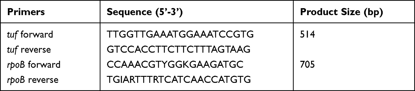 |
Table 1 Sequences of Oligonucleotides Used in This Study |
Besides, the phylogenetic trees were generated based on tuf gene. The length of the compared sequences was 506 bp. Following alignment with Clustal W, the evolutionary analyses were conducted in MEGA version 7.0 to create radial trees using the Maximum Likelihood method based on the Tamura-Nei model.17
Identification by MALDI-TOF MS and Result Interpretation
All the isolates were identified by VITEK MS (bioMérieux, Marcy-l’Etoile, France) and Bruker Biotyper (Bruker, Bremen Germany) following the manufacturers’ instructions. For the two MALDI-TOF MS systems, bacterial samples were prepared by direct deposit method and the bacterial identification of all isolates was performed in IVD settings to ensure the excitation energies under the optimum condition as follows.
For VITEK MS, colonies were picked from blood agar plates with a 1 μL plastic loop and spotted onto the disposable target plates (VITEK MS, bioMérieux). One microliter of the matrix solution (α-cyano-4-hydroxycinnamic acid, VITEK MS CHCA) was added onto smears. After air drying at room temperature, the target plates were then loaded into the mass spectrometer and the main spectrum profiles of isolates were obtained with identification standard settings (linear positive mode, 2000–20,000 Da) by VITEK MS IVD database version 3.2 containing 15,556 microbial strains comprising 1316 species. The calibration and quality control of every group of 16 samples was performed using Escherichia coli ATCC 8739 and S. pneumoniae ATCC49619 was also used as control strains. A confidence value, the percent probability, was calculated by the software to reflect the concordance of the observed spectrum with the VITEK MS database. A high-confidence result is obtained with a confidence value above 99%, a low-confidence result is obtained with a confidence value between 60% and 99% and a no identification is obtained when the confidence value is below 60% by VITEK MS.
For Bruker Biotyper, we performed the identification by using the manufacturer’s recommended direct transfer followed by the addition of formic acid. A single isolated colony was deposited on a polished steel MSP 96 target, and then 1 μL of a 70% formic acid solution was added to the bacterial spot. After being dried, the target was overlaid with 1 μL of a saturated α-cyano-4-hydroxycinnamic acid (HCCA) matrix solution (Bruker Daltonics). The target plate was analyzed by using a Microflex LT mass spectrometer (Bruker Daltonics) and the protein profile of each spot with m/z values of 1960 to 20,137 was analyzed by the IVD version of the Bruker Biotyper software package (version 3.0). The Bruker Biotyper database included 5989 entries of all microorganisms comprising 2371 species. Calibration was done by following the manufacturer’s instructions and using the manufacturer’s recommended bacterial test standard (Bruker Daltonics) and a Bacterial Test Standard (BTS) was used to calibrate the instrument before each acquisition session and S. pneumoniae ATCC49619 was used as control strains during bacterial identification. Data were interpreted by application of the manufacturer’s standard criteria.18 In short, species identification is obtained when scores are above 2.0 and genus identification is obtained when the score is between 1.7 and 2.0. If scores are lower than 1.7, no identification was assigned.
Statistical Analysis
Statistical analyses were conducted by using SPSS, version 25.0 (SPSS Inc, Chicago, IL, USA). A value of P ≤ 0.05 was considered statistically significant.
Results
Identification by Molecular Sequencing
Based on tuf or rpoB gene sequencing results, all enrolled 277 isolates were correctly identified at the species level. Eighty-four of the 277 strains were identified as S. pneumoniae and 193 strains were other VGS from the five different groups including S. anginosus group (n=91, 47.2%), S. mitis group (n=80, 41.5%), S. bovis group (n=11, 5.7%), S. salivarius group (n=10, 5.2%), and S. mutans group (n=1, 0.5%) (Table 2). Figure 1 shows the phylogenetic trees generated by tuf gene. Besides, all S. pneumoniae isolates were further confirmed with optochin sensitivity and bile solubility tests.
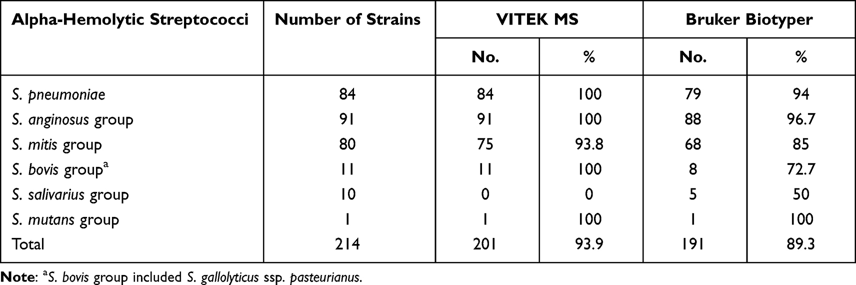 |
Table 2 Identified Accuracy Rate of Streptococci by Two MALDI-TOF Systems |
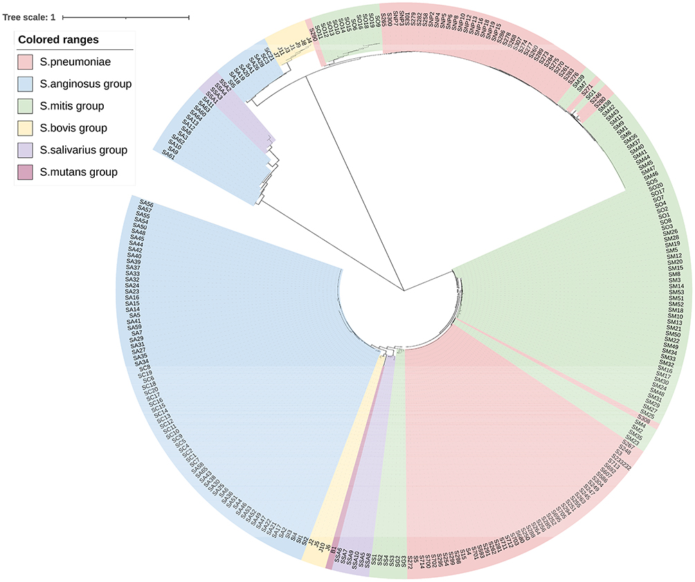 |
Figure 1 The molecular phylogenetic tree of viridans group streptococci isolates based on tuf gene. The evolutionary history was inferred by using the Neighbor-Joining method. The optimal tree with the sum of branch length = 5.29069834 is shown. The tree is drawn to scale, with branch lengths in the same units as those of the evolutionary distances used to infer the phylogenetic tree. The evolutionary distances were computed using the Maximum Composite Likelihood method2 and are in the units of the number of base substitutions per site. Evolutionary analyses were conducted in MEGA7. The length of the compared sequences was 506 bp. |
Identification by VITEK MS
Among 277 strains enrolled in this study, 262 strains (94.6%, 262/277) were correctly identified at the species level by VITEK MS with a probability score range of 99–100% (Table 2). All 84 clinical strains of S. pneumoniae were identified correctly. For the other VGS isolates, the VITEK MS gave high-confidence (99–100%) identification at the species level for 100% of S. anginosus group, S. bovis group and S. mutans group. However, for VITEK MS system, S. mitis and S. oralis isolates were identified as a slashline “S. mitis / S. oralis” with a high-confidence value. A similar phenomenon also occurred in S. salivarius, which identified as “S. salivarius ssp. salivarius / S. salivarius ssp. thermophilus / S. vestibularis” but with low-confidence. One S. mitis strain and 10 S. salivarius strains were correctly identified with confidence values between 60% and 99%. Two S. oralis strains were not identified by VITEK MS system (Table 3). Furthermore, one S. mitis and one S. oralis were misidentified as S. parasanguinis and S. pneumoniae, respectively (Table 4).
 |
Table 3 Identification Results of MALDI-TOF Systems for Alpha-Hemolytic Streptococci |
 |
Table 4 Error Identification of Streptococci by Two MALDI-TOF MS Systems |
Identification by Bruker Biotyper
The Bruker Biotyper correctly identified to the species level 89.9% (249/277) of the tested streptococci. An additional 17 strains were identified with scores between 1.7 and 2.0, so in total, 266 strains (96.0%) were identified with scores ≥1.70. The identified accuracy rates to the species level were 96.7% S. anginosus group, 94.0% for S. pneumoniae, 85.0% for S. mitis group, 72.7% for S. bovis group, and 50.0% for S. salivarius group (Table 2). Among S. pneumoniae, two strains were correctly tested with a score range of 1.7–2.0 and 1 strain was misidentified to Klebsiella aerogenes with low-confidence. Two strains were unsuccessfully identified with no mass peak and further confirmed as S. pneumoniae by optochin sensitivity and bile solubility testing. Two S. mitis strains were misidentified to S. pneumoniae with optochin-resistance and bile insolubility and the other three were incorrectly identified to S. anginosus or S. oralis. Bruker Biotyper misidentified one S. gallolyticus and one S. salivarius as E. coli and S. pneumoniae, respectively, but with high scores >2.0 (Table 4). Moreover, there were no identification results for two isolates of S. pneumoniae and one isolate of S. salivarius by Bruker Biotyper (Table 3).
Comparison Analysis of Two MALDI-TOF Systems
Among the two MALDI-TOF systems tested in this study, VITEK MS showed superiority in identifying all alpha-hemolytic streptococci compared to Bruker Biotyper systems (P<0.05). Especially, VITEK MS showed better identification results than Bruker Biotyper for S. mitis group including S. pneumoniae and S. bovis group, but for other VGS isolates two MALDI-TOF MS systems showed comparable identification performance. The false-negative values of VITEK MS were 5.0% in identification of S. mitis group, while the false-negative values of Bruker Biotyper were 3.6%, 6.3%, and 9.1% in identification of S. pneumoniae, S. mitis group, and S. bovis group, respectively. Notably, the false-positive values of VITEK MS and Bruker Biotyper in the identification of S. pneumoniae were 1.2% and 3.6%, respectively. Furthermore, VITEK MS was able to identify S. gallolyticus to the subspecies level with high-confidence (S. gallolyticus ssp. pasteurianus), while the Bruker Biotyper only gave the species level for S. gallolyticus. However, VITEK MS was poorly differentiating the subspecies of S. salivarius from S. vestibularis, while the other Bruker Biotyper systems could correctly identify them. Among the misidentified organisms, data showed that the Bruker Biotyper was more likely to misidentify S. mitis as other VGS species, probably owing to the reusable of the biotyper target plate and incomplete cleaning of protein crystallization.
Discussion
The difficulty of proper treatment of infectious diseases is the accurate diagnosis of pathogenic bacteria. According to previous papers described, the clinical significance of S. pneumoniae compared to other VGS strains is obviously different and accurate differentiation of this species appeared particularly important. Meanwhile, more and more clinical laboratories are attempted to search for rapid, comprehensive, and accurate identification methods for VGS isolates and commercial MALDI-TOF MS systems are recommended for routine identification of the VGS isolates instead of common biochemical reactions. However, the commercial MALDI-TOF MS systems have different diagnostic performance for the identification of streptococci.19 In this study, we evaluated the performance of VITEK MS and Bruker Biotyper in identification of clinical VGS isolates by using sequencing as the reference method; 94.6% and 89.9% of the VGS isolates were correctly identified at the species level, respectively. The reason why two MALDI-TOF MS systems have different performance is that identification is highly dependent on the quality of the databases and they have their own database involving bacteria and fungi. Previous studies have reported that several bacteria were correctly identified by using RUO database (Research Use Only), whereas in contrast using the IVD database, none of them could be identified.20,21 At present, the identification of VGS isolates is often problematic including misidentification or low-confidence identification. Therefore, it is crucial to choose an efficient database of MALDI-TOF MS that meets the needs of one’s own laboratory and update the database timely, which will significantly improve the identification performance.
Concerning the identification of S. pneumoniae, VITEK MS can accurately identify pneumococcal isolates with 100% accuracy, which was higher than Bruker Biotyper. Similar previous studies have reported that the sensitivity of VITEK MS system for the identification of S. pneumoniae is >99%.22,23 Furthermore, 94% of the S. pneumoniae isolates were correctly identified at the species level using the Bruker Biotyper database with 5989 entries, which is slightly lower than other reports. Other papers have reported 100% correct species assignment to S. pneumoniae using a more updated library and their identification was confirmed by peak analysis.11,24 It is worth noting that sometimes Bruker Biotyper may erroneously identify S. pneumoniae, which can have important consequences in a clinical setting. Even so, we still conclude that VITEK MS and Bruker Biotyper systems can correctly differentiate S. pneumoniae isolates from other VGS isolates. It is probably linked to their respective algorithms, which may efficiently detect the specific mass/charge peak profiles of S. pneumoniae (2937.5 and 5877 m/z) compared to other closely related species, as recently highlighted by Werno et al.25 Noteworthy, since S. pneumoniae isolates frequently originated from respiratory samples and other VGS groups can have similar colony morphologies, we recommend that optochin sensitivity or bile solubility tests and gene-based analysis should be performed for further confirmation if unsuccessfully identified.
For other non-pneumococcal VGS isolates, two MALDI-TOF MS systems have different accuracy and the misidentified rate for S. mitis group, especially for S. mitis and S. oralis, is higher than for other VGS groups, in particular for Bruker Biotyper. A study also pointed out the similar phenomenon with misidentifications of the S. mitis group frequently occurring, in particular for S. pneumoniae, S. mitis, and S. oralis.26 The main reason is that according to the analysis of 16S rRNA and housekeeping gene sequences, S. pneumoniae, S. mitis and S. oralis are highly related and S. mitis or S. oralis is often misidentified as S. pneumoniae by MALDI-TOF MS. Furthermore, the clinical significance of S. mitis group can vary in different body sites. In some cases, it is necessary to accurately differentiate all members of the group, because of their presence in blood cultures of patients with endocarditis which often leads to treatment strategies that differ from S. pneumoniae given their high penicillin resistance rates.27 However, it is worth noting that VITEK MS can accurately identify the S. mitis group and it only displays the combination result “S. mitis / S. oralis” in the current database. Therefore, considering to the same treatment for S. mitis/S. oralis, clinical microbiologists can make no distinction among them but at least differentiate S. pneumoniae from other non-pneumococcal strains in S. mitis group.
In addition, the two MALDI-TOF MS systems always provide satisfactory identification rates for S. anginosus and S. mutans groups but not for S. bovis and S. salivarius groups. S. gallolyticus is the more common species of S. bovis group. S. gallolyticus has three subspecies, subsp. pasteurianus, subsp. gallolyticus, and subsp. macedonicus. Previous studies have shown that S. gallolyticus subsp. gallolyticus is associated with clinical infectious endocarditis, gastrointestinal disorders, colon cancer, and chronic liver disease, and S. gallolyticus subsp. pasteurianus is related to meningitis.9 S. gallolyticus subsp. macedonicus is not pathogenic and have been systematically isolated from milk and fermented dairy products worldwide.28 Therefore, it is important to differentiate the three subspecies of S. gallolyticus. In this study, VITEK MS system correctly identified S. gallolyticus to the subspecies level while Bruker Biotyper could not differentiate. For the S. salivarius group, 8 S. salivarius strains were used for evaluation and the two MALDI-TOF MS systems showed different identification performance. Species-level identification was impossible with the VITEK MS for S. salivarius group and gave the combined result of “S. salivarius ssp. salivarius / S. salivarius ssp. thermophilus / S. vestibularis”, because Streptococcus vestibularis was not included in Vitek MS IVD system. However, Bruker Biotyper could identify S. salivarius with approximately 50% accuracy.
This study still has some limitations: 1) Even though the VGS isolates were from two hospitals in Shanghai, possible selection bias in the group/species distribution of isolates from the same geographic location may exist. When the isolates were poorly identified by MALDI-TOF, it may reflects the particular clone which could be not differentiated by MALDI-TOF; 2) Owing to the low isolation rate of S. mutans group, no more isolates were included in this study.
Conclusions
In summary, this study demonstrated that VITEK MS and Bruker Biotyper systems allowed discrimination for most VGS isolates, even though these two systems have different bacterial database, which would influence the identification performance. However, Bruker Biotyper could produce more misidentifications and VITEK MS system. Therefore, it is crucial to be familiar with the performance of MALDI-TOF MS systems before these systems can start being used in clinical microbiology and should update the databases timely, which can help us to identify VGS strains to species level, especially when strains are isolated from sterile body fluids.
Acknowledgments
We thank all members of the clinical laboratory of Shanghai Children’s Hospital for their cooperation and technical help.
Funding
This work was supported by the foundation of Shanghai Municipal Key Clinical Specialty (shslczdzk06902), Shanghai Municipal Commission of Health and Family Planning (201940253), and Shanghai “Rising Stars of Medical Talents” Youth Development Program (Youth Medical Talents –Clinical Laboratory Practitioner Program).
Disclosure
All authors declare no conflicts of interest in this work.
References
1. Jorgensen JH, Pfaller MA. Manual of Clinical Microbiology. Vol. 1.
2. Zhao W, Pan F, Wang B, et al. Epidemiology characteristics of Streptococcus pneumoniae from children with pneumonia in Shanghai: a retrospective study. Front Cell Infect Microbiol. 2019;9:258. doi:10.3389/fcimb.2019.00258
3. Rachina S, Zakharenkov I, Dekhnich N, et al. Aetiology of severe community-acquired pneumonia and antimicrobial susceptibility of Streptococcus pneumoniae in adults in Russia. J Antimicrob Chemother. 2021;76(5):1368–1370. doi:10.1093/jac/dkab014
4. Kawamura Y, Hou XG, Sultana F, Miura H, Ezaki T. Determination of 16S rRNA sequences of Streptococcus mitis and Streptococcus gordonii and phylogenetic relationships among members of the genus Streptococcus. Int J Syst Bacteriol. 1995;45(2):406–408. doi:10.1099/00207713-45-2-406
5. Angeletti S, Dicuonzo G, Avola A, et al. Viridans Group Streptococci clinical isolates: MALDI-TOF mass spectrometry versus gene sequence-based identification. PLoS One. 2015;10(3):e0120502. doi:10.1371/journal.pone.0120502
6. Raddaoui A, Ben Tanfous F, Achour W, Baaboura R, Ben Hassen A. Description of a novel mutation in the atpC gene in optochin-resistant Streptococcus pneumoniae strains isolates from Tunisia. Int J Antimicrob Agents. 2018;51(5):803–805. doi:10.1016/j.ijantimicag.2017.12.029
7. Ercibengoa M. Assessment of the optochin susceptibility test to differentiate Streptococcus pneumoniae from other viridans Group Streptococci. Clin Lab. 2021;67(3). doi:10.7754/Clin.Lab.2020.200438
8. Al Majid F, Aldrees A, Barry M, Binkhamis K, Allam A, Almohaya A. Streptococcus anginosus group infections: management and outcome at a tertiary care hospital. J Infect Public Health. 2020;13(11):1749–1754. doi:10.1016/j.jiph.2020.07.017
9. Dekker JP, Lau AF, Kraft CS. An update on the Streptococcus bovis group: classification, identification, and disease associations. J Clin Microbiol. 2016;54(7):1694–1699. doi:10.1128/JCM.02977-15
10. Agnes A, Biondi A, Belia F, et al. Association between colorectal cancer and Streptococcus gallolyticus subsp. pasteuranus (former S. bovis) endocarditis: clinical relevance and cues for microbiota science. Case report and review of the literature. Eur Rev Med Pharmacol Sci. 2021;25(1):480–486.
11. Marin M, Cercenado E, Sanchez-Carrillo C, et al. Accurate differentiation of Streptococcus pneumoniae from other species within the Streptococcus mitis group by peak analysis using MALDI-TOF MS. Front Microbiol. 2017;8:698. doi:10.3389/fmicb.2017.00698
12. Yahiaoui RY, Goessens WH, Stobberingh EE, Verbon A. Differentiation between Streptococcus pneumoniae and other viridans group streptococci by matrix-assisted laser desorption/ionization time of flight mass spectrometry. Clin Microbiol Infect. 2020;26(8):1088e1081–1088 e1085. doi:10.1016/j.cmi.2019.11.024
13. Chen JH, She KK, Wong OY, et al. Use of MALDI biotyper plus ClinProTools mass spectra analysis for correct identification of Streptococcus pneumoniae and Streptococcus mitis/oralis. J Clin Pathol. 2015;68(8):652–656. doi:10.1136/jclinpath-2014-202818
14. Zhou M, Yang Q, Kudinha T, et al. Using Matrix-Assisted Laser Desorption Ionization-Time of Flight (MALDI-TOF) complemented with selected 16S rRNA and gyrB genes sequencing to practically identify clinical important Viridans Group Streptococci (VGS). Front Microbiol. 2016;7:1328. doi:10.3389/fmicb.2016.01328
15. Clinical and Laboratory Standards Institute. Interpretative Criteria for Identification of Bacteria and Fungi by DNA Target Sequencing; Approved Guideline. Wayne, PA: CLSI document MM18-A Clinical and Laboratory Standards Institute; 2008.
16. Wessels E, Schelfaut JJ, Bernards AT, Claas EC. Evaluation of several biochemical and molecular techniques for identification of Streptococcus pneumoniae and Streptococcus pseudopneumoniae and their detection in respiratory samples. J Clin Microbiol. 2012;50(4):1171–1177. doi:10.1128/JCM.06609-11
17. Kumar S, Stecher G, Tamura K. MEGA7: molecular evolutionary genetics analysis version 7.0 for bigger datasets. Mol Biol Evol. 2016;33(7):1870–1874. doi:10.1093/molbev/msw054
18. Schulthess B, Bloemberg GV, Zbinden A, et al. Evaluation of the bruker MALDI biotyper for identification of fastidious gram-negative rods. J Clin Microbiol. 2016;54(3):543–548. doi:10.1128/JCM.03107-15
19. Fan WT, Qin TT, Bi RR, Kang HQ, Ma P, Gu B. Performance of the matrix-assisted laser desorption ionization time-of-flight mass spectrometry system for rapid identification of streptococci: a review. Eur J Clin Microbiol Infect Dis. 2017;36(6):1005–1012. doi:10.1007/s10096-016-2879-2
20. Wattal C, Oberoi JK, Goel N, Raveendran R, Khanna S. Matrix-assisted laser desorption ionization time of flight mass spectrometry (MALDI-TOF MS) for rapid identification of micro-organisms in the routine clinical microbiology laboratory. Eur J Clin Microbiol Infect Dis. 2017;36(5):807–812. doi:10.1007/s10096-016-2864-9
21. Wilen CB, McMullen AR, Burnham CA. Comparison of sample preparation methods, instrumentation platforms, and contemporary commercial databases for identification of clinically relevant mycobacteria by matrix-assisted laser desorption ionization-time of flight mass spectrometry. J Clin Microbiol. 2015;53(7):2308–2315. doi:10.1128/JCM.00567-15
22. Branda JA, Markham RP, Garner CD, Rychert JA, Ferraro MJ. Performance of the vitek MS v2.0 system in distinguishing Streptococcus pneumoniae from nonpneumococcal species of the Streptococcus mitis group. J Clin Microbiol. 2013;51(9):3079–3082. doi:10.1128/JCM.00824-13
23. Dubois D, Segonds C, Prere MF, Marty N, Oswald E. Identification of clinical Streptococcus pneumoniae isolates among other alpha and nonhemolytic streptococci by use of the Vitek MS matrix-assisted laser desorption ionization-time of flight mass spectrometry system. J Clin Microbiol. 2013;51(6):1861–1867. doi:10.1128/JCM.03069-12
24. Harju I, Lange C, Kostrzewa M, Maier T, Rantakokko-Jalava K, Haanpera M. Improved differentiation of Streptococcus pneumoniae and other S. mitis group Streptococci by MALDI biotyper using an improved MALDI biotyper database content and a novel result interpretation algorithm. J Clin Microbiol. 2017;55(3):914–922. doi:10.1128/JCM.01990-16
25. Werno AM, Christner M, Anderson TP, Murdoch DR. Differentiation of Streptococcus pneumoniae from nonpneumococcal streptococci of the Streptococcus mitis group by matrix-assisted laser desorption ionization-time of flight mass spectrometry. J Clin Microbiol. 2012;50(9):2863–2867. doi:10.1128/JCM.00508-12
26. Stevenson LG, Drake SK, Murray PR. Rapid identification of bacteria in positive blood culture broths by matrix-assisted laser desorption ionization-time of flight mass spectrometry. J Clin Microbiol. 2010;48(2):444–447. doi:10.1128/JCM.01541-09
27. Carvalho MG, Steigerwalt AG, Thompson T, Jackson D, Facklam RR. Confirmation of nontypeable Streptococcus pneumoniae-like organisms isolated from outbreaks of epidemic conjunctivitis as Streptococcus pneumoniae. J Clin Microbiol. 2003;41(9):4415–4417. doi:10.1128/JCM.41.9.4415-4417.2003
28. Papadimitriou K. Novel insight into the pathogenicity of Streptococcus gallolyticus subsp. gallolyticus belonging to the Streptococcus bovis/Streptococcus equinus complex. Virulence. 2018;9(1):662–665. doi:10.1080/21505594.2018.1432932
 © 2023 The Author(s). This work is published and licensed by Dove Medical Press Limited. The
full terms of this license are available at https://www.dovepress.com/terms
and incorporate the Creative Commons Attribution
- Non Commercial (unported, 3.0) License.
By accessing the work you hereby accept the Terms. Non-commercial uses of the work are permitted
without any further permission from Dove Medical Press Limited, provided the work is properly
attributed. For permission for commercial use of this work, please see paragraphs 4.2 and 5 of our Terms.
© 2023 The Author(s). This work is published and licensed by Dove Medical Press Limited. The
full terms of this license are available at https://www.dovepress.com/terms
and incorporate the Creative Commons Attribution
- Non Commercial (unported, 3.0) License.
By accessing the work you hereby accept the Terms. Non-commercial uses of the work are permitted
without any further permission from Dove Medical Press Limited, provided the work is properly
attributed. For permission for commercial use of this work, please see paragraphs 4.2 and 5 of our Terms.