
Back to Journals » Journal of Pain Research » Volume 19
Parasternal Intercostal Plane Blocks for Enhanced Recovery After Cardiac Surgery: A Systematic Review of Technical Refinements and Evidentiary Support
Authors He YN  , Lin PR, Wang S
Received 4 March 2026
Accepted for publication 4 July 2026
Published 23 July 2026 Volume 2026:19 600493
DOI https://doi.org/10.2147/JPR.S600493
Checked for plagiarism Yes
Review by Single anonymous peer review
Peer reviewer comments 2
Editor who approved publication: Dr Karina Gritsenko
Yi-Na He,1,2 Pei-Rong Lin,1 Sheng Wang1,3
1Department of Anesthesiology, Beijing Anzhen Hospital, Capital Medical University, Beijing, People’s Republic of China; 2Department of Anesthesiology, Beijing Anzhen Nanchong Hospital of Capital Medical University & Nanchong Central Hospital, Nanchong, Sichuan, People’s Republic of China; 3Department of Anesthesiology, Linzhi People’s Hospital, Linzhi, Tibet, People’s Republic of China
Correspondence: Sheng Wang, Email [email protected]
Background: Parasternal intercostal plane (PIP) blocks provide opioid-sparing analgesia within cardiac Enhanced Recovery After Surgery (ERAS) pathways, but non-standardized nomenclature and ambiguous anatomical targets limit their adoption.
Methods: A systematic search of PubMed, Cochrane, and Embase databases was conducted. Inclusion criteria encompassed randomized controlled trials and observational studies reporting on PIP blocks in adult cardiac surgery to synthesize evidence on their efficacy, technical refinements, and safety.
Results: This review integrated 20 clinical studies (2019– 2025). Adopting the El-Boghdadly terminology resolves descriptive inconsistencies by distinguishing superficial (SPIP) and deep (DPIP) techniques. SPIP targets the pectoralis major-intercostal interface (T2–T6), while DPIP accesses the intercostal-transversus thoracis plane (T1–T6). PIP blocks reduced 12-hour postoperative pain (average 1.21-point reduction) and opioid use (average 30.34 milligram morphine equivalents (MME) reduction), though analgesic benefits diminished after 24 hours. SPIP provides a safer profile with minimal internal mammary artery (IMA) injury risk but limited cephalad spread. DPIP achieves broader dermatomal coverage but positions the needle 3– 5 mm from the IMA; thus, it should be applied with caution or replaced by SPIP in post-CABG patients. Recovery outcomes demonstrated earlier extubation and shorter intensive care unit stays. However, bupivacaine concentrations approached neurotoxic thresholds ≥ 2.0 μg/mL in 7.1% of DPIP cases.
Conclusion: PIP blocks provide effective opioid-sparing analgesia, requiring meticulous anatomical execution. SPIP is favored for safety, whereas DPIP demands real-time ultrasound guidance and caution in patients with IMA grafts. Future research should optimize dermatomal coverage, refine individualized dosing, and evaluate long-term outcomes.
Keywords: parasternal intercostal plane block, enhanced recovery after surgery, ERAS, cardiac surgery, standardization of nomenclature, pain management, opioid-sparing analgesia
Introduction
Effective perioperative pain management remains a central challenge in Enhanced Recovery After Surgery (ERAS) pathways for cardiac surgery. Median sternotomy and internal mammary artery (IMA) harvest produce substantial somatic and neuropathic nociception. To establish the clinical burden, standard post-sternotomy pain scores often reach 6 to 8 out of 10 on the Numerical Rating Scale (NRS) in the early postoperative period, underscoring the critical need for ERAS pathways and regional blocks. Furthermore, in association with nociception, asymmetric sternal retraction during IMA harvest frequently causes stretching and nerve compression of the brachial plexus, leading to neuropathic pain in the upper extremities.1 This specifically highlights why a simple plane block—which targets only the anterior cutaneous branches of the thoracic intercostal nerves—may not be a silver bullet for all types of post-cardiac surgery pain. These pain-related burdens may hinder early mobilization, delay respiratory recovery, and compromise overall rehabilitation outcomes.
Parasternal intercostal plane (PIP) blocks—comprising the superficial parasternal intercostal plane (SPIP) and deep parasternal intercostal plane (DPIP) approaches—provide targeted analgesia by anesthetizing the anterior cutaneous branches of the thoracic intercostal nerves. Despite expanding interest, the clinical integration of PIP blocks has been slowed by persistent issues: non-standardized nomenclature, ambiguous anatomical depth boundaries that reduce study reproducibility, and incomplete dosing safety evidence for local anesthetic systemic toxicity in cardiac surgery populations. Terms such as “Parasternal block”, “Pecto-intercostal fascial block”, and “Parasternal intercostal block” have been used interchangeably in the literature, often without clear alignment to anatomical planes. Similarly, definitions of “superficial” versus “deep” injection sites vary among studies, obscuring interpretation and hindering reproducibility, and robust data on safe dosing thresholds in hemodynamically vulnerable cardiac surgery patients remains scarce.
These ambiguities are particularly consequential in cardiac surgery, where unique anatomical considerations—sternal division, IMA dissection, and parasternal tissue disruption—influence block performance and safety. Although the DPIP approach offers wider predicted coverage of T1–T6 dermatomes, needle passage occurs within 3–5 millimeters of the IMA, raising concerns regarding potential vascular injury. The SPIP approach enhances procedural safety by avoiding the IMA but often provides insufficient cephalad spread to reliably cover upper thoracic dermatomes (T1–2), where sternotomy-related pain is most pronounced. This insufficient cephalad spread is fundamentally due to compartmental boundaries created by the superficial cervical fascia and clavicular attachments, which act as physical barriers that restrict the upward migration of the injectate.
To clarify these issues and support evidence-based application of PIP blocks in cardiac ERAS programs, this review synthesizes anatomic, technical, and clinical data through a systematic search of PubMed, Cochrane, and Embase databases. We integrated 20 clinical investigations between 2019 and 2025 (Table 1). These studies were chosen based on specific inclusion criteria encompassing randomized controlled trials and observational studies reporting on block efficacy, safety, and pharmacokinetics in adult cardiac surgery. Exclusion criteria were: 1) non-English language publications; 2) case reports, case series with fewer than 10 patients, review articles, letters, and editorials; 3) studies focusing exclusively on pediatric or non-cardiac surgical populations; 4) studies without reported pain or opioid consumption outcomes. We examine the evolution of PIP nomenclature, define the anatomical planes relevant to SPIP and DPIP, describe technical refinements aimed at improving consistency and safety, especially regarding IMA safety, and evaluate the analgesic efficacy and limitations of current approaches. Finally, we outline key research priorities needed to optimize thoracic wall analgesia for contemporary cardiac surgery. The study selection process is summarized in accordance with the PRISMA 2020 statement (Figure 1).
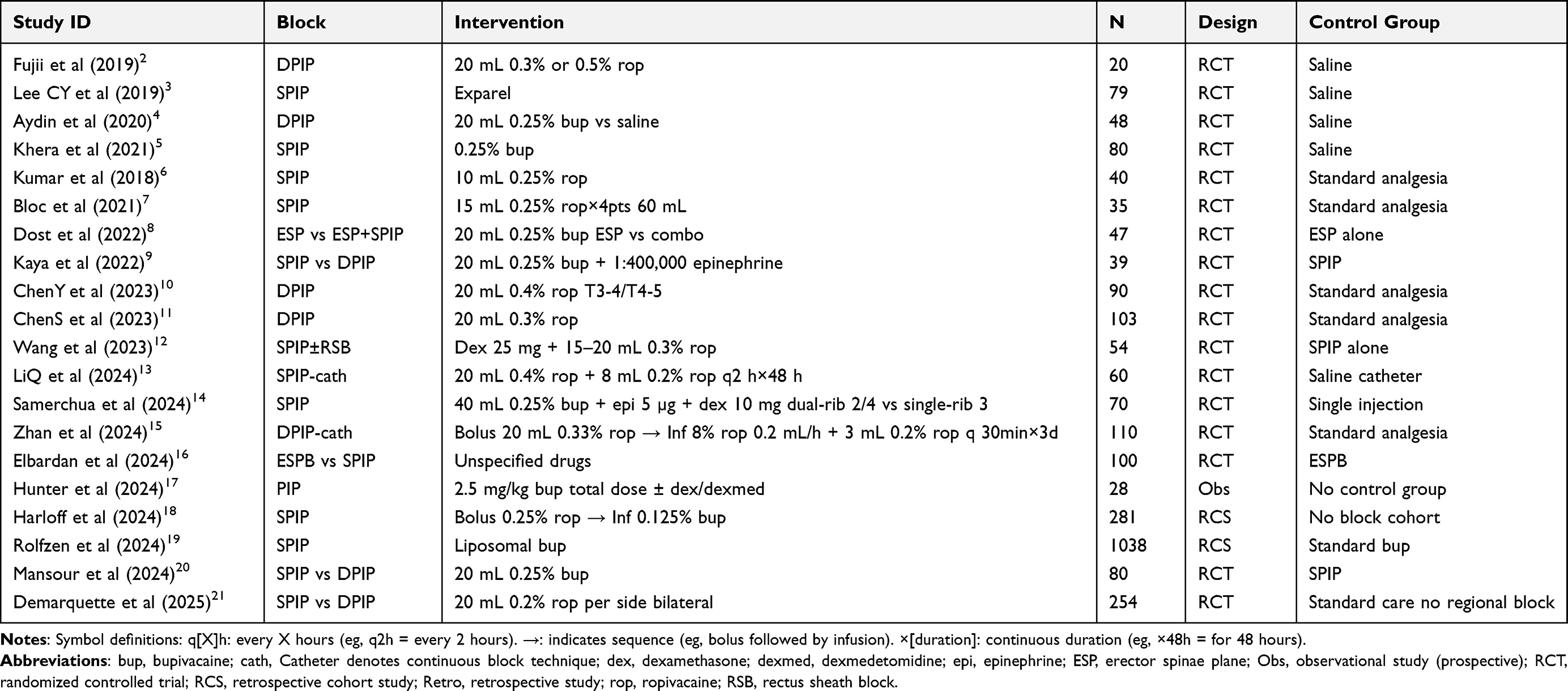 |
Table 1 Characteristics of Included Studies |
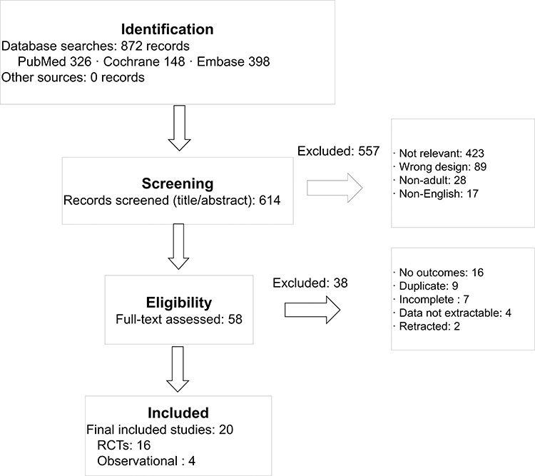 |
Figure 1 PRISMA 2020 flow diagram illustrating the study selection process. |
Standardization of Nomenclature and Anatomical Precision
Variability in the nomenclature used to describe parasternal regional techniques has posed a persistent barrier to their broader clinical adoption and to the synthesis of high-quality evidence. Early literature frequently employed overlapping terms—such as “Parasternal intercostal block”, “Parasternal block”, or “Pecto-Intercostal Fascial Block”—often without clear delineation of the targeted anatomical plane or proper fascial considerations at the expense of muscular layer interrelationships.1,22 This inconsistency complicates study interpretation, contributes to heterogeneity in systematic reviews, and obscures the relationship between injection site, spread of local anesthetic, and clinical effect.
These challenges were highlighted by Haskins and Memtsoudis, who described the proliferation of terms in this anatomical region as an “alphabet soup”.22–24 Much of the confusion stems from three recurrent issues: imprecise definition of intercostal levels, inconsistent identification of fascial planes, and lack of a scalable naming system capable of incorporating future refinements. To address these limitations, the 2021 ASRA-ESRA Delphi consensus led by El-Boghdadly et al proposed a unified terminology framework grounded in the depth of injection.25 Recent methodological reviews emphasize that such Delphi consensus studies are vital in health sciences for standardizing clinical practices and influencing structural healthcare frameworks.14,26 Under this system “PIP block” serves as the overarching term with two clearly defined subtypes (Figure 2):
- SPIP block: Injection in the precise plane between the deep fascia of the pectoralis major muscle and the superficial aspect of the external or internal intercostal muscles.
- DPIP block (formerly known as Transversus Thoracis Muscle Plane Block, TTPB): Injection within the exact deep plane bordered by the internal intercostal muscles and the transversus thoracis muscle (TTM). Including this parenthetical note bridges the gap for clinicians trained before 2021, aligning the historical TTPB nomenclature with the modern DPIP classification.
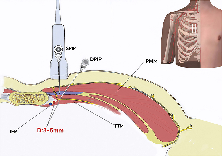 |
Figure 2 Sonographic Anatomy for Parasternal Intercostal Plane Block. Transducer placement: Parasternal 2 cm alignment with transverse imaging at the 3rd–4th intercostal space (ICS). 2 cm lateral to the sternal border. The white-boxed area in the top-right panel indicates the standard transducer placement position on the chest wall. Tissue stratification (superficial-to-deep): PMM, IICM, TTM. Target fascial planes for block: SPIP: Local anesthetic injection (blue-shaded area) within the PMM–IICM superficial plane, targeting terminal anterior cutaneous nerve branches. DPIP: Needle tip (white trajectory line) advanced to the IICM–TTM deep plane, anesthetizing primary anterior cutaneous nerve trunks. Top-right panel: Horizontal cross-section of the chest wall. Safety margin (D): A 3–5 mm clearance between needle path and posterior border of internal mammary artery/vein (IMA/V) is mandated. Abbreviations: PMM, Pectoralis major muscle; IICM, Internal intercostal muscle; TTM, Transversus thoracis muscle; SPIP, Superficial Parasternal Intercostal Plane Block; DPIP, Deep Parasternal Intercostal Plane Block; D, Distance. |
Early PIP block studies exhibited significant terminology ambiguity. This standardization of nomenclature framework offers three core benefits: enhancing clinical study reproducibility, improving literature retrieval precision, and reducing inter-study anatomical targeting variability. Notably, historical references to “Parasternal Block” may encompass both intercostal and pectoral muscle elements, requiring meticulous technique deconstruction and anatomical correlation analysis when integrating evidence.
Anatomical Mechanisms and Technical Refinement of PIP Blocks
Both the superficial and deep variants of the PIP blocks have demonstrated clear clinical benefits in thoracic and breast surgery, with their analgesic effects supported by many RCTs,27,28 yet DPIP blocks remain under investigation in cardiac surgery. This predominantly stems from the specificity of postoperative cardiac pain, which is constituted by four distinct and independent contributing factors: bilateral intercostal nerve damage from sternum splitting, parietal pleural irritation from chest drains, a systemic inflammatory cascade from cardiopulmonary bypass, and visceral irritation from the pericardium. These pain sources differ significantly from those encountered in unilateral video-assisted thoracoscopic surgery (VATS) or partial mastectomies. Given these unique anatomical and physiological features, special approaches are needed for using the thoracic wall blocks in heart surgery.
Anatomical Foundations of PIP Blocks
Establishing the anatomical foundation for the thoracic wall nerve block necessitates prioritized consideration of three critical factors: neuroanatomical distribution patterns, fascial compartment targeting accuracy, and procedural-related neural compromise risks. The anteromedial thoracic wall, extending from the midclavicular line to the sternal border, comprises layered structures including skin, subcutaneous fat, pectoralis major, the intercostal muscles, costal cartilages, ribs, and the TTM, all innervated by branches of the T2–T6 intercostal nerves.29 Within the intercostal compartment—bounded by the innermost intercostal muscle and endothoracic fascia—the intercostal nerves course anteromedially toward the sternum before dividing into anterior cutaneous branches. These branches traverse the intercostal musculature and pectoralis major to supply the skin and soft tissues adjacent to the sternum.
This neural arrangement supports two distinct parasternal fascial targets (Figure 2):
- SPIP: the plane between the deep fascia of the pectoralis major muscle and the superficial aspect of the external or internal intercostal muscles.30
- DPIP: the plane between the internal intercostal muscles and the TTM (with caution for adjacent internal mammary vessels), which carries inherent risks of iatrogenic injury to the internal mammary vessels (IMA/IMV) during needle advancement.31,32
Hilton’s law33 states that the nerve supplying a muscle that moves a joint also innervates the joint capsule and the overlying skin. Importantly, this principle applies exclusively to articular structures (eg, costosternal and costochondral joints) and does not extend to the bony sternum itself, whose innervation derives from separate periosteal nerve plexuses. When applied to the anterior chest wall, this principle explains why blockade of intercostal nerve branches at two distinct fascial planes—the pectoralis-intercostal interface (SPIP) and the intercostal-TTM plane (DPIP)—can attenuate nociceptive transmission from sternotomy. However, the clinical significance of this anatomical mechanism remains incompletely defined, particularly for deep sternal pain and substernal drain discomfort following cardiac surgery.
Neural Targeting and Diffusion Constraints of SPIP Blocks
Anatomical Targeting
The SPIP block targets the anterior cutaneous branches of the T2–T6 intercostal nerves. These branches typically ascend 1.5–2.0 cm lateral to the sternal border within a fascial layer located between the deep surface of the pectoralis major muscle and the superficial margin of the intercostal musculature. Ultrasound-guided deposition of local anesthetic, typically 20 mL of 0.25% bupivacaine, results in a longitudinal “fusiform” spread along this plane (Figure 3).
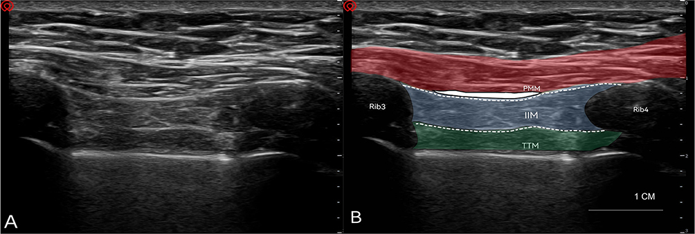 |
Figure 3 Parasagittal parasternal ultrasound image. (A) Pre-annotation; (B) Post-annotation. The in-plane view demonstrates the parasternal intercostal block plane between the third and fourth ribs. A 1-cm scale bar is included for depth calibration. PMM, Pectoralis Major Muscle (red overlay); IICM, Internal Intercostal Muscle (blue overlay); TTM, Transversus Thoracis Muscle (green overlay). White dashed lines indicate target fascial layers for blockade. SPIP, Superficial Parasternal Intercostal Plane Block (upper dashed line, injection site); DPIP, Deep Parasternal Intercostal Plane Block (lower dashed line, injection site). |
Diffusion Constraints
Cadaveric studies consistently demonstrate that SPIP injections provide limited longitudinal diffusion. A cadaver study reported that a single SPIP injection typically anesthetized two intercostal segments, while a dual-injection technique extended coverage to approximately three levels.34 Findings from Samerchua et al30 corroborated these results, reporting mean spreads of 2.7 segments (single injection), 3.8 segments (dual injection), and 5.3 segments (triple injection), with substantial interindividual variability. Douglas et al similarly observed segmental coverage ranging from one to four interspaces across specimens.33
Despite incremental benefits from dual or multisite injections, SPIP spread consistently displayed a cephalad limitation with minimal extension into the T1–T2 dermatomes. In nearly all SPIP specimens, injectate failed to reach the uppermost thoracic segments, aligning with clinical reports of residual incisional pain in the manubrial region. Douglas et al proposed two anatomical explanations: supraclavicular nerve dominance in manubrial innervation, which remains unaddressed by SPIP and is supported by anatomical evidence of supraclavicular nerve distribution35; or compartmental boundaries created by the superficial cervical fascia and clavicular structures, which limit cephalad dye migration.33,36 These anatomical boundaries may explain persistent T1–T2 pain patterns following sternotomy despite technically successful SPIP block performance consistent with observations from Fujii et al.2
Transverse spread across the sternum is also restricted. Sternal osteofascial attachments limit cross-midline injectate migration, with <15% contralateral spread observed in anatomical studies.2 Ipsilateral spread, however, is robust; approximately 75% of specimens displayed medial pectoral nerve uptake, demonstrating partial overlap with the distribution of interpectoral fascial plane blocks.37 In this respect, SPIP shares mechanistic similarities with pectoral plane blocks (PECS I/II), which have shown early extubation and reduced opioid consumption in sternotomy patients.6,38 This fascial continuity may partially explain the clinical analgesic benefit observed with SPIP despite its dermatomal limitations.
Core Risks of DPIP Blocks
Influence of TTM Anatomical Variants on Operative Techniques
Effective DPIP administration requires an appreciation of the substantial interindividual variability in TTM morphology. Postmortem studies demonstrate considerable variation in TTM costal insertions across ribs 2–6, with cephalad attachments most commonly at ribs 2–3 and caudal extensions reaching rib 6.38 The TTM may be composed of two to five distinct muscular slips.38 These anatomical variations influence the spread of local anesthetic and may limit injectate distribution to the 4th–6th intercostal spaces. The posterior border of the TTM may also serve as a partial barrier to diffusion, helping retain local anesthetic near the anterior intercostal nerve branches but concurrently restricting cephalad or caudad spread. Visualization challenges may arise following coronary artery bypass grafting (CABG); surgical disruption, edema, and altered tissue echogenicity from IMA harvest can impair identification of the TTM on ultrasound, favoring the use of SPIP in the postoperative period.39
IMA Injury: The Central Risk
The most significant hazard of DPIP blockade is inadvertent IMA injury. Originating from the subclavian artery, the IMA courses vertically approximately 1–1.5 cm lateral to the sternal border (Figure 4), lying posterior to the first six costal cartilages. Ultrasound-guided studies recommend maintaining a lateral insertion point roughly 2 cm from the sternal margin to reduce the risk of vascular puncture. Cadaveric data show that the needle trajectory for DPIP blocks commonly passes within 3–5 mm of the IMA (Figure 2).33 At the 2nd–3rd intercostal spaces, the IMA is separated from the pleura only by a thin layer of endothoracic fascia, further increasing procedural risk margins.30 Because the vessels lie deep to the costal cartilages, direct external compression is rarely effective in the event of IMA injury, necessitating prompt surgical or interventional radiology support in cases of hemorrhage.23
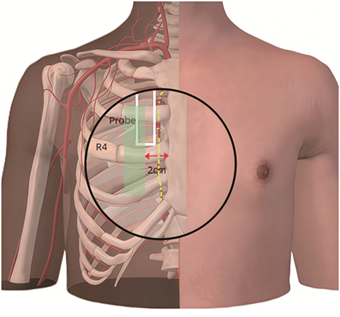 |
Figure 4 IMA Surface Projection and Needle Insertion Safety Zone. Ultrasound transducer transversely positioned at the 3rd–4th intercostal space, 2 cm lateral to the sternal margin (white-bordered area). The green rectangle demarcates the safety zone. Right IMA originates from the subclavian artery and descends vertically 1.5 cm lateral to the sternal border (yellow dashed line). Cutaneous needle insertion site should be maintained 2 cm lateral to the sternum to avoid IMA injury, demarcating the safety zone. |
Pneumothorax, Pleural Puncture, and Infection Risk
While IMA injury remains the primary vascular concern with deep injections, a comprehensive safety profile must also account for respiratory and infectious complications. The proximity of the DPIP plane to the highly vascularized and delicate parietal pleura introduces the inherent risk of pleural puncture and subsequent pneumothorax. Regarding pulmonary safety, Demarquette et al21 reported a significantly higher incidence of pneumothorax in the DPIP group (11.8%) compared with standard care (2.4%; p = 0.032), whereas the SPIP group showed no significant difference from controls, providing high-level evidence supporting the more favorable safety profile of SPIP. Furthermore, performing repetitive fascial plane penetrations in extreme proximity to a fresh median sternotomy wound carries an underlying risk of introducing superficial skin flora into the deep mediastinal space, potentially precipitating devastating complications such as deep sternal wound infections or mediastinitis. Consequently, strict adherence to aseptic techniques and continuous in-plane visualization of the needle tip are non-negotiable requirements.
Guidance for Block Selection in Special Populations
Clinicians must tailor their block selection based on patient-specific risk factors, balancing analgesic needs against procedural hazards:
Patients with Prior CABG (IMA grafts): Altered anatomy and postoperative scarring from previous sternotomies and IMA harvesting make deep-plane identification extremely hazardous. Even following left IMA harvest, residual ipsilateral perforating branches remain highly susceptible to mechanical injury, and the contralateral intact IMA is vulnerable during bilateral block performance. Although major RCTs have not reported accidental IMA punctures, this safety record is entirely predicated on meticulous Doppler mapping and continuous real-time ultrasound guidance. Therefore, DPIP is not strictly contraindicated in post-CABG patients, but it represents an advanced, technically demanding intervention. For routine postoperative analgesia in this cohort, SPIP is overwhelmingly prioritized to avoid deep-plane neurovascular risks.
Anticoagulated Patients: Cardiac surgical patients are frequently placed on stringent postoperative anticoagulation protocols. SPIP is favored over DPIP in these scenarios because the superficial plane allows for effective and immediate manual compression in the event of iatrogenic bleeding. Conversely, deep bleeding from a DPIP block behind the costal cartilages cannot be effectively compressed externally.
Obese Patients (BMI > 30): Excessive adipose tissue severely degrades ultrasound beam penetration, making deeper structures like the TTM and the pleura difficult to reliably identify. In obese cohorts, SPIP provides a safer, more superficial, and far more reliable sonographic target.
Clinical Decision-Making and Risk Mitigation
Clinicians must carefully weigh the analgesic benefits of DPIP blocks against their inherent vascular risks. When considering DPIP in the intraoperative setting, practitioners should ensure that appropriate resources are available for immediate management of bleeding. Performing DPIP pre-sternotomy may allow for rapid open-chest access and hemorrhage control during surgery; however, this approach may shorten the duration of postoperative analgesia. The decision to use preoperative versus postoperative DPIP should therefore be individualized.
For patients undergoing CABG procedures requiring IMA grafting, DPIP should be applied with caution or replaced by SPIP. The DPIP block is technically more demanding than the superficial PIP block, making real-time ultrasound guidance mandatory (eg, continuous in-plane visualization of the needle tip at all times, preoperative Doppler identification of the IMA trajectory) to achieve the safety profile reported in clinical trials. Even after left IMA harvest, residual ipsilateral perforating branches and musculophrenic branches remain at risk of injury, and the contralateral IMA remains vulnerable during bilateral parasternal needle advancement. For routine postoperative analgesia in CABG patients, SPIP is preferred because it avoids deep needle placement near the IMA and mitigates the risk of catastrophic vascular compromise (Table 2).
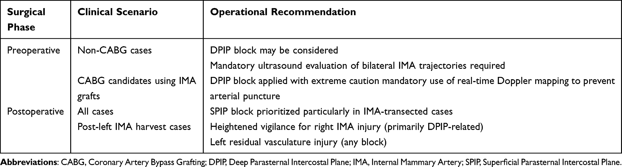 |
Table 2 Clinical Decision Advice Parasternal Intercostal Plane Blocks |
Comparative Analysis of Block Techniques
Selecting between SPIP and DPIP blocks requires careful consideration of anatomical constraints, procedural risk, and analgesic goals. As detailed in Table 3, although both techniques target the anterior cutaneous branches of the intercostal nerves, they differ significantly in injection site, spread characteristics, technical complexity, and safety profile.
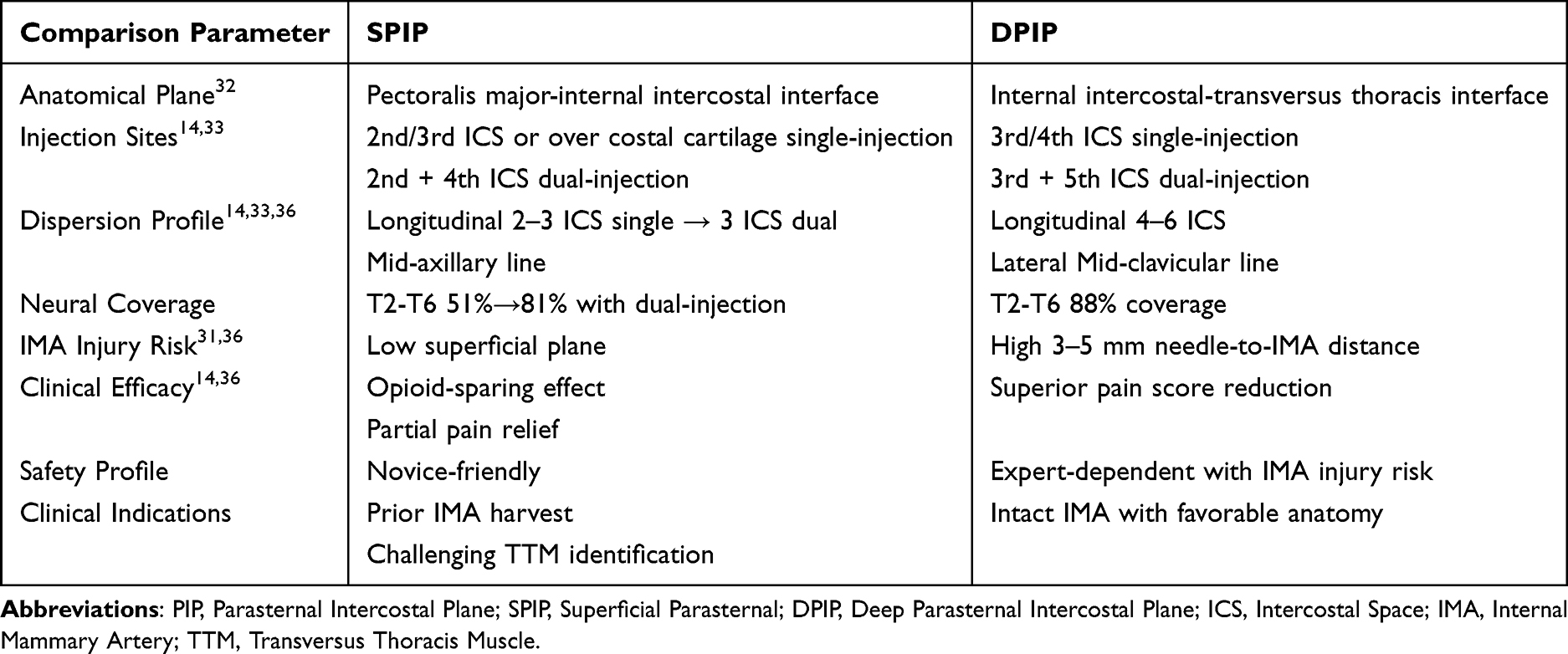 |
Table 3 Comparative Analysis of PIP Block Techniques |
Cadaveric studies have clarified the distinct injectate behavior of SPIP and DPIP blocks, showing that although both techniques can achieve multilevel distribution with appropriate injection strategies, DPIP generally produces more continuous and predictable longitudinal spread along the deep parasternal plane, while SPIP often requires higher volumes or multilevel injections to approximate similar coverage.30,33 These results are reinforced by anatomical evaluations demonstrating that DPIP reliably deposits injectate near the transversus thoracis muscle where the anterior cutaneous branches run, whereas SPIP predominantly fills the superficial fascial layers with less consistent direct neural staining.34,40 Douglas et al further confirmed that DPIP provides denser parasternal spread directly over the target nerves, while SPIP tends to extend more laterally with less focused deep-plane engagement.33
Safety considerations additionally differentiate these approaches. The internal mammary artery (IMA) lies close to the deep parasternal plane, creating a potential risk of vascular injury during DPIP — especially in patients with prior IMA graft harvest, which may distort deep tissue planes and complicate sonoanatomy.31 Because SPIP is performed in a more superficial and visually accessible layer, it may offer a comparatively wider safety margin, particularly when operator experience is limited or when mediastinal anatomy has been altered by previous surgical intervention13. This positional proximity of DPIP to both pleura and the IMA underscores the need for refined ultrasound skills and careful trajectory planning during deep-plane injections.31,40
Clinically, parasternal intercostal blocks as a group—whether superficial or deep—have been shown to improve post-sternotomy analgesia, reduce opioid consumption, and enhance respiratory recovery in cardiac surgery patients, although most published trials evaluate the techniques collectively rather than comparing SPIP and DPIP individually.41 When the anatomical and cadaveric evidence is integrated with current clinical findings, DPIP appears better suited for achieving dense midline sternotomy analgesia due to its more consistent engagement of anterior cutaneous branches33,40 whereas SPIP remains a technically simpler and potentially safer alternative in scenarios where vascular risk, pleural proximity, or altered anatomy raise concerns.31,34
In summary, DPIP offers more predictable deep-plane blockade of anterior cutaneous intercostal branches but occupies a higher-risk anatomic corridor,33,40 while SPIP provides a safer superficial trajectory with potentially less consistent deep nerve engagement unless multiple injection levels are used.30,34 Although both techniques confer meaningful clinical benefit after median sternotomy, the available evidence does not definitively support the superiority of either approach, making patient anatomy, surgical context, and operator experience central to individualized decision-making.41
Existing Evidence on PIP Block Techniques for ERAS in Cardiac Surgery
PIP blocks—encompassing both SPIP and DPIP variants—have been increasingly investigated as components of multimodal analgesia for cardiac surgery within ERAS pathways. Most available studies focus on postoperative pain scores, opioid consumption, recovery parameters, and complication profiles. Although evidence supports meaningful analgesic benefit in the early postoperative period, the magnitude, duration, and consistency of these effects vary across block types and study designs.
Postoperative Numerical Rating Scale Assessments
The Numerical Rating Scale remains the most widely used measure of postoperative pain following cardiac surgery. A meta-analysis by Li et al demonstrated significant short-term analgesic benefit across three PIP block categories—SPIP (five studies), DPIP/TTMP (five studies), and mixed techniques (two studies).42 Compared with controls, PIP blocks reduced pain at extubation, with mean differences of −1.92 (95% CI −2.31 to −0.76, p < 0.05) for SPIP, −1.63 for DPIP, and −2.31 (p < 0.05) for mixed techniques.42
A recent meta-analysis by Medeiros et al43 also confirmed that PIP blocks significantly lower pain scores at 12 (mean difference −1.21 points) and 24 hours (MD −0.69 points) post-surgery and reduced overall 24-hour opioid consumption by 30.34 milligram morphine equivalents (MME), though with substantial inter-study heterogeneity (I2 ranging from 72% to 98%). While Medeiros et al43 focused purely on the quantitative meta-analysis of analgesic outcomes and MME reduction, our review uniquely synthesizes the qualitative anatomical constraints, safety parameters such as precise IMA clearance risks, and technical refinements necessary for safe clinical application.
Pain after sternotomy is typically most severe during the first 72 postoperative hours;10 however, analgesic benefits of single-injection PIP techniques diminish after the initial 24-hour period as local anesthetic effects wane. Kaya et al9 found no significant differences in 24-hour morphine use between SPIP and DPIP (13.89 ± 6.80 mg versus 15.08 ± 7.42 mg, p = 0.608), though SPIP prolonged the time to first analgesic request (660 minutes versus 240 minutes, p = 0.002). Aydin et al4 found that compared to the control group, the DPIP group showed significantly lower resting and active movement pain scores at 12 hours post-surgery (p < 0.001). Similarly, Rolfzen et al19 reported comparable 72-hour pain scores after liposomal bupivacaine parasternal blocks versus controls.
Overall, current techniques reliably reduce acute pain within the first 24 hours but provide limited coverage during the high-pain 24–72-hour window, and there is insufficient evidence to support an impact on chronic pain development.
Opioid-Sparing Analgesia
Opioid reduction is a key goal of ERAS. In the same meta-analysis, Li et al42 reported that PIP blocks significantly decreased opioid requirements both intraoperatively (−530.5 µg sufentanil, 95% CI −709.9 to −351.1, p < 0.05) and postoperatively (−447.8 µg sufentanil, 95% CI −525.2 to −370.5, p < 0.05). Aydin et al4 similarly demonstrated substantially lower 24-hour opioid consumption in the DPIP group (median 255 µg versus 465 µg, p < 0.001).
However, findings remain inconsistent. Rolfzen et al19 observed no reduction in opioid use during the first 72 hours after liposomal bupivacaine parasternal blocks. Kaya et al9 reported comparable 24-hour morphine consumption between SPIP and DPIP, although SPIP significantly delayed the need for rescue analgesia. Mansour et al20 noted a clear benefit of SPIP over DPIP: 24-hour morphine use averaged 4.8 ± 1.0 mg with SPIP versus 7.8 ± 2.0 mg with DPIP, p < 0.05. These findings suggest that opioid-sparing effects differ by block type and that SPIP may offer longer early analgesic duration in some settings.
Postoperative Recovery Parameters
Regional anesthesia enhances recovery trajectories through opioid minimization and anti-inflammatory mechanisms.
Extubation Timing
Improved pain management may accelerate ventilatory recovery. Cameron et al44 demonstrated a 113-minute reduction in extubation time with SPIP (p < 0.001). Wang et al12 found shorter extubation times with PIFB (9.4 ± 4.1 hours versus 12.1 ± 4.6 hours, p = 0.031). Zhan et al15 reported dramatically shorter extubation with continuous DPIP (3.4 versus 9.2 hours, p = 0.03), though the control group’s notably extended extubation time substantially exceeded standard ERAS benchmarks (ie, >6 hours)—a deviation from optimal perioperative care protocols that may have potentially exaggerated the magnitude of the observed analgesic and functional recovery benefits associated with the continuous DPIP intervention. Chen Y et al15 likewise reported DPIP-associated reductions in extubation times (difference >60 minutes, p < 0.01).
ICU and Hospital Length of Stay
Zhan et al15 observed a 46% reduction in ICU stay (1.4 versus 2.6 hours, p < 0.01), while Li et al42 reported a 2.02-day reduction in overall hospital length of stay (95% CI −2.96 to −1.09, p < 0.05). Nevertheless, Kaya et al9 found no differences in extubation time or length of stay, illustrating center-specific variability and the influence of institutional protocols.
Postoperative Complications
Nerve block techniques present a dual profile in complication management, conferring benefits including reduced postoperative nausea and vomiting (PONV) and postoperative cognitive dysfunction (POCD), while being associated with potential risks such as local anesthetic systemic toxicity (LAST).
PONV and POCD
PIP blocks appear to reduce PONV. Li et al42 reported a significant reduction in PONV (OR 0.23, 95% CI 0.10–0.52, p < 0.05). DPIP has demonstrated further reductions in PONV, likely through improved pain management.10 Aydin et al4 found fewer nausea events (2/24 versus 9/24, p = 0.04) and significantly less pruritus (6/24 versus 18/24, p < 0.001).
Chen et al11 reported a POCD incidence of 15.4% with DPIP compared with 31.4% in controls. Khera et al5 observed a trend toward reduced delirium with SPIP, though not statistically significant. These benefits may be mediated by reduced inflammatory response and improved insulin sensitivity. Bloc et al7 reported that the SPIP group attenuated inflammatory response with decreased cytokines (IL-8, IL-18, IL-23, IL-33, MCP-1) over postoperative day 7 (p < 0.05). These findings align with established links between systemic inflammation, insulin resistance, and neurocognitive decline.45–47
LAST
Systemic absorption varies notably by technique. Hunter et al17 found that 7.1% of patients receiving DPIP with 2.5 mg/kg bupivacaine exceeded the neurotoxic plasma threshold (≥2.0 µg/mL). Dexmedetomidine appeared to accelerate absorption and shorten time-to-peak concentration, potentially heightening early toxicity risk. Conversely, epinephrine slowed systemic uptake during SPIP even at 2 mg/kg bupivacaine, maintaining plasma concentrations within safe limits even at higher doses. Importantly, hemodynamic parameters were not monitored in these studies despite the enhanced cardiotoxic vulnerability of cardiac surgery patients. Future pharmacokinetic investigations should incorporate comprehensive cardiovascular monitoring.
Block Timing and Local Anesthetic Strategies: Critical Factors in Cardiac Surgical Analgesia Optimization
Optimizing PIP block performance in cardiac surgery requires careful attention to both the timing of block administration and the selection of local anesthetic strategies. Despite increasing clinical use, fundamental questions regarding safe dosing, pharmacokinetics, and technique refinement remain unresolved.
Undefined Local Anesthetic Dosing Safety
Current evidence does not establish clear maximum safe dosing for SPIP versus DPIP blocks. A meta-analytic review supports the need for individualized dosing strategies, as pharmacokinetic profiles vary widely across techniques. Maximos et al48 evaluated postoperative bilateral SPIP blocks performed at the 3rd and 6th intercostal spaces using 2 mg/kg bupivacaine with epinephrine 5 µg/mL. Peak plasma concentrations remained low (0.32 µg/mL total, 0.019 µg/mL unbound), approximately 5–20 times below established toxicity thresholds, with peak levels occurring at 120–180 minutes. These data suggest that epinephrine-containing mixtures may limit systemic uptake in SPIP applications.48
In contrast, Hunter et al17 investigated intraoperative DPIP blocks performed with bupivacaine 2.5 mg/kg combined with dexamethasone or dexmedetomidine in cardiac surgery patients. Mean peak plasma concentrations were higher (0.60 µg/mL), and 7.1% of patients exceeded the neurotoxic threshold (>2.0 µg/mL). Adjuvant use accelerated absorption significantly (Tmax 10 minutes versus 22.86 minutes, p=0.004), raising concern for early-onset systemic toxicity. Importantly, neither study incorporated continuous hemodynamic monitoring, and Maximos et al48 did not account for the influence of CPB on uptake kinetics present in 13 of 18 cases. These gaps underscore the need for more rigorous pharmacokinetic and safety evaluations.
Strategies for Enhancing PIP Block Efficacy
Several pharmacological and technical strategies have been proposed to prolong analgesia and expand coverage. Adjunct agents—such as dexmedetomidine, clonidine, and dexamethasone—have demonstrated the ability to prolong block duration and enhance analgesia.13,17 Similarly, continuous catheter techniques have translated these adjuvant benefits into robust clinical gains. Li et al13 demonstrated that programmed intermittent bolus delivery of bilateral SPIP catheter infusions (initial 0.4% ropivacaine bolus followed by 0.2% ropivacaine 8 mL every 2 hours) extended coverage to 48 hours, reduced morphine consumption by 66.8% (25.34 ± 3.11 mg versus 76.28 ± 7.72 mg, 95% CI −81.9 to −20.0, p=0.002), and decreased nausea/vomiting by 75% (3/29 versus 12/29, p=0.007). Harloff et al18 similarly reported lower pain scores and opioid use with continuous bilateral SPIP blocks in a large cardiac surgery cohort (n=125, 44% receiving continuous blocks). Liposomal bupivacaine may be suitable for certain chest wall plane blocks (PECS I/II, PIF), but data do not support its use for achieving 72-hour analgesia with single-shot PIP injections. Reported benefits may reflect the drug’s inherent 24–48-hour activity rather than true interval extension.43
Current Optimization Strategies
Injection Technique Refinement: Multisite Unilateral Injection
Recent work by Samerchua et al14 highlights the value of multisite SPIP injections. Compared with a single injection, dual-site SPIP significantly improved T2–T6 dermatomal coverage and increased overall block success (81% versus 51%, RR 1.6, 95% CI 1.2–2.0, p<0.001). Triple-site injections further enhanced coverage, including occasional extension to T1, T7 and T8 dermatomes. Cadaveric modeling supports an optimal pattern of superficial approach injections at the 2nd, 4th and 5th intercostal spaces, as well as deep approach injections at the 3rd and 5th intercostal spaces. These approaches may compensate for the limited longitudinal spread of standard single-shot SPIP injections.14
Anatomical Synergy Through Combined Blocks
Horizontal Combination (Dermatomal Expansion)
Adding SPIP to an erector spinae plane (ESP) block augments coverage of anterior thoracic dermatomes. Dost et al8 demonstrated that combining ESP with SPIP significantly reduced rescue analgesia requirements (30.4% versus 87.5%, p<0001). This benefit likely reflects complementary targeting of posterior, lateral, and anterior chest wall innervation. Elbardan et al16 also reported that SPIP and ESP blocks provided comparable clinical outcomes when used individually after cardiac surgery, supporting their interchangeable use depending on operator expertise and patient anatomy.
Vertical Combination: Multiplanar Coverage
Neither SPIP nor DPIP reliably spreads to the rectus sheath,36 limiting their ability to address substernal drain pain. Combining SPIP with rectus sheath block has demonstrated improved analgesia in this region. Wang et al12 found significantly reduced opioid consumption at 24 hours (23.3 ± 17.7 mg versus 38.1 ± 22.4 mg, p=0.010) and 48 hours (47.1 ± 27.1 mg versus 72.5 ± 37.6 mg, p=0.006), although pain scores and inflammatory markers did not differ significantly.
Discussion
Clinical Implications
This systematic review synthesizes current evidence on PIP blocks for cardiac surgery ERAS pathways, with two core clinical implications. First, standardized nomenclature (SPIP/DPIP per the 2021 Delphi consensus) is essential for consistent clinical practice and evidence synthesis. Second, block selection should balance analgesic efficacy and safety: SPIP is preferred for routine use due to its favorable safety profile, while DPIP may be considered for patients requiring broader dermatomal coverage when sufficient ultrasound expertise is available.
Special Population Considerations
For special clinical populations, current evidence supports prioritizing SPIP as the safer option in patients with prior CABG, therapeutic anticoagulation, or obesity (BMI > 30), as detailed in Anatomical Mechanisms and Technical Refinement of PIP Blocks. C.4. However, dedicated high-quality trials focusing on these high-risk cohorts remain scarce, and current recommendations are largely extrapolated from general cardiac surgery populations.
Limitations
Notwithstanding the above clinical implications, the current evidence base has notable methodological limitations. Interpretation of the existing PIP block literature in cardiac surgery is complicated by substantial methodological heterogeneity. Variations in study design, terminology, and procedural execution often contribute more to inconsistent results than true differences in block efficacy. Several recurrent issues limit the ability to draw definitive conclusions about the analgesic value of SPIP and DPIP techniques.
A central problem involves misclassification of block types. Some studies use terms such as “Superficial parasternal block” or “TTM plane block” inconsistently, while others conflate SPIP with DPIP techniques or include blocks outside the intended fascial planes under the PIP umbrella. For example, Cameron et al44 included three studies that were not true SPIP techniques, introducing bias into their pooled effect estimates. Similarly, Xue et al47 noted that 5 of 9 DPIP studies lacked adequate blinding and none reported predefined study protocols—issues that threaten internal validity.
Significant clinical heterogeneity also restricts generalizability, given unstandardized surgical categories (CABG vs non-CABG, on-pump vs off-pump), diverse analgesic regimens, varying local anesthetic types/concentrations/doses, inconsistent block timing (pre- vs postoperative), and technical variables like single-site versus multi-site injections. The effectiveness of long-acting agents like liposomal bupivacaine is questionable,44 and there is a lack of research on optimal dosing.
Critical data gaps persist in high-risk populations. Few studies report outcomes in obese patients (BMI > 30), despite the increased technical difficulty and altered pharmacokinetic profiles associated with increased adiposity. Similarly, there is insufficient safety data for anticoagulated patients, a population at theoretical risk for occult IMA injury. Finally, the multifactorial nature of post-sternotomy pain—which includes contributions from sternal retraction, mediastinal drainage, and variations in IMA harvesting—requires tailored regional strategies that remain incompletely evaluated in current trials.
Together, these methodological limitations underscore the need for rigorously designed, transparently reported trials using standardized nomenclature, harmonized block techniques, and consistent outcome definitions. Without such standardization, comparisons across studies will continue to be confounded by nonuniform practices rather than reflecting true differences in block performance.49
Future Directions
Debate surrounding PIP blocks centers on the balance between achieving broad analgesic coverage and ensuring procedural safety. Deep parasternal blocks (DPIP) offer the potential for more extensive dermatomal anesthesia (T1–T6), yet their proximity to the IMA raises important safety concerns. Conversely, the superficial approach (SPIP) provides a wider safety margin but often delivers incomplete cephalad coverage. The optimal integration of these techniques into cardiac ERAS pathways remains an active area of investigation.
High-quality patient-centered research is essential
Demarquette et al21 compared SPIP and DPIP using the Quality of Recovery-15 (QoR-15) metric and found no improvement in early recovery with single-shot blocks, highlighting the need for future research on optimized regimens (eg, continuous infusion, multisite injection) focusing on global patient recovery outcomes. In Hunter’s pharmacokinetic analysis,17 7.1% of cardiac surgery patients reached toxic plasma concentrations (>2.0 µg/mL) following DPIP block—an effect likely influenced by hemodynamic alterations after cardiopulmonary bypass. Sepolvere’s predictive model31 (R2 = 0.81), which incorporates BMI and incision length, may provide a basis for individualized dosing algorithms that account for anatomical and metabolic variability among patients.
Safety in High-Risk Cohorts and Long-Term Outcomes
Critical evidence gaps remain. Obesity-related challenges in needle trajectory and anticoagulation-associated bleeding risks are insufficiently addressed—Samerchua et al30 notably excluded these variables. Regarding chronic pain prevention, current studies predominantly report outcomes within ≤7 days (eg, Chen’s cognitive assessment)10, leaving long-term trajectories unclear. Multicenter registries are needed to capture 5-year data on chronic pain and cognitive outcomes.
Additionally, while our review comprehensively incorporated all relevant published data available at the time of analysis, it is important to acknowledge that several studies from a single research group (Zhang Y et al, cited herein as Refs15) reported effect sizes for outcomes such as extubation time and ICU length of stay that appear disproportionately large relative to the broader literature. The recent retraction of one article from this group due to methodological concerns further highlights the need for critical evaluation.
Although this does not invalidate all findings from the group, the influence of these outlier studies on pooled estimates—particularly within the meta-analyses referenced42,50—warrants a cautious interpretation of these quantitative results.
Conclusions
PIP blocks, standardized as SPIP and DPIP techniques, serve as valuable opioid-sparing analgesic options within ERAS pathways for cardiac surgery. SPIP offers a favorable safety profile but limited cephalad spread, notably at T1–T2, while DPIP provides broader dermatomal coverage (T1–T6), yet carries significant procedural risks due to its proximity to the IMA, requiring extreme caution; it is not recommended for routine clinical practice in CABG patients with IMA grafts. Both achieve substantial early analgesic benefit, with a reported range of 30% to 50% pain reduction at peak postoperative pain (4–8 hours), reflecting inter-study heterogeneity, but wane after 24 hours. The tentative duration of single-shot analgesia generally ranges from 12 to 24 hours, heavily dependent on the local anesthetic volume, concentration, and the use of perineural adjuvants. Emerging optimization strategies include multisite SPIP injections, anatomy-adjusted dosing, and combination blocks, alongside a need for individualized dosing to mitigate LAST risk, particularly with DPIP. Importantly, PIP blocks must be integrated into comprehensive multimodal analgesic regimens rather than evaluated as isolated interventions. Ultimately, future progress hinges on high-quality comparative trials and real-world validation to firmly establish their optimal role, dosing, safety, and long-term impact on chronic postsurgical pain prevention.
Funding
This study was supported by the Major Project of the Ministry of Science and Technology (MOST) for the 2030 Agenda, Grant No. 2023ZD0504404, titled “Construction of an Adaptive Technology System for Organ Protection in the Perioperative Period of Type A Aortic Dissection Anesthesia”.
Disclosure
The authors report no conflicts of interest in this work.
References
1. Hosobuchi Y. The majority of unmyelinated afferent axons in human ventral roots probably conduct pain. Pain. 1980;8(2):167–17. doi:10.1016/0304-3959(88)90004-8
2. Fujii S, Roche M, Jones PM, Vissa D, Bainbridge D, Zhou JR. Transversus thoracis muscle plane block in cardiac surgery: a pilot feasibility study. Reg Anesth Pain Med. 2019;44(5):556–560. doi:10.1136/rapm-2018-100178
3. Lee CY, Robinson DA, Johnson CA, et al. A randomized controlled trial of liposomal bupivacaine parasternal intercostal block for sternotomy. Ann Thorac Surg. 2019;107(1):128–134. doi:10.1016/j.athoracsur.2018.06.081
4. Aydin ME, Ahiskalioglu A, Ates I, et al. Efficacy of ultrasound-guided transversus thoracic muscle plane block on postoperative opioid consumption after cardiac surgery: a prospective, randomized, double-blind study. J Cardiothorac Vasc Anesth. 2020;34(11):2996–3003. doi:10.1053/j.jvca.2020.06.044
5. Khera T, Murugappan KR, Leibowitz A, et al. Ultrasound-guided pecto-intercostal fascial block for postoperative pain management in cardiac surgery: a prospective, randomized, placebo-controlled trial. J Cardiothorac Vasc Anesth. 2021;35(3):896–903. doi:10.1053/j.jvca.2020.07.058
6. Kumar KN, Kalyane RN, Singh NG, et al. Efficacy of bilateral pectoralis nerve block for ultrafast tracking and postoperative pain management in cardiac surgery. Ann Card Anaesth. 2018;21(3):333–338. doi:10.4103/aca.ACA_15_18
7. Bloc S, Perot BP, Gibert H, et al. Efficacy of parasternal block to decrease intraoperative opioid use in coronary artery bypass surgery via sternotomy: a randomized controlled trial. Reg Anesth Pain Med. 2021;46(8):671–678. doi:10.1136/rapm-2020-102207
8. Dost B, Kaya C, Turunc E, Dokmeci H, Yucel SM, Karakaya D. Erector spinae plane block versus its combination with superficial parasternal intercostal plane block for postoperative pain after cardiac surgery: a prospective, randomized, double-blind study. BMC Anesthesiol. 2022;22(1):295. doi:10.1186/s12871-022-01832-0
9. Kaya C, Dost B, Dokmeci O, Yucel SM, Karakaya D. Comparison of ultrasound-guided pecto-intercostal fascial block and transversus thoracic muscle plane block for acute poststernotomy pain management after cardiac surgery: a prospective, randomized, double-blind pilot study. J Cardiothoracic Vascular Anesth. 2022;36(8):2313–2321. doi:10.1053/j.jvca.2021.09.041
10. Chen Y, Li Q, Liao Y, et al. Preemptive deep parasternal intercostal plane block for perioperative analgesia in coronary artery bypass grafting with sternotomy: a randomized, observer-blind, controlled study. Ann Med. 2023;55(2):2302983. doi:10.1080/07853890.2024.2302983
11. Chen S, Zhang H, Zhang Y. Effect of transverse thoracic muscle plane block on postoperative cognitive dysfunction after open cardiac surgery: a randomized clinical trial. J Cellular Molecular Medi. 2023;27(7):976–981. doi:10.1111/jcmm.17710
12. Wang L, Jiang L, Jiang B, et al. Effects of pecto-intercostal fascial block combined with rectus sheath block for postoperative pain management after cardiac surgery: a randomized controlled trial. BMC Anesthesiol. 2023;23(1):90. doi:10.1186/s12871-023-02044-w
13. Li Q, Liao Y, Wang X, Zhan M, Xiao L, Chen Y. Efficacy of bilateral catheter superficial parasternal intercostal plane blocks using programmed intermittent bolus for opioid-sparing postoperative analgesia in cardiac surgery with sternotomy: a randomized, double-blind, placebo-controlled trial. J Clin Anesth. 2024;95:111430. doi:10.1016/j.jclinane.2024.111430
14. Samerchua A, Sroiwong C, Lapisatepun P, et al. Successful cutaneous sensory blockade following single-injection and double-injection techniques of ultrasound-guided superficial parasternal intercostal plane block: a randomized clinical trial. Reg Anesth Pain Med. 2024:rapm–2024–105736. doi:10.1136/rapm-2024-105736
15. Zhan Y, Li L, Chen S, Peng Y, Zhang Y. Randomized clinical trial of continuous transversus thoracis muscle plane block for patients undergoing open heart valve replacement surgery. J Cell Mol Med. 2024;28(7):e18184. doi:10.1111/jcmm.18184
16. Elbardan IM, Abdelkarime EM, Elhoshy HS, Mohamed AH, ElHefny DA, Bedewy AA. Comparison of erector spinae plane block and pectointercostal facial plane block for enhanced recovery after sternotomy in adult cardiac surgery. J Cardiothorac Vasc Anesth. 2024;38(3):691–700. doi:10.1053/j.jvca.2023.12.006
17. Hunter C, Kendall MC, Chen TH, Apruzzese P, Maslow A. Serum levels of bupivacaine after bilateral ultrasound-guided deep parasternal intercostal plane block in cardiac surgery with median sternotomy. J Cardiothorac Vasc Anesth. 2024;38(11):2675–2683. doi:10.1053/j.jvca.2024.06.006
18. Harloff MT, Vlassakov K, Sedghi K, et al. Efficacy of opioid-sparing analgesia after median sternotomy with continuous bilateral parasternal subpectoral plane blocks. J Thorac Cardiovasc Surg. 2024;167(6):2157–2169.e4. doi:10.1016/j.jtcvs.2023.02.018
19. Rolfzen ML, Shostrom V, Black T, Liu H, Heiser N, Markin NW. Association between single-injection regional analgesia and postoperative pain in cardiac surgery patients: a single-center retrospective cohort study. J Cardiothorac Vasc Anesth. 2024;38(10):2334–2340. doi:10.1053/j.jvca.2024.06.033
20. Mansour MA, Mahmoud HE, Fakhry DM, Kassim DY. Comparison of the effects of transversus thoracic muscle plane block and pecto-intercostal fascial block on postoperative opioid consumption in patients undergoing open cardiac surgery: a prospective randomized study. BMC Anesthesiol. 2024;24(1):63. doi:10.1186/s12871-024-02432-w
21. Demarquette A, Jeanneteau A, Blanchard-Daguet A, et al. Impact of superficial and deep parasternal blocks on recovery after cardiac surgery with sternotomy: a randomised controlled trial. Br J Anaesth. 2025;135(3):764–771. doi:10.1016/j.bja.2025.05.043
22. Haskins SC, Memtsoudis SG. Fascial plane blocks for cardiac surgery: new frontiers in analgesia and nomenclature. Anesthesia Analg. 2020;131(1):125–126. doi:10.1213/ANE.0000000000004744
23. Kelava M, Alfirevic A, Bustamante S, Hargrave J, Marciniak D. Regional anesthesia in cardiac surgery: an overview of fascial plane chest wall blocks. Anesthesia Analg. 2020;131(1):127. doi:10.1213/ANE.0000000000004682
24. Yamauchi M, Sato Y. Complex issues in new ultrasound-guided nerve blocks: how to name, where to inject, and how to publish. J Anesth. 2018;32(2):283–287. doi:10.1007/s00540-018-2452-5
25. El-Boghdadly K, Wolmarans M, Stengel AD, et al. Standardizing nomenclature in regional anesthesia: an ASRA-ESRA Delphi consensus study of abdominal wall, paraspinal, and chest wall blocks. Reg Anesth Pain Med. 2021;46(7):571–580. doi:10.1136/rapm-2020-102451
26. Schifano J, Niederberger M. How Delphi studies in the health sciences find consensus: a scoping review. Syst Rev. 2025;14(1):14. doi:10.1186/s13643-024-02738-3
27. Shelley BG, Anderson KJ, Macfarlane AJR. Regional anaesthesia for thoracic surgery: what is the PROSPECT that fascial plane blocks are the answer? Anaesthesia. 2022;77(3):252–256. doi:10.1111/anae.15612
28. Jack JM, McLellan E, Versyck B, Englesakis MF, Chin KJ. The role of serratus anterior plane and pectoral nerves blocks in cardiac surgery, thoracic surgery and trauma: a qualitative systematic review. Anaesthesia. 2020;75(10):1372–1385. doi:10.1111/anae.15000
29. Sakamoto H, Akita K, Sato T. An anatomical analysis of the relationships between the intercostal nerves and the thoracic and abdominal muscles in man. I. Ramification of the intercostal nerves. Acta Anat. 1996;156(2):132–142. doi:10.1159/000147838
30. Samerchua A, Leurcharusmee P, Supphapipat K, et al. Optimal techniques of ultrasound-guided superficial and deep parasternal intercostal plane blocks: a cadaveric study. Reg Anesth Pain Med. 2023:rapm–2023–104595. doi:10.1136/rapm-2023-104595
31. Sepolvere G, Tognù A, Tedesco M, Coppolino F, Cristiano L. Avoiding the internal mammary artery during parasternal blocks: ultrasound identification and technique considerations. J Cardiothorac Vasc Anesth. 2021;35(6):1594–1602. doi:10.1053/j.jvca.2020.11.007
32. Chin KJ. An anatomical basis for naming plane blocks of the anteromedial chest wall. Reg Anesth Pain Med. 2017;42(3):414–415. doi:10.1097/AAP.0000000000000575
33. Harbell M, Nelson JA, Langley NR, Seamans DP, Craner R. Anatomical evaluation of the superficial parasternal intercostal plane block. Reg Anesth Pain Med. 2024;rapm–2024–105818. doi:10.1136/rapm-2024-105818
34. Liu XG, Pang RP, Zhou LJ, Wei XH, Zang Y. Neuropathic pain: sensory nerve injury or motor nerve injury? Adv Exp Med Biol. 2016;904:59–75. doi:10.1007/978-94-017-7537-3_5
35. Nathe T, Tseng S, Yoo B. The anatomy of the supraclavicular nerve during surgical approach to the clavicular shaft. Clin Orthop Relat Res. 2011;469(3):890–894. doi:10.1007/s11999-010-1608-x
36. Douglas RN, Kattil P, Lachman N, et al. Superficial versus deep parasternal intercostal plane blocks: cadaveric evaluation of injectate spread. Br J Anaesth. 2024;132(5):1153–1159. doi:10.1016/j.bja.2023.08.014
37. Rubin JE, Ng V, Chung J, et al. Efficacy of parasternal peripheral nerve catheters versus no block for median sternotomy: a single-centre retrospective study. BJA Open. 2024;11:100288. doi:10.1016/j.bjao.2024.100288
38. Versyck B, Groen G, van Geffen GJ, Van Houwe P, Bleys RL. The pecs anesthetic blockade: a correlation between magnetic resonance imaging, ultrasound imaging, reconstructed cross-sectional anatomy and cross-sectional histology. Clin Anat. 2019;32(3):421–429. doi:10.1002/ca.23333
39. Harbell MW, Langley NR, Seamans DP, Kraus MB, Carey FJ, Craner RC. Deep parasternal intercostal plane nerve block: an anatomical study. Reg Anesth Pain Med. 2024;49(3):179–183. doi:10.1136/rapm-2023-104716
40. Jelev L, Hristov S, Ovtscharoff W. Variety of transversus thoracis muscle in relation to the internal thoracic artery: an autopsy study of 120 subjects. J Cardiothorac Surg. 2011;6:11. doi:10.1186/1749-8090-6-11
41. Behr AU, Chan VWS, Stecco C. Living versus cadaver fascial plane injection. Reg Anesth Pain Med. 2019;rapm–2019–100893. doi:10.1136/rapm-2019-100893
42. Li J, Lin L, Peng J, He S, Wen Y, Zhang M. Efficacy of ultrasound-guided parasternal block in adult cardiac surgery: a meta-analysis of randomized controlled trials. Minerva Anestesiol. 2022;88(9). doi:10.23736/S0375-9393.22.16272-3
43. Medeiros HJS, Rodrigue ACLF, Mueller A, Korn E, Sabouri AS. Analgesic efficacy of parasternal intercostal plane block for midline sternotomy in adult cardiac surgery: a systematic review and meta-analysis of randomized controlled trials. J Biol Methods. 2024;12(1):e99010033. doi:10.14440/jbm.2024.0070
44. Cameron MJ, Long J, Kardash K, Yang SS. Superficial parasternal intercostal plane blocks in cardiac surgery: a systematic review and meta-analysis. Can J Anesth. 2024;71(6):883–895. doi:10.1007/s12630-024-02726-0
45. Karanovic N, Supe-Domic D, Sodic L, et al. Effects of dexamethasone on early cognitive decline after cardiac surgery: a randomised controlled trial. Euro J Anaesthesiol. 2017;34(11):776–784. doi:10.1097/EJA.0000000000000647
46. Tang N, Jiang R, Wang X, et al. Insulin resistance plays a potential role in postoperative cognitive dysfunction in patients following cardiac valve surgery. Brain Res. 2017;1657:377–382. doi:10.1016/j.brainres.2016.12.027
47. Cibelli M, Fidalgo AR, Terrando N, et al. Role of interleukin-1β in postoperative cognitive dysfunction. Ann Neurol. 2010;68(3):360–368. doi:10.1002/ana.22082
48. Maximos S, Vaillancourt-Jean É, Mouksassi S, et al. Peak plasma concentration of total and free bupivacaine after erector spinae plane and pectointercostal fascial plane blocks. Can J Anaesth. 2022;69(9):1151–1159. doi:10.1007/s12630-022-02260-x
49. Cameron MJ, Long J, Yang SS, Kardash K. How many patients are needed to detect a difference in pain with parasternal blocks? Anesth Analg. 2025;141(1):210–211. doi:10.1213/ANE.0000000000007355
50. Xue -J-J, Cui -Y-Y, Busse JW, et al. Transversus thoracic muscle plane block for pain during cardiac surgery: a systematic review and meta-analysis. Int J Surg. 2023;109(8):2500–2508. doi:10.1097/JS9.0000000000000470
 © 2026 The Author(s). This work is published and licensed by Dove Medical Press Limited. The
full terms of this license are available at https://www.dovepress.com/terms
and incorporate the Creative Commons Attribution
- Non Commercial (unported, 4.0) License.
By accessing the work you hereby accept the Terms. Non-commercial uses of the work are permitted
without any further permission from Dove Medical Press Limited, provided the work is properly
attributed. For permission for commercial use of this work, please see paragraphs 4.2 and 5 of our Terms.
© 2026 The Author(s). This work is published and licensed by Dove Medical Press Limited. The
full terms of this license are available at https://www.dovepress.com/terms
and incorporate the Creative Commons Attribution
- Non Commercial (unported, 4.0) License.
By accessing the work you hereby accept the Terms. Non-commercial uses of the work are permitted
without any further permission from Dove Medical Press Limited, provided the work is properly
attributed. For permission for commercial use of this work, please see paragraphs 4.2 and 5 of our Terms.