Back to Journals » Clinical Ophthalmology » Volume 20
Parameter-Dependent Agreement and Proportional Bias Between a Domestically Developed Scheimpflug-Based Imaging System and Pentacam HR
Authors Cui W ![]() , Liu K, Kong F, Wu J
, Liu K, Kong F, Wu J
Received 26 January 2026
Accepted for publication 4 May 2026
Published 15 May 2026 Volume 2026:20 599050
DOI https://doi.org/10.2147/OPTH.S599050
Checked for plagiarism Yes
Review by Single anonymous peer review
Peer reviewer comments 2
Editor who approved publication: Dr Sotiria Palioura
Wei Cui,1– 3,* Kun Liu,1– 3,* Fanqin Kong,1– 3 Jie Wu1– 3
1Eye Institute of Shandong First Medical University, Qingdao Eye Hospital of Shandong First Medical University, Qingdao, People’s Republic of China; 2State Key Laboratory Cultivation Base, Shandong Provincial Key Laboratory of Ophthalmology, Qingdao, People’s Republic of China; 3School of Ophthalmology, Shandong First Medical University, Qingdao, People’s Republic of China
*These authors contributed equally to this work
Correspondence: Jie Wu, Email [email protected]
Purpose: Accurate assessment of anterior segment parameters is essential for clinical decision-making and ophthalmic research. With the increasing availability of domestically developed anterior segment imaging systems, independent evaluation of their measurement characteristics relative to established devices is required. This study aimed to compare anterior segment measurements obtained using Scansys TA517, a domestically developed Scheimpflug-based imaging system, with those from Pentacam HR.
Methods: A total of 214 eyes were examined using both devices during the same visit. Anterior chamber depth (ACD), anterior chamber angle (ACA), anterior chamber volume (ACV), white-to-white corneal diameter (WTW), pupil diameter (PD), and the ACD/WTW ratio were analyzed. Inter-device differences were evaluated using Bland–Altman analysis, and proportional bias was assessed by linear regression of inter-device differences against the mean of the two measurements.
Results: Inter-device differences varied substantially across parameters. ACD and the ACD/WTW ratio demonstrated minimal mean bias, narrow limits of agreement, and no proportional bias, indicating high inter-device stability and reliability. In contrast, ACA and ACV exhibited larger bias, wider limits of agreement, and significant proportional bias. WTW and PD showed intermediate stability with evidence of magnitude-dependent discrepancies.
Conclusion: In conclusion, inter-device agreement between Scansys TA517 and Pentacam HR is parameter-dependent. Linear and ratio-based parameters demonstrated greater stability than angle- and volume-based measurements, and proportional bias was present for several parameters. These findings highlight the importance of parameter-specific and device-specific interpretation when comparing anterior segment measurements across imaging systems.
Keywords: anterior segment imaging, scheimpflug imaging, device comparison, bland–altman analysis, proportional bias, pentacam HR
Introduction
Accurate assessment of anterior segment anatomy is essential for a wide range of clinical and research applications in ophthalmology, including refractive surgery planning, glaucoma evaluation, and intraocular lens implantation. Over the past two decades, Scheimpflug-based imaging systems have become widely adopted for non-contact, three-dimensional analysis of anterior segment structures, enabling quantitative measurement of parameters such as anterior chamber depth (ACD), angle, volume, and corneal dimensions.1–4
Among commercially available Scheimpflug-based devices, Pentacam HR has been extensively validated and is commonly regarded as a reference system for anterior segment analysis in both clinical practice and research settings. Numerous studies have demonstrated its repeatability and agreement across a variety of anterior segment parameters, supporting its widespread use as a benchmark for device-comparison studies.5–8
In parallel with these developments, domestically developed anterior segment imaging systems have been increasingly introduced into clinical practice in recent years. While such systems offer potential advantages in accessibility and cost, independent evaluation of their measurement characteristics relative to established reference devices remains limited.9–11
Although both systems are based on Scheimpflug imaging, differences in camera resolution, image acquisition protocols, and segmentation algorithms may contribute to measurement variability between devices. Method-comparison studies commonly assess inter-device agreement using correlation coefficients or Bland–Altman analysis. However, correlation alone does not adequately reflect agreement at the individual measurement level, and Bland–Altman analysis assumes that inter-device differences remain constant across the measurement range. Violation of this assumption, known as proportional bias, may lead to systematic magnitude-dependent discrepancies that are not captured by conventional agreement metrics.12,13
Furthermore, previous studies have suggested that inter-device agreement may vary substantially across different anterior segment parameters, with linear distance measurements often demonstrating higher stability than angle- or volume-based parameters. These findings underscore the importance of parameter-specific evaluation when interpreting measurements obtained from different imaging systems.14,15
Methods
This cross-sectional agreement study was conducted at the Qingdao Eye Hospital of Shandong First Medical University, a tertiary ophthalmic center, between February and October 2023. Consecutive patients scheduled for corneal refractive surgery and undergoing routine preoperative anterior segment examination were enrolled. A total of 214 eyes were included in the final analysis. All measurements were obtained from the same eyes using both devices during the same visit.
Anterior segment imaging was performed using two Scheimpflug-based devices: the Scansys TA517 3D Anterior Segment Analyzer (Mediworks, Shanghai, China), a domestically developed system designed for three-dimensional reconstruction of anterior segment structures, and the Pentacam HR (Oculus Optikgeräte GmbH, Wetzlar, Germany), which served as the reference device. Both systems acquire multiple high-resolution Scheimpflug images to quantify anterior segment parameters.
All examinations were performed by experienced technicians in accordance with the manufacturers’ recommended protocols under identical examination conditions. Subjects were instructed to maintain steady fixation, and scans with poor image quality or acquisition errors were excluded from analysis.
The analyzed parameters included ACD, anterior chamber angle (ACA), anterior chamber volume (ACV), white-to-white corneal diameter (WTW), and pupil diameter (PD). In addition, the ratio of ACD to WTW (ACD/WTW) was calculated as a normalized parameter to assess inter-device measurement stability.
For each parameter, the inter-device difference was calculated as the value obtained from Scansys minus the corresponding value obtained from Pentacam HR. The mean of the two measurements was defined as the average of Scansys and Pentacam HR values and was used for Bland–Altman analysis and proportional bias assessment.
Statistical analyses were performed using SPSS software (version 26; IBM Corp., Armonk, NY, USA).
Continuous variables are presented as mean ± standard deviation. Inter-device differences were evaluated using paired comparisons. Agreement between Scansys and Pentacam HR was assessed using Bland–Altman analysis, with calculation of mean difference and 95% limits of agreement (mean difference ± 1.96 standard deviations).
To further characterize inter-device measurement behavior, proportional bias was assessed by linear regression of inter-device differences against the mean of the two measurements. A regression slope significantly different from zero was considered indicative of proportional bias.
All statistical tests were two-sided, and a P value < 0.05 was considered statistically significant. Paired comparisons between devices were performed using paired t-tests or Wilcoxon signed-rank tests, depending on data normality. Inter-device stability was classified by integrating the magnitude of mean bias and the width of the limits of agreement across parameters. Parameters were categorized as demonstrating high, moderate, or low stability based on their relative ranking among all analyzed parameters. In addition, intraclass correlation coefficients (ICCs) were calculated to assess inter-device reliability.
This study adhered to the tenets of the Declaration of Helsinki and was approved by the Ethics Committee of Qingdao Eye Hospital of Shandong First Medical University [approval number QYLS 2026(10)]. The study was conducted prospectively between February and October 2023, with all measurements obtained during routine clinical examinations. Ethical approval was obtained subsequently, and the use of anonymized clinical data for research purposes was approved by the ethics committee.
Results
Inter-Device Differences Across Anterior Segment Parameters
A total of 214 eyes were included in the analysis. Statistically significant inter-device differences were observed for all anterior segment parameters measured by Scansys and Pentacam HR, indicating the presence of systematic measurement bias rather than purely random variation.
As summarized in Table 1, the magnitude and dispersion of inter-device differences varied substantially across parameters. ACD and the ACD/WTW ratio demonstrated minimal mean differences with relatively narrow 95% limits of agreement, whereas ACA and ACV exhibited larger mean differences and wider limits of agreement. WTW and PD showed intermediate levels of bias and dispersion.
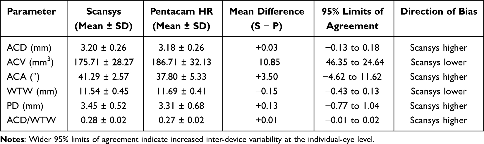 |
Table 1 Inter-Device Differences and Agreement Metrics for Anterior Segment Parameters Measured by Scansys and Pentacam HR |
These parameter-dependent patterns of inter-device bias are visually summarized in Figure 1, which illustrates increasing bias magnitude and variability from linear or ratio-based parameters to angle- and volume-based measurements.
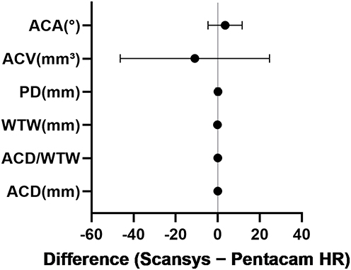 |
Figure 1 Inter-device differences across anterior segment parameters. Mean inter-device differences and 95% limits of agreement for anterior segment parameters measured by Scansys and Pentacam HR. Points indicate mean differences (Scansys − Pentacam HR), and error bars represent the 95% limits of agreement. The vertical line denotes zero difference. |
Parameter-Dependent Stability of Inter-Device Measurements
To further characterize the consistency of measurements across parameters, inter-device stability was classified based on the relative magnitude of mean bias and the width of the limits of agreement. As shown in Table 2, ACD and the ACD/WTW ratio were categorized as demonstrating high inter-device stability. WTW and PD showed moderate stability, whereas ACA and ACV were classified as low stability due to larger bias magnitude and increased dispersion.
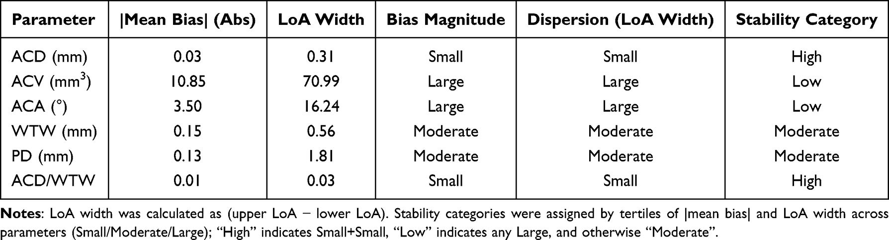 |
Table 2 Parameter-Dependent Stability of Inter-Device Measurements Between Scansys and Pentacam HR |
This stability classification highlights that inter-device agreement is not uniform across anterior segment parameters and suggests that certain measurements are inherently more susceptible to device-related variability.
ICCs indicated excellent agreement for ACD (ICC = 0.95), and good agreement for ACD/WTW (ICC = 0.88) and WTW (ICC = 0.89). Moderate agreement was observed for PD (ICC = 0.69) and ACV (ICC = 0.77), whereas ACA showed poor agreement (ICC = 0.38).
Bland–Altman Analysis for Parameters Without Proportional Bias
Bland–Altman plots for parameters without proportional bias are presented in Figure 2. For ACD and the ACD/WTW ratio, inter-device differences remained relatively constant across the measurement range, with no evident magnitude-dependent trends. These findings indicate that the observed differences for these parameters are largely independent of measurement size.
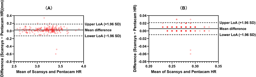 |
Figure 2 Bland–Altman analysis for parameters without proportional bias. Bland–Altman plots for (A) ACD and (B) the ACD/WTW ratio measured by Scansys and Pentacam HR. The solid line denotes the mean difference, and dashed lines represent the 95% limits of agreement. |
Proportional Bias Analysis
Linear regression analysis of inter-device differences against the mean of the two measurements revealed significant proportional bias for several parameters (Table 3). Specifically, ACA, ACV, WTW, and PD demonstrated magnitude-dependent inter-device discrepancies, as indicated by regression slopes significantly different from zero. In contrast, no proportional bias was observed for ACD or the ACD/WTW ratio.
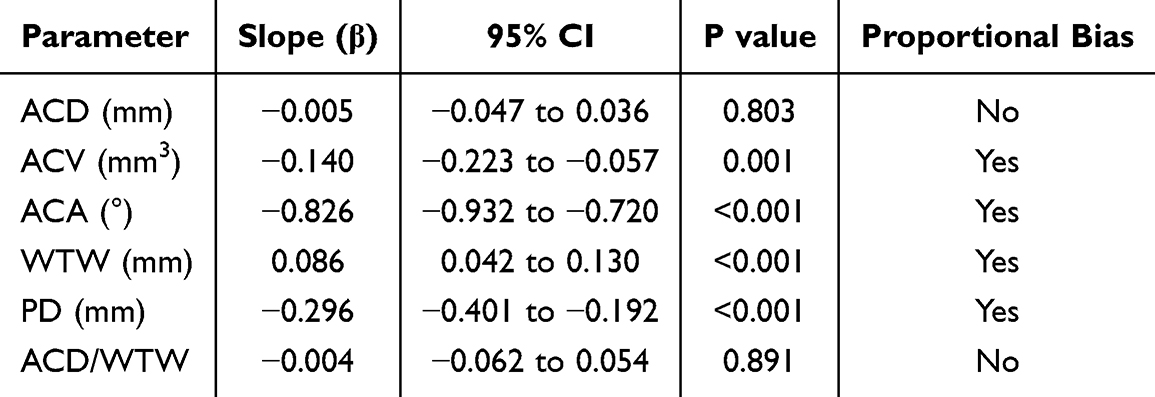 |
Table 3 Proportional Bias Between Scansys and Pentacam HR Measurements Assessed by Regression Analysis |
The presence of proportional bias for ACA, ACV, and PD is visually demonstrated in Figure 3, where regression lines illustrate systematic changes in inter-device differences across the measurement range.
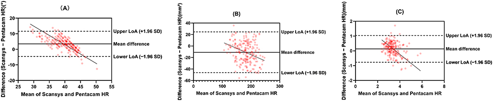 |
Figure 3 Bland–Altman plots showing proportional bias. Bland–Altman plots for (A) ACA, (B) ACV, and (C) PD measured by Scansys and Pentacam HR. Differences between devices (Scansys − Pentacam HR) are plotted against the mean of the two measurements. The solid line denotes the mean difference, dashed lines indicate the 95% limits of agreement, and regression lines indicate proportional bias. |
Discussion
Overview and Rationale
With the increasing availability of domestically developed anterior segment imaging systems, independent evaluation of their measurement characteristics relative to established reference devices has become increasingly important. In the present study, we performed a systematic comparison between Scansys, a domestically developed Scheimpflug-based corneal topography system, and Pentacam HR, focusing on inter-device differences, parameter-dependent stability, and proportional bias across multiple anterior segment parameters.16
Parameter-Dependent Inter-Device Differences
Our results demonstrated that inter-device differences between Scansys and Pentacam HR were not uniform across parameters but instead exhibited a clear parameter-dependent pattern. Linear and ratio-based parameters, such as ACD and the ACD/WTW ratio, showed minimal mean bias and relatively narrow limits of agreement. In contrast, angle- and volume-based parameters, particularly ACA and ACV, exhibited larger mean differences and wider limits of agreement. The ICC results further support this parameter-dependent pattern, demonstrating higher reliability for linear and ratio-based parameters and lower reliability for angle-based measurements such as ACA.
Similar parameter-dependent variability has been reported in previous comparisons between Pentacam and other anterior segment imaging devices, where linear distance measurements generally showed higher reproducibility than parameters derived from angular or volumetric reconstruction.5,17,18
Stability Classification and Interpretation
By integrating the magnitude of mean bias and the width of the limits of agreement, we further classified inter-device stability across parameters. This analysis demonstrated that ACD and the ACD/WTW ratio exhibited high inter-device stability, whereas ACA and ACV showed low stability, with WTW and PD demonstrating intermediate stability.
This finding underscores an important methodological consideration: high correlation or statistically significant agreement at the group level does not necessarily translate into acceptable agreement at the individual-eye level. Wide limits of agreement indicate substantial variability that may limit the interpretability or interchangeability of measurements obtained from different devices.12
Proportional Bias and Magnitude-Dependent Discrepancies
Beyond absolute agreement metrics, we further assessed proportional bias by examining the relationship between inter-device differences and measurement magnitude. Significant proportional bias was identified for ACA, ACV, WTW, and PD, indicating that measurement discrepancies for these parameters varied systematically across the measurement range.
Proportional bias represents a violation of the assumption that measurement differences remain constant across values and has been highlighted as an important consideration in method-comparison studies. Similar magnitude-dependent discrepancies have been reported in anterior segment imaging studies, particularly for parameters influenced by segmentation boundaries, pupil dynamics, or three-dimensional reconstruction algorithms.19,20
The inclusion of the ACD/WTW ratio warrants brief discussion. By normalizing ACD to WTW, this ratio may partially reduce the influence of absolute scaling differences between devices. In the present study, ACD/WTW demonstrated minimal mean bias, narrow limits of agreement, and no proportional bias, suggesting improved robustness compared with absolute measurements. These findings support the potential utility of normalized parameters for reducing inter-device variability, although further validation in different populations and imaging platforms is required.
Implications for Device-Specific Interpretation
From a practical perspective, the presence of parameter-dependent bias and proportional bias suggests that measurements obtained from Scansys and Pentacam HR should not be assumed to be universally interchangeable, particularly for angle- and volume-based parameters. These findings suggest that ACA and ACV measurements may not be directly interchangeable between devices, particularly when values approach clinical decision thresholds, and should therefore be interpreted with caution. Device-specific interpretation may therefore be necessary when applying threshold-based criteria or comparing measurements across studies using different imaging platforms.
Similar cautions have been raised in previous anterior segment imaging studies, which emphasized that device-specific calibration and interpretation are essential when measurements approach clinically relevant thresholds.10,21–23
Limitations
Several limitations of this study should be acknowledged. This was a single-center study, and the findings may not be directly generalizable to other populations or imaging conditions. The study population consisted exclusively of candidates for refractive surgery, which may limit the generalizability of the findings to other populations, particularly those with pathological anterior segment conditions. Intra-device repeatability and interobserver variability were not evaluated in this study. Although proportional bias was identified for several parameters, no attempt was made to derive correction equations, as the primary aim was to characterize measurement behavior rather than to establish conversion models. Only two anterior segment imaging systems were evaluated, and the observed patterns may not apply to other platforms.
Conclusions
In conclusion, this study provides an independent measurement-based evaluation of a domestically developed anterior segment imaging system relative to an established reference device. Inter-device differences between Scansys and Pentacam HR were parameter-dependent, with linear and ratio-based parameters demonstrating higher stability than angle- and volume-based measurements. The presence of proportional bias for several parameters further highlights the importance of device-specific interpretation when comparing anterior segment measurements across imaging platforms. ACD and ACD/WTW may be considered interchangeable between devices, whereas ACA, ACV, and PD should be interpreted with caution due to the presence of proportional bias.
Disclosure
The authors report no conflicts of interest in this work.
References
1. Cetin EN, Akbulut S, Ekici Tekin Z, et al. Corneal and lenticular clarity in children with inflammatory disease as assessed by Scheimpflug imaging. Photodiagnosis Photodyn Ther. 2022;39:103032. doi:10.1016/j.pdpdt.2022.103032
2. Winegarner A, Miki A, Kumoi M, et al. Anterior segment Scheimpflug imaging for detecting primary angle closure disease. Graefes Arch Clin Exp Ophthalmol. 2019;257(1):161–7. doi:10.1007/s00417-018-4171-x
3. Shajari M, Khalil S, Mayer WJ, et al. Comparison of 2 laser fragmentation patterns used in femtosecond laser-assisted cataract surgery. J Cataract Refract Surg. 2017;43(12):1571–1574. doi:10.1016/j.jcrs.2017.09.027
4. Hwang ES, Perez-Straziota CE, Kim SW, Santhiago MR, Randleman JB. Distinguishing highly asymmetric keratoconus eyes using combined scheimpflug and spectral-domain OCT analysis. Ophthalmology. 2018;125(12):1862–1871.
5. Phu J, Tong J, Kalloniatis M. Intra-session repeatability of anterior chamber depth across the chamber width using Pentacam Scheimpflug imaging in healthy subjects. Ophthalmic Physiol Opt. 2021;41(6):1273–1284. doi:10.1111/opo.12880
6. Saito A, Kamiya K, Fujimura F, et al. Comparison of angle-to-angle distance using three devices in normal eyes. Eye. 2020;34(6):1116–1120. doi:10.1038/s41433-019-0653-2
7. Piccinini AL, Golan O, Hafezi F, et al. Higher-order aberration measurements: comparison between Scheimpflug and dual Scheimpflug-Placido technology in normal eyes. J Cataract Refract Surg. 2019;45(4):490–494. doi:10.1016/j.jcrs.2018.11.015
8. Feldman RM, Kim G, Chuang AZ, et al. Comparison between the CASIA SS-1000 and Pentacam in measuring corneal curvatures and corneal thickness maps. BMC Ophthalmol. 2023;23(1):10. doi:10.1186/s12886-023-02768-w
9. Huang X, Lin X, Yang Y, et al. Comparison of a new scheimpflug camera and swept-source optical coherence tomographer for measurements of anterior segment parameters. Ophthalmol Ther. 2023;12(6):3187–3198.
10. Khorrami-Nejad M, Khodaparast M, Abdulkadhim IA, et al. A comparison of Scansys and Sirius tomography in healthy eyes. BMC Ophthalmol. 2024;24(1):138. doi:10.1186/s12886-024-03389-7
11. Xu W, Zhai C, Yusufu M, et al. Repeatability and agreement between a reference Scheimpflug tomographer and a low-cost Scheimpflug system. J Cataract Refract Surg. 2023;49(6):614–619. doi:10.1097/j.jcrs.0000000000001168
12. Bland JM, Altman DG. Statistical methods for assessing agreement between two methods of clinical measurement. Lancet. 1986;1(8476):307–310.
13. Sadler WA. Using the variance function to generalize Bland-Altman analysis. Ann Clin Biochem. 2019;56(2):198–203. doi:10.1177/0004563218806560
14. Cheng SM, Zhang J-S, Li -T-T, et al. Repeatability and agreement of two swept-source optical coherence tomographers for anterior segment parameter measurements. J Glaucoma. 2022;31(7):602–608. doi:10.1097/IJG.0000000000001989
15. Chan TCY, Yu MCY, Chiu V, et al. Comparison of two novel swept-source optical coherence tomography devices to a partial coherence interferometry-based biometer. Sci Rep. 2021;11(1):14853. doi:10.1038/s41598-021-93999-8
16. Baradaran-Rafii A, Motevasseli T, Yazdizadeh F, et al. Comparison between two scheimpflug anterior segment analyzers. J Ophthalmic Vis Res. 2017;12(1):23–29. doi:10.4103/jovr.jovr_104_16
17. Tañá-Rivero P, Rodríguez-Carrillo MD, Ruíz-Santos M, et al. Agreement between angle-to-angle distance and aqueous depth obtained with two different optical coherence tomographers and a scheimpflug camera. J Refract Surg. 2021;37(2):133–140. doi:10.3928/1081597X-20201013-01
18. Li X, Zhou Y, Young CA, et al. Comparison of a new anterior segment optical coherence tomography and Oculus Pentacam for measurement of anterior chamber depth and corneal thickness. Ann Transl Med. 2020;8(14):857. doi:10.21037/atm-20-187
19. Hafner M, Herold TR, Deiters V, et al. Quantitative comparison of a novel wide-field OCT-angiography device with ultrawide-field fluorescein angiography in detecting retinal nonperfusion in vascular retinopathies. BMC Ophthalmol. 2025;25(1):642.
20. Yalçinkaya Çakir G, Kirgiz A, Uzar I, et al. Refractive error prediction after phacoemulsification surgery: lenstar LS 900 versus Nidek AL-scan. J Refract Surg. 2025;41(3):e257–e263. doi:10.3928/1081597X-20250207-03
21. Zhou H, Bao T, Li S, et al. Repeatability and agreement of three biometers measuring corneal keratometry and astigmatism in eyes with cataract and high myopia. Photodiagnosis Photodyn Ther. 2025;54:104652. doi:10.1016/j.pdpdt.2025.104652
22. Salouti R, Kamalipour A, Masihpour N, et al. Effect of photorefractive keratectomy on agreement of anterior segment variables obtained by a swept-source biometer vs a Scheimpflug-based tomographer. J Cataract Refract Surg. 2020;46(9):1229–1235. doi:10.1097/j.jcrs.0000000000000252
23. Han SU, Ryu S, Jung H, et al. Analysis of keratometric measurements in accordance with axial length in an aged population. Sci Rep. 2022;12(1):4087. doi:10.1038/s41598-022-08194-0
 © 2026 The Author(s). This work is published and licensed by Dove Medical Press Limited. The
full terms of this license are available at https://www.dovepress.com/terms
and incorporate the Creative Commons Attribution
- Non Commercial (unported, 4.0) License.
By accessing the work you hereby accept the Terms. Non-commercial uses of the work are permitted
without any further permission from Dove Medical Press Limited, provided the work is properly
attributed. For permission for commercial use of this work, please see paragraphs 4.2 and 5 of our Terms.
© 2026 The Author(s). This work is published and licensed by Dove Medical Press Limited. The
full terms of this license are available at https://www.dovepress.com/terms
and incorporate the Creative Commons Attribution
- Non Commercial (unported, 4.0) License.
By accessing the work you hereby accept the Terms. Non-commercial uses of the work are permitted
without any further permission from Dove Medical Press Limited, provided the work is properly
attributed. For permission for commercial use of this work, please see paragraphs 4.2 and 5 of our Terms.