
Back to Journals » Advances in Medical Education and Practice » Volume 16
Orthopedic Trainees’ Perception of the Educational Utility of Patient-Specific 3D-Printed Anatomical Models: A Questionnaire-Based Observational Study
Received 18 May 2025
Accepted for publication 2 August 2025
Published 9 August 2025 Volume 2025:16 Pages 1399—1409
DOI https://doi.org/10.2147/AMEP.S534099
Checked for plagiarism Yes
Review by Single anonymous peer review
Peer reviewer comments 3
Editor who approved publication: Dr Sateesh Arja
Xiaoyang Zhou,* Kaijun Yi,* Yihua Shi
Department of Orthopedics, Xiangyang No.1 People’s Hospital, Hubei University of Medicine, Xiangyang, 441000, People’s Republic of China
*These authors contributed equally to this work
Correspondence: Yihua Shi, Department of Orthopedics, Xiangyang No.1 People’s Hospital, Hubei University of Medicine, Xiangyang, 441000, People’s Republic of China, Tel +86 0710 3420011, Fax +86 0710 3420176, Email [email protected]
Background: Traditional orthopedic teaching methods have inherent limitations in conveying complex three-dimensional anatomical relationships essential for surgical planning and execution. Three-dimensional (3D) printing technology offers a potential solution to these educational challenges, but systematic evaluation of its specific educational impact in orthopedic residency training remains limited.
Purpose: This study aimed to evaluate the educational efficacy of in-house 3D-printed patient-specific anatomical models in orthopedic training through assessment of three core domains: anatomical comprehension, surgical planning proficiency, and clinical teaching utility.
Methods: In this analytical observational study, paper-based questionnaires were distributed to 145 orthopedic residents at Hubei University of Medicine who participated in clinical teaching sessions using 3D-printed anatomical models between January 2025 and March 2025. Participants rated their experiences on a 10-point Likert scale. Data were analyzed using descriptive statistics.
Results: The response rate was 81.4% (n=118). A majority (85.6%) of residents reported enhanced understanding of complex anatomical structures. First-year residents demonstrated higher satisfaction (mean score 7.9) compared to more advanced trainees (mean scores 7.3 and 6.9). Small group settings (4– 6 participants) were preferred by 76.3% of respondents. Physical manipulation of models received the highest educational value rating (mean score 8.1). Primary limitations included production time (45.8%), material durability (38.6%), and limited model varieties (35.6%). Nearly half (43.2%) of residents requested more frequent practice sessions.
Conclusion: 3D-printed anatomical models significantly enhance orthopedic resident education, particularly for complex structures and junior trainees. Small-group, instructor-guided implementation maximizes educational benefits. When strategically integrated into existing curricula, in-house production enables widespread access across training levels with minimal resource constraints.
Keywords: 3D printing, orthopedic residency training, patient-specific anatomical models, educational assessment, tactile learning, surgical planning
Introduction
Orthopedic surgical education faces unique challenges due to the inherently three-dimensional (3D) nature of musculoskeletal pathology.1 Traditional teaching methods—such as two-dimensional (2D) imaging, textbook illustrations, and occasional cadaveric dissection—often fall short in conveying the complex spatial relationships essential for effective surgical planning and execution.2 These limitations become especially evident when teaching intricate fracture patterns, joint mechanics, and surgical approaches, where a precise understanding of spatial anatomy directly impacts clinical outcomes.3,4 Many residents report difficulty in translating 2D knowledge into 3D surgical competence, resulting in a persistent gap between theoretical understanding and practical skills—a gap that may ultimately affect patient care.5
A critical technological advance bridging this gap is the digital reconstruction of 3D anatomical models from standard 2D imaging modalities, primarily via the process of image segmentation.6 Medical image segmentation involves labeling each pixel or voxel in a dataset according to anatomical and tissue boundaries, thus enabling creation of accurate 3D representations from stacks of 2D images such as CT or MRI.7 Recent advances, including semi-automated and deep learning-based segmentation methods, have significantly improved both the efficiency and accuracy of this process.8 For example, convolutional neural networks can now automatically segment complex joint anatomy from multi-sequence 3D MRI, producing highly detailed digital models that are suitable for visualization, surgical simulation, and 3D printing.9
3D printing technology has emerged as a promising solution to the challenges in orthopedic education by enabling the creation of patient-specific anatomical models (PAMs).10 The process begins with the reconstruction of digital 3D models from conventional 2D medical images—such as CT scans—using segmentation techniques.11 These digital representations are then transformed into highly accurate, tangible replicas of actual pathologies through 3D printing.12 Unlike traditional educational tools, PAMs provide residents with a multisensory, hands-on learning experience that bridges the gap between conceptual knowledge and practical skills, supplementing visual learning with essential tactile feedback.13 The multisensory nature of 3D-printed models supplements visual learning with tactile feedback, an important consideration given the diverse learning preferences among medical trainees.14 This interactive approach aligns with educational theory, which supports that multimodal learning enhances knowledge retention and skill transfer—particularly in procedural specialties where spatial understanding is critical.15 By enabling deeper comprehension of complex fracture patterns and joint mechanics, 3D printing technology significantly enriches orthopedic education and advances operative training.
Within orthopedic curricula, the potential applications of 3D-printed models span multiple educational contexts. In case-based discussions, these models can facilitate demonstration of fracture patterns and reduction techniques that are difficult to visualize through imaging alone.16 During preoperative planning sessions, they provide platforms for collaborative problem-solving between attending surgeons and residents, potentially enhancing resident participation in surgical decision-making.17 Additionally, these models may improve communication with patients during informed consent processes, offering residents valuable experience in explaining complex procedures to non-medical audiences.18 Despite these theoretical advantages, systematic evaluation of educational outcomes associated with 3D-printed models in orthopedic training remains limited, with most evidence derived from small-scale studies or anecdotal reports.19
Previous research has consistently reported high satisfaction rates with 3D-printed models across various surgical specialties, confirming their value as educational tools.20 However, most studies have focused primarily on resident satisfaction rather than objective measures of knowledge acquisition or skill development.21 Critical questions remain regarding which specific orthopedic concepts benefit most from these models, how best to implement them within residency curricula, and how perceptions or educational needs may differ across training levels.22 Additionally, large-scale adoption of 3D-printed models is often limited by high costs and restricted access to printing facilities.23 These gaps in knowledge and implementation hinder the evidence-based integration of 3D printing technology into orthopedic education, potentially limiting its full educational impact.24
This study addresses critical gaps in orthopedic education research by evaluating the educational value of a low-cost, in-house 3D printing initiative integrated into a structured teaching methodology for residents. Using a structured questionnaire, we assess three core domains: enhancement of anatomical comprehension, improvement in surgical planning proficiency, and practical utility in clinical teaching. By analyzing both subjective feedback and quantifiable outcomes across different training levels and subspecialty areas, this study aims to establish evidence-based guidelines for effectively incorporating 3D-printed models into surgical curricula. The findings offer practical, replicable insights for residency programs seeking to optimize training with limited resources, while contributing meaningful evidence to the evolving landscape of technological innovation in medical education.
Aim
The aim of this study was to evaluate the educational efficacy of in-house 3D-printed PAMs in orthopedic resident training through structured questionnaire assessment focused on three core domains: enhancement of anatomical comprehension and spatial reasoning; improvement in surgical planning proficiency and interdisciplinary collaboration; and practical utility in clinical teaching contexts and skill development. By analyzing both subjective feedback and quantifiable outcomes across training levels and subspecialty areas, this investigation sought to establish evidence-based guidelines for effectively incorporating 3D-printed models into orthopedic surgical curricula.
Materials and Methods
Study Design and Population
Paper-based questionnaires were distributed to 145 orthopedic residents at Hubei University of Medicine who participated in clinical teaching sessions utilizing 3D-printed anatomical models. The questionnaire assessed their learning experience across multiple dimensions: anatomical structure comprehension, surgical planning capabilities, and overall educational satisfaction. Participants had a 2-month completion window, and reminders were sent to non-responders after 2 weeks. Completed questionnaires were submitted to a designated collection box to maintain confidentiality. Questionnaire completion constituted implied consent to participate.
3D Model Creation and Printing
Patient-specific 3D models were created from anonymized patient computed tomography (CT) scans, which were stored in the standard DICOM format. The anatomical regions of interest were then isolated using a semi-automated segmentation process in Mimics software (Materialise, Leuven, Belgium), a commonly used commercial software package for medical image processing. This process, performed by a trained technician, required approximately 2–4 hours per model. Once segmentation was complete, the data were converted into a stereolithography (STL) file format, which is standard for 3D printing applications. The physical models were then printed using a Fused Deposition Modeling (FDM) printer with polylactic acid (PLA) material, a common and cost-effective approach for producing educational models. The models were printed at a high resolution (eg, 12.5 µm for the x/y axes) to ensure anatomical fidelity. The average printing time was 8–12 hours, depending on the model’s size and complexity. The models selected for this study represented a range of complex orthopedic cases commonly encountered in residency, including comminuted acetabular fractures, severe spinal deformities, and complex tibial plateau fractures.
Educational Intervention
The teaching sessions were delivered in a structured format lasting 60–90 minutes. Each session involved a small group of 4–6 residents and was led by an attending surgeon. The surgeon first demonstrated key anatomical features and surgical principles using the model, which was followed by a period of supervised, hands-on practice by the residents. This study was designed to evaluate this single, standardized teaching format and did not include a comparison to other lecture forms, durations, or group sizes.
Questionnaire
The study utilized a 20-question survey to assess the educational value of 3D-printed models in clinical teaching and their efficacy in resident training. The educational value section assessed improvements in understanding anatomical structures, spatial relationships, and pathological changes. The training effectiveness section evaluated the models’ utility in surgical approach comprehension, preoperative planning, and case-based learning. A 10-point Likert scale (1 = not effective, 10 = highly effective) was used for all questions. The questionnaire included open-ended fields for qualitative feedback. The survey underwent pilot testing with five senior residents to ensure clarity and relevance before distribution. The full questionnaire is provided as Supplementary Figure 1.
Data Analysis
This study was designed as a descriptive observational survey to gather resident perceptions. As such, a formal a priori power analysis to determine sample size was not performed. All data from the completed questionnaires were analyzed using descriptive statistics in GraphPad Prism 8.0.2. Quantitative data from the 10-point Likert scale questions are presented as means, while categorical data are presented as percentages. No inferential statistical models were used, as the objective was to summarize and describe resident feedback rather than to test a specific hypothesis.
Results
Participant Demographics and Participation
Of 145 distributed questionnaires to orthopedic residents participating in teaching sessions between January 2025 and March 2025, 118 completed responses were received, for a response rate of 81.4%. The respondents included 42.4% first-year residents (n=50), 37.3% second-year residents (n=44), and 20.3% third-year residents (n=24), as shown in Figure 1A. The distribution of teaching session participation by sub-specialty is shown in Figure 1B, with trauma accounting for the largest share (45.8%).
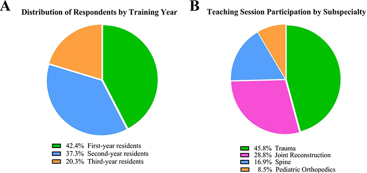 |
Figure 1 Participant distribution analysis (n=118). (A) Distribution of respondents by training year. (B) Participation in teaching sessions by orthopedic subspecialty. |
Perceived Educational Impact and Satisfaction
The educational impact of 3D-printed models was positive, with 85.6% of residents reporting enhanced understanding of complex anatomical structures. The effectiveness of 3D models varied across different teaching aspects, with spinal deformity corrections receiving a mean score of 7.7 and complex fracture patterns a score of 7.6 (Figure 2A). Interestingly, as shown in Figure 2B, first-year residents reported higher satisfaction rates (mean score 7.9) compared to second-year (mean score 7.3) and third-year residents (mean score 6.9), suggesting these models might be most beneficial during early surgical training.
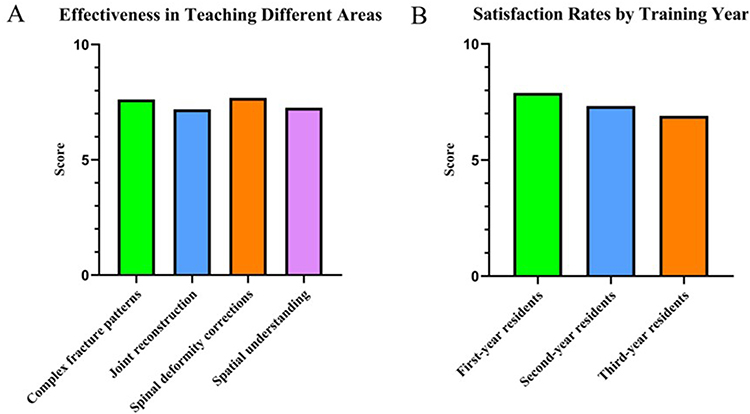 |
Figure 2 Educational impact assessment of 3D-printed anatomical models (n=118). (A) Mean effectiveness scores by orthopedic concept. (B) Mean satisfaction scores by training level. |
Key Learning Outcomes and Limitations
Residents identified key educational advantages of 3D-printed models, with tactile manipulation receiving the highest mean effectiveness score (8.1), followed by visualization of fracture patterns (7.9) and contributions to preoperative planning (7.2), as detailed in Figure 3A. Figure 3B illustrates the percentage of positive responses for these learning outcomes. The primary limitations reported by residents included the lengthy production process—encompassing both model segmentation (2–4 hours) and printing time (8–12 hours)—as well as material durability (38.6%) and limited model varieties (35.6%) (Figure 3C).
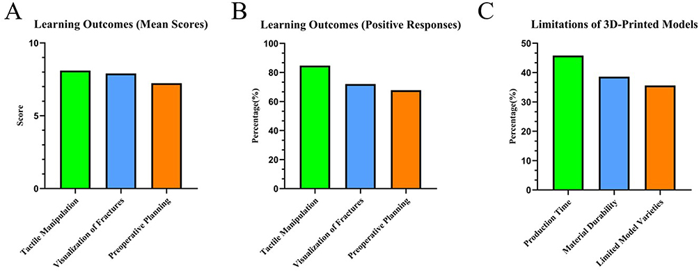 |
Figure 3 Learning outcomes and limitations of 3D-printed models (n=118). (A) Mean effectiveness scores for different learning outcomes. (B) Percentage of positive responses for learning outcomes. (C) Reported limitations of the models. |
Feedback on the Educational Environment
The educational environment significantly influenced teaching effectiveness. As shown in Figure 4A, small groups (4–6 participants) were preferred by 76.3% of residents, and a structured format (60–90 minutes with instructor-led demonstration followed by supervised hands-on practice) received positive feedback from 82.2%. Figure 4B presents the mean satisfaction ratings for various teaching environment elements, with optimal group size (8.4) and instructor guidance (8.2) rated highest. Despite overall positive feedback, 43.2% of participants recommended incorporating regular sessions with extended practice time, particularly for complex anatomical regions and challenging surgical approaches—aligning with the lower ratings for session frequency and practice duration.
 |
Figure 4 Educational environment assessment for 3D-printed model teaching (n=118). (A) Resident preferences for teaching format. (B) Mean satisfaction ratings for teaching environment elements. |
In summary, 3D-printed anatomical models provide substantial educational benefits for orthopedic surgical training, like the pelvis and acetabulum (see Figure 5 for an example of such a model), with impact varying by experience level. Junior residents reported higher satisfaction than seniors. Models were most valuable for three key purposes: enhancing tactile understanding of anatomy, visualizing complex fracture patterns, and improving preoperative planning. The small-group, instructor-guided teaching approach proved effective, though residents consistently requested more frequent and longer hands-on practice sessions, especially for complex anatomical regions. These findings suggest 3D-printed models deliver maximum benefit when integrated into a comprehensive educational framework with appropriate group sizes, structured guidance, and sufficient practice opportunities tailored to resident experience.
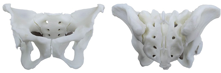 |
Figure 5 3D-printed pelvic model shown in anterior (left) and posterior (right) views. This white polymer model highlights key anatomical features including the acetabulum, sacroiliac joints, and sacral foramina. The patient-specific model enables residents to visualize complex spatial relationships and provides tactile learning for pelvic trauma surgery and fracture fixation techniques. |
Discussion
This study demonstrates that in-house, patient-specific 3D-printed anatomical models are perceived by orthopedic residents as valuable educational tools, with the strongest reported benefits among junior trainees and in the context of complex anatomical structures. Our findings are closely aligned with the study’s objectives: to evaluate the educational efficacy of 3D-printed models in three domains—anatomical comprehension, surgical planning, and clinical teaching utility. The results show consistent, positive resident feedback across these areas, with quantitative data confirming enhanced understanding of anatomy (85.6% of residents), high ratings for tactile learning, and clear preferences for small-group, instructor-guided sessions. The findings are relevant both to the stated aims and to the growing body of literature exploring technological innovation in surgical education. Most notably, junior residents reported the greatest educational benefit, suggesting 3D models are particularly effective in bridging the gap between abstract anatomical concepts and practical understanding during foundational training years. This is consistent with prior research indicating that physical models can support the development of spatial reasoning skills that are challenging to acquire through conventional 2D resources alone.25
The educational advantages of 3D-printed models can be understood through frameworks such as experiential learning theory and cognitive load theory.26,27 By providing tangible, 3D representations, these models help reduce the mental effort required to mentally reconstruct anatomical relationships from 2D images—a process that can otherwise increase extraneous cognitive load, especially for novice learners.28,29 The multisensory experience afforded by 3D models—integrating visual, tactile, and spatial modalities—enhances information encoding, retention, and the transfer of knowledge to clinical practice,30 while haptic feedback from physical manipulation is particularly valuable for developing procedural skills and addressing gaps left by traditional, primarily visual teaching methods.31,32 While our study interprets its findings through established educational theories, a more nuanced application is needed. Cognitive load theory highlights the limited capacity of working memory and underscores the importance of instructional design that reduces unnecessary cognitive load while promoting meaningful schema-building.33 In this context, 3D-printed models likely help redistribute cognitive effort by alleviating the burden of mentally reconstructing spatial relationships, allowing learners—especially those in the early stages of training—to focus more effectively on clinically relevant reasoning and skill development. However, it is important to recognize that physical models do not eliminate cognitive load entirely; rather, they optimize its allocation to enhance learning efficiency. To validate these theoretical benefits, future research should directly assess cognitive load during educational interventions, using objective tools such as subjective rating scales or dual-task paradigms, rather than relying solely on learner perceptions.
The findings highlight that the educational environment and implementation format significantly influence learning outcomes with 3D-printed models. A strong preference for small-group teaching (76.3%) and structured instructional approaches (82.2%), along with high satisfaction ratings for instructor guidance (mean score 8.2), indicate that these models are most effective when embedded in collaborative, guided learning experiences rather than self-directed study. This preference aligns with social constructivist learning theories, which emphasize knowledge construction through peer interaction and expert scaffolding. However, lower satisfaction with session frequency (mean score 6.5) suggests that regular, ongoing integration throughout the curriculum—rather than isolated sessions—would further maximize educational impact. Collectively, these results support the incorporation of 3D-printed models into formal, curriculum-based teaching sessions to optimize their educational utility.
The observed preference for small-group learning environments (typically 4–6 participants) reflects core principles of medical education, where knowledge is often constructed through peer interaction and collaborative problem-solving.34 Both orthopedic35 and neurosurgical36 training contexts have shown that 3D models enhance the understanding of complex anatomy, yet our findings add nuance by highlighting the varying impact across training levels and the importance of balancing instructor guidance with opportunities for independent exploration.37 Participants reported that the educational benefits of 3D models were most pronounced during structured 60–90 minute sessions that combined initial instructor demonstration with supervised, hands-on practice—an approach that received notably positive feedback.38 At the same time, residents expressed a desire for more frequent and extended sessions, highlighting a key implementation challenge that underscores the need for thoughtful curriculum integration.39 These results suggest that 3D-printed models are most effective when incorporated into collaborative, instructor-led learning experiences, with an emphasis on small-group formats. Strategic integration should ensure that these models are utilized throughout residency, with greater emphasis during foundational years and more targeted application as residents advance and focus on subspecialty interests.40 However, a key limitation of our study is the reliance on self-reported perceptions of effectiveness rather than objective performance metrics. While participants strongly agreed that 3D models improved their understanding, previous research has shown that this perceived benefit does not always translate into statistically significant gains in knowledge retention or test scores.41 This underscores the need for future research to incorporate objective assessments, such as pre- and post-intervention testing or performance in simulated surgical tasks, to validate the perceived benefits reported in our study.
Implementation considerations must balance educational benefits against resource constraints, faculty availability, and competing priorities within residency curricula.42 The development of accessible model libraries allowing supervised independent access outside structured teaching represents a potential solution to address the desire for additional practice without proportionately increasing faculty commitment.43 This approach aligns with adult learning principles by providing learner-directed opportunities while maintaining appropriate educational oversight.44 Programs implementing these models should consider strategic curriculum mapping to identify high-yield anatomical regions where three-dimensional understanding proves most challenging through conventional teaching methods.45 The acetabulum, pelvis, and complex articular surfaces emerged in our study as areas where residents reported maximum benefit from model interaction, suggesting that initial implementation efforts might prioritize these structures for optimal educational impact and resource utilization. Our study intentionally focused on complex cases such as comminuted acetabular fractures and severe spinal deformities, as the educational value of 3D models is most justified in these non-standard scenarios where traditional 2D imaging is least effective. The limitation of ‟model varieties” noted by residents likely reflects a desire to apply this valuable tool to an even broader range of challenging cases encountered during training.
The rapidly evolving landscape of 3D-printing technology presents both opportunities and challenges for wider implementation in orthopedic education.46 Decreasing production costs and increasing printing efficiency make adoption increasingly feasible, even for programs with limited resources.47 However, important considerations remain regarding model durability, anatomical accuracy, and material properties.48 While our white polymer models excelled in demonstrating structural relationships, they could not replicate the tactile properties of bone, potentially limiting their utility for certain aspects of surgical simulation such as drilling, cutting, or hardware fixation.49 Resident feedback echoed these concerns, particularly highlighting the practical limitations of 3D-printed models—most notably the significant time required for both model segmentation (2–4 hours per model) and printing (an additional 8–12 hours), as well as ongoing challenges with material durability. These issues are well documented in the field and can present barriers to broader adoption. Nevertheless, the primary contribution of our work is demonstrating the feasibility and perceived educational value of a low-cost, in-house workflow. By utilizing FDM printers and PLA material, we addressed significant cost barriers that often limit access to this technology in educational settings. This experience aligns with existing research indicating that affordable, low-cost models can deliver clinical and educational value comparable to more expensive alternatives. Ultimately, a comprehensive cost-benefit analysis must weigh not only direct production expenses but also potential downstream effects, including improvements in surgical efficiency, reductions in complications, and enhanced resident confidence that may translate to better patient outcomes.50 While the educational return on investment appears substantial based on resident feedback, more rigorous quantification through objective performance metrics would further strengthen the case for widespread implementation.51
A central limitation of our PLA models is that, while they effectively demonstrate anatomical relationships, they cannot replicate the biomechanical properties of real tissue, limiting their usefulness for realistic surgical simulations involving drilling or cutting. Advanced multi-material printers, such as the Stratasys Digital Anatomy printer, represent an important future direction by producing models with variable densities and textures that closely mimic real bone and soft tissue, offering a higher-fidelity platform for procedural training. Other limitations of our study include reliance on resident self-reporting rather than objective performance measures and the focus on short-term educational outcomes. Future research should incorporate pre- and post-intervention assessments, operative performance metrics, and longitudinal studies to evaluate knowledge retention and application in clinical practice. Despite these limitations, our findings indicate that 3D-printed patient-specific anatomical models are valuable educational tools for orthopedic training, particularly when combined with effective group sizes, structured guidance, and hands-on practice. These models address gaps in understanding complex three-dimensional anatomy and spatial relationships, ultimately improving educational outcomes and surgical preparation. Looking ahead, integrating 3D printing with technologies such as virtual or augmented reality, sensor-embedded models, and more advanced multi-material printing could further enhance orthopedic education by providing realistic tactile feedback and interactive visualization. While our study demonstrates the value of a basic, in-house 3D printing program, future work should explore these advanced and blended approaches to further enrich surgical training.
Conclusion
This study’ s findings indicate that orthopedic residents perceive 3D-printed, patient-specific anatomical models as a valuable addition to their training, with the strongest benefits reported by junior trainees and for understanding complex anatomical structures. Residents identified the models as being most useful for enhancing tactile anatomical understanding, visualizing complex fracture patterns, and improving preoperative planning.
The results further suggest that the educational effectiveness of these models is optimized when implemented within a framework of small-group settings, structured instructor guidance, and adequate hands-on practice. While this study is limited to resident perception and does not measure objective skill acquisition, it provides a replicable, evidence-based framework for institutions seeking to implement low-cost, in-house 3D printing to enrich the resident learning experience.
Highlights
- 3D-printed anatomical models significantly enhanced understanding of complex orthopedic structures among 85.6% of residents, with physical manipulation receiving the highest educational value rating (8.1/10).
- First-year residents demonstrated substantially greater educational benefit (mean score 7.9) compared to third-year trainees (6.9), highlighting the technology’s particular value in early training stages.
- Small-group teaching (4-6 participants) with instructor guidance was preferred by 76.3% of residents, while in-house production addressed key implementation barriers of cost and customization.
Abbreviations
2D, two-dimensional; 3D, three-dimensional; CT, computed tomography; FDM, Fused Deposition Modeling; PAMs, patient-specific anatomical models; PLA, polylactic acid; STL, stereolithography.
Data Sharing Statement
The datasets used and/or analyzed during the current study are available from the corresponding author on reasonable request.
Ethics Approval
The experimental protocol and procedures received approval from the Ethical Committee of Biomedical Basic Research of Xiangyang No.1 People’s Hospital (Review Approval Number: XYYYE20250027).
Consent for Publication
All authors have approved the final manuscript and provide their consent for publication.
Acknowledgments
We thank all of the participants for their willingness to participate in the study and the time that they devoted to the study.
Author Contributions
All authors made a significant contribution to the work reported, whether that is in the conception, study design, execution, acquisition of data, analysis and interpretation, or in all these areas; took part in drafting, revising or critically reviewing the article; gave final approval of the version to be published; have agreed on the journal to which the article has been submitted; and agree to be accountable for all aspects of the work.
Funding
This work, in part, was supported by grants from the Foundation of Health Commission of Hubei Province (NO. WJ2023F075).
Disclosure
The authors declare no competing interest in this work.
References
1. Levitt EB, Paul KD, Vatsia SK, et al. Benefits of an orthopedic education research collaborative: an innovative approach. Cureus. 2023;15(2):e34903. doi:10.7759/cureus.34903
2. Yan M, Huang J, Ding M, et al. Three-dimensional printing model enhances correct identification and understanding of pelvic fracture in medical students. J Surg Educ. 2023;80(3):331–337. doi:10.1016/j.jsurg.2022.10.016
3. Weidert S, Andress S, Linhart C, et al. Correction to: 3D printing method for next‑day acetabular fracture surgery using a surface filtering pipeline: feasibility and 1‑year clinical results. Int J Comput Assist Radiol Surg. 2021;16(4):703–704. doi:10.1007/s11548-021-02348-7
4. Chen Z, Zhang Y, Yan Z, et al. Artificial intelligence assisted display in thoracic surgery: development and possibilities. J Thorac Dis. 2021;13(12):6994–7005. doi:10.21037/jtd-21-1240
5. Elias A, Benady A, Golden E, Segal O, Dadia S. In situ cryoablation of sacral giant cell tumor using three-dimensional (3D) model: a case report. J Orthop. 2022;30:46–50. doi:10.1016/j.jor.2022.02.007
6. Xiao H, Teng X, Liu C, et al. A review of deep learning-based three-dimensional medical image registration methods. Quant Imaging Med Surg. 2021;11(12):4895–4916. doi:10.21037/qims-21-175
7. Hesamian MH, Jia W, He X, Kennedy P. Deep learning techniques for medical image segmentation: achievements and challenges. J Digit Imaging. 2019;32(4):582–596. doi:10.1007/s10278-019-00227-x
8. Zhou SK, Greenspan H, Davatzikos C, et al. A review of deep learning in medical imaging: imaging traits, technology trends, case studies with progress highlights, and future promises. Proc IEEE Inst Electr Electron Eng. 2021;109(5):820–838. doi:10.1109/JPROC.2021.3054390
9. Li M, Punithakumar K, Major PW, et al. Temporomandibular joint segmentation in MRI images using deep learning. J Dent. 2022;127:104345. doi:10.1016/j.jdent.2022.104345
10. Meyer-Szary J, Luis MS, Mikulski S, et al. The role of 3D printing in planning complex medical procedures and training of medical professionals-cross-sectional multispecialty review. Int J Environ Res Public Health. 2022;19(6):3331. doi:10.3390/ijerph19063331
11. Cong B, Zhang H. Innovative 3D printing technologies and advanced materials revolutionizing orthopedic surgery: current applications and future directions. Front Bioeng Biotechnol. 2025;13:1542179. doi:10.3389/fbioe.2025.1542179
12. Burnard JL, Parr W, Choy WJ, Walsh WR, Mobbs RJ. 3D-printed spine surgery implants: a systematic review of the efficacy and clinical safety profile of patient-specific and off-the-shelf devices. Eur Spine J. 2020;29(6):1248–1260. doi:10.1007/s00586-019-06236-2
13. Rama M, Schlegel L, Wisner D, et al. Using three-dimensional printed models for trainee orbital fracture education. BMC Med Educ. 2023;23(1):467. doi:10.1186/s12909-023-04436-5
14. Babaei S, Toofaninejad E, Kalantarion M, Taghirad HD, Mohammadi SF. From touch to skill: how tactile feedback is transforming simulation-based medical education. Med Teach. 2025;47(2):370. doi:10.1080/0142159X.2024.2415497
15. Sinou N, Sinou N, Filippou D. Virtual reality and augmented reality in anatomy education during COVID-19 pandemic. Cureus. 2023;15(2):e35170. doi:10.7759/cureus.35170
16. Assink N, Reininga I, Ten Duis K, et al. Does 3D-assisted surgery of tibial plateau fractures improve surgical and patient outcome? A systematic review of 1074 patients. Eur J Trauma Emerg Surg. 2022;48(3):1737–1749. doi:10.1007/s00068-021-01773-2
17. Delgado B, Campos M, Lobos D, et al. Training in lumbar pedicle screw instrumentation using a 3D-printed model: from validation to transferability onto a cadaver model. J Am Acad Orthop Surg. 2024. doi:10.5435/JAAOS-D-24-00699
18. Ozturk AM, Sirinturk S, Kucuk L, et al. Multidisciplinary assessment of planning and resection of complex bone tumor using patient-specific 3D model. Indian J Surg Oncol. 2019;10(1):115–124. doi:10.1007/s13193-018-0852-5
19. Wang C, Zhang L, Qin T, et al. 3D printing in adult cardiovascular surgery and interventions: a systematic review. J Thorac Dis. 2020;12(6):3227–3237. doi:10.21037/jtd-20-455
20. Khorsandi D, Fahimipour A, Abasian P, et al. 3D and 4D printing in dentistry and maxillofacial surgery: printing techniques, materials, and applications. Acta Biomater. 2021;122:26–49. doi:10.1016/j.actbio.2020.12.044
21. Maglara E, Angelis S, Solia E, et al. Three-dimensional (3D) printing in orthopedics education. J Long Term Eff Med Implants. 2020;30(4):255–258. doi:10.1615/JLongTermEffMedImplants.2020036911
22. Coxe FR, Stauffer TP, Ast MP. Virtual reality simulation in orthopedic surgery education improves immediate procedural skill and knowledge acquisition, but evidence on cost-effectiveness and skill retention remains lacking. Curr Rev Musculoskelet Med. 2025;18(10):363–378. doi:10.1007/s12178-025-09973-8
23. Capitani P, Zampogna B, Monaco E, et al. The role of virtual reality in knee arthroscopic simulation: a systematic review. Musculoskelet Surg. 2023;107(1):85–95. doi:10.1007/s12306-021-00732-9
24. Angelo RL, Gallagher AG. Comment on: systematic review of the current status of cadaveric simulation for surgical training. Br J Surg. 2020;107(4):467. doi:10.1002/bjs.11502
25. Sharmin N, Chow AK, King S. Effect of teaching tools in spatial understanding in health science education: a systematic review. Can Med Educ J. 2023;14(4):70–88. doi:10.36834/cmej.74978
26. Goyal S, Chua C, Chen YS, Murphy D, O ‘Neill GK. Utility of 3D printed models as adjunct in acetabular fracture teaching for Orthopaedic trainees. BMC Med Educ. 2022;22(1):595. doi:10.1186/s12909-022-03621-2
27. van Merriënboer JJ, Sweller J. Cognitive load theory in health professional education: design principles and strategies. Med Educ. 2010;44(1):85–93. doi:10.1111/j.1365-2923.2009.03498.x
28. Huang Z, Song W, Zhang Y, et al. Three-dimensional printing model improves morphological understanding in acetabular fracture learning: a multicenter, randomized, controlled study. PLoS One. 2018;13(1):e0191328. doi:10.1371/journal.pone.0191328
29. Wu AM, Wang K, Wang JS, et al. The addition of 3D printed models to enhance the teaching and learning of bone spatial anatomy and fractures for undergraduate students: a randomized controlled study. Ann Transl Med. 2018;6(20):403. doi:10.21037/atm.2018.09.59
30. Shams L, Seitz AR. Benefits of multisensory learning. Trends Cognit Sci. 2008;12(11):411–417. doi:10.1016/j.tics.2008.07.006
31. Baskaran V, Štrkalj G, Štrkalj M, Di Ieva A. Current applications and future perspectives of the use of 3D printing in anatomical training and neurosurgery. Front Neuroanat. 2016;10:69. doi:10.3389/fnana.2016.00069
32. Bizzotto N, Sandri A, Regis D, Romani D, Tami I, Magnan B. Three-dimensional printing of bone fractures: a new tangible realistic way for preoperative planning and education. Surg Innov. 2015;22(5):548–551. doi:10.1177/1553350614547773
33. Young JQ, Van Merrienboer J, Durning S, Ten Cate O. Cognitive load theory: implications for medical education: AMEE guide no. 86. Med Teach. 2014;36(5):371–384. doi:10.3109/0142159X.2014.889290
34. Mills S, Cioletti A, Gingell G, Ramani S. Training residents in virtual advance care planning: a new twist in telehealth. J Pain Symptom Manage. 2021;62(4):691–698. doi:10.1016/j.jpainsymman.2021.03.019
35. Tejo-Otero A, Buj-Corral I, Fenollosa-Artés F. 3D printing in medicine for preoperative surgical planning: a review. Ann Biomed Eng. 2020;48(2):536–555. doi:10.1007/s10439-019-02411-0
36. Sadeghi AH, Taverne Y, Bogers A, Mahtab E. Immersive virtual reality surgical planning of minimally invasive coronary artery bypass for Kawasaki disease. Eur Heart J. 2020;41(34):3279. doi:10.1093/eurheartj/ehaa518
37. Blohm JE, Salinas PA, Avila MJ, Barber SR, Weinand ME, Dumont TM. Three-dimensional printing in neurosurgery residency training: a systematic review of the literature. World Neurosurg. 2022;161:111–122. doi:10.1016/j.wneu.2021.10.069
38. Morone PJ, Shah KJ, Hendricks BK, Cohen-Gadol AA. Virtual, 3-dimensional temporal bone model and its educational value for neurosurgical trainees. World Neurosurg. 2019;122:e1412–e1415. doi:10.1016/j.wneu.2018.11.074
39. Barber SR, Jain S, Son YJ, Chang EH. Virtual functional endoscopic sinus surgery simulation with 3D-printed models for mixed-reality nasal endoscopy. Otolaryngol Head Neck Surg. 2018;159(5):933–937. doi:10.1177/0194599818797586
40. Langridge B, Momin S, Coumbe B, Woin E, Griffin M, Butler P. Systematic review of the use of 3-dimensional printing in surgical teaching and assessment. J Surg Educ. 2018;75(1):209–221. doi:10.1016/j.jsurg.2017.06.033
41. Virani FR, Chua EC, Timbang MR, Hsieh TY, Senders CW. Three-dimensional printing in cleft care: a systematic review. Cleft Palate Craniofac J. 2022;59(4):484–496. doi:10.1177/10556656211013175
42. Al-Mohrej OA, Alsadoun NF, Alshaalan FN, et al. Research activities and critical appraisal skills among Saudi orthopedic residents. BMC Med Educ. 2021;21(1):311. doi:10.1186/s12909-021-02772-y
43. Bhatt A, Gonzales H, Pallin R, Barnhorst A. Rising rates of adolescent firearm suicide and the clinician’s role in addressing firearms. J Am Acad Child Adolesc Psychiatry. 2023;62(6):614–617. doi:10.1016/j.jaac.2022.07.820
44. Reubenson A, Elkins MR. Clinical education of physiotherapy students. J Physiother. 2022;68(3):153–155. doi:10.1016/j.jphys.2022.05.012
45. Girotto MC, Lucena RL, Schwartsmann CR, Ungaretti Neto A, Introini GO, Spinelli LF. Use of 3D printing in planning the reconstruction of total hip arthroplasty: a teaching tool. Rev Bras Ortop. 2021;56(6):809–812. doi:10.1055/s-0041-1726064
46. Meng M, Wang J, Huang H, Liu X, Zhang J, Li Z. 3D printing metal implants in orthopedic surgery: methods, applications and future prospects. J Orthop Translat. 2023;42:94–112. doi:10.1016/j.jot.2023.08.004
47. Tian Y, Chen C, Xu X, et al. A review of 3D printing in dentistry: technologies, affecting factors, and applications. Scanning. 2021;2021:9950131. doi:10.1155/2021/9950131
48. Martínez Quiñones JV, Orduna Martínez J, Pinilla Arias D, Bernal Lecina M, Consolini Rossi F, Arregui Calvo R. Systematic review of the utility and limits of 3D printing in spine surgery. Neurocirugia. 2024;35(1):30–40. doi:10.1016/j.neucir.2023.06.001
49. Nikhil A, Gugjoo MB, Das A, Ahmad SM, Kumar A. 3D-printed-cryogel-impregnated functionalized scaffold augments bone regeneration in critical tibia fracture in goat. Adv Healthc Mater. 2024;13(32):e2402619. doi:10.1002/adhm.202402619
50. Yao Y, Shapiro MG. Using ultrasound to 3D-print materials. Science. 2023;382(6675):1126. doi:10.1126/science.adl5887
51. Yan M, Huang J, Ding M, Wang J, Song D. 3D-printed model is a useful addition in orthopedic resident education for the understanding of tibial plateau fractures. Sci Rep. 2024;14(1):24880. doi:10.1038/s41598-024-76217-z
 © 2025 The Author(s). This work is published and licensed by Dove Medical Press Limited. The
full terms of this license are available at https://www.dovepress.com/terms
and incorporate the Creative Commons Attribution
- Non Commercial (unported, 4.0) License.
By accessing the work you hereby accept the Terms. Non-commercial uses of the work are permitted
without any further permission from Dove Medical Press Limited, provided the work is properly
attributed. For permission for commercial use of this work, please see paragraphs 4.2 and 5 of our Terms.
© 2025 The Author(s). This work is published and licensed by Dove Medical Press Limited. The
full terms of this license are available at https://www.dovepress.com/terms
and incorporate the Creative Commons Attribution
- Non Commercial (unported, 4.0) License.
By accessing the work you hereby accept the Terms. Non-commercial uses of the work are permitted
without any further permission from Dove Medical Press Limited, provided the work is properly
attributed. For permission for commercial use of this work, please see paragraphs 4.2 and 5 of our Terms.