
Back to Journals » Clinical, Cosmetic and Investigational Dentistry » Volume 18
Oral Rehabilitation Under General Anesthesia in a Child with Cerebral Palsy and Epilepsy: A Case Report
Authors Fitriani CM, Primarti RS  , Ahmad M I, Andisetyanto P, Pratidina NB
Received 19 March 2026
Accepted for publication 5 June 2026
Published 9 July 2026 Volume 2026:18 608801
DOI https://doi.org/10.2147/CCIDE.S608801
Checked for plagiarism Yes
Review by Single anonymous peer review
Peer reviewer comments 4
Editor who approved publication: Dr Renan Dal Fabbro
Cut Meutia Fitriani,1 Risti Saptarini Primarti,2 Iwan Ahmad M,2 Prima Andisetyanto,2 Naninda Berliana Pratidina2
1Pediatric Dentistry Specialist Program, Faculty of Dentistry, Universitas Padjadjaran, Bandung, West Java, Indonesia; 2Department of Pediatric Dentistry, Faculty of Dentistry, Universitas Padjadjaran, Bandung, West Java, Indonesia
Correspondence: Risti Saptarini Primarti, Department of Pediatric Dentistry, Faculty of Dentistry, Universitas Padjadjaran, Bandung, West Java, Indonesia, Email [email protected]
Background: Cerebral palsy (CP) is a non-progressive neurodevelopmental disorder that often coexists with epilepsy and oromotor dysfunction, leading to poor oral hygiene, high caries burden, and limited cooperation during dental care. Comprehensive dental rehabilitation under general anesthesia (GA) may be required to deliver safe and definitive treatment in children with extensive disease and significant treatment barriers.
Objective: This case report aimed to describe comprehensive oral rehabilitation under general anesthesia in a child with cerebral palsy and epilepsy, and to evaluate short-term mother-reported outcomes after treatment.
Methods: A clinical case report was prepared based on history taking, extraoral and intraoral examination, perioperative documentation, and mother-reported follow-up. Tooth notation followed the Federation Dentaire Internationale (FDI) two-digit system. Radiographic examination was not performed because the patient was unable to maintain the required posture.
Case Report: A 9-year-old girl with spastic cerebral palsy and tonic-clonic epilepsy presented with fever and multiple untreated carious lesions. Examination revealed mixed dentition, poor oral hygiene, limited mouth opening, reversible pulpitis in teeth 62, 63, 72, 26, 16, 36, 46, and 83, and pulpal necrosis with non-restorable root remnants in teeth 85, 84, 75, 74, 55, and 54. Single-session full-mouth rehabilitation under GA included scaling and prophylaxis, composite restorations of teeth 62, 63, 72, 26, 16, 46, and 83, pulpotomy of tooth 36, extractions of teeth 85, 84, 75, 74, 55, and 54 and topical fluoride application. Transient postoperative facial edema occurred and was managed conservatively. At 1-month follow-up, the mother reported improved feeding comfort, pain-free sleep, and a 2-kg weight gain.
Conclusion: Full-mouth dental rehabilitation under GA provided a feasible approach for managing extensive dental disease in a child with CP and epilepsy when conventional chairside treatment was not possible. The intervention enabled completion of definitive dental care in a single session and was associated with mother-reported improvements in feeding comfort, sleep, and short-term weight gain. Longer follow-up and standardized outcome measures are needed to evaluate sustained oral health, function, and quality-of-life benefits in similar patients.
Keywords: cerebral palsy, epilepsy, general anesthesia, oral rehabilitation, pediatric dentistry, case report
Introduction
Cerebral palsy (CP) is a group of permanent disorders of movement and posture caused by non-progressive disturbances in the developing fetal or infant brain. Motor impairment may be accompanied by sensory, perceptual, cognitive, communication, and behavioral difficulties. These associated impairments can reduce functional independence and limit access to healthcare throughout childhood.1 Epilepsy is a common neurological comorbidity in children with CP. A recent systematic review reported an overall epilepsy prevalence of approximately 38% in children and adolescents with CP, with variation according to CP severity.2 Feeding difficulties, oromotor dysfunction, and parent-dependent daily care may further compromise nutrition and oral hygiene.3
Children with CP often present with a high oral disease burden and unmet dental treatment needs. Compared with peers, they have a higher risk of caries in the primary dentition, poorer gingival health, and malocclusion patterns that may interfere with plaque control.3,4 Coexisting epilepsy can complicate dental management because stress, pain, sleep disruption, and missed antiseizure medication may increase seizure risk.4 Conventional chairside care may also be limited by cognitive and communication difficulties, involuntary movements, reduced mouth opening and exaggerated gag reflex.5 In patients with oromotor impairment, meticulous airway protection is essential, as the risk of aspiration increases during procedures involving water spray or dental debris. Anxiety and physiologic stress associated with dental treatment may also precipitate seizures in vulnerable patients, highlighting the importance of structured perioperative planning and maintaining antiseizure therapy.6 Selected pediatric patients with special health care needs may be unable to tolerate or safely receive treatment in the dental chair, making comprehensive oral rehabilitation under general anesthesia (GA) a well-established option. GA allows definitive care to be completed in a single session with airway protection, analgesia, and multidisciplinary perioperative planning.7
This case focuses on single-session full-mouth dental rehabilitation under GA in a child with both CP and epilepsy, with attention to decision-making when radiographs were not feasible and to short-term mother-reported outcomes, including feeding comfort, sleep, and weight gain.
Case Report
The case report was written in accordance with the CARE case report guidelines.
Patient Information
A 9-year-old girl was brought by her mother to Dadi Keluarga Hospital, Ciamis, with a 3-day history of high-grade fever. The pediatrician noted multiple untreated carious lesions and considered a possible odontogenic focus. The patient weighed 20 kg and was 130 cm tall. Past medical history included spastic cerebral palsy and tonic-clonic epilepsy diagnosed in early childhood (reported diagnosis at 3 years in the medical record narrative). The seizure type was reported by the mother as tonic-clonic. The most recent seizure occurred during a febrile episode that required hospitalization, and the longest reported seizure duration was approximately 10 seconds. Detailed information regarding seizure frequency, medication doses, and treatment adherence was not available in the accessible medical record. Electroencephalography (EEG) was reported to confirm epilepsy, and pharmacotherapy included valproic acid and Latropil as documented. Prenatal history was reported as unremarkable; the mother denied medication use, systemic illness, smoking, and attempted abortion during pregnancy. The father denied alcohol use but reportedly smoked frequently near the mother. Psychosocial history included school attendance at a special education school (SLB). Cognitive/adaptive functioning were limited; the child had difficulty following simple instructions, sustaining attention, developing social communication, and articulating speech clearly. Most daily activities required assistance from parents/caregivers. Family history did not reveal relatives with similar conditions.
Patient Examination
Extraoral assessment revealed a convex facial profile with generally symmetrical facial proportions, incompetent lip seal, and hypotonic lips. Postural control was poor with a tendency to sway; lower limbs appeared stiff, consistent with diplegic involvement. Strabismus was observed (Figure 1). Intraoral examination, performed in an operating-room setting due to limited cooperation showed mixed dentition, extensive caries, poor oral hygiene, and restricted mouth opening. Reversible pulpitis was diagnosed in teeth 62, 63, 72, 26, 16, 36, 46, and 83 based on clinical findings and intraoperative assessment. Pulpal necrosis with non-restorable tooth structure/root remnants was identified in teeth 85, 84, 75, 74, 55, and 54. Tooth notation follows the Federation Dentaire Internationale (FDI) two-digit system (Figure 2).
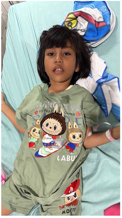 |
Figure 1 Preoperative extraoral clinical appearance showing postural instability, hypotonic lips, and strabismus. Patient-identifying details have been removed. |
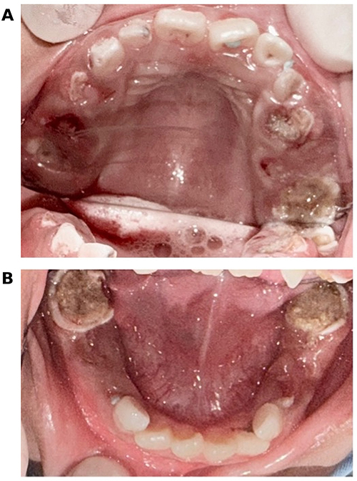 |
Figure 2 (A and B) Preoperative intraoral condition showing mixed dentition, extensive carious lesions, poor oral hygiene, and limited mouth opening. |
Diagnostic Assessment
Diagnostic assessment was based on extraoral and intraoral physical examination, clinical dental evaluation, and intraoperative findings. Although radiographs were planned, imaging could not be obtained because the patient was unable to maintain the required upright positioning due to postural instability. Clinical decisions were therefore validated using visible caries extent, remaining tooth structure, signs of root remnants, mobility, soft-tissue findings, and intraoperative pulpal status. This approach has important limitations because the absence of radiographs restricts assessment of periapical or furcation pathology, root morphology, developing permanent successors, and the full depth of carious lesions. Consequently, diagnoses such as reversible pulpitis, pulpal necrosis, and the indication for pulpotomy should be interpreted with caution and within the limits of clinical assessment alone. The pediatrician considered an odontogenic source as a potential contributor to the patient’s fever; however, other possible causes of fever could not be completely excluded from the available documentation.
Therapeutic Intervention
Treatment under general anesthesia comprised preventive, restorative, vital pulp, and surgical components. One day before the procedure, the patient attended the hospital for preoperative assessment, including laboratory testing, chest radiography, and evaluation for symptoms of upper respiratory tract infection, such as cough and rhinorrhea. On the day of the procedure, the patient was instructed to fast for 6 hours before general anesthesia. Preoperative vital signs were assessed, and the patient was re-evaluated for cough, rhinorrhea, or other symptoms that could compromise anesthetic safety. Prophylactic ampicillin was administered 30 minutes before the procedure according to institutional protocol. Intraoperatively, full-mouth scaling and prophylaxis were performed for both arches, followed by composite restorations of teeth 62, 63, 72, 26, 16, 46, and 83, pulpotomy of tooth 36, extractions of teeth 85, 84, 75, 74, 55, and 54, and topical fluoride application (Figure 3). Transient facial edema was observed on the day of surgery and managed conservatively with prescriptions and parental instructions. The patient’s clinical timeline and postoperative course are summarized in Table 1.
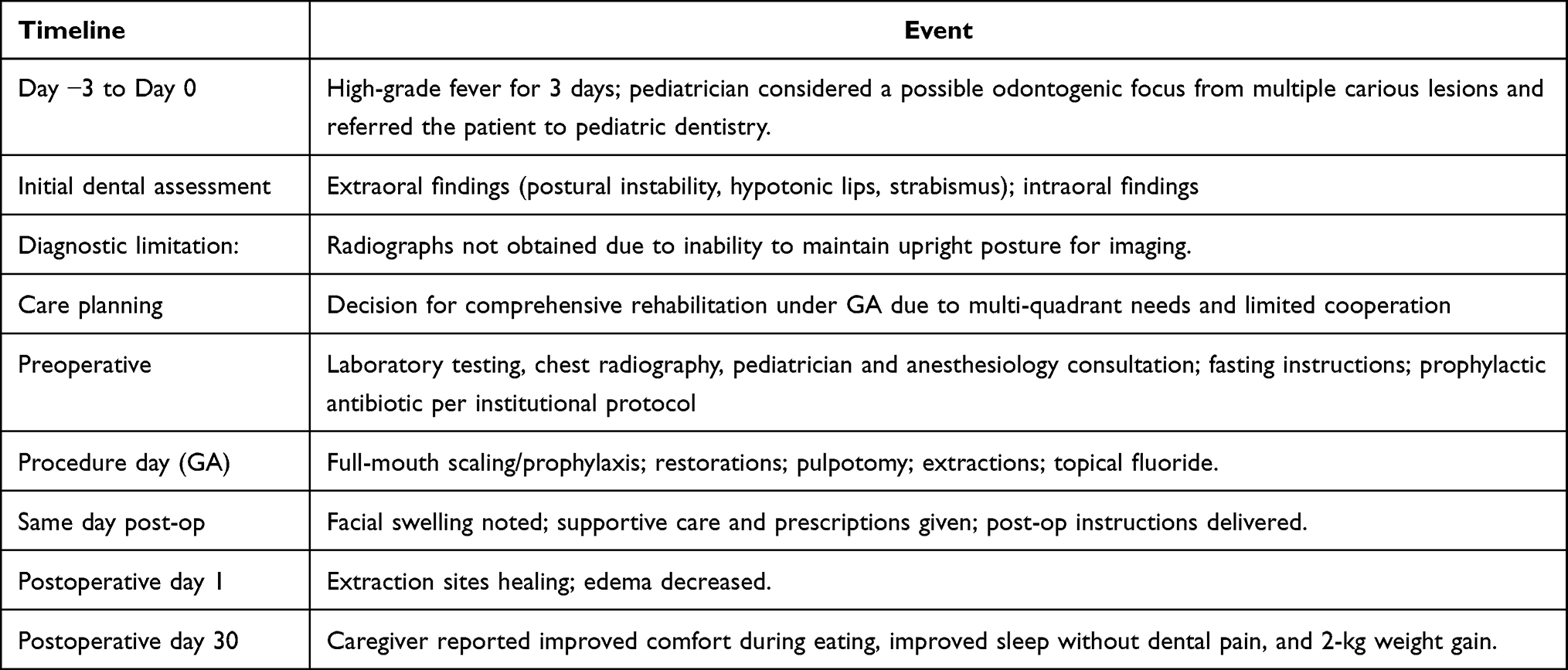 |
Table 1 Historical Timeline of Clinical Presentation, Treatment, and Follow-Up |
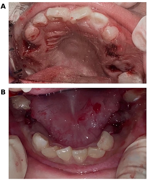 |
Figure 3 Intraoperative/postoperative condition after full-mouth oral rehabilitation under general anesthesia, including restorations, pulpotomy, extractions, prophylaxis, and fluoride application. |
Follow-Up Outcomes
The patient tolerated the procedure without intraoperative complications; however, facial swelling/edema was noted on the day of surgery. Postoperatively, maintenance intravenous Ringer’s lactate was administered and the patient was prescribed oral amoxicillin 250 mg three times daily after meals (No. X) and paracetamol 250 mg three times daily after meals as needed (No. VI). Parental instructions included avoiding manipulation of the extraction sites, avoiding hot foods and vigorous rinsing for 48 hours, and maintaining a soft diet for 72 hours before returning to a regular diet, assisting daily toothbrushing, supervising oral hygiene, and returning for scheduled review or earlier if fever, increasing swelling, bleeding, or pain occurred. At postoperative day 1, the extraction sites showed satisfactory healing and facial edema had decreased. By day 30, the patient’s mother reported improved comfort during eating, improved sleep without dental pain, and a 2-kg weight gain, with no additional follow-up diagnostic testing documented. At the 30-day follow-up, a limited frontal labial intraoral photograph showed satisfactory healing; however, full intraoral assessment could not be documented because the patient was unable to open the mouth adequately (Figure 4).
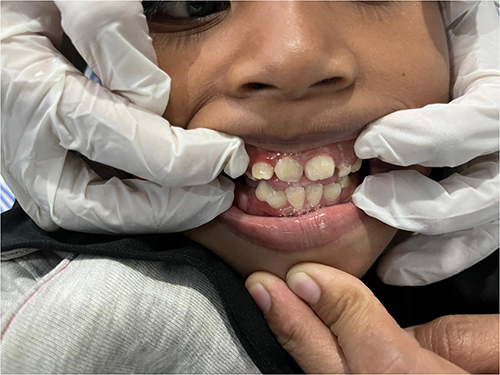 |
Figure 4 Thirty-day postoperative frontal labial intraoral photograph showing satisfactory healing. Patient-identifying details have been removed. |
Discussion
Cerebral palsy (CP) refers to a spectrum of permanent disorders affecting movement and posture that limit activity and result from non-progressive disturbances in the developing fetal or infant brain. Motor dysfunction is often accompanied by impairments in sensation and perception, cognitive and communication deficits, and behavioral disturbances, all of which can influence everyday functioning and access to health services.1 The etiology of CP is multifactorial and may involve prenatal, perinatal, and early postnatal contributors, including hypoxic–ischemic encephalopathy (HIE), infection or inflammation, cerebrovascular events, and congenital malformations. In this case, the reported birth history may be interpreted as suggestive of perinatal compromise. However, a definitive causal relationship between perinatal hypoxia/asphyxia and CP cannot be established from this case alone.2
Children with CP frequently experience increased caries risk and poorer gingival health compared with peers. Contributing factors include reduced manual dexterity, caregiver dependence, drooling/dysphagia, dietary modification, malocclusion, and barriers to routine dental services. Restricted mouth opening and atypical oral reflexes can further limit examination and preventive delivery. In the present patient, extensive caries, poor oral hygiene, and restricted mouth opening were consistent with high caries risk and reduced feasibility of incremental chairside treatment.3,4
Epilepsy coexisting with CP increases the complexity of dental treatment planning and requires seizure-safe perioperative management.2 Preoperative planning should clarify seizure type, seizure frequency, date of the last seizure, known triggers, medication doses, adherence, and the plan for maintaining antiseizure therapy during fasting and recovery.6,8,9 Missed or delayed antiseizure medication doses may increase breakthrough seizure risk; therefore, coordination with pediatrics/neurology and anesthesia is important to maintain therapeutic dosing, avoid drug interactions, and ensure prompt resumption of medication after recovery.6,9 The team should also be prepared for perioperative seizures, including airway protection, oxygenation, physiologic monitoring, rescue medication availability.6,9
Appropriate timing of antiseizure medications aims to maintain therapeutic drug levels throughout the perioperative period, as missed or delayed doses can increase the likelihood of breakthrough seizures. Preoperative planning typically includes confirming the child’s usual dosing schedule, administering the regular morning dose with a small sip of water when permitted, and establishing an alternative route (eg, intravenous) if oral intake is restricted or postoperative nausea/vomiting is anticipated.6 Postoperative home care is particularly important in children with CP because daily oral hygiene is often parent-dependent. Parents/caregivers should receive simple and practical instructions on assisted toothbrushing, safe positioning, plaque control, fluoride use when indicated, diet modification, and recognition of postoperative warning signs. Behavioral and emotional approaches such as short instructions, tell-show-do when feasible, positive reinforcement, parental presence, gradual desensitization, and distraction can support future dental visits and improve adherence to home self-care routines.6,9
Chairside management can be difficult or unsafe when cooperation is limited by cognitive or communication impairment, involuntary movements/spasticity, restricted mouth opening, and exaggerated gag reflex.4,5 Airway protection is critical during water- and debris-generating procedures in children with oromotor dysfunction because aspiration risk may be increased.4 This case required extensive multi-quadrant treatment including extractions and pulpotomy, making staged chairside completion unlikely to succeed. GA is a recognized approach for selected pediatric patients with special health care needs who cannot safely or effectively undergo required dental procedures in the clinic, enabling definitive care in a single session with controlled airway management and effective analgesia.7,10
Therapeutic decisions prioritized infection control, pain relief, and function. Teeth with pulpal necrosis and non-restorable root remnants were extracted due to poor prognosis and persistent infection risk. Teeth diagnosed with reversible pulpitis were restored conservatively, and pulpotomy was performed for one molar to preserve function where indicated. Scaling, prophylaxis and topical fluoride were included to reduce plaque burden and enhance caries resistance in a high-risk, caregiver-dependent child.7 Facial edema on the day of surgery was considered a transient postoperative finding that may have reflected local inflammatory response after multiple extractions and extensive oral treatment. Because it decreased by postoperative day 1 and no worsening systemic signs were documented, it was managed conservatively. Short-term benefits reported by the patient’s mother (improved feeding comfort, improved sleep, and 2-kg weight gain at one month) are consistent with the functional impact of eliminating oral pain and infection sources.
Strengths of this case include comprehensive single-session management under GA and documented short-term parent-reported functional improvement. Key limitations include the inability to obtain dental radiographs, incomplete documentation of epilepsy details, and short follow-up duration.7
Patient Perspective
The patient’s perspective was not directly recorded because of communication limitations. The patient’s mother reported that after treatment the child appeared more comfortable while eating, slept without signs of dental pain, and gained weight over the following month. Future follow-up should include structured parent/caregiver questions regarding appetite, sleep quality, irritability, oral hygiene burden, and difficulty following postoperative and home-care instructions.
Data Sharing Statement
The original contributions presented in the study are included in the article; further inquiries can be directed to the corresponding author.
Informed Consent Statement
Institutional approval was not required for publication of this single-patient case report according to the available documentation. Written informed consent for publication of the case details and accompanying images was obtained from the patient’s mother.
Funding
This research received no external funding.
Disclosure
The authors declare no conflicts of interest in this work.
References
1. Patel DR, Neelakantan M, Pandher K, Merrick J. Cerebral palsy in children: a clinical overview. Transl Pediatr. 2020;9(suppl 1):S125–8. doi:10.21037/tp.2020.01.01
2. Gong C, Liu A, Lian B, et al. Prevalence and related factors of epilepsy in children and adolescents with cerebral palsy: a systematic review and meta-analysis. Front Pediatr. 2023;11:1189648. doi:10.3389/fped.2023.1189648
3. Bensi C, Costacurta M, Docimo R. Oral health in children with cerebral palsy: a systematic review and meta-analysis. Spec Care Dentist. 2020;40(5):401–411. doi:10.1111/scd.12506
4. Harbi T, Rhbeini Y, Harbi S, Albargi A. Dental management of children with cerebral palsy: a review article. Int J Med Dev Ctries. 2023;7:1.
5. Wasnik M, Chandak S, Kumar S, George M, Gahold N, Bhattad D. Dental management of children with cerebral palsy: a review. J Oral Res Rev. 2020;12(1):52–58. doi:10.4103/jorr.jorr_19_19
6. Perks A, Cheema S, Mohanraj R. Anaesthesia and epilepsy. Br J Anaesth. 2012;108(4):562–571. doi:10.1093/bja/aes027
7. American Academy of Pediatric Dentistry. Management of dental patients with special health care needs. Pediatr Dent. 2018;40(6):237–242.
8. Oprea AD, Keshock MC, O’Glasser AY, et al. Preoperative management of medications for neurologic diseases: society for Perioperative Assessment and Quality Improvement consensus statement. Mayo Clin Proc. 2022;97(2):375–396. doi:10.1016/j.mayocp.2021.11.010
9. Cote CJ, Wilson S. Guidelines for monitoring and management of pediatric patients before, during, and after sedation for diagnostic and therapeutic procedures. Pediatr Dent. 2019;41(4):259–260.
10. Liviyanti D, Pratama AD. Effect of neurodevelopmental treatment frequency on functional abilities in children with cerebral palsy: a literature review. Majalah Ilmiah Fisioterapi Indonesia. 2025;13(02):319–326. doi:10.24843/mifi.000000447
 © 2026 The Author(s). This work is published and licensed by Dove Medical Press Limited. The
full terms of this license are available at https://www.dovepress.com/terms
and incorporate the Creative Commons Attribution
- Non Commercial (unported, 4.0) License.
By accessing the work you hereby accept the Terms. Non-commercial uses of the work are permitted
without any further permission from Dove Medical Press Limited, provided the work is properly
attributed. For permission for commercial use of this work, please see paragraphs 4.2 and 5 of our Terms.
© 2026 The Author(s). This work is published and licensed by Dove Medical Press Limited. The
full terms of this license are available at https://www.dovepress.com/terms
and incorporate the Creative Commons Attribution
- Non Commercial (unported, 4.0) License.
By accessing the work you hereby accept the Terms. Non-commercial uses of the work are permitted
without any further permission from Dove Medical Press Limited, provided the work is properly
attributed. For permission for commercial use of this work, please see paragraphs 4.2 and 5 of our Terms.
Recommended articles

Fracture Rates in Children with Cerebral Palsy: A Danish, Nationwide Register-Based Study
Granild-Jensen JB, Pedersen AB, Kristiansen EB, Langdahl B, Møller-Madsen B, Søndergaard C, Farholt S, Vestergaard ET, Rackauskaite G
Clinical Epidemiology 2022, 14:1405-1414
Published Date: 22 November 2022
Successful Anesthetic Management of Patient with Severe Scoliosis Due to Cerebral Palsy Who Underwent Dental Surgery
Bani Hani D, Alhowary A, Gharaibeh S, Al-zoubi R
International Medical Case Reports Journal 2025, 18:1423-1427
Published Date: 6 November 2025