")
Back to Journals » Clinical, Cosmetic and Investigational Dermatology » Volume 17
Oral Malignant Acanthosis Nigricans: An Early Diagnostic Sign for Ovarian Carcinoma: A Case Report
Authors Liu Y, Wu X , Chen S, Meng W
Received 3 November 2023
Accepted for publication 25 January 2024
Published 7 February 2024 Volume 2024:17 Pages 359—363
DOI https://doi.org/10.2147/CCID.S447977
Checked for plagiarism Yes
Review by Single anonymous peer review
Peer reviewer comments 2
Editor who approved publication: Dr Jeffrey Weinberg
Yang Liu,* Xiaoli Wu,* Siting Chen, Wenxia Meng
Stomatological Hospital, School of Stomatology, Southern Medical University, Guangzhou, 510280, People’s Republic of China
*These authors contributed equally to this work
Correspondence: Wenxia Meng, Stomatological Hospital, School of Stomatology, Southern Medical University, Guangzhou, 510280, People’s Republic of China, Email [email protected]
Abstract: Acanthosis nigricans (AN) is a dermatological condition characterised by the symmetrical development of velvety, hyperpigmented plaques predominantly in intertriginous areas such as the axillae, neck, inframammary regions, and groin. The malignant variant of AN is frequently associated with internal malignancies, particularly gastric adenocarcinoma, accounting for 55– 61% of cases. Patients exhibiting characteristic skin lesions are commonly initially evaluated in dermatology departments. This case report details a rare instance of a patient diagnosed with malignant acanthosis nigricans, presenting with only a mild form of florid oral papillomatosis concomitant with ovarian carcinoma. The early identification and management of these subtle clinical manifestations enabled timely intervention for the tumor, resulting in patient survival. There are few reported cases of malignant acanthosis nigricans associated with ovarian cancer. Oral medicine specialists should be cognisant of conditions manifesting as extensive oral papillary hyperplasia, and the possibility of an underlying malignant disease should be considered, particularly in cases of elderly-onset AN presenting exclusively with oral lesions.
Keywords: malignant acanthosis nigricans, ovarian cancer, papillary hyperplasia
Introduction
Malignant acanthosis nigricans (MAN), a rare dermatological disorder affecting both cutaneous and mucosal surfaces, requires prompt diagnosis and treatment to mitigate serious consequences.1 The cutaneous and mucosa lesion itself is benign, yet it is significant because it represents a cutaneous marker for internal malignancy. Previous studies have demonstrated an association between MAN and cancers of multiple systems, such as gastric, ovary, bladder, hepatobiliary tract, kidney, throat, and breast. MAN predominantly affects Asian females.2 Crucially, MAN often precedes the detection of the initial tumor, and patients frequently succumb to rapid cancer progression. Characteristic manifestations of MAN include symmetrical, dark, coarse, thickened, and velvety papillary lesions, predominantly affect the flexural areas of the skin.3 Patients with these features are commonly first evaluated in dermatology departments. Additionally, primary lesions of MAN may present on the oral mucosa. It is imperative for dental specialists to be conversant with both typical and atypical clinical presentations of MAN to facilitate early diagnosis. This article reports a case of MAN manifesting as mild papillomatosis, which completely resolved following treatment of the underlying malignancy, with no recurrence observed during over three years of follow-up.
Case Presentation
A 67-year-old previously healthy Chinese woman was evaluated in our clinic, presenting with diffuse and extensive oral mucosal prominence. Oral examination revealed velvety, widespread, reddish papillary lesions on the hard palate, buccal mucosa, gingiva, and the mucosal surface of the upper lip (Figure 1). General examination indicated no evidence of bone damage or perforation. Dermatological assessment showed mild pigmentation around the mouth, nape of the neck, back of the hands, and around the armpits (Figure 2). She had not sought hospital treatment previously as her symptoms were mild. Moreover, she had no significant medical or family history and was not on any medication.
 |
Figure 1 Clinical appearance of the oral cavity revealed extensive papillomatosis of the labial mucosa (a), buccal mucosa (b and c), tongue (d), gingiva (e), and hard palate (f). |
 |
Figure 2 Atypical skin manifestations around the mouth (a), posterior neck (b), back of the hand (c), and armpit (d). |
Histological analysis demonstrated papillary proliferation of the squamous epithelium (Figure 3). Elevated tumor marker levels were noted, including carbohydrate antigen (CA)_125 at 100 U/mL, CA_153 at 159.2 U/mL, cytokeratin fragment 21–1 (CYFRA21_1) at 4.97 ng/mL, and neuron-specific enolase (NSE) at 19.72 ng/mL. In contrast, CA_199, CA_724, and squamous cell carcinoma antigen (SCC) levels were within normal ranges. A systemic positron emission tomography/computed tomography scan raised strong suspicions of lymph node metastasis from ovarian cancer, which was confirmed histopathologically as regional lymph node metastasis of high-grade serous ovarian cancer. Correlating clinical and histopathological findings led to a diagnosis consistent with MAN. Following surgical and chemotherapeutic interventions, the patient’s oral mucosa and skin lesions associated with ovarian cancer completely resolved, with no recurrence observed over a three-year follow-up period (Figure 4). This case underscores that the abrupt onset of oral macular papillomatosis in older women may be indicative of ovarian malignancy.
 |
Figure 3 An incisional diagnostic biopsy of oral mucosa showed squamous epithelial papillary hyperplasia. (HE, ×200). |
 |
Figure 4 The lesions of the oral mucosa (a): labial mucosa; (b and c): buccal mucosa; (d): tongue; (e): gingiva; and (f): hard palate and skin (g): skin around the mouth; (h): posterior neck; (i): back of the hand were healed with pigmentation, with no recurrence over a period of three years. |
Discussion
The etiology of AN remains elusive despite extensive literature delineating its pathogenesis, clinical presentation, and histopathology characteristics. Research suggests a potential association between AN lesions and various factors, including metabolized cytokine-like peptides produced by the malignancies, pharmaceutical agents, and inflammatory states.4,5 Clinically, AN shares features with a spectrum of developmental, reactive, and syndromic disorders, encompassing florid cutaneous papillomatosis (FCP), linear verrucous epidermal nevus (LVEN), focal dermal hypoplasia (FDH), and lesions related to human papillomavirus (HPV), such as focal epithelial hyperplasia, squamous papillomas, condyloma, and verruca vulgaris. Accurate clinical differentiation among these entities is imperative. For a comprehensive discussion on differential diagnosis, refer to our preceding review.2
The lesions characteristic of oral AN are typically benign; however, the primary clinical concern pertains to their paraneoplastic origins. Literature reports suggest that such lesions resolve following the treatment of the associated malignancies (Table 1). Yet, there is a dearth of documentation regarding the prognosis of oral lesions in cases of ovarian cancer-related MAN. In this instance, we detail the inaugural report on the outcome following surgical and chemotherapeutic intervention for ovarian cancer, which culminated in the complete resolution of oral mucosa and skin lesions, with no recurrence observed throughout an extensive follow-up period exceeding three years. The paraneoplastic mucosal presentations of MAN typically manifest extensively and abruptly, often coinciding with the initial medical consultation, which may, at times, be delayed.6 A considerable number of patients with MAN succumb rapidly to cancer progression.7 Early detection of associated tumors is crucial to afford patients the opportunity for timely and effective treatment, thereby enhancing survival prospects. In the present case, the patient’s lesions were so subtle that they could have been easily overlooked or misdiagnosed. Owing to the prompt recognition and management of these mild clinical features, the patient was able to receive timely oncological treatment and consequently survived.
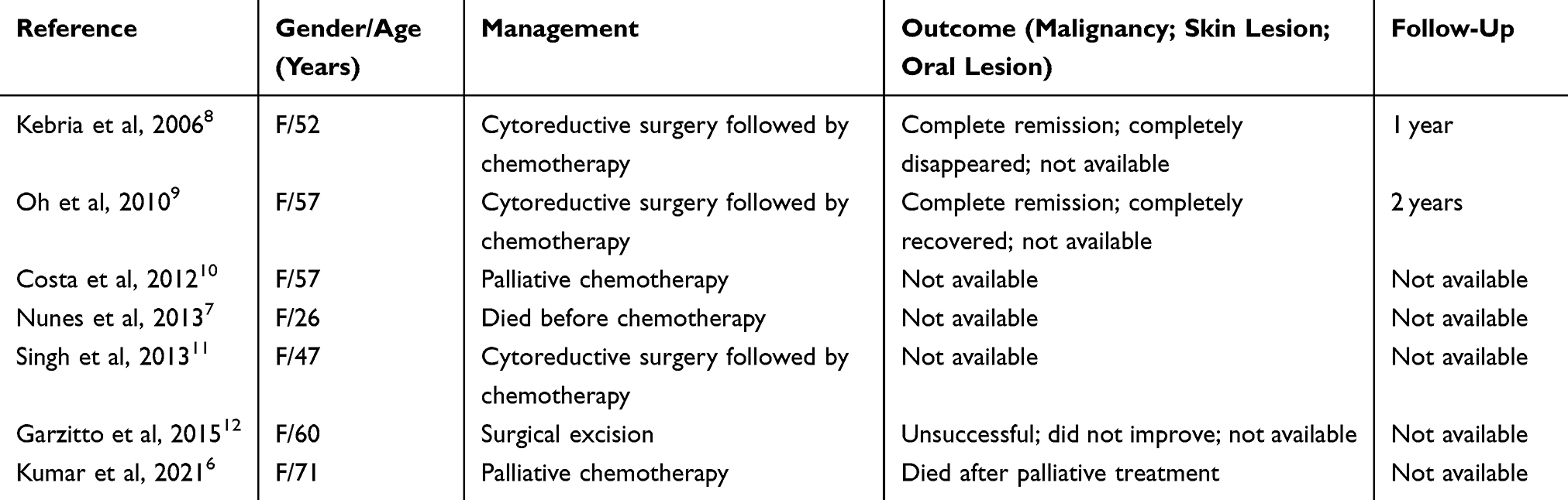 |
Table 1 Case Reports of Malignant Acanthosis Nigricans with Ovarian Cancer and the Treatment of Tumor and Outcome |
Through this case, we observe that stomatologists must remain vigilant for the potential of underlying malignant conditions when treating patients presenting with oral papillary hyperplasia. It is imperative that these dental specialists guide patients to seek prompt medical evaluation. Additionally, stomatologists ought to monitor for mucosal lesions that might signal tumor recurrence and advocate for regular oral examinations. Even after comprehensive treatment of the tumor, when oral lesions have fully subsided, long-term follow-up been essential to mitigate the risk of recurrence.
Conclusion
We present a case of malignant acanthosis nigricans that was atypical, with pronounced oral manifestations but without significant skin lesions. Post-surgical and chemotherapeutic treatment for ovarian cancer, the patient’s oral mucosa exhibited complete healing, with no recurrence over a period of three years. This case underscores several key learnings: Dental specialists must be attentive to oral signs of paraneoplastic syndromes and facilitate timely intervention to ensure early diagnosis and treatment.
Consent Statement
The patient had given written informed consent for the publication of his clinical details and accompanying images. Institutional approval is not required for this case study.
Acknowledgments
The authors acknowledge the Nonprofit Industry Research Specific Fund of National Health and Family Planning Commission of China (No.201502018) for financial support.
Disclosure
Yang Liu and Xiaoli Wu are co-first authors for this study. The authors have no conflicts of interest to declare for this work.
References
1. Sinha S, Schwartz RA. Juvenile acanthosis nigricans. J Am Acad Dermatol. 2007;57(3):502–508. doi:10.1016/j.jaad.2006.08.016
2. Liu Y, Xu X, Yang Y, et al. Malignant acanthosis nigricans and diseases with extensive oral papillary hyperplasia. Clin Exp Dermatol. 2022;47(4):651–657. doi:10.1111/ced.14995
3. Kutlubay Z, Engin B, Bairamov O, Tüzün Y. Acanthosis nigricans: a fold (intertriginous) dermatosis. Clin Dermatol. 2015;33(4):466–470. doi:10.1016/j.clindermatol.2015.04.010
4. Patel NU, Roach C, Alinia H, Huang WW, Feldman SR. Current treatment options for acanthosis nigricans. Clin Cosmet Invest Dermatol. 2018;11:407–413. doi:10.2147/CCID.S137527
5. Daye M, Temiz SA, Işık B, Durduran Y. Relationship between acanthosis nigricans, acrochordon and metabolic syndrome in patients with lichen planus. Int J Clin Pract. 2021;75(10):e14687. doi:10.1111/ijcp.14687
6. Kumar P, Mukundan MK, Sehrawat A, Sundriyal D. Tripe palms and malignant acanthosis nigricans: more than a diagnostic pointer. Cancer Rep. 2021;4(1):e1307. doi:10.1002/cnr2.1307
7. Nunes MC, Moreira DR, Ferrari TC. Cardiac metastasis from yolk sac tumor: case report and review. Exp Hematol Oncol. 2013;2(1):13. doi:10.1186/2162-3619-2-13
8. Kebria MM, Belinson J, Kim R, Mekhail TM. Malignant acanthosis nigricans, tripe palms and the sign of Leser-Tre’lat, a hint to the diagnosis of early stage ovarian cancer: a case report and review of the literature. Gynecologic Oncol. 2006;101(2):353–355. doi:10.1016/j.ygyno.2005.12.024
9. Oh CW, Yoon J, Kim CY. Malignant acanthosis nigricans associated with ovarian cancer. Case Rep Dermatol. 2010;2(2):103–109. doi:10.1159/000317116
10. Costa MC, Martinez NS, Belicha MG, Leal F. Acanthosis nigricans and ”tripe palm” as paraneoplastic manifestations of metastatic tumor. Anais Brasil De Dermatol. 2012;87(3):498–500. doi:10.1590/S0365-05962012000300030
11. Singh SK, Rai T. A rare case of malignant acanthosis nigricans in a lady with ovarian cancer. Indian Dermatol Online J. 2013;4(2):125–127. doi:10.4103/2229-5178.110640
12. Garzitto A, Ricceri F, Pescitelli L, Tripo L, Prignano F. Vitiligo masks malignant acanthosis nigricans in a woman with ovarian cancer. Int J Dermatol. 2015;54(11):1300–1302. doi:10.1111/ijd.12358
 © 2024 The Author(s). This work is published and licensed by Dove Medical Press Limited. The full terms of this license are available at https://www.dovepress.com/terms.php and incorporate the Creative Commons Attribution - Non Commercial (unported, v3.0) License.
By accessing the work you hereby accept the Terms. Non-commercial uses of the work are permitted without any further permission from Dove Medical Press Limited, provided the work is properly attributed. For permission for commercial use of this work, please see paragraphs 4.2 and 5 of our Terms.
© 2024 The Author(s). This work is published and licensed by Dove Medical Press Limited. The full terms of this license are available at https://www.dovepress.com/terms.php and incorporate the Creative Commons Attribution - Non Commercial (unported, v3.0) License.
By accessing the work you hereby accept the Terms. Non-commercial uses of the work are permitted without any further permission from Dove Medical Press Limited, provided the work is properly attributed. For permission for commercial use of this work, please see paragraphs 4.2 and 5 of our Terms.