")
Back to Journals » International Journal of Nanomedicine » Volume 14
On the importance of modeling gold nanoparticles distribution in dose-enhanced radiotherapy
Authors Rasouli FS, Masoudi SF, Asadi S
Received 5 May 2019
Accepted for publication 2 July 2019
Published 29 July 2019 Volume 2019:14 Pages 5865—5874
DOI https://doi.org/10.2147/IJN.S214517
Checked for plagiarism Yes
Review by Single anonymous peer review
Peer reviewer comments 4
Editor who approved publication: Dr Thomas Webster
Fatemeh S Rasouli,1 S Farhad Masoudi,1 Somayeh Asadi2
1Department of Physics, K.N. Toosi University of Technology, Tehran, Iran; 2Department of Mechanical Engineering, Politecnico Di Milano, Milan, Italy
Purpose: To investigate the effect of precise modeling for Monte Carlo simulations of gold nanoparticles (GNPs) dose-enhanced radiotherapy, two models characterized by their distribution of GNPs in a simulated macroscopic cubic tumor were introduced. The motivation was the widely documented tendency of GNPs to localize around the cell nucleus.
Methods: The introduced models composed of 2.7×10⁷ ellipsoid cells, each of them containing a centrally located nucleus as the target for dose evaluation. In the first model, the spheres of GNP are homogeneously distributed in the whole tumor volume, and in the latter, GNPs are localized in the cytoplasms surrounded the nuclei.
Results: The results achieved through applying Monte Carlo radiation transports using the Mont Carlo N-Particle eXtended code (MCNPX) show an underestimation of nuclear dose enhancement caused by homogeneous model compared with that of heterogeneous distribution. By investigating various quantities, it was found that subcellular location of GNPs strongly governs the sensitivity of dose enhancement to the number and concentration of GNPs targeted in the tumor. Other obvious differences are revealed by studying the dose enhancement curves in depth of the tumor. While the heterogeneous model predicts an approximately constant dose enhancement in depth for primary photon energies of 50 keV and more, the homogeneous model estimates an energy-dependent increase of about 11 to 30%.
Conclusion: It can be concluded that defining a model in accordance with the experimental observations can effectively account for accurate prediction of macroscopic dose enhancement in the target of interest.
Keywords: gold nanoparticles, dose-enhanced radiotherapy, cell model, tumor, Monte Carlo simulation
Introduction
The primary objective of an ideal tumor treatment is the delivery of a high dose to the tumor while sparing surrounding healthy tissue. Due to the secondary electrons produced through physical interactions between photons and tissue, radiotherapy provides benefitial conditions to deliver appropriate doses for treating tumors. Moreover, it has been shown that it would be possible to selectively increase the delivered dose to the target of interest during radiation. Owing to the ability of materials with a high atomic number to absorb ionizing radiation, using the high-Z constituent atoms of nanoparticles would result in a physically enhanced therapeutic efficacy of cancer treatment. For the primary energies below about 500 keV which irradiate to the medium loaded with these atoms, the radiation interaction is primarily via the photoelectric effect. Multiple photoelectrons emitted from atoms of these agents passively targeted in the tumor, as well as the secondaries produced along the primary radiation trajectory, are expected to result in dose enhancement. Extensive discussions on physical processes involved in the interactions of photons with high-Z nanoparticles can be found in the literature.1,2 Arising from both the chemical properties and high biocompatibility, gold nanoparticles (GNPs) are the most appropriate candidates among many tested so far.3,4
Transport of photons through a chosen target and their interactions with GNPs, as well as evaluations of dose enhancement, can be studied by in vitro or in vivo methods or Monte Carlo simulations. Although enhanced radiation therapy using GNPs is experimentally available, researchers working on the parameters governing the degree of dose enhancement are often involved in planning pre-clinical tests. Given that computer simulations using the Monte Carlo method allow satisfying simulation of experimental conditions and provide valuable insights into the scenarios which are difficult to create or measure experimentally, various Monte Carlo codes have been developed. There are a number of Monte Carlo-based published works discussing the potential of using GNPs to enhance the radiation efficacy and studying the effects of size, shape, and concentration of GNPs as well as the energy of irradiated photons on the dose delivered to the tumor.5–8
Studies which have focused on examining microscopic scenarios of dose distribution in nanoscale in the vicinity of a single GNP have revealed that the contribution of Auger electrons in the dose dominates in the vicinity of the nanoparticle and falls off rapidly beyond ~200 nm,9 resulting in a statistically insignificant effect on the overall dose enhancement in the cell.10 These studies do not give an insight into the energy deposition patterns in the presence of numerous GNPs, because of the complicated scattering processes which primary and secondary particles are involved in. On the other hand, a number of works have been dedicated to investigating the importance of modeling GNPs in tissue to achieve accurate and reliable dose calculations. They present a comparison between the results corresponding to simulating a uniform mixture of gold and water and those of modeling a medium with a given number of spherical GNPs homogeneously placed in the medium.11–13 Their results show an overestimation of dose enhancement caused by modeling a gold–water mixture rather than gold nanospheres.
Taking into account the widely documented observation that GNPs have a tendency to heterogenetically disperse in the medium, mostly in the cytoplasm around the nucleus,14,15 in a recently published study16 we investigated the effect of the distribution of nanoparticles in a single cell on nucleus dose enhancement. The results emphasized that an appropriate small-scale model of distribution of GNPs in the cell is is of high importance to accurately estimate the degree of dose enhancement for clinical application. While being interesting, the outcomes are limited to a model consisting of a single cell in micrometer dimensions. Inspired by these results, in the present work, we tried to extend our single cell model to a typical tumor of centimeter dimensions containing millions of cells. The results are expected to answer the question of whether focusing on precise modeling of the distribution of GNPs on the cellular level leads to a significant difference in dose enhancement on a macroscopic scale. This study is planned to investigate the sensitivity of the dose calculated over a tumor of realistic dimensions to the localization of GNPs in tissue. To address this issue, the work is structured as follows. A typical tumor volume filled with cells containing nucleus and cytoplasm is designed. For homogeneous and heterogeneous distribution of gold nanospheres in the tumor volume, the parameters governing the dose enhancement are investigated. The dose enhancement curves in the depth of the phantom are used to study the performance of the irradiated beam in the direction of irradiation.
Materials and methods
Simulations and particle tracking in the medium were carried out using the Monte Carlo N-Particle eXtended code (MCNPX). Owing to the fact that uncertainty of Monte Carlo results depends on the number of histories, an adequate number of particles were tracked to achieve a given accuracy and reliable results. In order to examine the dose enhancement in the presence of GNPs in the designed geometries, we used dose enhancement factor (DEF) notation, which is defined as the ratio of the dose to the target of interest in the presence of GNPs to that in the absence of these particles. To show the concentration of gold per gram of water, we used the notation of mgAu/gwater. Each GNP was considered to be a sphere of 30, 50, 70 or 100 nm in diameter, and was assumed to be composed of pure gold.
A cubic tumor of 1.2×1.2×1.2 cm3 was designed for transporting particles and evaluation of the deposited dose. This tumor is filled with 2.7×10⁷ cells, each modeled as described in our previous study:16 an ellipsoid with the semi-major axis of 18 µm and two equal semi-minor axes of 10 µm with a centrally located sphere of 7 µm radius as the nucleus. The volume delimited between this ellipsoid and the nucleus is named the cytoplasm. Each cell is surrounded by a cube of 40×40×40 µm3. Monoenergetic photons of 10–100 keV which are emitted from a point source located at 1.2 cm distance from the center of the tumor irradiate to the cells. In addition to the electron linac-based sources, these energies are also available by using brachytherapy sources such as 131Cs, 103Pd, and 125I. The primary particles were confined to emit in a right circular cone with the equal radius and height of 0.6 cm. As DNA in the nucleus is believed to be the biologically relevant target of radiation, the physical doses due to the irradiation of these photons in either the presence or absence of GNPs were scored in the nucleus of each cell. The data were averaged over all the nuclei located in the tumor.
Two patterns were used to disperse GNPs in cells of the cubic tumor:
- The homogeneous model, in which the spheres of GNPs are distributed homogeneously in the whole volume of the tumor, except in the nuclei. The uniform distribution of nanoparticles throughout both the cytoplasm and surrounding medium in this model can be considered equivalent to the assumption that all of the tumor volume is sensitive to absorbing GNPs, and accordingly to the ionizing radiation. Alongside the model in which all the target volume is considered as a fully homogeneous gold–water mixture, this model is also one of the most common geometries used for simulation of GNPs.7
- The heterogeneous model, in accordance with the observation that GNPs have a tendency to localize in the cytoplasm around the nuclei.
In these models, the nuclei and free spaces between the GNPs are defined as water. Moreover, the water model, which is considered as a fully homogeneous water material, was employed to test the dose enhancement due to the presence of GNPs in the models. The corresponding data were denoted as control. Figure 1 shows a schematic 3-D view of the geometries simulated.
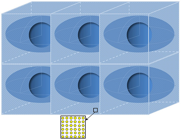 |
Figure 1 A schematic 3-D view of the simulated geometries. In the homogeneous model, spherical GNPs are homogeneously distributed in the whole medium. In the heterogeneous model, the spheres of GNPs have uniformly localized in the cytoplasm of each cell (ellipsoidal volumes). In both models, the nuclei (central spheres) are excluded to be filled with GNPs, and the free space between the GNPs is filled with water. Number of cells and dimensions are not to scale.Abbreviation: GNPs, gold nanoparticles. |
In order to examine the behavior of various quantities in our models, the deviation between the results corresponding to these data was calculated. If the data of the desired quantity for the heterogeneous model are denoted by Xhetro, and the corresponding value from the homogeneous model is characterized by Xhomo, the deviation is then represented by (Xhetro/Xhomo)−1.
Results and discussion
Figure 2 reports the energy dependence of the relative values of averaged physical doses deposited in the nuclei of the tumor for the models introduced, as well as those of control (no gold). These values have been calculated considering the reference value of the dose delivered to the water (control) for the primary photons of 10 keV. The values of DEF can be easily obtained by dividing the relative dose for the homogeneous/heterogeneous model of a given energy by its corresponding value in water (control). The results correspond to a typical concentration of 20 mgAu/gwater of 50 nm GNPs. This figure makes two points explicit: the DEFs are highly dependent on primary energies, and on the model used for localization of GNPs. In the homogeneous model, DEFs do not exceed ~1.72 and drop below unity for energies lower than 20 keV. These results most likely can be interpreted due to self-absorption within the nanoparticles arising from the distance between GNPs and the nuclei of the tumor. For the heterogeneous model, DEFs range between minimum and maximum values of 1.14 (for 10 keV photons) and about 8.8 (for 30 keV photons), respectively. While the figure shows that the DEFs corresponding to both models are highly dependent on the energies of primary photons, and approximately experience peaks and valleys on the same positions, it reveals an obvious difference between the DEFs due to the models simulated. The results suggest that radiotherapy can be enhanced significantly only if the GNPs are in proximity to the biological target, as occurs in realistic conditions. Table 1 reports the values of DEF as well as deviation from DEF of the heterogeneous model of those corresponding to the homogeneous model for the ten energies tested, and gives a deeper understanding of these differences. The values highlight the importance of defining the cellular location of GNPs in the target of interest on DEF, especially for energies below 40 keV.
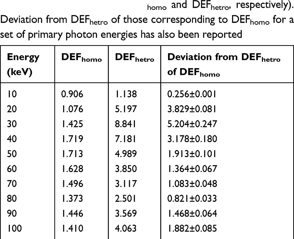 |
Table 1 The values of DEF for the homogeneous and heterogeneous models (denoted by DEFhomo and DEFhetro, respectively). Deviation from DEFhetro of those corresponding to DEFhomo for a set of primary photon energies has also been reported |
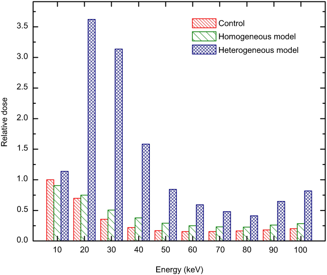 |
Figure 2 Energy dependence of the relative values of averaged physical doses deposited in the nuclei of the tumor for the models introduced (see Figure 1), and those of the control (no gold). The results correspond to the typical concentration of 20 mgAu/gwater of 50 nm GNPs, and the dose delivered to the water (control) for primary photons of 10 keV is considered as the reference value.Abbreviation: GNPs, gold nanoparticles. |
The study has been extended to other gold concentrations. For a given concentration, the DEFs follow the same behavior observed in Figure 2. However, more interesting results are obtained by comparing the sensitivity of the introduced models to variation of this quantity. For 50 nm GNPs with concentrations of 5, 10, 15, 20, 25, and 30 mgAu/gwater, the results are shown in Figure 3. These concentrations are equivalent to 6.84×10¹², 1.36×10¹³, 2.05×10¹³, 2.73×10¹³, 3.42×10¹³, and 4.10×10¹³ number of GNPs in the tumor volume. In both models, the more the concentration of GNPs increases, the more the degree of dose enhancement grows, due to the increment in the number of photon interactions. Furthermore, while there is a linear dependence of DEFs on the number of GNPs, the slopes of these straight lines are energy-dependent. In spite of these similar behaviors, the sensitivity of two models to the number of GNPs is not the same. In the homogeneous model, for the typical energy of 30 keV, deviation from the DEF corresponding to the concentration of 30 mgAu/gwater of that of 5 mgAu/gwater reaches about 37.5%. This deviation reaches 263% in the heterogeneous model. The results exhibit that the subcellular location of GNPs strongly governs the sensitivity of dose enhancement over all of the tumor to the number of GNPs targeted in the media.
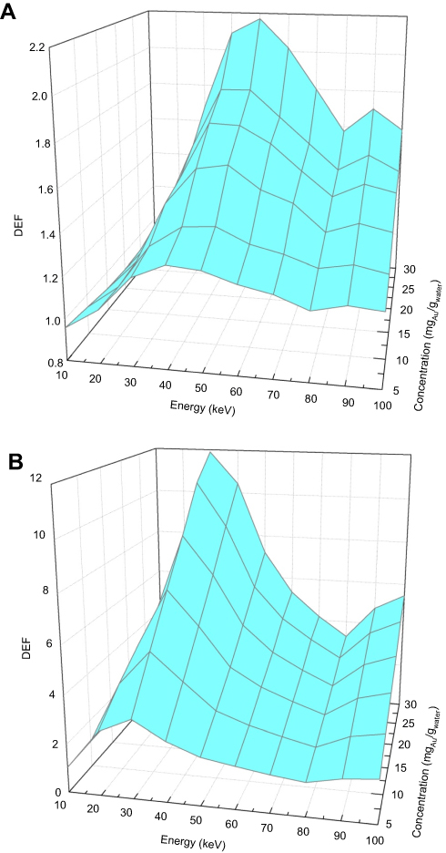 |
Figure 3 3-D plot to show the dependency of DEFs on both the concentration of 50 nm GNPs and primary photon energy for (A) the homogeneous model, and (B) the heterogeneous model. The relative errors, not shown in the plots, are less than 1%.Abbreviations: DEF, dose enhancement factor; GNPs, gold nanoparticles. |
Figure 4 reports deviation from DEF of the heterogeneous model of those corresponding to the homogeneous model for the ten energies tested, and for six different concentrations. As the figure shows, the more the concentration of GNPs increases, the more the DEFs deviate. This deviation depends on the primary energy of photons.
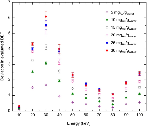 |
Figure 4 Deviation from the values of DEF for the tumor with heterogeneous model of those corresponding to the tumor with homogeneous model for a set of primary photon energies. The results are reported for six different concentrations of GNPs.Abbreviations: DEF, dose enhancement factor; GNPs, gold nanoparticles. |
To test the influence of GNP size on DEF in our models, four arbitrary sizes of 30, 50, 70 and 100 nm were chosen. These values refer to the diameters of each spherical GNP dispersed in the tumor. By keeping the concentration of GNPs in the homogeneous model constant, it was found that dose enhancement varies as a function of both the GNPs’ size and the incident photon energy. For a given energy, the values of DEF increase by increasing the size. For the typical concentration of 20 mgAu/gwater, this increment ranges between about 6.6% and 107% for 20 and 50 keV photons, respectively. More details can be found in Figure 5A. Unlike the homogeneous model, the values of DEF do not change significantly by size variation in the other introduced model. As Figure 5B shows, while dose enhancements are energy-dependent, they show a small change, on the order of a few percent, by changing the GNPs’ size. For the constant concentration mentioned, these variations range between about 0.44% and 6.6% for 90 keV and 30 keV primary photons, respectively. This effect can be understood as the result of increasing numbers of low-energy electrons trapped inside the GNPs and do not contribute to the dose delivered to the nuclei. It is worth noting that the sizes chosen in this work are only used to assess the effect of this parameter on physical dose enhancement, and are not necessarily beneficial in experimental studies. Other parameters such as performance of the GNPs in delivery to the target, their adsorption and internalization by cells, and intracellular uptake play influential roles on making GNPs more advantageous.
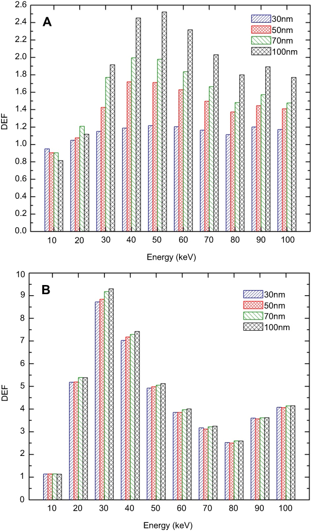 |
Figure 5 Size-energy dependence of DEFs evaluated in the nuclei of the tumor for (A) homogeneous model, and (B) heterogeneous model. The results correspond to the typical concentration of 20 mgAu/gwater. Abbreviation: DEF, dose enhancement factor. |
The foregoing results present dose enhancement averaged over all the nuclei in the tumor volume. It would also be worth while to study the dependency of DEFs on the depth of the tumor. The depth–DEF curves help to understand the performance of the irradiated beam in the direction of the height of the conceptual cone which encloses the primary photons. To address this issue, the tumor has been divided into twelve layers perpendicular to the source axis. The reported values have been averaged over the values of DEF calculated in the nuclei of all 225×104 cells located in each layer. The results corresponding to the mentioned condition for an arbitrary concentration of 20 mgAu/gwater of 50 nm GNPs for our two models subjected to the irradiation of various energies are summarized in Figure 6. According to this figure, the values of DEF in the depth of the phantom for primary energies of about 20 keV drop below unity. This is due to the fact that in these energies, the decrement in the fluence of photons overcomes the local energy deposition due to the generated photoelectrons. Moreover, there is a more interesting point about these curves: as Figure 6B shows, for energies greater than 30 keV, DEFs are approximately constant with depth, with negligible variations of about 0.3–4%. This can be justified by the mean energy of the secondary electrons in depth; as can be understood from Figure 2, for primary energies above 30 keV, the DEFs in the heterogeneous model experience a decrement in comparison with their maximum value. On the other hand, while by penetrating in depth of the tumor the mean energy of primary photons remains approximately constant, their number decreases significantly. Nevertheless, the mean energy of the secondary electrons corresponding to the primary photons of E>30 keV ranges between 30 and 40 keV in depth (see Figure 7), which results in multiple electron production followed by dose enhancement in each layer.
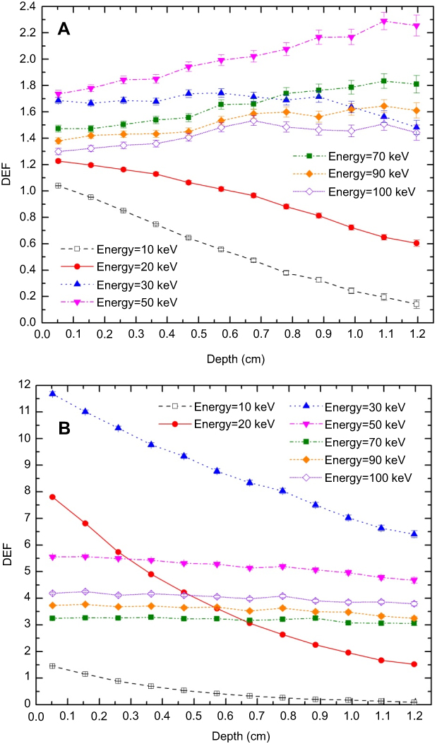 |
Figure 6 The values of DEF versus depth within the layers (each 148 mm3) perpendicular to the beam axis for (A) homogeneous model, and (B) heterogeneous model. The results correspond to the typical concentration of 20 mgAu/gwater of 50 nm GNPs and seven arbitrary incident photon energies.Abbreviation: DEF, dose enhancement factor. |
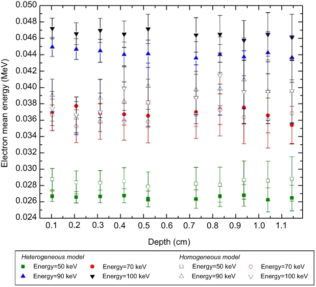 |
Figure 7 Mean energy of secondary electrons in depth of the tumor for primary photons of 50, 70, 90 and 100 keV irradiated to the layers (each 148 mm3) perpendicular to the beam axis for homogeneous (filled symbols) and homogeneous (open symbols) models. The results correspond to the typical concentration of 20 mgAu/gwater of 50 nm GNPs.Abbreviation: GNPs, gold nanoparticles. |
Except for primary photons below 30 keV which irradiated to the homogeneous model, the values of DEF increase by depth (Figure 6A). For example, the value of DEF averaged over the nuclei of the last layer of the tumor deviates from that of the first layer by about 30% and 11% for 50 and 100 keV photons, respectively. Similar to the case of the heterogeneous model, the mean energy of the secondary electrons ranges between 30 and 40 keV in depth (Figure 7). The electron cascade produced in depth dominates the decremented number of the primary photons in deep layers and results in DEF enhancement. While the heterogeneous model predicts an approximately constant value of DEF in depth, the homogeneous model estimates an energy-dependent increase. Strictly speaking, this model may result in a misleading conception about uniform dose enhancement in the tumor volume. Moreover, though for the homogeneous model the different primary energies result in approximately similar values of DEF in the first layers, the heterogeneous model behaves in a different way. For example, the values of DEF in the first layer of the homogeneous model range between about 1.3 and 1.7 for primary photons of 100 keV and 50 keV, respectively. This interval extends from 3.24 to 5.55 for the heterogeneous model, which is equivalent to a deviation of about 71.3%, more than 2.3 times that of the homogeneous model. These results emphasize that modeling GNPs embedded uniformly in the medium does not give a precise comprehension of the performance of depth–DEF curves in tissue.
Conclusion
The present work was devoted to investigating the effect of using a proposed model considering the widely documented observation that GNPs have a tendency to heterogenetically localize around the cell nucleus in dose-enhanced radiotherapy. We aimed to address the issue of whether precise modeling of the distribution of GNPs at a cellular level leads to a considerable difference on the dose delivered to a typical phantom at a macroscopic scale, and to clarify the importance of this detailed modeling on the computation of physical dose enhancement in nanoparticle radiation therapy. As the dose in the nucleus is an appropriate predictor of the radiosensitizing effect of GNPs combined with photons’ irradiation, the degree of dose enhancement was evaluated through the calculation in the nucleus of each constituent cell of the tumor and averaged over all the nuclei. The results show that the subcellular location of GNPs strongly governs the sensitivity of dose enhancement over all the tumor to various characteristics of GNPs targeted in the media. In conclusion, the prediction of the behavior of macroscopic dose enhancement depends highly on the modeling of nanoparticles, and geometry of localizing them in the medium plays a key role on improving the simulation of dose-enhanced radiotherapy.
Disclosure
The authors report no conflicts of interest in this work.
References
1. Butterworth KT, McMahon SJ, Currell FJ, Prise KM. Physical basis and biological mechanisms of gold nanoparticle radiosensitization. Nanoscale. 2012;4(16):4830–4838. doi:10.1039/c2nr31227a
2. Kuncic Z, Lacombe S. Nanoparticle radio-enhancement: principles, progress and application to cancer treatment. Phys Med Biol. 2018;63(2):02TR01. doi:10.1088/1361-6560/aabd50
3. Perrault SD, Walkey C, Jennings T, Fischer HC, Chan WC. Mediating tumor targeting efficiency of nanoparticles through design. Nano Lett. 2009;9(5):1909–19015. doi:10.1021/nl900031y
4. Laurent G, Bernhard C, Dufort S, et al. Minor changes in the macrocyclic ligands but major consequences on the efficiency of gold nanoparticles designed for radiosensitization. Nanoscale. 2016;8(23):12054–12065. doi:10.1039/C6NR01228K
5. Cho SH. Estimation of tumor dose enhancement due to gold nanoparticles during typical radiation treatments: a preliminary Monte Carlo study. Med Phys. 2005;32(6):2162. doi:10.1118/1.1998660
6. McMahon SJ, Hyland WB, Muir MF, et al. Nanodosimetric effects of gold nanoparticles in megavoltage radiation therapy. Radiother Oncol. 2011;100(3):412–416. doi:10.1016/j.radonc.2011.08.026
7. Lechtman E, Chattopadhyay N, Cai Z, Mashouf S, Reilly R, Pignol JP. Implications on clinical scenario of gold nanoparticle radiosensitization in regards to photon energy, nanoparticle size, concentration and location. Phys Med Biol. 2011;56(15):4631. doi:10.1088/0031-9155/56/15/001
8. Jones BL, Krishnan S, Cho SH. Estimation of microscopic dose enhancement factor around gold nanoparticles by Monte Carlo calculations. Med Phys. 2010;37(7 Part1):3809–3816. doi:10.1118/1.3455703
9. McMahon SJ, Hyland WB, Muir MF, et al. Biological consequences of nanoscale energy deposition near irradiated heavy atom nanoparticles. Sci Rep. 2011;1:18. doi:10.1038/srep00018
10. Douglass M, Bezak E, Penfold S. Monte Carlo investigation of the increased radiation deposition due to gold nanoparticles using kilovoltage and megavoltage photons in a 3D randomized cell model. Med Phys. 2013;40(7):071710. doi:10.1118/1.4771960
11. Zhang SX, Gao J, Buchholz TA, et al. Quantifying tumor-selective radiation dose enhancements using gold nanoparticles: a Monte Carlo simulation study. Biomed Microdevices. 2009;11(4):925. doi:10.1007/s10544-009-9309-5
12. Koger B, Kirkby C. A method for converting dose-to-medium to dose-to-tissue in Monte Carlo studies of gold nanoparticle-enhanced radiotherapy. Phys Med Biol. 2016;61(5):2014. doi:10.1088/0031-9155/61/5/2014
13. Martinov MP, Thomson RM. Heterogeneous multiscale Monte Carlo simulations for gold nanoparticle radiosensitization. Med Phys. 2017;44(2):644–653. doi:10.1002/mp.12061
14. Štefančíková L, Porcel E, Eustache P, et al. Cell localisation of gadolinium-based nanoparticles and related radiosensitising efficacy in glioblastoma cells. Cancer Nanotechnol. 2014;5(1):6. doi:10.1186/s12645-014-0006-6
15. Lechtman E, Mashouf S, Chattopadhyay N, et al. A Monte Carlo-based model of gold nanoparticle radiosensitization accounting for increased radiobiological effectiveness. Phys Med Biol. 2013;58(10):3075–3087. doi:10.1088/0031-9155/58/10/3075
16. Rasouli FS, Masoudi SF. Monte Carlo investigation of the effect of gold nanoparticles’ distribution on cellular dose enhancement. Radiat Phys Chem. 2019;158(1):6–12. doi:10.1016/j.radphyschem.2019.01.006
 © 2019 The Author(s). This work is published and licensed by Dove Medical Press Limited. The full terms of this license are available at https://www.dovepress.com/terms.php and incorporate the Creative Commons Attribution - Non Commercial (unported, v3.0) License.
By accessing the work you hereby accept the Terms. Non-commercial uses of the work are permitted without any further permission from Dove Medical Press Limited, provided the work is properly attributed. For permission for commercial use of this work, please see paragraphs 4.2 and 5 of our Terms.
© 2019 The Author(s). This work is published and licensed by Dove Medical Press Limited. The full terms of this license are available at https://www.dovepress.com/terms.php and incorporate the Creative Commons Attribution - Non Commercial (unported, v3.0) License.
By accessing the work you hereby accept the Terms. Non-commercial uses of the work are permitted without any further permission from Dove Medical Press Limited, provided the work is properly attributed. For permission for commercial use of this work, please see paragraphs 4.2 and 5 of our Terms.