Back to Journals » International Journal of Women's Health » Volume 14
Obstetric Performance Following Post-Traumatic Recurrent Fundal Uterine Rupture: A Case Report
Authors Worku M ![]() , Teloye MT
, Teloye MT ![]() , Ketema W
, Ketema W ![]()
Received 8 August 2022
Accepted for publication 13 October 2022
Published 15 October 2022 Volume 2022:14 Pages 1459—1463
DOI https://doi.org/10.2147/IJWH.S385397
Checked for plagiarism Yes
Review by Single anonymous peer review
Peer reviewer comments 2
Editor who approved publication: Professor Elie Al-Chaer
Misganaw Worku,1 Meles Tazeb Teloye,1 Worku Ketema2
1Department of Obstetrics and Gynecology, HawassaUniversity, Hawassa, Sidama Region, Ethiopia; 2Department of Pediatrics and Child Health, Hawassa University, Hawassa, Sidama Region, Ethiopia
Correspondence: Worku Ketema, Email [email protected]
Abstract: A case of recurrent post-traumatic fundal uterine rupture followed by an uncomplicated alive birth, unusual obstetric performance following extensive myometrial damage, happened in Hawassa University comprehensive specialized hospital. The first incident of fundal rupture was 5 years earlier than the second, after she sustained a road traffic accident at the gestational age of 31 weeks. There was associated polytrauma. The second uterine rupture was at 33 weeks of gestation, following a fall-down accident on a flat surface while performing routine household activities. The third pregnancy was an uncomplicated elective cesarean delivery, and the tubes were not ligated as the woman strongly wishes to preserve her future fertility. A single facility experience with this rare clinical scenario of obstetric performance is reported to stimulate interest in additional research into the subsequent obstetric performance of patients with recurrent fundal uterine rupture and resultant extensive myometrial damage.
Keywords: recurrent post-traumatic fundal uterine rupture, uncomplicated alive birth
Introduction
Rupture of the uterus remains a common obstetrical complication in underdeveloped countries and probably reflects the extent of access to obstetric care in these countries.1 Until recently, available conventional treatment modalities specifically for post-traumatic fundal uterine rupture, ie, hysterectomy, repair with sterilization, and repair with preservation of future fertility at the expense of a well-recognized volcano to the possibility of subsequent pregnancy on the premise that such uteri are structurally unfit to either withhold the fetus to maturity or withstand the pressures of labor.2 However, an increasing number of obstetricians are preserving fertility with the attendant risk of varying pregnancy outcomes.3,4 The literature review did not yield much on this new and practical case despite risky management options, particularly in 3rd world sub-Saharan countries like Ethiopia. This case is reported to motivate further reporting of this incident of totally unstudied and unreported clinical experiences in this area.
Ethical Review
After receiving authorization from the Hawassa University Comprehensive Specialized Hospital (HUCSH) Institutional Review Board, the mother offered informed consent for the publication of this case report (IRB).
Case Presentation
A 25-year-old G3P2 (none alive) woman presented at the antenatal booking clinic at the gestational age of 19+6 weeks. She had no complaints but was worried. Her past obstetric history revealed two previous episodes of uterine rupture, managed at the same hospital. The first rupture occurred 5 years earlier, at the gestational age of 31 weeks, after she sustained a road traffic accident (collision) and had musculoskeletal injuries (humeral fracture) and bruised abdominal wall. At laparotomy, a freshly dead stillborn female weighing 1.7 kg was extracted from the peritoneal cavity and the uterine laceration, which was longitudinal fundal with the extension to the right cornu, was repaired. The tubes were not ligated. She was transfused with two units of whole blood and sent home after two weeks in the orthopedics ward for musculoskeletal trauma with no proper counseling. She had a repeat uterine rupture in the following pregnancy at 33 weeks of gestation, 3 years prior to the current pregnancy. This occurred as a result of a fall on her abdomen on a flat surface. She was complaining of abdominal pain, vomiting, vaginal bleeding, and symptoms of acute blood loss. On presentation, she was tachycardic with an acute abdomen and fetal bradycardia (FHB = 40) and uterine rupture was diagnosed. Exploratory laparotomy revealed a dead male fetus weighing 2 kg in the peritoneal cavity completely extruded from a recurrent fundal laceration with extension to the right utero-tubal junction and laceration of anastomotic site vessels. Laceration was meticulously repaired, hemostasis secured, and the tubes were not ligated. She was transfused with two units of whole blood. The postoperative period was uneventful and she was discharged on the 5th postoperative day with documented written counseling for subsequent pregnancy.
Her gynecologic history is unremarkable. She has had an ANC follow-up at a similar tertiary hospital (8 contacts) and it was uneventful. The plan at ANC follow-up was to admit her at 33 weeks for dexamethasone and termination of the pregnancy via elective cesarean delivery at 34 weeks. Surprisingly, she came late by herself at 34 weeks of gestation.
On examination, she was of average body build. Her vital signs were normal. The symphysis-fundal height was 34 centimeters. The patient was admitted for termination after dexamethasone. Her hematocrit was 36.2%, WBC, 7300/cubic millimeter, and platelet count was 240,000/cubic millimeter of blood. The blood group was B-Rhesus-D positive. An ultrasound scan revealed no abnormalities. The placenta was fundal, and the amniotic fluid was found to be adequate.
On the 3rd day of admission, the patient had no complaints. Her vital signs were normal. The abdomen was soft. The fundal height was 34 centimeters, and a fetal kick could be felt. Dexamethasone was completed 48 hours back and she was prepared for elective cesarean delivery and operated. The findings were minor adhesion, which was released, healthy looking adnexa, and a newly formed lower uterine segment. To affect the delivery of a 2.3 kg male neonate with an APGAR score of 7 and 8 in the 1st and 5th minute, lower uterine segment transverse cesarean section was done. Placenta was delivered by gentle cord traction and upon mopping the uterus; there was a 2 cm by 2 cm fundal uterine defect with no residual myometrial thickness but intact serosa. The hysterectomy incision was closed. Upon opening of the intact serosa of the Cesarean scar defect (“NICHE”) at the fundus as shown in the figures below (Figures 1 and 2), the edge appears to be old and was trimmed and repaired in two layers. The tubes were not ligated, and the postoperative course was uneventful. She was sent home after being properly counseled about the gravity of her problem and the rarity of her obstetric performance. There was unacceptable neonatal morbidity after being admitted to the neonatal intensive care unit (NICU) for the diagnosis of respiratory distress syndrome (RDS) and being critically ill for the first 72 hours. Later on, there was neonatal meningitis, which was treated for 21 days and cured. Fortunately, both the mother and the newborn were discharged safely.
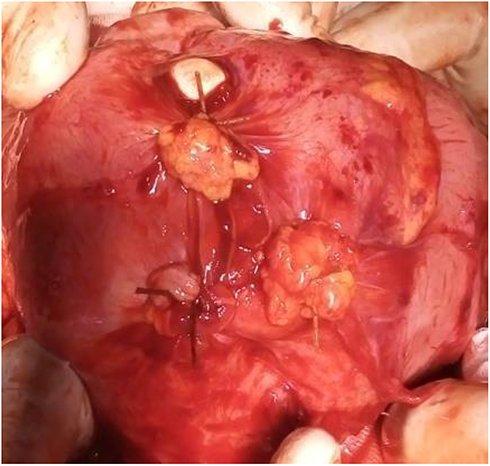 |
Figure 1 Intraoperative photographs of fundal niche with the surgeons glove visible. |
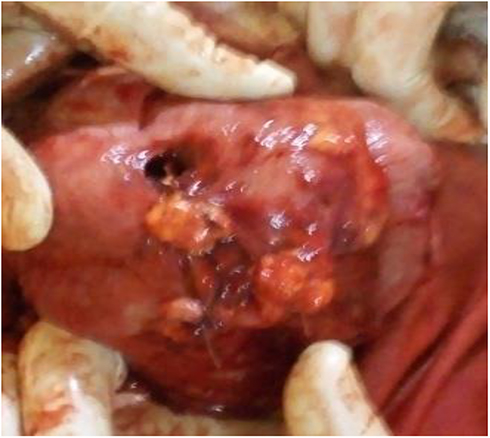 |
Figure 2 Intraoperative photographs of fundal niche with the surgeons glove visible and anterior uterine wall fibro-fatty adhesion (released) and lower uterine segment transverse cesarean section hysterectomy incision. |
Discussion
Post-traumatic uterine rupture is a rare but life-threatening cause of uterine rupture. At first glance, future fertility preservation appears to have a bleak prognosis due to extensive damage and an unacceptably high risk of recurrent rupture at progressively earlier gestation.5–7
When the uterine fundus is implicated in the rupture, the chance of recurrence appears to be highest.6 Given the modest proportion of unscarred uterine ruptures following trauma, extrapolating these results to assist management decisions in subsequent pregnancies following scarred uterine rupture appears plausible, and this is what we actually did. Data on future pregnancies following ruptured uterus repair is gathered primarily from a small case series of women who have undergone ruptured uterus repair. In these papers, the probability of recurrent rupture ranges from 22 to 100%.5–7 And ours is a rare case of a successful pregnancy following a tragic rupture of the uterus and loss of pregnancy.
The time of repeat rupture of a pregnant uterus depends greatly on the site of the previous scar and placental insertion, which could predispose to early rupture because of trophoblastic invasion of the scar. Early ultrasound localization of the placenta vis-à-vis a previous scar could thus be a useful prognostic factor.8 In this case, the rupture occurred earlier than at term (after a road traffic accident at 31 weeks of gestation). The recurrent rupture occurred at 33 weeks of gestation as well, following a fall down on her abdomen on a flat surface, and there was no optimal ANC follow up because the woman had poly-trauma with musculo-skeletal injuries at the time of the primary rupture and was admitted to the orthopedics ward and discharged after improvement with no proper counseling on future pregnancy prognosis and risk of recurrence. The third pregnancy ended in an uncomplicated elective cesarean delivery at 34+3 weeks of gestation, and the mother was discharged home with future fertility potential intact. This is definitely the only reported case of uncomplicated elective cesarean delivery after two previous post-traumatic fundal uterine ruptures. She had extensive myometrial damage and still remains fertile. The woman did not appear to have a significantly increased risk of subsequent rupture.
While hysterectomy might not always be feasible or desirable for both medical and socio-cultural reasons, repair of the ruptured uterus with conservation of reproductive function puts a big strain on both the obstetrician and the patient. There is a need for prospective studies to help formulate clearer guidelines.
There are limitations to this case report. Its rarity as a clinical scenario and its single-institution experience limit its potential to be generalized to large populations. The lack of specific guidelines for such incredibly rare but actual clinical cases of such obstetrics also places a burden on the clinicians managing such cases.
Conclusion
Recurrent post-traumatic fundal uterine rupture can give live birth after meticulous repair and close ANC follow-up at a tertiary hospital.
The risk of subsequent rupture did not appear to increase as it tended to stabilize after 3 uterine ruptures.
There is no societal consensus regarding recurrent fundal and extensive uterine rupture and their reproductive performance.
While hysterectomy might not always be feasible or desirable for both medical and socio-cultural reasons, repair of the ruptured uterus with conservation of reproductive function puts a burden both on the obstetrician and patient. So, there is a need for prospective studies that to help formulate clear guidelines.
Data Sharing Statement
The data used to support the findings of this study will be made available upon reasonable request from the corresponding author.
Acknowledgments
We would like to acknowledge all the managing teams who participated in the management of this mother throughout her stay in our hospital. We would also like to thank the staff at Hawassa University’s Neonatal Intensive Care Unit (NICU) for the mother’s adorable newborn’s care and treatment, which ended with a safe discharge.
Author Contributions
All authors significantly contributed to the work that has been reported, whether it be in the conception, study design, execution, data collection, analysis, and interpretation, or in all of these areas. They also participated in the article’s drafting, revision, or critical review, gave their final approval for the version that would be published, decided on the journal to which the article would be submitted, and agreed to be held responsible for every aspect of the work.
Disclosure
The authors declare that they have no conflicts of interest in this work.
References
1. Tuggy ML. Uterine-vesicular rupture during trial of labor. J Am Board Fam Pract. 1995;8(5):405–409.
2. Beazley J. Maternal injuries and complications. In: Dewhurst’s Textbook of Obstetrics and Gynecology for Post Graduates.
3. Downey GP, Tuck SM. Spontaneous uterine rupture during subsequent pregnancy following non-excision of an interstitial ectopic gestation. Br J Obstet Gynaecol. 1994;101(2):162–163. doi:10.1111/j.1471-0528.1994.tb13086.x
4. Odusoga OL, Adefuye PO, Oloyede OA, Fakoya TA, Olatunji AO. Uterine rupture: a major contributor to obstetric morbidity in Sagamu. Trop J Obstet Gynaecol. 2003;20(2):137–140.
5. Chibber R, El-Saleh E, Fadhli RA, et al. Uterine rupture and subsequent pregnancy outcome–how safe is it? A 25-year study. J Matern Fetal Neonatal Med. 2010;23(5):421–424. doi:10.3109/14767050903440489
6. Usta IM, Hamdi MA, Abu Musa AA, et al. Pregnancy outcome in patients with previous uterine rupture. Acta Obstet Gynecol Scand. 2007;86(2):172–176. doi:10.1080/00016340601089768
7. Lim AC, Kwee A, Bruinse HW. Pregnancy after uterine rupture: a report of 5 cases and a review of the literature. Obstet Gynecol Surv. 2005;60(9):613–617. doi:10.1097/01.ogx.0000176677.26657.6c
8. Matsuzaki S, et al. Maternal and fetal outcomes after prior mid-trimester uterine rupture: a systematic review with our experience. Medicina. 2021;57(12):1294.
 © 2022 The Author(s). This work is published and licensed by Dove Medical Press Limited. The
full terms of this license are available at https://www.dovepress.com/terms
and incorporate the Creative Commons Attribution
- Non Commercial (unported, 3.0) License.
By accessing the work you hereby accept the Terms. Non-commercial uses of the work are permitted
without any further permission from Dove Medical Press Limited, provided the work is properly
attributed. For permission for commercial use of this work, please see paragraphs 4.2 and 5 of our Terms.
© 2022 The Author(s). This work is published and licensed by Dove Medical Press Limited. The
full terms of this license are available at https://www.dovepress.com/terms
and incorporate the Creative Commons Attribution
- Non Commercial (unported, 3.0) License.
By accessing the work you hereby accept the Terms. Non-commercial uses of the work are permitted
without any further permission from Dove Medical Press Limited, provided the work is properly
attributed. For permission for commercial use of this work, please see paragraphs 4.2 and 5 of our Terms.