
Back to Journals » International Journal of Nanomedicine » Volume 20
Novel Platelet-Like PLGA-PSP Nanoparticles Based on Platelet-Specific Peptides Alleviated Traumatic Hemorrhage
Authors Fu W, Wei H, Ren D, Zheng Y, Zhang J, Mu S, Yang L
Received 8 May 2025
Accepted for publication 30 August 2025
Published 19 September 2025 Volume 2025:20 Pages 11491—11504
DOI https://doi.org/10.2147/IJN.S539137
Checked for plagiarism Yes
Review by Single anonymous peer review
Peer reviewer comments 2
Editor who approved publication: Dr Kamakhya Prakash Misra
Wenda Fu, Hua Wei, Dongxia Ren, Yan Zheng, Jin Zhang, Shijie Mu, Longfei Yang
Department of Transfusion Medicine, Tangdu Hospital, Fourth Military Medical University, Xi’an, Shaanxi, 710038, People’s Republic of China
Correspondence: Longfei Yang; Email [email protected] Shijie Mu, Email [email protected]
Background: Platelets play a crucial role in regulating coagulation. However, balancing the supply and clinical demands of platelet concentrates is a tough challenge. It is urgent to explore a novel platelet substitute with biosafety and efficacy in traumatic hemorrhage.
Methods: The platelet-like PLGA-PSP nanoparticles were synthesized by covalently coupling the linear platelet-specific peptides including CBP, VBP and FMP to the active carboxyl functional group of PLGA-PEG nanoparticles. The biosafety of the platelet-like PLGA-PSP nanoparticles was assessed. Subsequently, in vitro experiments were conducted to verify the effects of PLGA-PSP nanoparticles on platelet adhesion, aggregation and activation. Furthermore, the hemostatic efficacy of PLGA-PSP nanoparticles was confirmed in the tail vein, liver and femoral artery hemorrhage of normal and thrombocytopenic mice.
Results: We successfully designed and synthesized the non-toxic PLGA-PSP nanoparticles with specific hemostatic ability that significantly induced platelet adhesion and aggregation without triggering unexpected platelet activation. Moreover, the application of PLGA-PSP nanoparticles was demonstrated to effectively reduce the bleeding time and blood loss in the tail vein, liver and femoral artery of both normal and thrombocytopenic mice.
Conclusion: The novel platelet-like PLGA-PSP nanoparticles present a promising therapeutic option for the rapid hemostasis of traumatic hemorrhage based on the biosafety and efficacy.
Keywords: traumatic hemorrhage, platelet substitute, platelet-specific peptide, PLGA-PSP nanoparticle
Introduction
Traumatic hemorrhage is the major cause of prehospital death in both military and civilian settings,1 and transfusion of platelet components remains the primary treatment for traumatic hemorrhage. Platelets are blood cells continuously produced from megakaryocytes mainly in the bone marrow and typically have a lifespan of 7–10 days in circulation.2 They play a critical role in various pathophysiological processes, such as traumatic hemostasis, bone marrow transplantation, leukemia, tumor development and metastasis, and host defense.3–5 Traditionally, platelet concentrates for transfusion are predominantly sourced from blood donations.6 However, the existing platelet concentrates fail to fully meet clinical requirements due to their single source, the limited shelf life of 5 to 7 days, development of storage lesions, and risk of bacterial contamination.7,8 Therefore, it is necessary to explore platelet substitutes for the treatment of traumatic hemorrhage.
In recent years, the advent of novel biomaterials has greatly propelled the research on hemostatic capability.9–12 Naturally derived hemostatic biomaterials such as collagen, gelatin, and thrombin can promote platelet activation and aggregation to achieve rapid hemostasis, but their clinical application is limited by high cost and potential pathogenic risk.13 Synthetically derived hemostatic biomaterials, typically based on cyanoacrylate and polyurethane,14,15 are widely used in various hemostasis operations because of the low immunogenicity and customizable properties. However, the toxic side effects and non-anticipatory aggregate from these hemostatic materials cause local irritation and inflammation.9 Hence, there is an urgent need to develop a novel platelet substitute that combines biosafety and therapeutic efficacy for the management of traumatic hemorrhage.
The development of nanomedicine offered promising solution for the traumatic hemostasis.16 Specifically, poly lactide-co-glycolic acid (PLGA) and polyethylene glycol (PEG) nanoparticles have garnered significant attention because of the enhanced biocompatibility and drug penetration in vivo.16,17 Artificial platelets have been developed using peptide-decorated nanoparticles to mimic the platelets adhesion or aggregation mechanism for homeostatic application.18 The self-assembled peptides have the advantages of low immunogenicity, good biocompatibility and modifiability.19,20 Considering the development and clinical application of hemostatic biomaterials, it is difficult for a single biomaterial to meet the complicated hemostasis requirements. Inspired by the physiological structure of platelets, we designed and synthesized platelet specific polypeptides-decorated PLGA nanoparticles (PLGA-PSP) based on the linear platelet-specific peptides: collagen-binding peptide (CBP), von Willebrand Factor binding peptide (VBP) and fibrinogen-mimetic peptide (FMP). The biosafety and hemostatic efficacy of PLGA-PSP nanoparticles in targeting vascular injury sites were systematically evaluated through in vitro and in vivo, and further clarified the possible hemostatic mechanism, promising to provide a new strategy for clinical therapy of traumatic hemorrhage.
Materials and Methods
Materials
PLGA 10K-PEG 3400-NHS was provided by Ruixi Biological Technology (Xi’an, China). The peptides TRYLRIHPQSWVHQI (VBP), [GPO]7 (CBP) and HHLGGAKQAGDV (FMP) were custom-synthesized by Jitai Peptide Technology (Jiangsu, China). N, N-Dimethylformamide (DMF), triethylamine, busulfan, adenosine 5’-diphosphate (ADP) sodium salt, thrombin and dialysis tube were purchased from Sigma-Aldrich (Merck KGaA, Germany). Dimethyl sulfoxide (DMSO) was purchased from Solarbio (Beijing, China). Cell plasma membrane staining kit with Dil (red fluorescence) and Calcein AM cell viability assay kit (green fluorescence) were obtained from Beyotime (Shanghai, China). The collagen-coated 96-well plate and cell counting kit were obtained from WHB Scientific (Shanghai, China) and 7Sea Biotech (Shanghai, China), respectively.
PLGA-PSP Synthesis
The liner peptides assembled into nanoparticles were produced as follow, 100 mg PLGA 10K-PEG 3400-NHS and VBP, CBP, FMP (1.1 eq) were dissolved in 3 mL DMF solution, respectively, then triethylamine (3.0 eq) was added and reacted at room temperature for 12 h. Linear peptides conjugation PLGA 10K-PEG 3400-VBP-NH2, PLGA 10K-PEG 3400-CBP-NH2 and PLGA 10K-PEG 3400-FMP-NH2 were obtained and transferred to dialysis tubing (molecular weight cut-off 2000 Da) for 24 h, the dialysis solution was collected and freeze-dried to obtain the products. Next, 3 mg of each linear peptide conjugation was dissolved in 500 µL DMSO solution, the mixture was added to 4.5 mL double distilled water drop by drop, and stirred continuously (500 r/min) for 2 h at room temperature, and dialysis was performed overnight with double distilled water (molecular weight cut-off 3500 Da). The characterization of synthesized PLGA-PSP nanoparticles was verified by the UV-vis absorption spectrum (Epoch, BioTeK, USA), Nanoparticle Tracking Analysis (NTA, Zetaview, Particle Metrix, Germany) and Transmission Electron Microscopy (TEM, HT7800/HT7700, HITACHI, Japan).
Mice
Age- and weight-matched Kunming (KM) male mice were purchased from the Animal Center of Fourth Military Medical University (Xi’an, China). All mice were maintained under specific pathogen-free conditions and offered food and water ad libitum. The animals were fed adaptively for one week before being used in experiments. All experimental procedures were approved by the Ethics Committee of Fourth Military Medical University and strictly followed the National Research Council’s guidelines for the care and use of laboratory animals.
Platelet-Rich Plasma (PRP) Preparation
This study was approved by the Second Affiliated Hospital of the Fourth Military Medical University and conformed to the principles outlined in the Declaration of Helsinki. All volunteers provided informed consent. Peripheral venous blood samples were collected from healthy volunteers with no medication use within the preceding 14 d, PRP was then isolated by centrifugation at 1500 g for 15 min under ambient temperature conditions. The platelet counts in PRP were monitored using automatic blood cell analyzer (XN-350, Sysmex, Japan), and counts were maintained at 150,000/μL (lower limit of normal human platelet count).
Cytotoxicity Assays
Human Umbilical Vein Endothelial Cells (HUVEC) were obtained from the American Type Culture Collection. Cells in the logarithmic growth phase were harvested and cultured in a humidified incubator at 37°C with 5% CO2. HUVECs were seeded in 96-well plates at a density of 3,000 cells per well. Following adhesion, cells were treated with nanoparticles 10 µL (1.8 mg/mL) and maintained in a humidified incubator for 24 h, 48 h and 72 h, added 10 μL/well 7Sea-cell counting kit solution and continue to incubate in the incubator for 2 h, the formazan intensity (cell metabolic viability) was quantified with UV-visible spectrometry at 450 nm (Epoch, BioTeK, USA).
Erythrocyte Osmotic Fragility Assays
The freshly isolated erythrocytes from the peripheral blood of volunteers were suspended in PBS at a density of 6×10⁸ cells/mL, the osmotic fragility of erythrocytes was measured with a series of osmotic fragility solutions (0.2–0.9% NaCl). 50 µL erythrocyte suspension treated with 10 µL nanoparticles (1.8 mg/mL) was added to 450 µL NaCl, and the cell suspension was allowed to equilibrate at 37°C for 30 min. After centrifugation at 800 g for 10 min, the supernatant was determined for absorbance (A) at 540 nm. Erythrocytes treated with double distilled water were regarded as 100% hemolysis. The rate of hemolysis was calculated using the following formula:21

Measurement of Platelet Adhesion
The PRP was further centrifuged at 13,000 g for 5 min to obtain the platelets precipitated at the bottom. The platelets were washed by PBS for 3 times and stained with Calcein AM cell viability assay kit at room temperature for 30 min away from light. 10 µL nanoparticles (1.8 mg/mL) were mixed with cell plasma membrane staining kit with DiI and were added to 96-well collagen-coated plates, incubated at room temperature and away from light for 20 min. Finally, platelets with fluorescent labeling were placed in 96-well plates and incubated with nanoparticles for 5–10 min at room temperature, washed and observed by immunofluorescence microscopy (Eclipse Ti2-A, Nikon, Japan).
Detection of Platelet Aggregation
50 µL PRP suspension and 10 µL nanoparticles (1.8 mg/mL) were placed into 96-well plates and incubated at room temperature for 5 min. Next, ADP with final concentration of 20 µM was added and mixed thoroughly. Then the mixture was poured on microscopy slides, and platelet aggregation was observed under optical microscope (Eclipse ci, Nikon, Japan).
Analysis of Platelet Activation
The PRP was further centrifuged to obtain platelets precipitated at the bottom and washed by PBS for 3 times. The platelets (2×10⁷/mL,100 µL) were incubated with 10 µL nanoparticles (1.8 mg/mL) at 37°C for 15 min, thrombin 0.1 U/mL was added and incubated for 5 min. Then the treated platelets were co-incubated with PE-conjugated anti-CD62p antibodies for 15 min in the dark, and cells were collected and analyzed by flow cytometry (FACSCalibur, BD, USA).
Tail-Transection Bleeding Hemostasis Mice Model
100 µL nanoparticles were injected via mice tail vein at a concentration of 200/nL.22 A 5 mm distal tail amputation was performed using sterile surgical blades after administered for 5 min, the transected tail was immediately immersed in 37°C normal saline (0.9% NaCl). Bleeding time was quantitatively measured from moment of transection until complete hemostasis using a digital stopwatch and collected blood was subsequently analyzed by the automatic blood cell analyzer. Mice were euthanized at the end of experiments, major organs (liver, lung, kidney and spleen) were fixed in 4% paraformaldehyde and routinely paraffin embedded and processed. Hematoxylin-eosin (H&E) staining was performed on organs section. The inflammation infiltration and thrombus in organs were assessed by optical microscope (Eclipse ci, Nikon, Japan).
Liver Bleeding Hemostasis Mice Model
The mice were administered 100 µL nanoparticles (200/nL) through tail vein and anesthetized with isoflurane after 20 min. Midline laparotomy was performed using a sterile scalpel to expose the hepatic lobes, a 3 mm incision was created on the left hepatic lobe using scissors. The pre-weighed sterile absorbent gauze was placed in the abdominal cavity to collect hepatic hemorrhage, upon achieving complete hemostasis at the hepatic injury site, both bleeding time and blood loss were recorded by a digital stopwatch.
Femoral Artery Bleeding Hemostasis Mice Model
The mice were anesthetized with isoflurane for 20 min after injecting 100 µL nanoparticles (200/nL) through tail vein. The skin and muscle at the groin of the mice were removed with a medical scalpel, and the femoral artery was exposed, a femoral artery hemorrhage model was established by puncturing of the mid-femoral arterial segment with a 26-gauge needle, the bleeding time and blood loss were recorded by a digital stopwatch.
Statistical Analysis
All data presented as means ± standard from three separate experiments were analyzed using GraphPad Prism version 9. Statistical differences were evaluated using two-tailed Student’s t-test or two-way ANOVA. p<0.05 was considered statistically significant.
Results
Synthesis and Characterization of PLGA-PSP Nanoparticles
The platelet-like PLGA-PSP nanoparticles consisted of PLGA block copolymer cores conjugated with PEG arms terminated by CBP, VBP and FMP (Figure 1A and B). The UV-vis absorption spectrum of PLGA-PSP nanoparticles increased significantly in the 201–250 nm region compared to PLGA-PEG, which was consistent with the strong absorption characteristics of the three platelet-specific peptides including PLGA-FMP, PLGA-VBP and PLGA-CBP in this wavelength range (Figure 1C). Particularly at ~204 nm and ~230 nm, the absorption spectrum of PLGA-PSP nanoparticles exhibits two absorption peaks, corresponding to the characteristic absorption peaks of peptides PLGA-FMP, PLGA-VBP, and PLGA-CBP in the respective wavelength bands. The UV-vis absorption spectrum indicated that the three platelet-specific peptides have been successfully conjugated to the PLGA-PSP structure. The average particle size of the platelet-like PLGA-PSP nanoparticles was 120 nm, which was consistent with the expected size of artificially synthesized platelets (Figure 1D). TEM showed that the PLGA-PSP nanoparticles had morphologically flexible vesicles similar to natural platelets (Figure 1E), and still had relatively complete vesicle-like structures after 6 months of conventional storage at 4°C (Figure 1F).
 |
Figure 1 Synthesis and characterization of PLGA-PSP nanoparticles. (A) The synthesis and scheme of liner PLGA-FMP. (B) The process for synthesis of PLGA-PSP nanoparticles. (C) Ultraviolet spectra of PLGA-PEG, PLGA-FMP, PLGA-VBP, PLGA-CBP and PLGA-PSP nanoparticles. The red and blue arrows indicated characteristic absorption peaks of PLGA-PSP nanoparticles at ~204 nm and ~230 nm compared with PLGA-PEG, respectively. The red circle indicated absorption peaks of PLGA-PSP nanoparticles, the blue circle indicated absorption peaks of peptides PLGA-FMP, PLGA-VBP and PLGA-CBP. (D) Particle size detection of PLGA-PSP nanoparticles by NTA technology. (E-F) TEM images of freshly prepared PLGA-PSP nanoparticles and PLGA-PSP nanoparticles stored for 6 months at 4°C, respectively. Scale bar = 100 nm. |
Biosafety Analysis of PLGA-PSP Nanoparticles
The biosafety of the synthesized PLGA-PSP nanoparticles was verified by in vitro experiments and found that it was not cytotoxicity for human umbilical vein endothelial cells (Figure 2A). Erythrocyte osmotic fragility is an indirect method to assess oxidative stress,21,23 the effect of PLGA-PSP nanoparticles on erythrocytes osmotic fragility was further investigated, and the results showed that there was no change in the osmotic fragility curve after 24 h (Figure 2B) and 48 h (Figure 2C) of PLGA-PSP nanoparticles co-culture with erythrocytes. In addition, we also studied the effect of intravenous injection of PLGA-PSP nanoparticles on the microcirculation of tissues and organs in mice. Histopathological analysis (Figure 2D) showed that PLGA-PSP nanoparticles did not cause inflammation and microthrombus in major organs (liver, lung, kidney and spleen). These data indicated that PLGA-PSP nanoparticles were non-cytotoxic, did not cause oxidative damage to erythrocyte, as well as micro-thrombosis and inflammatory reactions in tissues and organs, and may be safe for human beings.
 |
Figure 2 Biosafety analysis of PLGA-PSP nanoparticles. (A) Cytotoxicity assessment of PLGA-PSP nanoparticles co-cultured with HUVEC for 24 h, 48 h and 72 h (N=5), respectively. (B and C) Osmotic fragility curves by PLGA-PSP nanoparticles co-cultured with erythrocytes for 24 h and 48 h (N=5), respectively. (D) H&E staining of liver, lung, kidney and spleen were performed at 5 min after tail intravenous injection. Scale bar = 0.2 mm. |
PLGA-PSP Nanoparticles Enhanced Collagen-Induced Platelet Adhesion
Adhesion to the injured vascular site is an important step for platelets to play a hemostatic role, which is a rapid process that takes place within seconds.24 To determine whether PLGA-PSP nanoparticles have an impact on platelet adhesion, immunofluorescence staining was used to detect the effects of PLGA-PSP nanoparticles on platelet adhesion. The polypropylene plate was coated with collagen to simulate collagen exposure at the damaged site after vascular injury. We observed that PLGA-PEG nanoparticles showed comparable effects to PBS, failing to induce platelet adhesion. In contrast, PLGA-PSP nanoparticles demonstrated significant platelet adhesive effects (Figure 3). The results suggested that PLGA-PEG nanoparticles had no significant effect on platelet adhesion, but PLGA-PSP nanoparticles played an important role in platelet adhesion to the vascular injured site.
 |
Figure 3 PLGA-PSP nanoparticles enhanced collagen-induced platelet adhesion. Platelet adhered to collagen-coated surfaces was assessed by immunofluorescence staining of platelet (green) and nanoparticles (red). Representative images were shown for the end of the experiment for each condition. Scale bar = 5 μm. All images were taken at the same magnification. |
PLGA-PSP Nanoparticles Promoted ADP-Induced Platelet Aggregation
Upon vascular injury, the platelets adhering to the damaged site would recruit the neighboring platelets to aggregate and form blood clots to reduce bleeding, therefore we further explored the effect of PLGA-PSP nanoparticles on platelet aggregation. Data from platelet aggregation assays showed that PLGA-PSP nanoparticles with “resting” platelets, no spontaneous platelet aggregation was recorded for any of the groups tested as compared with the PBS (Figure 4A). Platelets can be activated by numerous stimuli from various origins, such as the platelets themselves that secrete activating agonists like ADP after adhesion to the endothelium.25 Considering that ADP occurs naturally in human body and plays a role in maintaining normal hemostasis and thrombosis, a concentration of 20 µM was chosen to stimulate platelet aggregation in our studies.26 Consistent with our expectation, PLGA-PSP nanoparticles could significantly promote platelet aggregation compared with the PLGA-PEG nanoparticles and PBS (Figure 4B). These results indicated that PLGA-PSP nanoparticles promoted platelet aggregation in the presence of platelet stimulants, without causing spontaneous platelet aggregation.
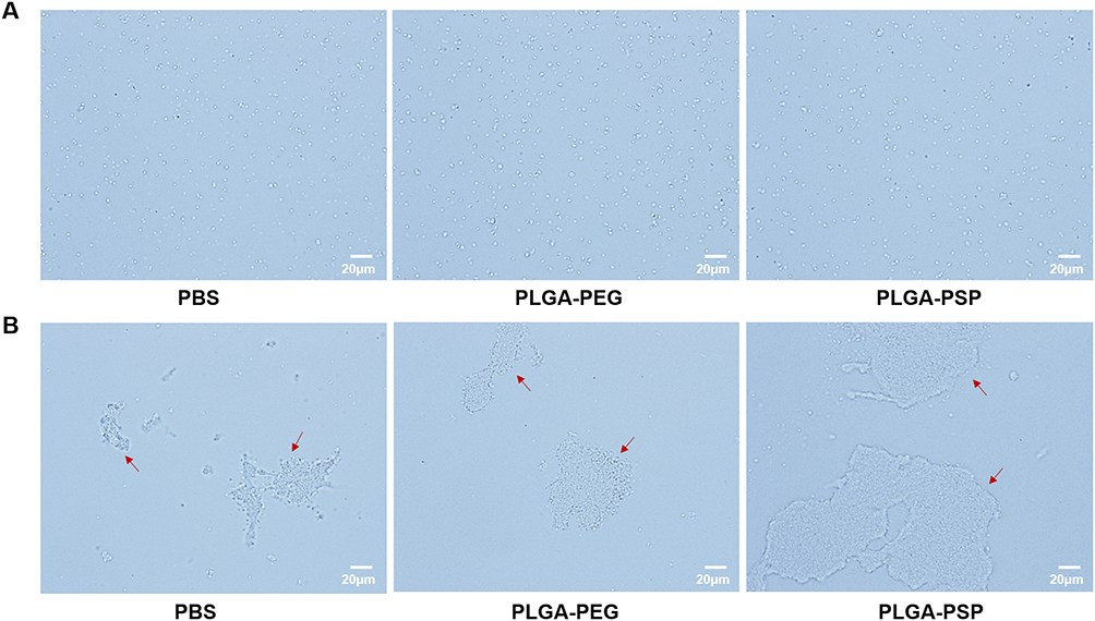 |
Figure 4 PLGA-PSP nanoparticles promoted ADP-induced platelet aggregation. (A) Platelet aggregation at rest was evaluated under an optical microscope. Scale bar = 20 μm. (B) Platelet aggregation in the presence of platelet activating agonists ADP under an optical microscope. The red arrow indicated aggregated platelet. Scale bar = 20 μm. |
PLGA-PSP Nanoparticles Did Not Trigger Thrombin-Induced Platelet Activation
P-selectin (CD62p) is a membrane protein stored in platelet α-granules and transferred to the membrane surface and plasma with platelet activation, forms a marker of activation coupled degranulation.27,28 To investigate the effect of PLGA-PSP nanoparticles on platelet activation, flow cytometry was used to detect the expression of P-selectin on platelet surface. Not surprisingly, in the presence of platelet activating agonists like thrombin, PLGA-PSP nanoparticles failed to affect platelet activation (Figure 5A and B). The results showed that the artificial synthetic PLGA-PSP nanoparticles did not trigger platelet activation.
 |
Figure 5 PLGA-PSP nanoparticles did not trigger thrombin-induced platelet activation. (A) Flow cytometry was used to investigate thrombin-induced platelet activation, the expression of P-selectin on platelet surface was detected by CD62p (N=5). (B) Statistical results of PE-CD62p fluorescence intensity based on (A), N=5. |
PLGA-PSP Nanoparticles Enhanced Hemostasis in Traumatic Hemorrhage Model of Mice
The hemostatic ability of PLGA-PSP nanoparticles in vivo was explored by traumatic hemorrhage model in mice. In the experiments, bleeding time and blood loss were measured at 5 min after tail intravenous administered. PLGA-PEG nanoparticles showed no decrease in clipped tails of bleeding time compared with PBS, while PLGA-PSP nanoparticles treated mice significantly shorten the bleeding time (p < 0.01, Figure 6A). We deemed that red blood cell (RBC) represented the blood loss. Consistent with the tail-transection bleeding time, the blood loss (p < 0.01, Figure 6B) decreased significantly in PLGA-PSP nanoparticles treated mice. In addition to injured veins bleeding which is a type of traumatic hemorrhage, we also studied visceral and artery hemorrhage, respectively. PLGA-PSP nanoparticles significantly improved the bleeding time (p < 0.001, Figure 6C) and blood loss (p < 0.01, Figure 6D) of liver injury. Surprisingly, the liver bleeding time in the PLGA-PEG nanoparticles treated mice was significantly reduced by 30% compared to PBS (p < 0.05). Moreover, compared with PBS, PLGA-PSP nanoparticles significantly improved the femoral artery bleeding time (p < 0.01, Figure 6E) and blood loss (p < 0.05, Figure 6F), while there was only a downward trend without statistical significance compared with PLGA-PEG nanoparticles of bleeding time. Whether PLGA-PEG nanoparticles contributed to hemostasis at the site of visceral and artery injury remains to be studied. Overall, the results demonstrated that PLGA-PSP nanoparticles enhanced hemostasis in traumatic hemorrhage model of mice.
 |
Figure 6 PLGA-PSP nanoparticles enhanced hemostasis in traumatic hemorrhage model of mice. Bleeding time (A) and blood loss (B) were evaluated after tail intravenous injection of PBS, PLGA-PEG and PLGA-PSP nanoparticles at 5 min. Liver bleeding time (C) and blood loss (D) were assessed at 20 min after tail vein injection of PBS, PLGA-PEG and PLGA-PSP nanoparticles. Femoral artery bleeding time (E) and blood loss (F) were shown at 20 min after administration of PBS, PLGA-PEG and PLGA-PSP nanoparticles. N=5. *p < 0.05, **p < 0.01, ***p < 0.001. |
PLGA-PSP Nanoparticles Accelerated Hemostasis in Traumatic Hemorrhage Model of Thrombocytopenic Mice
In order to investigate whether PLGA-PSP nanoparticles had the same hemostatic effect in traumatic hemorrhage of thrombocytopenic animals, the hemostatic efficacy of PLGA-PSP nanoparticles further evaluated in mice that were rendered thrombocytopenic by intraperitoneally administration with busulfan (2 mg/kg) once a day for 14 d (Figure 7A). The thrombocytopenic mice model was successfully established when the platelet counts decreased by 50~60% compared with normal mice (p < 0.0001, Figure 7B). The hemostatic effect of PLGA-PSP nanoparticles was assessed by tail-transection bleeding of thrombocytopenic mice. As expected, the tail vein bleeding time of PLGA-PSP nanoparticles treated mice was reduced by 40% compared with PBS (p < 0.001, Figure 7C), and only decreased trends of the blood loss were observed compared with PLGA-PEG nanoparticles (Figure 7D). Meanwhile, PLGA-PSP nanoparticles were also found to have the same hemostatic effect on liver bleeding time (p < 0.0001, Figure 7E) and the blood loss (p < 0.01, Figure 7F) in thrombocytopenia mice. Moreover, compared with the PBS, PLGA-PSP nanoparticles significantly reduced the femoral artery bleeding time (p < 0.05, Figure 7G) and blood loss (p < 0.05, Figure 7H) in thrombocytopenia mice. These data proved that PLGA-PSP nanoparticles had a good hemostatic ability on traumatic hemorrhage model of thrombocytopenia mice.
 |
Figure 7 PLGA-PSP nanoparticles accelerated hemostasis in traumatic hemorrhage model of thrombocytopenic mice. (A) Scheme showing traumatic hemorrhage model of thrombocytopenic mice. (B) Platelet counts in normal (wild-type) mice and busulfan induced thrombocytopenic mice. Bleeding time (C) and blood loss (D) in thrombocytopenia mice were evaluated after tail intravenous injection of PBS, PLGA-PEG and PLGA-PSP nanoparticles at 5 min. Liver bleeding time (E) and blood loss (F) were assessed at 20 min after tail vein injection of PBS, PLGA-PEG and PLGA-PSP nanoparticles. Femoral artery bleeding time (G) and blood loss (H) were shown at 20 min after administration of PBS, PLGA-PEG and PLGA-PSP nanoparticles. N=10. *p < 0.05, **p < 0.01, ***p < 0.001, ****p < 0.0001. |
Discussion
In recent years, platelet concentrates have struggled to meet the clinical needs of traumatic hemorrhage due to the increased transfusion of platelet components and limited storage period of platelets. Development of the efficient hemostatic materials is essential for the management of traumatic hemorrhage.29 Therefore, the purpose of this study was to design and optimize a novel biosafety and efficacy platelet-like hemostatic material that can be administered intravenously. PLGA-PSP nanoparticles were synthesized by covalently coupling the platelet-specific peptides (CBP, VBP and FMP) to the surface of PLGA-PEG nanoparticles, mimicking the physical properties and biological functions of natural platelets. The biosafety and hemostatic ability of the nanoparticles were subsequently verified by in vivo and in vitro experiments.
Within the wide range of polymeric materials that can be used to synthesize nanocarriers, PLGA is widely used for nanoparticle synthesis which composed of poly(lactic acid) (PLA) and poly(glycolic acid) (PGA) monomers.30 PLGA nanoparticles are ideal for constructing intravenously administered drug carriers because of the good biocompatibility, biodegradability and targeted drug delivery capabilities.31 However, the hydrophobic surface of PLGA nanoparticles makes them to be easily recognized as hetero-antigen by organisms, leading to premature clearance by neutrophils and macrophages from the bloodstream,32 and the negative charge on the surface impedes their interaction with negatively charged biological barriers.33 PEG is a polymer with excellent hydrophilicity, which has the ability to undergo several polymer conformations to evade neighboring protein molecules.34 The “stealth” effect of PLGA nanoparticles can be achieved by attaching PEG chains on the surface.32 Coupling the non-platelet alternative coagulants Arg-Gly-Asp (RGD) sequence to PLGA surface via 4600 Da PEG had a greatest platelet adhesion and aggregation.35 When the PEG arm length exceeds 5000 Da, the polymer chain has enough repeat units to fold over and shield functional domains.34 Considering these factors, PEG with a molecular weight 4300 Da was selected to synthesize PLGA-PEG nanoparticles.
The resting platelet is a generally biconvex discoid structure of 2–3 μm in diameter.36 Upon vascular injury, the glycoprotein GP Ib on the surface of platelets reversibly bind to the damaged endothelium by interacting with von Willebrand Factor (vWF) secreted by endothelial cells, then the glycoprotein GP Ia/IIa forms stable adhesion by binding to collagen. Subsequently, platelet activation occurs and fibrinogen-mediated interactions with the integrin GP IIb/IIIa on the platelet surface, other locally activated platelets are recruited to aggregate, ultimately achieving effective hemostasis at the wound site.27 It was reported that platelet-specific peptides was conjugated to a lipid-PEG molecule via amide to form lipid-PEG-peptides conjugates.37 Inspired by the hemostatic function of natural platelets, PLGA-PSP are peptide-decorated nanoparticles targeted to sites of damaged vascular. CBP and VBP bind to exposed collagen and vWF, respectively, and then FMP promoted platelet recruitment and aggregation at the injury site (Figure 8). Our data showed that PLGA-PSP nanoparticles were non-toxic, injectable and did not cause microthrombi in organisms. We simulated collagen exposure after vascular endothelial cells damaged in vitro. In the presence of PLGA-PSP nanoparticles, both activated platelets and PLGA-PSP nanoparticles bind to injured endothelium, as well as to combine with each other. The platelet adhesion in the PLGA-PSP nanoparticles treatment group was observed by immunofluorescence staining. In addition, we also observed that incubation PLGA-PSP nanoparticles with “resting” platelets failed to result in any apparent platelet aggregation. Studies had shown that PLGA-PEG nanoparticles with platelets stimulated by ADP did not appear to influence the aggregation of the platelets,38 while PLGA-PSP nanoparticles in our data can significantly induce platelet aggregation without affecting platelet activation function.
 |
Figure 8 Schematic illustration of synthesis and hemostatic function of artificial platelet-like PLGA-PSP nanoparticles. |
The hemostatic efficacy of peptide-related materials has been recently demonstrated in a variety of traumatic hemorrhage animal models.39,40 Liposomes modified with RGD peptide effectively activated platelets, augmented hemostasis and shortened the bleeding time of mice tail.41 Functionally decorated platelet-like nanoparticles (PLNs) decreased the tail bleeding time of mice by 65%, but increased the possibility of inducing pulmonary microthrombus.42 Synthetic Platelet (SP) nanoparticles reduced blood loss by 35% in von Willebrand disease type 2B mice.43 Prophylactically administered SynthoPlateTM efficiently improved the hemostatic defect in thrombocytopenia mice22 and elevated survival rate in a porcine model of traumatic arterial hemorrhage.44 In this study, we optimized and synthesized PLGA-PSP nanoparticles which significantly improved sub-acute bleeding (tail transection) in normal and thrombocytopenia mice. After observing the promising hemostatic effects, we focused on evaluating the application of PLGA-PSP nanoparticles for hemostasis in liver injury, a mouse model of visceral bleeding that better reflects the rescue of traumatic hemorrhage.40 In parallel, we observed improved bleeding time and reduced blood loss in both normal and thrombocytopenia mouse models of liver bleeding following intravenous administration of PLGA-PSP nanoparticles. Similarly, hemostatic effect of PLGA-PSP nanoparticles was validated in mice model of massive acute bleeding, which also shown positive hemostatic ability in both normal and thrombocytopenic mouse models of femoral artery bleeding. However, due to the large diameter and high flow rate of the femoral artery, the hemostatic effect was less pronounced compared to tail transection and liver injury models. Above all, these studies provided strong evidence toward the effectiveness of PLGA-PSP nanoparticles in vivo. In the following study, the hemostatic effect of PLGA-PSP nanoparticles will be further verified in other rodents and large animals.
Our study provided evidences for the hemostasis of PLGA-PSP nanoparticles in traumatic hemorrhage, but there are some limitations. Firstly, although PLGA-PEG nanoparticles have been reported to exhibit excellent biodegradability, the metabolic cycle of PLGA-PSP nanoparticles in vivo remains unexplored. Furthermore, the optimal therapeutic concentration and administration timing of PLGA-PSP nanoparticles for traumatic hemorrhage were not investigated in this study. These limitations will also be the main research direction of our future work.
Rapid hemostatic effect, injectability, non-cytotoxicity, non-immunogenicity and absence of microthrombus formation are the critical requirements for artificial mimic platelets.35 PLGA nanoparticles used in this study were approved by FDA, which offer a new intravenous administration strategy for clinical therapy of traumatic hemorrhage. The novel platelet-like PLGA-PSP nanoparticles have the potential to be developed as clinically safe and effective hemostatic materials in the context of long-term shortage of platelet supply. However, hemostasis is only the initial step in wound healing process, which comprises multiple dynamic and intricately intertwined stages, including inflammation, proliferation and remodeling.45,46 In the future, we will further optimize PLGA-PSP nanoparticles, with a focus on achieving rapid hemostasis and high antibacterial performance.
Conclusion
In summary, inspired by the physiological structure of platelets, PLGA-PSP nanoparticles were synthesized in our study by covalently coupling the platelet-specific peptides (CBP, VBP and FMP) to the surface of PLGA-PEG nanoparticles. We found that PLGA-PSP nanoparticles had a good biological safety and significantly induced platelet adhesion and aggregation without triggering unexpected platelet activation. Moreover, the application of PLGA-PSP nanoparticles was also demonstrated to effectively reduce the bleeding time and blood loss in the tail vein, liver and femoral artery of both normal and thrombocytopenic mice. Therefore, PLGA-PSP nanoparticles can achieve rapid hemostasis in traumatic hemorrhage without side effects. However, wound healing progresses through four dynamically overlapping phases without distinct boundaries, our subsequent research will focus on developing multifunctional nanoparticles that combine robust antibacterial properties with hemostatic efficacy to address subsequent healing challenges. The novel platelet-like PLGA-PSP nanoparticles emerge as a promising therapeutic platform for managing traumatic hemorrhage.
Funding
This study was supported by research grants from the National Natural Science Foundation of China (82570283) and the Academic Development Program of Tangdu Hospital (2024JSYX018).
Disclosure
The authors declare that they have no competing interests.
References
1. Huang Z, Zhang D, Tong L, et al. Protonated-chitosan sponge with procoagulation activity for hemostasis in coagulopathy. Bioact Mater. 2024;41:174–192. doi:10.1016/j.bioactmat.2024.07.012
2. Van der Meijden PEJ, Heemskerk JWM. Platelet biology and functions: new concepts and clinical perspectives. Nat Rev Cardiol. 2019;16(3):166–179. doi:10.1038/s41569-018-0110-0
3. Fang X, Li Y, Wang Y, et al. Platelet-derived biomaterials for targeted drug delivery and tissue repair. J Mater Chem B. 2025;13(5):1573–1585. doi:10.1039/D4TB02477J
4. Gao F, Zhou X, Shi J, et al. Eltrombopag treatment promotes platelet recovery and reduces platelet transfusion for patients with post-transplantation thrombocytopenia. Ann Hematol. 2020;99(11):2679–2687. doi:10.1007/s00277-020-04106-2
5. Berenger JB, Saillard C, Sannini A, et al. Prophylactic versus restrictive platelet transfusion strategy in patients with haematological malignancies in the ICU setting, a propensity-score analysis. J Crit Care. 2024;83:154817. doi:10.1016/j.jcrc.2024.154817
6. Drexler C, Macher S, Lindenau I, et al. High-dose intravenous versus oral iron in blood donors with iron deficiency: the IronWoMan randomized, controlled clinical trial. Clin Nutr. 2020;39(3):737–745. doi:10.1016/j.clnu.2019.03.025
7. Pozy E, Brown AC. Transfusion biomaterials for hemostasis. J Thromb Haemost. 2025;23(3):790–803. doi:10.1016/j.jtha.2024.11.030
8. Lee HJ, Oh SH, Jo SY, et al. Platelet inventory management program: development and practical experience. Ann Lab Med. 2021;41(1):95–100. doi:10.3343/alm.2021.41.1.95
9. Guo Y, Cheng N, Sun H, et al. Advances in the development and optimization strategies of the hemostatic biomaterials. Front Bioeng Biotechnol. 2022;10:1062676. doi:10.3389/fbioe.2022.1062676
10. Leng F, Liu J, Du E, et al. Recent progress in polysaccharide microsphere-based hemostatic material for intravascular and extravascular hemostasis: a review. Int J Biol Macromol. 2025;300:140280. doi:10.1016/j.ijbiomac.2025.140280
11. Ma Y, Da G, He L, et al. Multifunctional chitosan/cellulose/tannic acid biocomposite sponge with excellent biocompatibility for rapid hemostasis. Biomacromolecules. 2025;26(4):2467–2478. doi:10.1021/acs.biomac.4c01840
12. Yang J, Wang T, Zhang L, et al. Injectable hemostatic hydrogel adhesive with antioxidant, antibacterial and procoagulant properties for hemorrhage wound management. J Colloid Interface Sci. 2024;673:395–410. doi:10.1016/j.jcis.2024.05.207
13. Li L, Du Y, Yin Z, et al. Preparation and the hemostatic property study of porous gelatin microspheres both in vitro and in vivo. Colloids Surf B Biointerfaces. 2020;187:110641. doi:10.1016/j.colsurfb.2019.110641
14. Overby RJ, Feldman DS. Influence of poly(ethylene glycol) end groups on poly(ethylene glycol)-albumin system properties as a potential degradable tissue scaffold. J Funct Biomater. 2018;10(1):1. doi:10.3390/jfb10010001
15. Lumsden AB, Heyman ER. Closure medical surgical sealant study g. prospective randomized study evaluating an absorbable cyanoacrylate for use in vascular reconstructions. J Vasc Surg. 2006;44(5):1002–1009. doi:10.1016/j.jvs.2006.06.039
16. Crisafulli E, Scalzone A, Tonda-Turo C, et al. Multimodal layer-by-layer nanoparticles: a breakthrough in gene and drug delivery for osteosarcoma. J Mater Chem B. 2024;12(48):12540–12552. doi:10.1039/D4TB01541J
17. Wu K, Yu B, Li D, et al. Recent advances in nanoplatforms for the treatment of osteosarcoma. Front Oncol. 2022;12:805978. doi:10.3389/fonc.2022.805978
18. Yang PP, Zhang K, He PP, et al. A biomimetic platelet based on assembling peptides initiates artificial coagulation. Sci Adv. 2020;6(22):eaaz4107. doi:10.1126/sciadv.aaz4107
19. Rad-Malekshahi M, Lempsink L, Amidi M, et al. Biomedical applications of self-assembling peptides. Bioconjug Chem. 2016;27(1):3–18. doi:10.1021/acs.bioconjchem.5b00487
20. Treacy NJ, Clerkin S, Davis JL, et al. Growth and differentiation of human induced pluripotent stem cell (hiPSC)-derived kidney organoids using fully synthetic peptide hydrogels. Bioact Mater. 2023;21:142–156. doi:10.1016/j.bioactmat.2022.08.003
21. Ye M, Li H, Luo H, et al. Potential antioxidative activity of homocysteine in erythrocytes under oxidative stress. Antioxidants. 2023;12(1):202. doi:10.3390/antiox12010202
22. Shukla M, Sekhon UD, Betapudi V, et al. In vitro characterization of SynthoPlate (synthetic platelet) technology and its in vivo evaluation in severely thrombocytopenic mice. J Thromb Haemost. 2017;15(2):375–387. doi:10.1111/jth.13579
23. Sharma B, Rai DK, Rai PK, et al. Determination of erythrocyte fragility as a marker of pesticide-induced membrane oxidative damage. Methods Mol Biol. 2010;594:123–128.
24. Bergmeier W, Stefanini L. Platelets at the vascular interface. Res Pract Thromb Haemost. 2018;2(1):27–33. doi:10.1002/rth2.12061
25. la Harpe KM D, Kondiah PPD, Choonara YE, et al. The hemocompatibility of nanoparticles: a review of cell-nanoparticle interactions and hemostasis. Cells. 2019;8(10):1209. doi:10.3390/cells8101209
26. Zhou X, Zhou X, Zhu R, et al. The mechanism of oleic acid inhibiting platelet activation stimulated by collagen. Cell Commun Signal. 2023;21(1):278. doi:10.1186/s12964-023-01276-0
27. Tyagi T, Jain K, Gu SX, et al. A guide to molecular and functional investigations of platelets to bridge basic and clinical sciences. Nat Cardiovasc Res. 2022;1(3):223–237. doi:10.1038/s44161-022-00021-z
28. Mehic D, Machacek J, Schramm T, et al. Platelet function and soluble P-selectin in patients with primary immune thrombocytopenia. Thromb Res. 2023;223:102–110. doi:10.1016/j.thromres.2023.01.012
29. Sharma A, Verma C, Singh P, et al. Alginate based biomaterials for hemostatic applications: innovations and developments. Int J Biol Macromol. 2024;264(Pt 2):130771. doi:10.1016/j.ijbiomac.2024.130771
30. Hernandez-Parra H, Cortes H, Romero-Montero A, et al. Polymeric nanoparticles decorated with fragmented chitosan as modulation systems for neuronal drug uptake. Carbohydr Polym. 2024;336:122121. doi:10.1016/j.carbpol.2024.122121
31. Villate A, Barreto GP, Nicolas MS, et al. Development, characterization and in vitro gastrointestinal release of PLGA nanoparticles loaded with full-spectrum cannabis extracts. AAPS Pharm Sci Tech. 2024;25(5):120. doi:10.1208/s12249-024-02836-4
32. Sheffey VV, Siew EB, Tanner EEL, et al. PLGA’s plight and the role of stealth surface modification strategies in its use for intravenous particulate drug delivery. Adv Healthc Mater. 2022;11(8):e2101536. doi:10.1002/adhm.202101536
33. Qian L, Zheng J, Wang K, et al. Cationic core-shell nanoparticles with carmustine contained within O(6)-benzylguanine shell for glioma therapy. Biomaterials. 2013;34(35):8968–8978. doi:10.1016/j.biomaterials.2013.07.097
34. Gref R, Luck M, Quellec P, et al. ‘Stealth’ Corona-core nanoparticles surface modified by polyethylene glycol (PEG): influences of the corona (PEG chain length and surface density) and of the core composition on phagocytic uptake and plasma protein adsorption. Colloids Surf B Biointerfaces. 2000;18(3–4):301–313. doi:10.1016/S0927-7765(99)00156-3
35. Bertram JP, Williams CA, Robinson R, et al. Intravenous hemostat: nanotechnology to halt bleeding. Sci Transl Med. 2009;1(11):11ra22. doi:10.1126/scitranslmed.3000397
36. Li L, Wang S, Han K, et al. Quantifying shear-induced margination and adhesion of platelets in microvascular blood flow. J Mol Biol. 2023;435(1):167824. doi:10.1016/j.jmb.2022.167824
37. Hickman DA, Pawlowski CL, Sekhon UDS, et al. Biomaterials and advanced technologies for hemostatic management of bleeding. Adv Mater. 2018;30(4). doi:10.1002/adma.201700859
38. Bakhaidar R, O’Neill S, Ramtoola Z. PLGA-PEG nanoparticles show minimal risks of interference with platelet function of human platelet-rich plasma. Int J Mol Sci. 2020;21(24):9716. doi:10.3390/ijms21249716
39. Sekhon UDS, Swingle K, Girish A, et al. Platelet-mimicking procoagulant nanoparticles augment hemostasis in animal models of bleeding. Sci Transl Med. 2022;14(629):eabb8975. doi:10.1126/scitranslmed.abb8975
40. Dyer MR, Hickman D, Luc N, et al. Intravenous administration of synthetic platelets (SynthoPlate) in a mouse liver injury model of uncontrolled hemorrhage improves hemostasis. J Trauma Acute Care Surg. 2018;84(6):917–923. doi:10.1097/TA.0000000000001893
41. Alipour M, Baneshi M, Hosseinkhani S, et al. Recent progress in biomedical applications of RGD-based ligand: from precise cancer theranostics to biomaterial engineering: a systematic review. J Biomed Mater Res A. 2020;108(4):839–850. doi:10.1002/jbm.a.36862
42. Anselmo AC, Modery-Pawlowski CL, Menegatti S, et al. Platelet-like nanoparticles: mimicking shape, flexibility, and surface biology of platelets to target vascular injuries. ACS Nano. 2014;8(11):11243–11253. doi:10.1021/nn503732m
43. Roullet S, Luc N, Rayes J, et al. Efficacy of platelet-inspired hemostatic nanoparticles on bleeding in von Willebrand disease murine models. Blood. 2023;141(23):2891–2900. doi:10.1182/blood.2022018956
44. Hickman DA, Pawlowski CL, Shevitz A, et al. Intravenous synthetic platelet (SynthoPlate) nanoconstructs reduce bleeding and improve ‘golden hour’ survival in a porcine model of traumatic arterial hemorrhage. Sci Rep. 2018;8(1):3118. doi:10.1038/s41598-018-21384-z
45. Lei XX, Liu ML, Lu CF, et al. A self-hygroscopic, rapidly self-gelling polysaccharide-based sponge with robust wet adhesion for non-compressible hemorrhage control and infected wounds healing. Bioact Mater. 2025;46:311–330. doi:10.1016/j.bioactmat.2024.12.016
46. Cheng Y, Liu X, Fan F, et al. An effective drug-free hydrogel for accelerating the whole healing process of bacteria-infected wounds. Biomater Sci. 2025;13(3):758–776. doi:10.1039/D4BM01467G
 © 2025 The Author(s). This work is published and licensed by Dove Medical Press Limited. The
full terms of this license are available at https://www.dovepress.com/terms
and incorporate the Creative Commons Attribution
- Non Commercial (unported, 4.0) License.
By accessing the work you hereby accept the Terms. Non-commercial uses of the work are permitted
without any further permission from Dove Medical Press Limited, provided the work is properly
attributed. For permission for commercial use of this work, please see paragraphs 4.2 and 5 of our Terms.
© 2025 The Author(s). This work is published and licensed by Dove Medical Press Limited. The
full terms of this license are available at https://www.dovepress.com/terms
and incorporate the Creative Commons Attribution
- Non Commercial (unported, 4.0) License.
By accessing the work you hereby accept the Terms. Non-commercial uses of the work are permitted
without any further permission from Dove Medical Press Limited, provided the work is properly
attributed. For permission for commercial use of this work, please see paragraphs 4.2 and 5 of our Terms.