Back to Archived Journals » Open Access Journal of Sports Medicine » Volume 13
No Significant Change in MRI Abnormalities or Back Pain Prevalence in the Thoraco-Lumbar Spine of Young Elite Skiers Over a 2-Year Follow-Up
Authors Witwit WA, Hebelka H, Swärd Aminoff A, Abrahamson J, Todd C ![]() , Baranto A
, Baranto A
Received 29 March 2022
Accepted for publication 25 July 2022
Published 18 August 2022 Volume 2022:13 Pages 69—76
DOI https://doi.org/10.2147/OAJSM.S366548
Checked for plagiarism Yes
Review by Single anonymous peer review
Peer reviewer comments 4
Editor who approved publication: Prof. Dr. Andreas Imhoff
Wisam A Witwit,1– 3 Hanna Hebelka,1,4 Anna Swärd Aminoff,1 Josefin Abrahamson,1,2 Carl Todd,1 Adad Baranto1,2
1Institute of Clinical Sciences at Sahlgrenska Academy, University of Gothenburg, Gothenburg, Sweden; 2Department of Orthopaedics, Sahlgrenska University Hospital, Gothenburg, Sweden; 3Department of Neuroradiology, Yale University, New Haven, CT, USA; 4Department of Radiology, Sahlgrenska University Hospital, Gothenburg, Sweden
Correspondence: Wisam A Witwit, Institute of Clinical Sciences at Sahlgrenska Academy, University of Gothenburg, Gothenburg, Sweden, Email [email protected]
Background: Young athletes are at increasing risk for spinal column injuries due to overloading the spine with excessive sports activities, with potential development of complications later in life.
Purpose: The purpose of this 2-year follow-up study of young elite skiers and non-athletes was to investigate any potential change in the thoraco-lumbar findings on MRI and to outline any change in back pain prevalence with continuing sporting activity and age.
Study Design: Longitudinal cross-sectional study.
Methods: MRI of the thoraco-lumbar spine was performed on 30 skiers (mean age 20 years, female 43%) and 16 non-athletes (mean age 19, female 75%), available for the 2-year follow-up. The intervertebral discs were evaluated for signal, height, bulge/herniation, and additionally according to Pfirrmann classification, and the endplates were graded according to endplate defect score. Any of the following disc findings was defined as disc degenerative change: reduced signal, reduced height, bulge, or herniation. All participants answered a specific back pain questionnaire.
Results: No significant difference in spinal column abnormalities, nor back pain, was found between baseline and 2-year follow-up in neither skiers nor controls. There was significantly higher prevalence of disc degenerative changes in skiers (73%) than in non-athletes (44%, p=0.05). Skiers (63%) had significantly more Pfirrmann grade ≥ 3 discs compared to non-athletes (25%) (p=0.03). There was no significant difference in number of endplates with score ≥ 4 between skiers and non-athletes (50% vs 38%, p=0.40) nor in lifetime prevalence of back pain between skiers (46%) and non-athletes (40%).
Conclusion: There was no significant change over time of the spinal column MRI abnormalities, nor back pain prevalence, during a 2-year follow-up of skiers and non-athletes. Young skiers had significantly higher prevalence of spine abnormalities compared with non-athletes. There was no significant difference of the back pain lifetime prevalence in skiers compared with non-athletes.
Keywords: spinal column, intervertebral disc, athletes, Pfirrmann grade
Introduction
There has been a growing interest in competitive sports among young individuals in recent decades.1,2 It is a common belief that excessive training at a very young age is necessary to succeed as an athlete. This is based on the idea that athletic training actually improves physical conditioning, coordination, and balance.3–5
Excessive training often involves monotonous movements with repeated heavy loads and spinal column bending that can increase the potential risk for injuries.6 Overload injuries have become a growing concern and received increased attention particularly in youth sports in recent years. These injuries may lead to disturbed growth, altered morphology, and increased morbidity, which may lead to an early cessation of an athlete’s sporting career.2,7–11 Endplate modic changes can also develop in children with potential association with prolonged symptoms.12
Spinal column abnormalities, such as disc degeneration, and Schmorl’s nodes have been described in prior studies involving sports such as wrestling, gymnastics, tennis, and weight-lifting (26–90%).11,13–19 Variable results of back pain prevalence in young athletes have previously been reported, ranging between 1% and 94% with different sporting disciplines,8,19–28 with rowing and cross-country skiing both having a high lifetime prevalence of LBP.27
The change of spinal column findings and back pain over time is not thoroughly studied. Certain studies report that most spinal column abnormalities in young athletes develop during growth spurt.13,24 Moreover, there is a need for follow-up studies to investigate if any complications develop later in life.12,19,29 The present study is a longitudinal follow-up of a previously published baseline study of the same participants.19 The baseline study showed significantly higher prevalence of spinal column abnormalities in skiers (82%) compared with non-athletes (54%), while the lifetime prevalence of back pain was not statistically different between skiers (50%) and non-athletes (44%). Developing a better understanding on how these variables may change over time is important as such understanding may play a role in prevention as well as development of rehabilitation programs and treatment.
This 2-year follow-up study of young elite skiers and non-athletes aims to investigate and characterize any potential MRI changes in the thoraco-lumbar spine and back pain prevalence with continuing sporting activity and age.
Materials and Methods
Subjects
All participants in the baseline study,19 including 65 young elite skiers and 26 non-athletes, were asked to undergo a follow-up MRI of the thoraco-lumbar spine and answer follow-up questionnaires approximately 2 years after baseline examinations.
A total of 35 skiers and 10 non-athletes were not followed up from the baseline study two years earlier due to random dropouts, travel commitments, lack of interest to participate in more studies, and failure to attend the appointments. Eventually, there was a total of 30 young elite alpine and mogul skiers and 16 non-athletes available for the 2-year follow-up examination.
All skiers were practicing at Åre High School Ski Academy in Östersund, Sweden, and all non-athletic controls were high school students in Järpen and Östersund, Sweden. The inclusion criteria for the skiers were training and competing at elite level within the high school competitions. Inclusion criteria for the control group were no previous nor present participation in any organized sport activities, and no physical activity of more than 2 hours on a weekly basis.
Participants (skiers and non-athletes) were excluded in case of previous surgery or significant traumatic injury of the thoraco-lumbar spine, pelvis, or hip joints. In addition, the exclusion criteria included pregnancy and any history of systemic disease such as inflammatory arthritis. The date range for participants' enrollment in the study was between 2013 and 2017.
MRI Examinations
Similar to the baseline study, all MRI examinations were performed at the Department of Radiology at Östersund Hospital, Sweden. Images of the thoraco-lumbar spine T5 to S3 were obtained on 1.5 tesla MRI scanners (General Electric HDxt signa echo speed). The total examination time was about 10–15 minutes per individual.
The MRI protocol included sagittal T1 (TR560/TE20.8) weighted sequences and T2 (TR4463/TE110), with slice thickness 4 mm. An experienced radiologist performed the evaluation of the MRI examinations. MRI examinations were evaluated according to a standardized protocol as specified in Table 1.13 Intervertebral discs (T5/T6–L5/S1) were also graded according to Pfirrmann classification.30 Disc degeneration was defined as any of the following findings: disc signal loss, disc height loss, disc bulge, or disc herniation. The endplates were evaluated according to endplate defect score.31,32 The last-mentioned was not implemented in the baseline study.
 |
Table 1 The Standardized Protocol Used to Evaluate MRI Findings in the Thoraco-Lumbar Spine |
The data analysis included total number of discs graded Pfirrmann ≥3 and additionally the number of participants with one or more disc graded Pfirrmann ≥3. The total number of discs with MRI changes and additionally the number of individuals with at least one abnormality were registered.
Intra-observer agreement was analyzed after repeating all MRIs evaluation 6 months later. Inter-observer agreement measurements were performed in the baseline study, involving the same radiologists as in this follow-up, demonstrating high agreement.19
Questionnaire – Back Pain
All skiers and non-athletes answered a questionnaire developed by Swärd et al15 and Baranto et al.13 All details were specified in the baseline study.19 Back pain was self-assessed. Physical activity was investigated through questions about present and previous activity level.
Ethics Considerations
The study was approved, in keeping with the declaration of Helsinki, by the Regional Ethical Review Board, University of Gothenburg, Gothenburg, Sweden (ID number: 692–13). Informed consents were obtained from all participants and parents of those younger than 18 years prior to study commencement.
Statistical Analysis
The data were analysed using IBM SPSS Statistics for Windows, version 27 (Armonk, NY; IBM Corp.). The description of data was expressed in terms of mean, standard deviation for continuous data, and frequencies and percentages for categorical data. For comparison of continuous variables, the Student independent t-test was used. Pearson’s chi-square test was used to evaluate associations between groups. McNemar test was used for comparison between baseline and follow-up for categorical variables.
The intra-observer reliability of the measurements was determined with the intraclass correlation coefficient (ICC, 2,1) (2-way random model, absolute agreement, single measures). To categorize the level of agreement among ICC values, we used the classification system proposed by Landis and Koch.33 All tests were two-tailed, and alpha was set at 0.05.
Results
Group Demographic Characteristics
The skiers' (n=30) and non-athletes' (n=16) mean age was 20 (SD 0.6) and 19 (SD 0.5) years, respectively. The mean body mass index was 22 for both groups; 43% of skiers were females compared with 75% of non-athletes.
Follow-Up of the Skiers and Non-Athletes
There was no significant change over time of the spine MRI findings, nor back pain prevalence, during a 2-year follow-up period of skiers and non-athletes. The findings at baseline and follow-up MRI studies are summarized in Tables 2 and 3. There were minimal findings, with no significant change over time, regarding stress fractures, apophyseal injuries, spondylolisthesis, high-intensity zone (HIZ), and vertebral abnormalities.
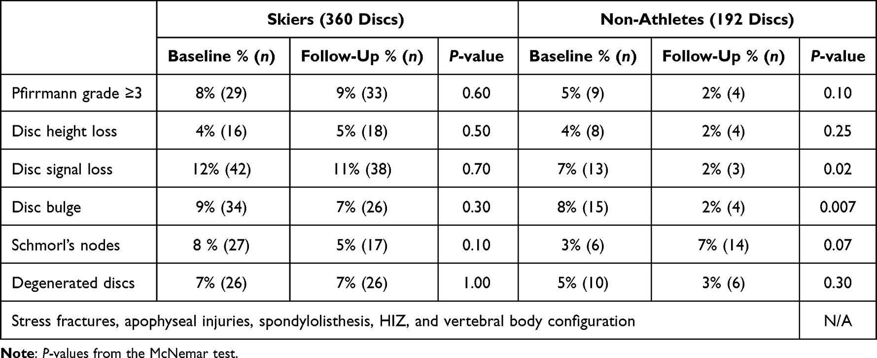 |
Table 2 The Findings on MRI at Baseline and 2-Year Follow-Up Based on Number of Discs |
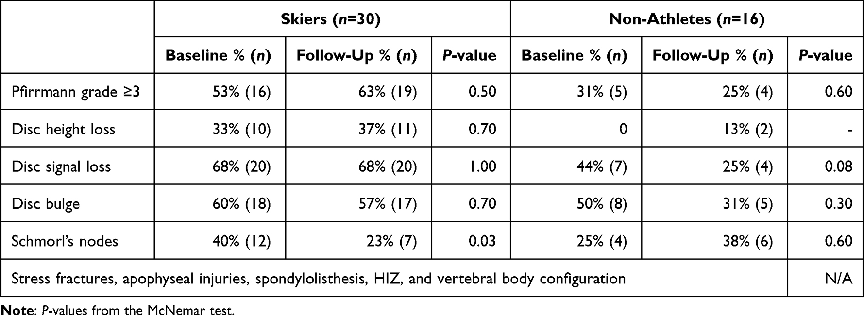 |
Table 3 The Findings on MRI at Baseline and 2-Year Follow-Up When Analyzed per Individual |
MRI Findings
The MRI findings are listed in Table 4. Intervertebral discs graded Pfirrmann ≥3 were seen in 63% of skiers compared to 25% of non-athletes (p=0.03). The number of discs graded Pfirrmann ≥3 was 1.1 on average (range 1–4) per skier and 0.3 (range 0–1) per non-athlete (Figure 1). Disc degenerative changes were more common in skiers than in non-athletes (73% vs 44%, respectively, p=0.05), when any of the following findings was considered as disc degenerative change: reduced signal, reduced height, disc bulge, or herniation.
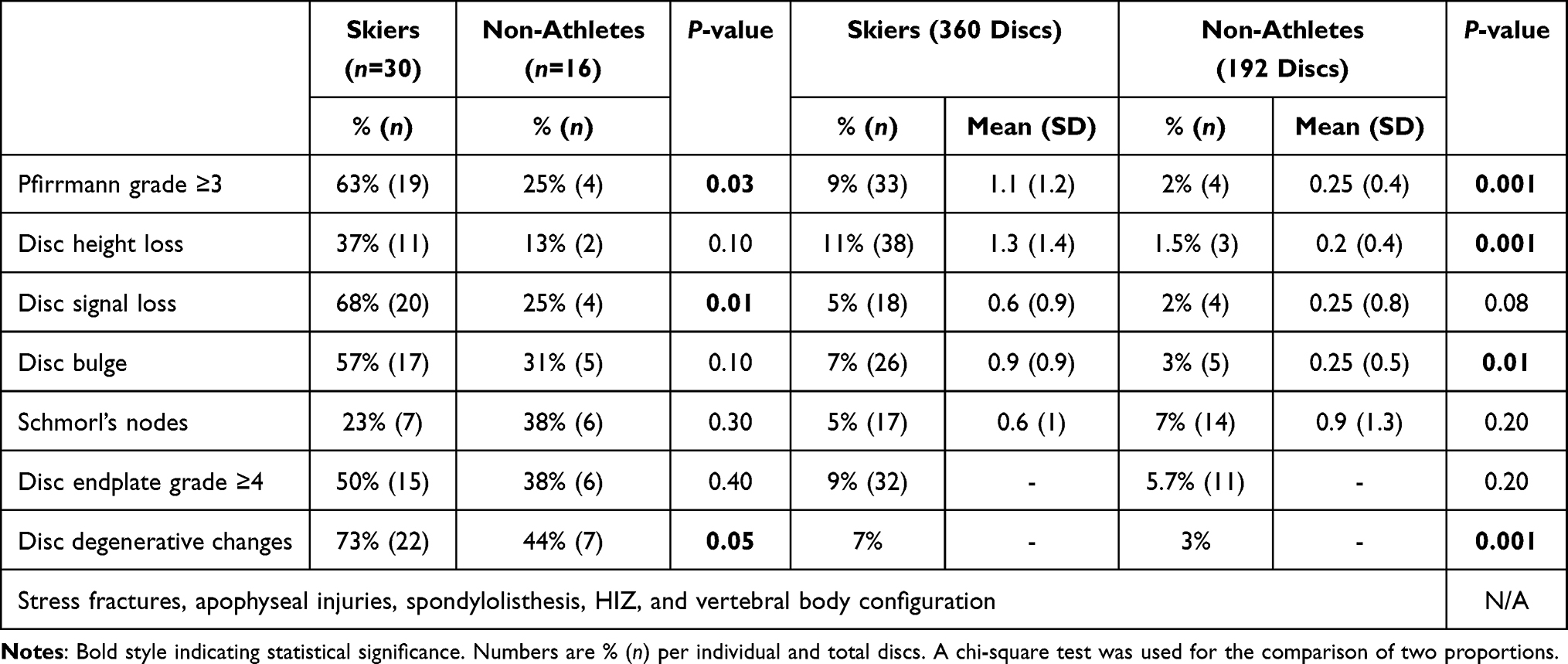 |
Table 4 The Findings on MRI in Skiers versus Non-Athletes When Analyzed as Groups of Individuals and When Analyzed According to the Total Number of Discs |
 |
Figure 1 MRI T2-weighted images of the thoraco-lumbar spine. (A) Athlete with disc signal and height loss at L4–L5 and L5–S1 (arrows). (B) Non-athlete without abnormalities. |
Back Pain
There was no statistically significant difference of the back pain lifetime prevalence in skiers (46%) compared with non-athletes (40%). There was no significant worsening over time. Back pain was associated with disc abnormalities in skiers (p=0.04) but not in non-athletes (p=0.70).
Validity
The intra-observer reliability was high, with Cohen’s kappa coefficient ranging between κ = 0.85 and κ = 0.95 for all parameters (Table 5).
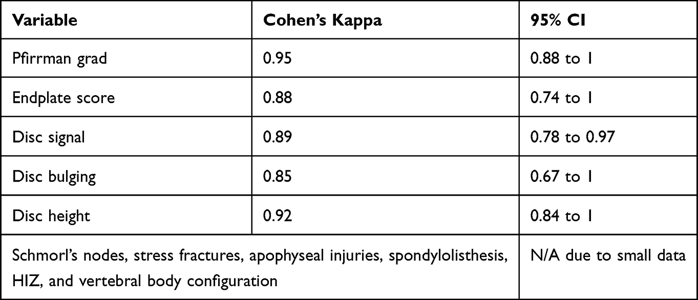 |
Table 5 Intra-Observer Reliability Cohen’s Kappa Values |
Discussion
In this longitudinal study, examining the interval change over time of spinal column abnormalities and back pain over a 2-year follow-up period, the occurrence of new MRI abnormalities was found to be rare and the change in back pain prevalence over time was not significant.
In contrast to the present study, prior longitudinal studies of athletes have reported a wide degree of deterioration of spine abnormalities on long-term follow-up. This could be related to the longer duration of overloading the spine with continuous sporting activity in addition to normal aging.24,34,35
Most spinal abnormalities were reported to occur during a growth spurt when the spine is most sensitive to injuries in young individuals.20,36,37 However, the average age of the participants at both baseline and follow-up was beyond the growth spurt age, 18 and 20 years, respectively. This could potentially explain the rare occurrence of new abnormalities in the present study.
The current findings of higher prevalence of spinal column abnormalities in young skiers compared with non-athletes are in concordance with several prior studies of young athletes such as wrestlers, gymnasts, weight-lifters, divers, skiers, long-distance runners, ice-hockey players, football players, and tennis players.11,15,19,24 This higher prevalence in athletes, with excessive and lengthy physical training at a very young age, is believed to be due to overloading the skeletally immature spine.20,36,37 Repetitive microtraumas of the discs and vertebrae are considered to be the main culprit for the reported abnormalities.11,15,19 It is suggested that frontal and lateral bending of the spine associated with skiing in addition to axial overload predisposes the discs to overuse injuries, which may eventually lead to back pain.6
There was association between back pain and disc abnormalities in skiers in the present study. Prior studies, including the baseline study, reported no such association.19,38,39 These findings suggest that spinal abnormalities may lead to complications like back pain later in life. Nonetheless, back pain could also be due to additional extra-spinal factors such as muscle strain and ligament sprain.22,27,40
There was no significant difference of the back pain lifetime prevalence in skiers compared with non-athletes. This could be a true representation of the general population, both athletes and non-athletes. However, alternative factors may include recall bias and the small size of the groups. A wide range of back pain prevalence was previously reported in young athletes.13,15,17,20,21,26 A 1-year prevalence of back pain was reported to range between 26% and 76% in a recent literature review of 43 papers.27
Further long-term follow-up projects with prospective design and with larger population groups are warranted. Incorporation of new imaging methods such as functional MRI and mapping analysis can be useful to achieve more objective quantitative estimates and to detect subtle abnormalities.41–43
Strengths and Limitations
The skiers and controls were within the same age-range and considered a valid representation for the adolescent population. The height and weight were very similar between the groups.
The small sample size and large number of dropouts on follow-up are the main limitations in the present study. All available age-matched athletes were invited to participate in the baseline study and to undergo follow-up 2 years later. However, some participants were travelling abroad or unable to attend the appointments. Another limitation is gender inequality as females constituted 43% of skiers and 75% of non-athletes; this is a potential confounder, although gender did not appear to significantly affect the results in prior studies.19,37 As this was an observational study, other unknown confounders such as nutrition, smoking, and participants' hobbies could have also biased the outcome. The back pain questionnaires are subject-reported information and may be affected by recall bias and difficult to measure objectively.
Conclusion
There was no significant change over time of the spinal column MRI abnormalities, nor back pain prevalence, during a 2-year follow-up of skiers and non-athletes. Young skiers had significantly higher prevalence of spine abnormalities compared with non-athletes. There was no significant difference of the back pain lifetime prevalence in skiers compared with non-athletes.
Disclosure
The authors report no conflicts of interest in this work.
References
1. Adirim TA, Cheng TL. Overview of injuries in the young athlete. Sports Med. 2003;33(1):75–81. doi:10.2165/00007256-200333010-00006
2. Hogan KA, Gross RH. Overuse injuries in pediatric athletes. Orthop Clin North Am. 2003;34(3):405–415. doi:10.1016/S0030-5898(03)00006-3
3. Gebhart JJ, Streit JJ, Bedi A, Bush-Joseph CA, Nho SJ, Salata MJ. Correlation of pelvic incidence with cam and pincer lesions. Am J Sports Med. 2014;42(11):2649–2653. doi:10.1177/0363546514548019
4. Morris WZ, Fowers CA, Yuh RT, Gebhart JJ, Salata MJ, Liu RW. Decreasing pelvic incidence is associated with greater risk of cam morphology. Bone Joint Res. 2016;5(9):387–392. doi:10.1302/2046-3758.59.BJR-2016-0028.R1
5. Todd CTO, Swärd L, Karlsson J, et al. Pelvic retroversion is associated with flat back and cam type femoro- acetabular impingement in young elite skiers. J Spine. 2016;5:326. doi:10.4172/2165-7939.1000326
6. Spörri J, Kröll J, Haid C, Fasel B, Müller E. Potential mechanisms leading to overuse injuries of the back in alpine ski racing: a descriptive biomechanical study. Am J Sports Med. 2015;43(8):2042–2048. doi:10.1177/0363546515588178
7. Mosley GE, Hoy RC, Nasser P, et al. Sex differences in rat intervertebral disc structure and function following annular puncture injury. Spine. 2019;44(18):1257–1269. doi:10.1097/BRS.0000000000003055
8. Bergstrom KA, Brandseth K, Fretheim S, Tvilde K, Ekeland A. Back injuries and pain in adolescents attending a ski high school. Knee Surg Sports Traumatol Arthrosc. 2004;12(1):80–85. doi:10.1007/s00167-003-0389-0
9. Florenes TW, Heir S, Nordsletten L, Bahr R. Injuries among World Cup freestyle skiers. Br J Sports Med. 2010;44(11):803–808. doi:10.1136/bjsm.2009.071159
10. Bono CM. Low-back pain in athletes. J Bone Jt Surg. 2004;86(2):382–396. doi:10.2106/00004623-200402000-00027
11. Goldstein JD, Berger PE, Windler GE, Jackson DW. Spine injuries in gymnasts and swimmers An Epidemiologic Investigation. Am J Sports Med. 1991;19(5):463–468. doi:10.1177/036354659101900507
12. Mallow GM, Zepeda D, Kuzel TG, et al. ISSLS PRIZE in Clinical Science 2022: epidemiology, risk factors and clinical impact of juvenile Modic changes in paediatric patients with low back pain. Eur Spine J. 2022;31(5):1069–1079. doi:10.1007/s00586-022-07125-x
13. Baranto A, Hellstrom M, Nyman R, Lundin O, Sward L. Back pain and degenerative abnormalities in the spine of young elite divers: a 5-year follow-up magnetic resonance imaging study. Knee Surg Sports Traumatol Arthrosc. 2006;14(9):907–914. doi:10.1007/s00167-005-0032-3
14. Sward L, Hellstrom M, Jacobsson B, Nyman R, Peterson L. Acute injury of the vertebral ring apophysis and intervertebral disc in adolescent gymnasts. Spine. 1990;15(2):144–148. doi:10.1097/00007632-199002000-00021
15. Sward L, Hellstrom M, Jacobsson B, Peterson L. Back pain and radiologic changes in the thoraco-lumbar spine of athletes. Spine. 1990;15(2):124–129. doi:10.1097/00007632-199002000-00015
16. Granhed H, Morelli B. Low back pain among retired wrestlers and heavyweight lifters. Am J Sports Med. 1988;16(5):530–533. doi:10.1177/036354658801600517
17. Thoreson O, Svensson K, Jonasson P, Kovac P, Sward L, Baranto A. Back pain and MRI abnormalities in the thoraco-lumbar spine of elite long distance runners A Cross Sectional Study. Med Res Arch. 2015;2(4).
18. Hellström M, Jacobsson B, Swärd L, Peterson L. Radiologic abnormalities of the thoraco-lumbar spine in athletes. Acta radiologica. 1990;31(2):127–132. doi:10.1177/028418519003100202
19. Witwit WA, Kovac P, Sward A, et al. Disc degeneration on MRI is more prevalent in young elite skiers compared to controls. Knee Surg Sports Traumatol Arthrosc. 2018;26(1):325–332. doi:10.1007/s00167-017-4545-3
20. Kulling FA, Florianz H, Reepschlager B, Gasser J, Jost B, Lajtai G. High prevalence of disc degeneration and spondylolysis in the lumbar spine of professional beach volleyball players. Orthop J Sports Med. 2014;2(4):2325967114528862. doi:10.1177/2325967114528862
21. Rachbauer F, Sterzinger W, Eibl G. Radiographic abnormalities in the thoracolumbar spine of young elite skiers. Am J Sports Med. 2001;29(4):446–449. doi:10.1177/03635465010290041101
22. Mortazavi J, Zebardast J, Mirzashahi B. Low Back Pain in Athletes. Asian J Sports Med. 2015;6(2):e24718. doi:10.5812/asjsm.6(2)2015.24718
23. Todd C, Aminoff AS, Agnvall C, et al. No difference in prevalence of spine and Hip pain in young Elite skiers. Knee Surg Sports Traumatol Arthrosc. 2018;26(7):1959–1965. doi:10.1007/s00167-017-4733-1
24. Baranto A, Hellstrom M, Cederlund CG, Nyman R, Sward L. Back pain and MRI changes in the thoraco-lumbar spine of top athletes in four different sports: a 15-year follow-up study. Knee Surg Sports Traumatol Arthrosc. 2009;17(9):1125–1134. doi:10.1007/s00167-009-0767-3
25. Horne J, Cockshott WP, Shannon HS. Spinal column damage from water ski jumping. Skeletal Radiol. 1987;16(8):612–616. doi:10.1007/BF00357108
26. Ogon M, Riedl-Huter C, Sterzinger W, Krismer M, Spratt KF, Wimmer C. Radiologic abnormalities and low back pain in elite skiers. Clin Orthop Relat Res. 2001;390(390):151–162. doi:10.1097/00003086-200109000-00018
27. Trompeter K, Fett D, Platen P. Prevalence of back pain in sports: a systematic review of the literature. Sports Med. 2017;47(6):1183–1207. doi:10.1007/s40279-016-0645-3
28. Ekşi M, Özcan-ekşi EE, Özmen BB, et al. Lumbar intervertebral disc degeneration, end-plates and paraspinal muscle changes in children and adolescents with low-back pain. J Pediatr Orthop B. 2022;31(1):93–102. doi:10.1097/BPB.0000000000000833
29. Zemková E, Kováčiková Z, Zapletalová L. Is there a relationship between workload and occurrence of back pain and back injuries in athletes? Front Physiol. 2020;11:894. doi:10.3389/fphys.2020.00894
30. Pfirrmann CW, Metzdorf A, Zanetti M, Hodler J, Boos N. Magnetic resonance classification of lumbar intervertebral disc degeneration. Spine. 2001;26(17):1873–1878. doi:10.1097/00007632-200109010-00011
31. Rade M, Määttä JH, Freidin MB, Airaksinen O, Karppinen J, Williams FMK. Vertebral endplate defect as initiating factor in intervertebral disc degeneration: strong association between endplate defect and disc degeneration in the general population. Spine. 2018;43(6):412–419. doi:10.1097/BRS.0000000000002352
32. Rajasekaran S, Venkatadass K, Naresh Babu J, Ganesh K, Shetty AP. Pharmacological enhancement of disc diffusion and differentiation of healthy, ageing and degenerated discs: results from in-vivo serial post-contrast MRI studies in 365 human lumbar discs. Eur Spine J. 2008;17(5):626–643. doi:10.1007/s00586-008-0645-6
33. Landis JR, Koch GG. The measurement of observer agreement for categorical data. Biometrics. 1977;33(1):159–174. doi:10.2307/2529310
34. Wang F, Cai F, Shi R, Wang XH, Wu XT. Aging and age related stresses: a senescence mechanism of intervertebral disc degeneration. OARS. 2016;24(3):398–408. doi:10.1016/j.joca.2015.09.019
35. Thoreson O, Ekström L, Hansson HA, et al. The effect of repetitive flexion and extension fatigue loading on the young porcine lumbar spine, a feasibility study of MRI and histological analyses. J Exp Orthop. 2017;4(1):16. doi:10.1186/s40634-017-0091-7
36. Rosendahl K, Strouse PJ. Sports injury of the pediatric musculoskeletal system. Radiol Med. 2016;121(5):431–441. doi:10.1007/s11547-015-0615-0
37. Wojtys EM, Ashton-Miller JA, Huston LJ, Moga PJ. The association between athletic training time and the sagittal curvature of the immature spine. Am J Sports Med. 2000;28(4):490–498. doi:10.1177/03635465000280040801
38. Witwit W, Thoreson O, Swärd Aminoff A, et al. Young football players have significantly more spinal changes on MRI compared to non-athletes. Transl Sport Med. 2020;3(4):288–295. doi:10.1002/tsm2.144
39. Thoreson O, Kovac P, Swärd A, Agnvall C, Todd C, Baranto A. Back pain and MRI changes in the thoraco-lumbar spine of young elite Mogul skiers. Scand J Med Sci Sports. 2017;27(9):983–989. doi:10.1111/sms.12710
40. Peacock N, Walker JA, Fogg R, Dudley K. Prevalence of low back pain in alpine ski instructors. J Orthop Sports Phys Ther. 2005;35(2):106–110. doi:10.2519/jospt.2005.35.2.106
41. Belavy DL, Brisby H, Douglas B, et al. Characterization of intervertebral disc changes in asymptomatic individuals with distinct physical activity histories using three different quantitative MRI techniques. J Clin Med. 2020;9(6):1841. doi:10.3390/jcm9061841
42. Lagerstrand K, Baranto A, Hebelka H. Different disc characteristics between young elite skiers with diverse training histories revealed with a novel quantitative magnetic resonance imaging method. Eur Spine J. 2021;30(7):2082–2089. doi:10.1007/s00586-021-06869-2
43. Waldenberg C, Hebelka H, Brisby H, Lagerstrand KM. Differences in IVD characteristics between low back pain patients and controls associated with HIZ as revealed with quantitative MRI. PLoS One. 2019;14(8):e0220952. doi:10.1371/journal.pone.0220952
 © 2022 The Author(s). This work is published and licensed by Dove Medical Press Limited. The
full terms of this license are available at https://www.dovepress.com/terms
and incorporate the Creative Commons Attribution
- Non Commercial (unported, 3.0) License.
By accessing the work you hereby accept the Terms. Non-commercial uses of the work are permitted
without any further permission from Dove Medical Press Limited, provided the work is properly
attributed. For permission for commercial use of this work, please see paragraphs 4.2 and 5 of our Terms.
© 2022 The Author(s). This work is published and licensed by Dove Medical Press Limited. The
full terms of this license are available at https://www.dovepress.com/terms
and incorporate the Creative Commons Attribution
- Non Commercial (unported, 3.0) License.
By accessing the work you hereby accept the Terms. Non-commercial uses of the work are permitted
without any further permission from Dove Medical Press Limited, provided the work is properly
attributed. For permission for commercial use of this work, please see paragraphs 4.2 and 5 of our Terms.