
Back to Journals » Journal of Inflammation Research » Volume 18
Nicotine Attenuates Chondrocyte Inflammation via the α7nAChR-Mediated Inhibition of the HMGB1/TLR4/NF-κB Signaling Pathway
Authors Lin S, Liu T, Song G, Pan H
Received 10 June 2025
Accepted for publication 3 November 2025
Published 19 November 2025 Volume 2025:18 Pages 16157—16168
DOI https://doi.org/10.2147/JIR.S545945
Checked for plagiarism Yes
Review by Single anonymous peer review
Peer reviewer comments 3
Editor who approved publication: Dr Ujjwol Risal
Silong Lin, Tao Liu, Gaoming Song, Hong Pan
Department of Orthopaedics, Anqing First People’s Hospital of Anhui Medical University, Anqing, Anhui, People’s Republic of China
Correspondence: Hong Pan, Email [email protected]
Background: Chondrocyte inflammation is a major driver of osteoarthritis (OA). The cholinergic anti-inflammatory pathway (CAP) exhibits significant anti-inflammatory effects, with the α 7 nicotinic acetylcholine receptor (α 7nAChR) playing a pivotal role. However, it remains unclear whether α 7nAChR alleviates chondrocyte inflammation through the regulation of the high mobility group box 1 (HMGB1)/TLR4/NF-κB signaling pathway.
Methods: An in vitro model of chondrocyte inflammation was established by lipopolysaccharide (LPS) treatment of mouse cartilage cells. The protective effects of various concentrations of nicotine (Nic) on chondrocytes were assessed using the CCK-8 assay. To verify the critical role of α 7nAChR, groups treated with methyllycaconitine (MLA, an α 7nAChR antagonist) and siRNA-α 7nAChR were included. Immunofluorescence was employed to observe the localization and expression of α 7nAChR, HMGB1, and P65. Additionally, qRT-PCR, Western blot, and ELISA were employed to detect the expression of inflammation-related factors, cytokines, and MMPs.
Results: The CCK-8 assay indicated that 1000 nmol/L Nic significantly restored chondrocyte proliferation activity reduced by LPS stimulation (P < 0.01). Immunofluorescence, Western blot, and qRT-PCR results demonstrated that Nic markedly enhanced α 7nAChR expression and inhibited LPS-induced nuclear translocation of HMGB1, as well as the expression of TLR4 and P65. The anti-inflammatory effects of Nic were significantly diminished following MLA treatment or siRNA-mediated knockdown of α 7nAChR (P < 0.01). Furthermore, ELISA assays demonstrated that Nic suppressed the secretion of IL-1β, TNF-α, MMP-2, and MMP-9, an effect dependent on α 7nAChR activation.
Conclusion: This study reveals that Nic alleviates chondrocyte inflammatory responses by activating α 7nAChR and inhibiting the HMGB1/TLR4/NF-κB signaling pathway. These findings not only enhance the understanding of CAP’s role in OA inflammation regulation but also provide a theoretical basis for exploring α 7nAChR as a potential anti-inflammatory target. However, since the research is currently limited to in vitro cellular studies, further animal experiments are necessary to validate its clinical translational potential.
Keywords: nicotine, α 7nAChR, HMGB1/TLR4/NF-κB pathway, chondrocyte inflammation, osteoarthritis
Introduction
Osteoarthritis (OA) is a prevalent degenerative joint disorder, impacting approximately 10% to 15% of the adult population worldwide.1 Recent data indicate that the global prevalence of OA was 7.96% in 2020, marking an increase of over 130% since 1990.2,3 The pathological characteristics of OA include articular cartilage degradation and synovial inflammation, with chondrocyte-mediated inflammatory responses recognized as key drivers of OA progression.4–6 In patients with OA, pro-inflammatory mediators such as tumor necrosis factor-alpha (TNF-α) and interleukin-1 beta (IL-1β) are significantly elevated in the cartilage. These cytokines activate chondrocytes to produce matrix-degrading enzymes, including matrix metalloproteinases (MMPs) and a disintegrin and metalloproteinase with thrombospondin motifs (ADAMTS). These enzymes degrade extracellular matrix (ECM) like type II collagen and proteoglycans, perpetuating a detrimental cycle that disrupts the equilibrium between cartilage degradation and repair.7–9
The cholinergic anti-inflammatory pathway (CAP), governed by vagal signaling, has recently been recognized as a crucial mechanism for chronic inflammatory responses.10,11 CAP functions through the release of acetylcholine from vagus nerve endings, which activates the α7 nicotinic acetylcholine receptors (α7nAChR) on immune cells.12,13 Activation of α7nAChR suppresses pro-inflammatory cytokine expression and dampens inflammatory cascades, demonstrating robust anti-inflammatory effects in diverse disease models, including sepsis and rheumatoid arthritis.14–16 Activation of α7nAChR downregulates Toll-like receptor 4 (TLR4) expression in macrophages and inhibits the production and release of high mobility group box 1 (HMGB1), thereby attenuating continuous TLR4/NF-κB pathway activation and mitigating local tissue damage and systemic inflammation.17,18 HMGB1, a prototypical damage-associated molecular pattern (DAMP), is upregulated in OA cartilage and synovium. By binding to TLR4, it activates NF-κB signaling, induces downstream cytokine release, and exacerbates local inflammation and tissue destruction.19 In summary, the α7nAChR-mediated cholinergic pathway exerts potent anti-inflammatory effects by repressing mediators such as HMGB1 and signaling cascades like TLR4/NF-κB. However, the role of the α7nAChR-HMGB1/TLR4/NF-κB axis in modulating chondrocyte inflammation has not been fully elucidated.
Nicotine (Nic) is a classic exogenous agonist of α7nAChR. Compared with newly developed agonists, Nic offers advantages such as a simple structure and widespread availability. Its immunomodulatory effects are complex and context-dependent, influenced by factors including site of action, route of administration, and dosage.20 Numerous investigations have demonstrated that Nic exerts significant anti-inflammatory effects in various diseases, notably in ulcerative colitis, sepsis, and rheumatoid arthritis, where it effectively downregulates inflammatory mediators like HMGB1 and TNF-α, ameliorates tissue damage, and improves survival rates.21–24 In collagen-induced arthritis (CIA) models, Nic suppresses Th17 cell-mediated inflammation and inhibits HMGB1 nuclear translocation in synovial tissues via α7nAChR activation, thereby alleviating synovitis and joint destruction.25,26 Furthermore, in monosodium iodoacetate-induced OA models, Nic has been shown to reduce joint pain and cartilage degradation through the α7nAChR-mediated PI3K/Akt–NF-κB pathway.27 However, these studies primarily focus on the effects of Nic on immune cells like macrophages, leaving its direct impact on chondrocyte inflammation, as well as the involvement of HMGB1 and related pathways, largely unexplored.
Currently, research on the direct anti-inflammatory effects of Nic on OA chondrocytes is limited, particularly regarding whether it alleviates chondrocyte inflammation by activating α7nAChR to inhibit the HMGB1/TLR4/NF-κB signaling cascade. Therefore, this study established an in vitro chondrocyte inflammation model induced by lipopolysaccharide (LPS) to examine how nicotine modulates inflammatory responses and to clarify the involvement of α7nAChR in regulating the HMGB1/TLR4/NF-κB axis. This research is expected to provide theoretical and experimental support for a deeper understanding of OA inflammatory mechanisms and the development of novel therapeutic strategies.
Materials and Methods
Cell Preparation
Primary chondrocytes were obtained in accordance with the protocol reported by Jiansen Lu et al.28 Costal cartilage was harvested from 72-hour-old neonatal male C57BL/6J mice under a stereomicroscope. After removing surrounding soft tissues with a cell scraper, the cartilage was washed with sterile PBS and minced into approximately 1 mm3 pieces using ophthalmic scissors. The tissue fragments were subjected to enzymatic digestion: initially with 0.25% trypsin (Cat. No. 25200072, Gibco, USA) for 30 minutes, followed by 0.1% type II collagenase (Cat. No. C6885, Sigma-Aldrich, USA) in medium containing 10% FBS (Cat. No. G8003, Servicebio, China) for an additional 4 to 6 hours. Then, the suspension was passed through a 200-mesh sieve and centrifuged to remove debris. The harvested chondrocytes were resuspended in DMEM/F12 (Cat. No. 11320033, Gibco, USA) supplemented with 10% FBS and seeded into 24-well plates. Cultures were maintained at 37 °C with 5% CO2. The first medium change occurred 24 hours post-seeding, followed by medium changes every 2–3 days until cells achieved nearly 80% confluency for subsequent experiments.
CCK-8 Assay
Cell proliferation under varying concentrations of Nic was evaluated through the CCK-8 assay. Post-treatment, cells were harvested and transferred into 96-well plates (5×10³ cells/mL). To minimize edge effects, PBS was added to the peripheral wells. Medium was refreshed every 24 hours. At 0, 24, 48, and 72 hours after the first medium change, the medium was replaced with fresh medium supplemented with 10% CCK-8 (Cat. No. G4103, Servicebio, China) and cells were incubated for an additional 2 hours. Absorbance was measured at OD450 using an ELISA reader.
LPS is a major pro-inflammatory inducer and is commonly used to establish arthritis models in chondrocytes.29,30 The experiment included five groups, each with six parallel samples: Control, LPS, LPS+Nic10, LPS+Nic100, and LPS+Nic1000. (1) Control group: cells were maintained under standard culture conditions without any experimental intervention. (2) LPS group: cells treated with 1 mg/L LPS (from E. coli 055:B5, Cat. No. L2880, Sigma-Aldrich, USA) for 9 hours. (3) LPS+Nic10 group: cells were pretreated with 10 nmol/L Nic (Cat. No. N3876, Sigma-Aldrich, USA) for 10 minutes, and without washing, subsequently treated with 1 mg/L LPS for 9 hours. (4) LPS+Nic100 group: cells were pretreated with 100 nmol/L Nic for 10 minutes, and without washing, subsequently treated with 1 mg/L LPS for 9 hours. (5) LPS+Nic1000 group: cells were pretreated with 1000 nmol/L Nic for 10 minutes, and without washing, subsequently treated with 1 mg/L LPS for 9 hours.
Grouping and Treatments
Primary chondrocytes were assigned to six groups, each with at least three replicates: Control, LPS, LPS+Nic, LPS+Nic+MLA, LPS+Nic+siRNA-α7nAChR, and LPS+Nic+siRNA-NC. (1) Control group: cells were maintained under standard culture conditions without any experimental intervention. (2) LPS group: cells treated with 1 mg/L LPS for 9 hours. (3) LPS+Nic group: cells pretreated with 1000 nmol/L Nic for 10 minutes, and without washing, subsequently treated with 1 mg/L LPS for 9 hours. (4) LPS+Nic+MLA group: cells pretreated with 1 μmol/L methyllycaconitine (MLA, Cat. No. 1029, Tocris Bioscience, UK) for 30 minutes, then with 1000 nmol/L Nic for 10 minutes, and without washing, subsequently treated with 1 mg/L LPS for 9 hours. (5) LPS+Nic+siRNA-α7nAChR group: cells transfected with siRNA-α7nAChR were plated into 6-well plates and maintained for 39 hours, then treated with 1000 nmol/L Nic for 10 minutes, and without washing, subsequently treated with 1 mg/L LPS for 9 hours. (6) LPS+Nic+siRNA-NC group: cells transfected with siRNA-NC were treated identically to the LPS+Nic+siRNA-α7nAChR group.
Immunofluorescence Staining
Cells were divided into four experimental groups: Control, LPS, LPS+Nic, and LPS+Nic+MLA (outlined in Section “Grouping and Treatments”). Chondrocytes were cultured on glass coverslips placed in 24-well plates. Once the cell confluence reached approximately 70%–80%, they were rinsed with sterile PBS and fixed using 4% paraformaldehyde for 30 minutes. After fixation, the cells were permeabilized with 0.3% Triton X-100 (Cat. No. T8787, Sigma-Aldrich, USA) and blocked with 5% BSA (Cat. No. G5001, Servicebio, China), then were incubated overnight at 4 °C with the primary antibody. The following day, cells were treated with Goat Anti-Rabbit IgG H&L (1:1000, Alexa Fluor® 488, ab150077, Abcam). Nuclei were counterstained with 20 μL DAPI (Cat. No. D9542, Sigma-Aldrich, USA) for 5 minutes, followed by PBS washing and mounting with 20 μL of antifade solution (Cat. No. P0126, Beyotime, China). Images were acquired using a fluorescence microscope. Six microscopic fields were randomly selected, and the mean fluorescence intensity was quantified using ImageJ software.
RNA Interference
Based on the target sequence of C57BL/6J mice from the GenBank database, siRNA-α7nAChR oligonucleotides were designed and synthesized, with siRNA-NC obtained as negative control (Genomeitech, Shanghai, China). The sequences were: siRNA-α7nAChR-sense-5′-AAGAGCUCCUGCUACAUCGAU-3′ and antisense-5′-AUCGAUGUAGCAGGAGCUCUU-3′. siRNA-NC-sense-5′-CGUACGCGGAAUACUUCGA-3′ and antisense 5′-UCGAAGUAUUCCGCGUACG-3′ (Genomeitech, China). 24 hours prior to transfection, chondrocytes were seeded into 6-well plates and cultured until reaching 70–80% confluence. The transfection procedure was carried out using Lipofectamine 2000 (Cat. No. 11668027, Invitrogen, USA) in accordance with the manufacturer’s protocol. Cells were harvested at 48 hours to extract proteins for assessing knockdown levels.
Enzyme-Linked Immunosorbent Assay (ELISA)
Culture supernatants from each group were collected and centrifuged to obtain cell-free supernatants. The levels of TNF-α (Cat. No. BMS607) and IL-1β (Cat. No. BMS6002, Thermo Fisher Scientific, USA), MMP-2 (Cat. No. MMP200), and MMP-9 (Cat. No. MMPT90, R&D Systems, USA) were then quantified through mouse-specific ELISA kits.
qRT-PCR
Total RNA was isolated using the TRIzol reagent (Cat. No. G3013, Servicebio, China). cDNA synthesis was conducted with the PrimeScript™ RT reagent Kit integrated with gDNA Eraser (Cat. No. RR047A, Takara, Japan). Quantitative real-time PCR was carried out utilizing the TB Green® Premix Ex Taq™ kit (Cat. No. RR420A, Takara, Japan), following the manufacturer’s instructions, with each sample run in triplicate. GAPDH served as the reference gene. The relative expression levels of mRNA were determined using the 2−ΔΔCt method. All primer sequences applied in this study are detailed in Table 1.
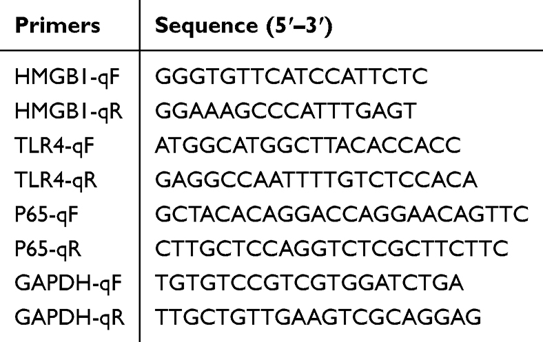 |
Table 1 Primer Sequences |
Western Blot (WB)
Chondrocytes were cultured in 6-well plates until they reached approximately 80% confluence. The cells were then subjected to the interventions described above. Upon completion of the treatments, the cells were immediately collected, and proteins were extracted using RIPA lysis buffer (Cat. No. R0020, Solarbio, China) containing both protease and phosphatase inhibitors (Cat. No. P1045, Beyotime, China). Protein samples were subjected to SDS-PAGE for electrophoretic separation and subsequently transferred onto PVDF membranes for immunoblotting analysis. The expression levels of HMGB1, TLR4, phosphorylated P65 (p-P65), and NF-κB (P65) were detected, with GAPDH serving as the loading control. Primary antibodies used included: GAPDH (1:2000, ab9485, Abcam), HMGB1 (1:1000, ab18256, Abcam), TLR4 (1:1000, ab13556, Abcam), p-P65 (1:500, ab86299, Abcam), and t-P65 (1:1000, ab16502, Abcam). The secondary antibody was horseradish peroxidase (HRP)-conjugated Goat Anti-Rabbit IgG H&L (1:5000, ab6721, Abcam).
Statistical Analysis
All experiments were repeated at least three times. Statistical analyses were performed using SPSS 19.0 and GraphPad Prism 5.0. Results are expressed as mean ± standard deviation (SD). The Shapiro–Wilk test was used to assess the normality of the data. For normally distributed data, comparisons between two groups were performed using Student’s t-test, while one-way analysis of variance (ANOVA) was used for comparisons among multiple groups. When significant differences were detected by ANOVA, Tukey’s post hoc test was applied to identify intergroup differences. Statistical significance was defined as P < 0.05.
Results
Effects of LPS and Nic on Chondrocyte Proliferation
To verify the cytotoxic effect of LPS and the protective effects of Nic at different concentrations, CCK-8 assays were conducted to monitor chondrocyte proliferation (Figure 1). At the initial time (0 h), no differences were observed (P > 0.05), indicating comparable starting conditions. As incubation time progressed, LPS markedly suppressed cell proliferation. At 24, 48, and 72 h, OD450 values in the LPS group were markedly lower than those observed in the Control group (P < 0.001), confirming the inhibitory effect of LPS. The LPS+Nic100 and LPS+Nic1000 groups showed significantly higher proliferation than the LPS group at 48 and 72 h (P < 0.05), with Nic1000 restoring proliferation to near-control levels at 72 h (P > 0.05). This suggests that 1000 nmol/L Nic provides superior protection against LPS-induced cytotoxicity compared to lower concentrations.
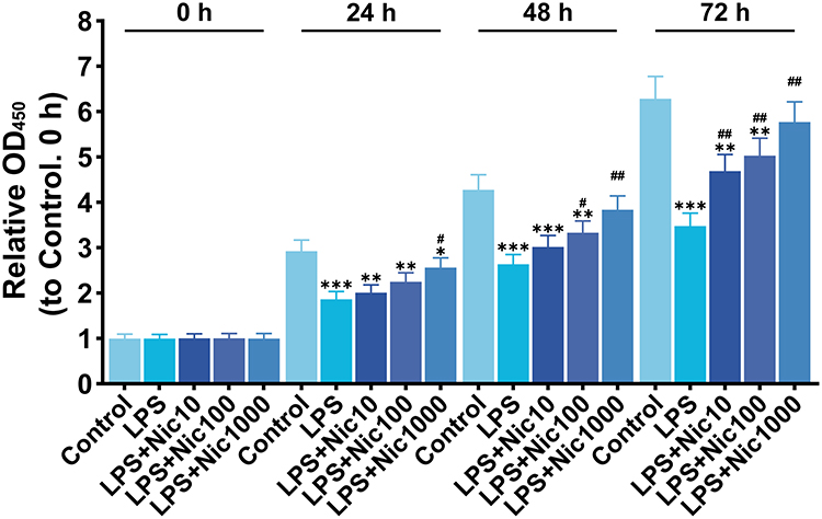 |
Figure 1 Effects of LPS and different concentrations of Nic on cell proliferation activity detected by CCK-8. n=5. (Data are presented as mean ± SD, and relative OD450 values were calculated using the Control group as the baseline. “*” indicates a significant difference compared with the Control group, and “#” indicates a significant difference compared with the LPS group. “*, #” represents P < 0.05, “**, ##” represents P < 0.01, and “***” represents P < 0.001). |
Nic Activates CAP and Regulates α7nAChR, HMGB1, and P65 Expression and Nuclear Localization
We assessed the cellular localization and expression of α7nAChR, HMGB1, and P65 using immunofluorescence, quantifying fluorescence intensity. DAPI staining was used to identify nuclei and evaluate nuclear translocation. Immunofluorescence revealed α7nAChR localized to the plasma membrane and HMGB1 to the nucleus in untreated chondrocytes. LPS stimulation significantly increased α7nAChR fluorescence intensity (P < 0.05), indicating CAP activation (Figure 2A). Nic treatment further amplified this effect relative to the LPS group (P < 0.01), yet the intervention of MLA prior to Nic effectively abolished it (no significant difference from LPS alone, P > 0.05). LPS also significantly elevated HMGB1 expression and induced its translocation from the nucleus to the cytoplasm. Nic effectively suppressed both the translocation and expression (P < 0.01), whereas MLA reversed Nic’s suppression, allowing HMGB1 levels to rise again (Figure 2B). Additionally, Nic significantly inhibited LPS-induced P65 nuclear translocation and downregulated its expression (P < 0.01), but MLA treatment disrupted this inhibitory effect (Figure 2C). These results indicate that LPS activates CAP in chondrocytes, Nic enhances this pathway, and Nic, via α7nAChR, suppresses HMGB1 and P65 expression and nuclear translocation.
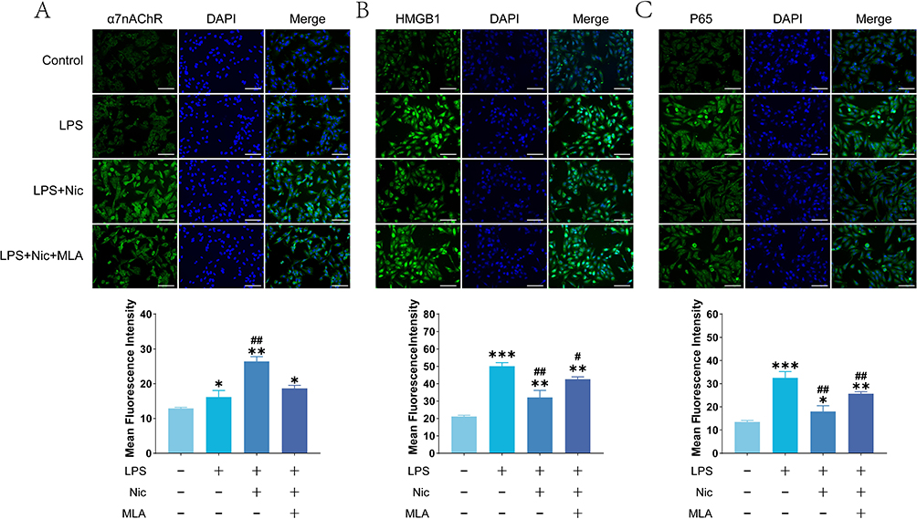 |
Figure 2 Immunofluorescence detection of α7nAChR, HMGB1, and P65 expression and nuclear translocation. (A) Representative immunofluorescence images of α7nAChR and the corresponding quantitative analysis (n=6). (B) Representative immunofluorescence images of HMGB1 and the corresponding quantitative analysis (n=6). (C) Representative immunofluorescence images of P65 and the corresponding quantitative analysis (n=6). Scale: 100 μm. (Data are presented as mean ± SD. “*” indicates a significant difference compared with the Control group, and “#” indicates a significant difference compared with the LPS group. “*, #” represents P < 0.05, “**, ##” represents P < 0.01, and “***” represents P < 0.001). |
Nic Regulates the Expression Levels of HMGB1, TLR4 and P65 via α7nAChR
To elucidate the essential role of α7nAChR in Nic-induced CAP activation and NF-κB regulation, RNA interference was performed. WB verified that α7nAChR expression was knocked down by 58% in comparison with the control group (Figure 3A and B). We then assessed expression levels of HMGB1, TLR4, and P65 via qRT-PCR and WB upon α7nAChR silencing. LPS stimulation significantly elevated HMGB1, TLR4, and P65 mRNA and protein levels, as well as P65 phosphorylation (P < 0.05), indicating NF-κB pathway activation and an inflammatory response (Figure 3C–K). Nic intervention in LPS-treated cells markedly suppressed these increases (P < 0.01). Crucially, this suppression was largely reversed when α7nAChR was inhibited—either by MLA or siRNA interference—with HMGB1, TLR4, and P65 levels rebounding significantly versus the LPS+Nic group. The siRNA-NC (negative control) group exhibited no significant difference from LPS+Nic, supporting the reliability of knockdown results. These findings underscore α7nAChR as a necessary mediator for Nic’s inhibition of the NF-κB signaling via the CAP pathway.
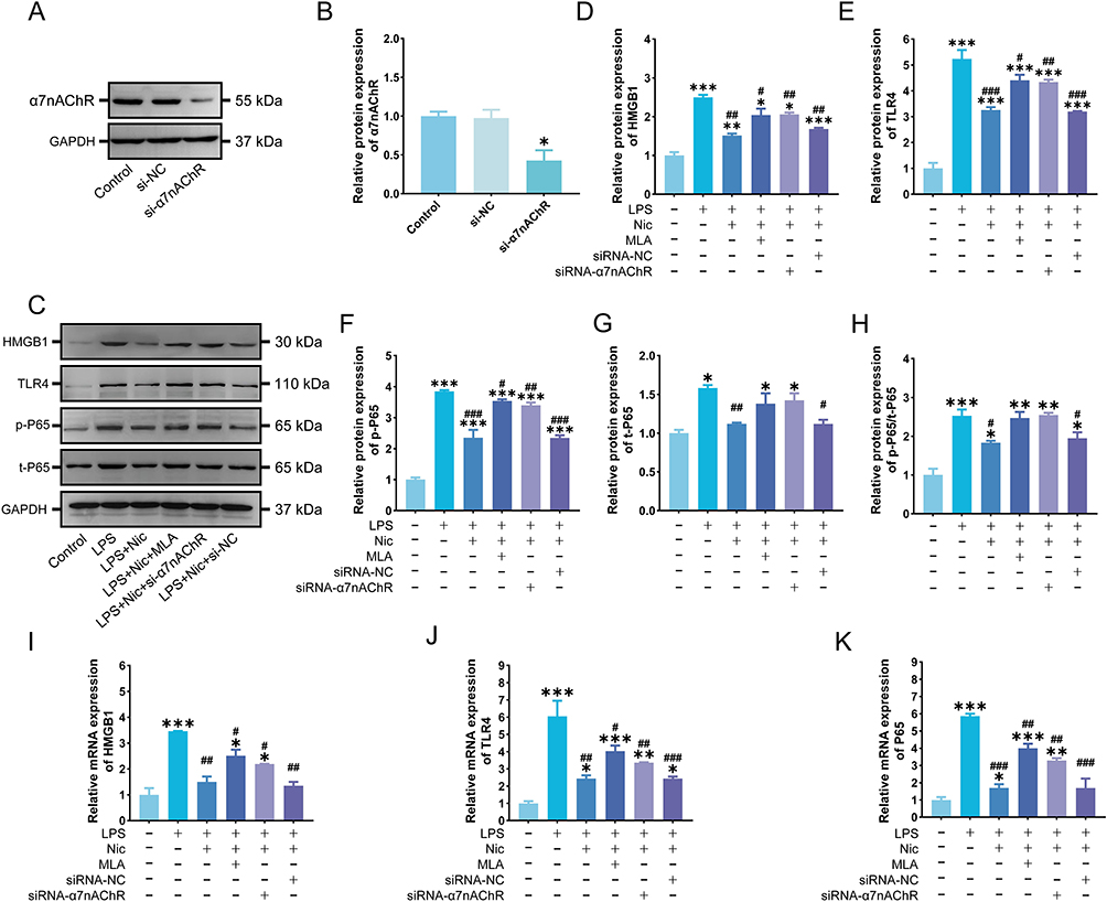 |
Figure 3 Expression analysis of key HMGB1/TLR4/NF-κB pathway proteins following α7nAChR knockdown. (A and B) WB and statistical results after si-α7nAChR transfection (n=3). (C–H) WB and statistical results of HMGB1, TLR4, p-P65, t-P65 and p-P65/t-P65 (n=3). (I–K) qRT-PCR statistical results of HMGB1, TLR4 and P65 (n=3). (Data are presented as mean ± SD, and relative protein expression was calculated using the Control group as the baseline. “*” indicates a significant difference compared with the Control group, and “#” indicates a significant difference compared with the LPS group. “*, #” represents P < 0.05, “**, ##” represents P < 0.01, and “***, ###” represents P < 0.001). |
Nic Can Inhibit LPS-Induced IL-1β and TNF-α Secretion
To further clarify the regulatory effects of Nic on inflammation via CAP activation, levels of IL-1β and TNF-α in the cell supernatants were assessed using ELISA (Figure 4A and B). The levels of both cytokines were significantly increased in the LPS group relative to those in the Control group (P < 0.01), indicating successful induction of an inflammatory response. Nic treatment significantly decreased the concentrations of IL-1β and TNF-α in LPS-stimulated cells compared with the LPS group (P < 0.05), suggesting an anti-inflammatory effect of Nic. When α7nAChR was specifically antagonized by MLA or silenced with siRNA-α7nAChR, the anti-inflammatory effect of Nic was markedly weakened: cytokine levels in both cases significantly higher than those in the LPS+Nic group and comparable to the LPS group (P > 0.05). Conversely, the LPS + Nic + siRNA-NC group exhibited cytokine levels comparable to those of the LPS + Nic group, further validating the reliability of the transfection. These findings indicate that Nic has a notable inhibitory effect on LPS-induced inflammation, which is dependent on α7nAChR expression. Blocking or silencing α7nAChR significantly diminishes the anti-inflammatory effect of Nic.
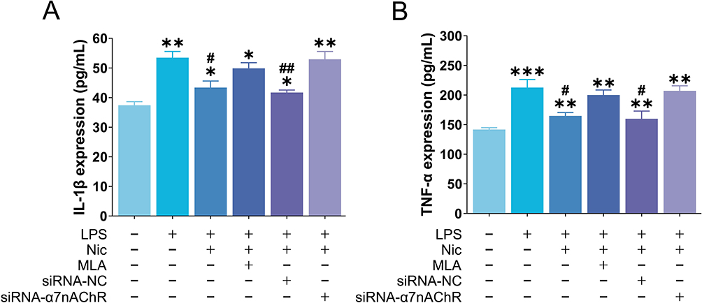 |
Figure 4 Results of ELISA detection of IL-1β and TNF-α levels in cell supernatants. (A and B) Statistical results of IL-1β and TNF-α expression levels detected by ELISA (n=3). (Data are presented as mean ± SD. “*” indicates a significant difference compared with the Control group, and “#” indicates a significant difference compared with the LPS group. “*, #” represents P < 0.05, “**, ##” represents P < 0.01, and “***” represents P < 0.001). |
Nic Can Inhibit LPS-Induced ECM Degradation
MMP-2 and MMP-9, members of the MMP family, are key enzymes responsible for degrading ECM components.31–34 They are excessively activated upon LPS stimulation, and their expression changes are closely associated with ECM remodeling (Figure 5A and B).35,36 Similar to the trends observed for IL-1β and TNF-α, the expression levels of MMP-2 and MMP-9 were significantly increased after LPS treatment (P < 0.01), indicating that ECM damage occurs during LPS-induced inflammatory responses. Nicotine intervention markedly reduced the expression of MMP-2 and MMP-9 (P < 0.05). However, this effect was reversed by MLA and siRNA-α7nAChR. These results suggest that nicotine attenuates LPS-induced ECM degradation, and that α7nAChR is likely involved in this process.
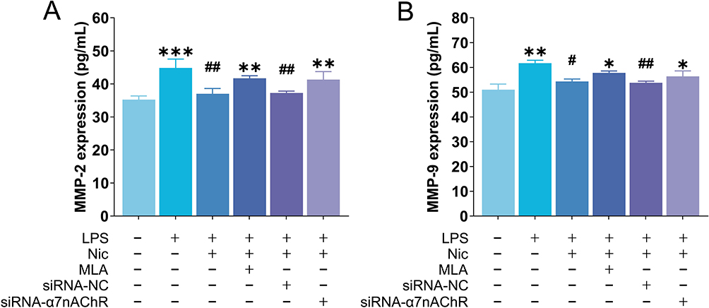 |
Figure 5 Results of ELISA detection of MMP-2 and MMP-9 levels in cell supernatants. (A and B) Statistical results of MMP-2 and MMP-9 expression levels detected by ELISA (n=3). (Data are presented as mean ± SD. “*” indicates a significant difference compared with the Control group, and “#” indicates a significant difference compared with the LPS group. “*, #” represents P < 0.05, “**, ##” represents P < 0.01, and “***” represents P < 0.001). |
Discussion
OA continues to jeopardize the health and quality of life of numerous patients. Research indicates that chondrocyte inflammation is a key driver of OA pathogenesis. The α7nAChR, a pivotal component of the CAP, plays a critical role in inflammatory regulation. It has been demonstrated to participate in bone remodeling during OA and exert significant anti-inflammatory effects. However, few studies have explored whether α7nAChR-mediated CAP modulates chondrocyte inflammation in OA. Therefore, we established an in vitro chondrocyte inflammation model using LPS stimulation and investigated Nic as an α7nAChR agonist. Our results show that Nic markedly alleviates chondrocyte inflammation by activating α7nAChR, with its mechanism linked to suppression of the HMGB1/TLR4/NF-κB inflammatory pathway.
Nic has long been restricted in clinical applications due to its association with addiction.37–39 However, recent studies reveal its biphasic, dose-dependent effects. While high doses of Nic exacerbate inflammation and adverse outcomes, moderate doses exhibit well-documented anti-inflammatory properties, mitigating joint inflammation and cartilage degradation. For instance, 0.1–1 μmol/L Nic reduced mechanical allodynia and synovitis in OA mice.20 Accordingly, we assessed chondrocyte proliferation under varying Nic concentrations via CCK-8 assay. LPS significantly suppressed proliferation, whereas Nic restored it dose-dependently, with 1 μmol/L Nic normalizing proliferation to control levels by 72 h.
CAP is a neuroimmune regulatory mechanism mediated by the vagus nerve. Acetylcholine released from vagus nerve endings binds to α7nAChR on the surface of immune cells such as macrophages, inhibiting the release of proinflammatory cytokines.40–43 Studies by Han et al showed that the selective α7nAChR agonist PHA-543613 can promote the secretion of IL-10 by macrophages through α7nAChR, and suppresses the production of TNF-α, IL-1β and IL-6 induced by LPS through IL-10/STAT3 signaling.44 In arthritis, α7nAChR-mediated CAP plays the same role. Studies in rat models of rheumatoid arthritis have found that the use of galantamine (a cholinesterase inhibitor) to increase endogenous acetylcholine levels can reduce joint inflammation and damage, while the use of α7nAChR antagonists will block this anti-inflammatory effect.45 This suggests that α7nAChR is an important receptor that mediates local anti-inflammatory responses within joint tissues. Liu et al observed that the level of α7nAChR in degenerated cartilage of OA patients was significantly reduced, accompanied by an imbalance between autophagy and apoptosis. The use of Nic can activate α7nAChR and protect joints, suggesting that α7nAChR is involved in the regulation of chondrocyte fate in OA.46 In contrast, the use of PNU-282987 (an α7nAChR agonist) in OA rats led to a reduction in joint concentrations of inflammatory mediators such as TNF-α and IL-1β, primarily through suppression of the NF-κB signaling and NLRP3 inflammasome pathways.47 Consistent with these results, we found through immunofluorescence and other techniques that Nic intervention can activate CAP and reduce the secretion of cellular inflammatory factors through suppressing the TLR4/NF-κB axis. In this regulation context, α7nAChR is an indispensable link. When MLA or siRNA-α7nAChR was used to antagonize α7nAChR expression, the suppression of Nic on the TLR4/NF-κB pathway was significantly reduced.
In addition, we found that Nic regulates the inflammation process through CAP, and also involves the regulation of HMGB1. Joint inflammation is not only mediated by classical cytokines, but also involves the participation of damage-associated pattern molecules (DAMPs). HMGB1 is one of the important DAMP molecules and is significantly increased in OA.48,49 High levels of HMGB1 can be detected in the articular cartilage, synovial tissue and synovial fluid of OA patients, and increasing concentrations are linked to more severe disease progression and symptomatology.50 HMGB1 is passively released from damaged or stressed chondrocytes, or actively secreted by synovial macrophages. HMGB1 can bind to TLR4, trigger the downstream NF-κB signaling cascade, promote the synthesis of pro-inflammatory mediators and amplify the inflammatory response.51,52 The use of HMGB1 antagonists such as A-box fragment peptides can competitively inhibit the binding of HMGB1 to the receptor, reduce IL-1β-induced chondrocyte MMP production in vitro, and reduce cartilage destruction in OA models.53 Further studies have shown that plant extracts such as Forsythoside B can inhibit the HMGB1/TLR4/NF-κB axis, attenuate IL-1β-induced chondrocyte inflammation and oxidative stress, and reduce cartilage degeneration in OA rats54 These studies suggest that HMGB1 is an amplifying factor of OA inflammation, and blocking its action or signaling pathway can reverse the proinflammatory effect of chondrocytes to alleviate OA symptoms. However, these studies have not fully explored the upstream regulatory factors of HMGB1. Therefore, this study further focused on the regulation of HMGB1 by α7nAChR, which not only confirmed the key position of the HMGB1/TLR4/NF-κB axis in the regulation of chondrocyte inflammation, but also proved that Nic can regulate this pathway through α7nAChR.
α7nAChR has been considered a potential anti-inflammatory drug target. Selective α7nAChR agonists are used to treat inflammation. GTS-21 (also known as DMXBA) is an orally effective partial α7nAChR agonist that can not only inhibit the production of pro-inflammatory cytokines within the synovial tissue of rheumatoid arthritis patients and reduce synovial inflammation, but also significantly inhibit NF-κB signaling in a colitis mouse model and enhance intestinal barrier function.55,56 Given that Nic can exert anti-inflammatory effects through α7nAChR and is easy to obtain, it also has the potential to treat OA. However, more research is still needed to rule out its addictiveness and confirm the dosage. This is also one of the shortcomings of this study. In the future, we will further extend our research to animal models.
Conclusion
In summary, this study reveals the role of the Nic-α7nAChR/HMGB1/TLR4/ NF-κB pathway in chondrocyte inflammation at the cellular and molecular levels, and this finding can enrich the understanding of the pathogenesis of OA inflammation. The confirmation of this mechanism has important theoretical and potential clinical translational value. The significance of this study is to provide new molecular targets and theoretical basis for the control of inflammation in OA. It is worth noting that although our research shows the positive effect of Nic on inhibiting inflammatory response, the experiments are based on the cellular level and clinical verification remains warranted.
Data Sharing Statement
The data that support the findings of this study are available on request from the corresponding author. The data are not publicly available due to privacy or ethical restrictions.
Ethics Approval
All animal experiments were conducted in accordance with the Guidelines for the Care and Use of Laboratory Animals of the Ministry of Science and Technology of the People’s Republic of China (2006) and the Laboratory Animal–Guidelines for Ethical Review of Animal Welfare (GB/T 35892-2018, China). This study was reviewed and approved by Animal Ethics Committee of Anhui Medical University (Approval Number: LLSC20221200).
Funding
This work was supported by the Key Project of Natural Science Research in Anhui Provincial Universities by the Education Department of Anhui Province(Award Number: 2022AH050749); Anhui Medical University Scientific Research Fund Project (Natural Sciences Category) (Award Number: 2022xkj094).
Disclosure
No potential conflict of interest was reported by the authors.
References
1. Ma Z, Wang D, Weng J, Zhang S, Zhang Y. BNIP3 decreases the LPS-induced inflammation and apoptosis of chondrocytes by promoting the development of autophagy. J Orthopaedic Surg Res. 2020;15(1):284. doi:10.1186/s13018-020-01791-7
2. Steinmetz JD, Culbreth GT, Haile LM, et al. Global, regional, and national burden of osteoarthritis, 1990–2020 and projections to 2050: a systematic analysis for the Global Burden of Disease Study 2021. Lancet Rheumatol. 2023;5(9):e508–e522. doi:10.1016/s2665-9913(23)00163-7
3. Wu R, Guo Y, Chen Y, Zhang J. Osteoarthritis burden and inequality from 1990 to 2021: a systematic analysis for the global burden of disease Study 2021. Sci Rep. 2025;15(1):8305. doi:10.1038/s41598-025-93124-z
4. Zhu R, Wang Y, Ouyang Z, et al. Targeting regulated chondrocyte death in osteoarthritis therapy. Biochem Pharmacol. 2023;215:115707. doi:10.1016/j.bcp.2023.115707
5. Musumeci G, Castrogiovanni P, Trovato FM, et al. Biomarkers of chondrocyte apoptosis and autophagy in osteoarthritis. Int J Mol Sci. 2015;16(9):20560–20575. doi:10.3390/ijms160920560
6. Jiang T, Su S, Tian R, et al. Immunoregulatory orchestrations in osteoarthritis and mesenchymal stromal cells for therapy. J Orthop Transl. 2025;55:38–54. doi:10.1016/j.jot.2025.08.009
7. Li H, Xie S, Qi Y, Li H, Zhang R, Lian Y. TNF-α increases the expression of inflammatory factors in synovial fibroblasts by inhibiting the PI3K/AKT pathway in a rat model of monosodium iodoacetate-induced osteoarthritis. Exp Ther Med. 2018;16(6):4737–4744. doi:10.3892/etm.2018.6770
8. Barlas IO, Sezgin M, Erdal ME, et al. Association of (−1,607) 1G/2G polymorphism of matrix metalloproteinase-1 gene with knee osteoarthritis in the Turkish population (knee osteoarthritis and MMPs gene polymorphisms). Rheumatol Int. 2009;29(4):383–388. doi:10.1007/s00296-008-0705-6
9. Li T, Peng J, Li Q, Shu Y, Zhu P, Hao L. The mechanism and role of ADAMTS protein family in osteoarthritis. Biomolecules. 2022;12(7):959. doi:10.3390/biom12070959
10. Su JC, Pan Q, Xu X, Wei X, Lei X, Zhang P. Structurally diverse steroids from an endophyte of Aspergillus tennesseensis 1022LEF attenuates LPS-induced inflammatory response through the cholinergic anti-inflammatory pathway. Chem Biol Interact. 2022;362:109998. doi:10.1016/j.cbi.2022.109998
11. Pavlov VA, Tracey KJ. The cholinergic anti-inflammatory pathway. Brain Behav Immun. 2005;19(6):493–499. doi:10.1016/j.bbi.2005.03.015
12. Xie Y, Tao S, Pan B, et al. Cholinergic anti-inflammatory pathway mediates diesel exhaust PM(2.5)-induced pulmonary and systemic inflammation. J Hazard Mater. 2023;458:131951. doi:10.1016/j.jhazmat.2023.131951
13. Keever KR, Yakubenko VP, Hoover DB. Neuroimmune nexus in the pathophysiology and therapy of inflammatory disorders: role of α7 nicotinic acetylcholine receptors. Pharmacol Res. 2023;191:106758. doi:10.1016/j.phrs.2023.106758
14. Wang W, Xu H, Lin H, Molnar M, Ren H. The role of the cholinergic anti-inflammatory pathway in septic cardiomyopathy. Int Immunopharmacol. 2021;90:107160. doi:10.1016/j.intimp.2020.107160
15. van Maanen MA, Vervoordeldonk MJ, Tak PP. The cholinergic anti-inflammatory pathway: towards innovative treatment of rheumatoid arthritis. Nat Rev Rheumatol. 2009;5(4):229–232. doi:10.1038/nrrheum.2009.31
16. Koopman FA, Schuurman PR, Vervoordeldonk MJ, Tak PP. Vagus nerve stimulation: a new bioelectronics approach to treat rheumatoid arthritis? Best Pract Res. 2014;28(4):625–635. doi:10.1016/j.berh.2014.10.015
17. Wang Z, Hou L, Yang H, et al. Electroacupuncture pretreatment attenuates acute lung injury through α7 nicotinic acetylcholine receptor-mediated inhibition of HMGB1 release in rats after cardiopulmonary bypass. Shock. 2018;50(3):351–359. doi:10.1097/shk.0000000000001050
18. Sitapara RA, Gauthier AG, Valdés-Ferrer SI, et al. The α7 nicotinic acetylcholine receptor agonist, GTS-21, attenuates hyperoxia-induced acute inflammatory lung injury by alleviating the accumulation of HMGB1 in the airways and the circulation. Mol Med. 2020;26(1):63. doi:10.1186/s10020-020-00177-z
19. Huang W, Tang Y, Li L. HMGB1, a potent proinflammatory cytokine in sepsis. Cytokine. 2010;51(2):119–126. doi:10.1016/j.cyto.2010.02.021
20. Zhang W, Lin H, Zou M, et al. Nicotine in inflammatory diseases: anti-inflammatory and pro-inflammatory effects. Front Immunol. 2022;13:826889. doi:10.3389/fimmu.2022.826889
21. Qin Z, Wan JJ, Sun Y, et al. Nicotine protects against DSS colitis through regulating microRNA-124 and STAT3. J Mol Med. 2017;95(2):221–233. doi:10.1007/s00109-016-1473-5
22. Wang H, Liao H, Ochani M, et al. Cholinergic agonists inhibit HMGB1 release and improve survival in experimental sepsis. Nat Med. 2004;10(11):1216–1221. doi:10.1038/nm1124
23. Datta S, Rahman MA, Koka S, Boini KM. High mobility group box 1 (HMGB1) mediates nicotine-induced podocyte injury. Front Pharmacol. 2024;15:1540639. doi:10.3389/fphar.2024.1540639
24. Li T, Zuo X, Zhou Y, et al. The vagus nerve and nicotinic receptors involve inhibition of HMGB1 release and early pro-inflammatory cytokines function in collagen-induced arthritis. J Clin Immunol. 2010;30(2):213–220. doi:10.1007/s10875-009-9346-0
25. Bruchfeld A, Goldstein RS, Chavan S, et al. Whole blood cytokine attenuation by cholinergic agonists ex vivo and relationship to vagus nerve activity in rheumatoid arthritis. J Internal Med. 2010;268(1):94–101. doi:10.1111/j.1365-2796.2010.02226.x
26. van Maanen MA, Lebre MC, van der Poll T, et al. Stimulation of nicotinic acetylcholine receptors attenuates collagen-induced arthritis in mice. Arthritis Rheum. 2009;60(1):114–122. doi:10.1002/art.24177
27. Teng P, Liu Y, Dai Y, Zhang H, Liu WT, Hu J. Nicotine attenuates osteoarthritis pain and matrix metalloproteinase-9 expression via the α7 nicotinic acetylcholine receptor. J Iimmunol. 2019;203(2):485–492. doi:10.4049/jimmunol.1801513
28. Lu J, Zhang H, Cai D, et al. Positive-feedback regulation of subchondral H-type vessel formation by chondrocyte promotes osteoarthritis development in mice. J Bone Miner Res. 2018;33(5):909–920. doi:10.1002/jbmr.3388
29. Li H, Xie S, Li H, Zhang R, Zhang H. LncRNA MALAT1 mediates proliferation of LPS treated-articular chondrocytes by targeting the miR-146a-PI3K/Akt/mTOR axis. Life Sci. 2020;254:116801. doi:10.1016/j.lfs.2019.116801
30. Zeng R, Lu X, Lin J, et al. FOXM1 activates JAK1/STAT3 pathway in human osteoarthritis cartilage cell inflammatory reaction. Exp Biol Med. 2021;246(6):644–653. doi:10.1177/1535370220974933
31. Niu H, Liu Z, Guan Y, Wen J, Dang Y, Guan J. Harnessing synergistic effects of MMP-2 Inhibition and bFGF to simultaneously preserve and vascularize cardiac extracellular matrix after myocardial infarction. Acta Biomater. 2025;191:189–204. doi:10.1016/j.actbio.2024.10.050
32. Zong L, Xu H, Zhang H, et al. A review of matrix metalloproteinase-2-sensitive nanoparticles as a novel drug delivery for tumor therapy. Int J Biol Macromol. 2024;262(Pt 2):130043. doi:10.1016/j.ijbiomac.2024.130043
33. Gregersen I, Scarth ME, Abdullah R, et al. Elevated interleukin 8 and matrix metalloproteinase 9 levels are associated with myocardial pathology in users of anabolic-androgenic steroids. Eur J Prev Cardiol. 2024;31(12):1469–1476. doi:10.1093/eurjpc/zwae126
34. Mazloomnejad R, Ahmadi A, Shirzad F, et al. Matrix metalloproteinase inhibitor-loaded extracellular matrix macromolecules enhance cartilage regeneration and inhibit degeneration in osteoarthritis. Int J Biol Macromol. 2025;326:147120. doi:10.1016/j.ijbiomac.2025.147120
35. Alekhmimi N, Ramadan Q, Cialla-May D, et al. In vivo near-infrared fluorescence resonance energy transfer (NIR-FRET) imaging of MMP-2 in ALI/ARDS in LPS-treated mice. ACS Omega. 2024;9(3):3609–3615. doi:10.1021/acsomega.3c07614
36. Fang Y, Lou C, Lv J, et al. Sipeimine ameliorates osteoarthritis progression by suppression of NLRP3 inflammasome-mediated pyroptosis through inhibition of PI3K/AKT/NF-κB pathway: an in vitro and in vivo study. J Orthop Transl. 2024;46:1–17. doi:10.1016/j.jot.2024.04.004
37. Vogel EA, Barrington-Trimis JL, Vassey J, Soto D, Unger JB. Young adults’ exposure to and engagement with tobacco-related social media content and subsequent tobacco use. Nicotine Tobacco Res. 2024;26(Supplement_1):S3–s12. doi:10.1093/ntr/ntad108
38. Patwardhan S. Confidence in nicotine for tobacco harm reduction-Bridging the policy-practice gap. Drug Test Anal. 2023;15(10):1205–1210. doi:10.1002/dta.3413
39. Gray N, Henningfield JE, Benowitz NL, et al. Toward a comprehensive long term nicotine policy. Tob Control. 2005;14(3):161–165. doi:10.1136/tc.2004.010272
40. Mehranfard D, Speth RC. Cholinergic anti-inflammatory pathway and COVID-19. BioImpacts. 2022;12(2):171–174. doi:10.34172/bi.2022.23980
41. Thompson DA, Tsaava T, Rishi A, et al. Optogenetic stimulation of the brainstem dorsal motor nucleus ameliorates acute pancreatitis. Front Immunol. 2023;14:1166212. doi:10.3389/fimmu.2023.1166212
42. Li SH, Li MY, Yuan TT, et al. Osthole activates the cholinergic anti-inflammatory pathway via α7nAChR upregulation to alleviate inflammatory responses. Chem Biodivers. 2024;21(4):e202400290. doi:10.1002/cbdv.202400290
43. Wu YJ, Wang L, Ji CF, Gu SF, Yin Q, Zuo J. The role of α7nAChR-mediated cholinergic anti-inflammatory pathway in immune cells. Inflammation. 2021;44(3):821–834. doi:10.1007/s10753-020-01396-6
44. Han QQ, Deng MY, Liu H, Ali U, Li XY, Wang YX. Cynandione A and PHA-543613 inhibit inflammation and stimulate macrophageal IL-10 expression following α7 nAChR activation. Biochem Pharmacol. 2021;190:114600. doi:10.1016/j.bcp.2021.114600
45. Gowayed MA, Rothe K, Rossol M, et al. The role of α7nAChR in controlling the anti-inflammatory/anti-arthritic action of galantamine. Biochem Pharmacol. 2019;170:113665. doi:10.1016/j.bcp.2019.113665
46. Liu Y, Xu S, Zhang H, et al. Stimulation of α7-nAChRs coordinates autophagy and apoptosis signaling in experimental knee osteoarthritis. Cell Death Dis. 2021;12(5):448. doi:10.1038/s41419-021-03726-4
47. Zhu X, Dai S, Xia B, Gong J, Ma B. Activation of the alpha 7 nicotinic acetylcholine receptor mitigates osteoarthritis progression by inhibiting NF-κB/NLRP3 inflammasome activation and enhancing autophagy. PLoS One. 2021;16(12):e0256507. doi:10.1371/journal.pone.0256507
48. Andersson U, Tracey KJ, Yang H. Post-translational modification of HMGB1 disulfide bonds in stimulating and inhibiting inflammation. Cells. 2021;10(12):3323. doi:10.3390/cells10123323
49. Ding L, Buckwalter JA, Martin JA. DAMPs synergize with cytokines or fibronectin fragment on inducing chondrolysis but lose effect when acting alone. Mediat Inflamm. 2017;2017:2642549. doi:10.1155/2017/2642549
50. Palumbo A, Atzeni F, Murdaca G, Gangemi S. The Role of Alarmins in Osteoarthritis Pathogenesis: HMGB1, S100B and IL-33. Int J Mol Sci. 2023;24(15):12143. doi:10.3390/ijms241512143
51. Xiong T, Huang S, Wang X, et al. n-3 polyunsaturated fatty acids alleviate the progression of obesity-related osteoarthritis and protect cartilage through inhibiting the HMGB1-RAGE/TLR4 signaling pathway. Int Immunopharmacol. 2024;128:111498. doi:10.1016/j.intimp.2024.111498
52. Hu Z, Xiao M, Cai H, Li W, Fang W, Long X. Glycyrrhizin regulates rat TMJOA progression by inhibiting the HMGB1-RAGE/TLR4-NF-κB/AKT pathway. J Cell Mol Med. 2022;26(3):925–936. doi:10.1111/jcmm.17149
53. Fu Y, Lei J, Zhuang Y, Zhang K, Lu D. Overexpression of HMGB1 A-box reduced IL-1β-induced MMP expression and the production of inflammatory mediators in human chondrocytes. Exp Cell Res. 2016;349(1):184–190. doi:10.1016/j.yexcr.2016.10.014
54. Li S, Li Y, Hou L, Tang L, Gao F. Forsythoside B alleviates osteoarthritis through the HMGB1/TLR4/NF-κB and Keap1/Nrf2/HO-1 pathways. J Biochem Mol Toxicol. 2024;38(1):e23569. doi:10.1002/jbt.23569
55. Ye Z, Zhu Y, Tang N, et al. α7 nicotinic acetylcholine receptor agonist GTS-21 attenuates DSS-induced intestinal colitis by improving intestinal mucosal barrier function. Mol Med. 2022;28(1):59. doi:10.1186/s10020-022-00485-6
56. Kim SW, Ding YS, Alexoff D, et al. Synthesis and positron emission tomography studies of C-11-labeled isotopomers and metabolites of GTS-21, a partial alpha7 nicotinic cholinergic agonist drug. N Med Biol. 2007;34(5):541–551. doi:10.1016/j.nucmedbio.2007.04.005
 © 2025 The Author(s). This work is published and licensed by Dove Medical Press Limited. The
full terms of this license are available at https://www.dovepress.com/terms
and incorporate the Creative Commons Attribution
- Non Commercial (unported, 4.0) License.
By accessing the work you hereby accept the Terms. Non-commercial uses of the work are permitted
without any further permission from Dove Medical Press Limited, provided the work is properly
attributed. For permission for commercial use of this work, please see paragraphs 4.2 and 5 of our Terms.
© 2025 The Author(s). This work is published and licensed by Dove Medical Press Limited. The
full terms of this license are available at https://www.dovepress.com/terms
and incorporate the Creative Commons Attribution
- Non Commercial (unported, 4.0) License.
By accessing the work you hereby accept the Terms. Non-commercial uses of the work are permitted
without any further permission from Dove Medical Press Limited, provided the work is properly
attributed. For permission for commercial use of this work, please see paragraphs 4.2 and 5 of our Terms.