Back to Journals » Drug Design, Development and Therapy » Volume 14
New Benzofuran N-Acylhydrazone Reduces Cardiovascular Dysfunction in Obese Rats by Blocking TNF-Alpha Synthesis
Authors Costa GC, Montagnoli TL ![]() , Da Silva JS
, Da Silva JS ![]() , Alencar AKN
, Alencar AKN ![]() , Reina Gamba LE
, Reina Gamba LE ![]() , Alves BEO
, Alves BEO ![]() , Silva MMC, Trachez MM
, Silva MMC, Trachez MM ![]() , Nascimento JHM
, Nascimento JHM ![]() , Pimentel-Coelho PM
, Pimentel-Coelho PM ![]() , Mendez-Otero R
, Mendez-Otero R ![]() , Lima LM
, Lima LM ![]() , Barreiro EJ
, Barreiro EJ ![]() , Sudo RT, Zapata-Sudo G
, Sudo RT, Zapata-Sudo G ![]()
Received 20 April 2020
Accepted for publication 22 June 2020
Published 17 August 2020 Volume 2020:14 Pages 3337—3350
DOI https://doi.org/10.2147/DDDT.S258459
Checked for plagiarism Yes
Review by Single anonymous peer review
Peer reviewer comments 2
Editor who approved publication: Dr Tuo Deng
Gizele Cabral Costa1 ,* Tadeu Lima Montagnoli2 ,* Jaqueline Soares Da Silva,2 Allan Kardec Nogueira de Alencar,2 Luis Eduardo Reina Gamba,2 Bryelle Eccard Oliveira Alves,1,2 Marina Moraes Carvalho da Silva,2 Margarete Manhães Trachez,2 José Hamilton M do Nascimento,2,3 Pedro Moreno Pimentel-Coelho,3 Rosália Mendez-Otero,3 Lidia Moreira Lima,2 Eliezer J Barreiro,2 Roberto Takashi Sudo,2 Gisele Zapata-Sudo1,2
1Programa de Pós-Graduação em Medicina (Cardiologia), Instituto do Coração Edson Saad, Universidade Federal do Rio de Janeiro, Rio de Janeiro 21941-902, Brazil; 2Programa de Pesquisa em Desenvolvimento de Fármacos, Instituto de Ciências Biomédicas, Universidade Federal do Rio de Janeiro, Rio de Janeiro 21941-902, Brazil; 3Instituto de Biofísica Carlos Chagas Filho, Universidade Federal do Rio de Janeiro, Rio de Janeiro 21941-902, Brazil
*These authors contributed equally to this work
Correspondence: Gisele Zapata-Sudo
Instituto de Ciências Biomédicas, Centro de Ciências da Saúde, Universidade Federal do Rio de Janeiro, Rio de Janeiro, 21941-590, Brazil
Tel/ Fax +55-21-39386505
Email [email protected]
Introduction: Diabetic obese patients are susceptible to the development of cardiovascular disease, including hypertension and cardiac dysfunction culminating in diabetic cardiomyopathy (DC), which represents a life-threatening health problem with increased rates of morbidity and mortality. The aim of the study is to characterize the effects of a new benzofuran N-acylhydrazone compound, LASSBio-2090, on metabolic and cardiovascular alterations in Zucker diabetic fatty (ZDF) rats presenting DC.
Methods: Male non-diabetic lean Zucker rats (ZL) and ZDF rats treated with vehicle (dimethylsulfoxide) or LASSBio-2090 were used in this study. Metabolic parameters, cardiovascular function, left ventricle histology and inflammatory protein expression were analyzed in the experimental groups.
Results: LASSBio-2090 administration in ZDF rats reduced glucose levels to 85.0 ± 1.7 mg/dL (p < 0.05). LASSBio-2090 also lowered the cholesterol and triglyceride levels from 177.8 ± 31.2 to 104.8 ± 5.3 mg/dL and from 123.0 ± 11.4 to 90.9 ± 4.8 mg/dL, respectively, in obese diabetic rats (p < 0.05). LASSBio-2090 normalized plasma insulin, insulin sensitivity and endothelial function in aortas from diabetic animals (p < 0.05). It also enhanced systolic and diastolic left-ventricular function and reverted myocardial remodeling by blocking the threefold elevation of TNF-α levels in hearts from ZDF rats.
Conclusion: LASSBio-2090 alleviates metabolic disturbance and cardiomyopathy in an obese and diabetic rat model, thus representing a novel strategy for the treatment of cardiovascular complications in obesity-associated type 2 diabetes mellitus.
Keywords: type 2 diabetes mellitus, diabetic cardiomyopathy, anti-TNF-α therapy, cardiac dysfunction, heart hypertrophy, Zucker diabetic fatty rat
Introduction
Diabetes mellitus is considered a group of metabolic disturbances of which the main characteristic is the high blood levels of glucose. Type 2 diabetes (T2DM) has become one of the biggest burdens in world health.1 This scenario is associated with increasingly prevalent overweight and obesity because these diseases are related to metabolic disturbances leading to T2DM.2–4 Obese T2DM patients have a worse prognosis and are significantly more susceptible to develop cardiovascular diseases, representing a life-threatening health problem with increased rates of morbidity and mortality.3,5
Multiple pathophysiological alterations are involved in the development of cardiovascular disease in obesity and T2DM such as insulin resistance, inflammation and oxidative stress.3,4 Increased concentrations of circulating free fatty acids promote metabolic abnormalities and systemic inflammation, and leads to vascular dysfunction and reduced endothelial nitric oxide production.4,6 Additionally, obesity is correlated with treatment-resistant hypertension, and obese patients may develop atypical responses to common anti-hypertensive drugs, including increased risk of renal dysfunction due to inhibition of the renin–angiotensin system.7
Among cardiovascular complications detected in T2DM subjects, diabetic cardiomyopathy (DC) is a heart disease promoted by insulin resistance in heart cells.8 DC develops independently of associated cardiovascular risk factors, such as coronaropathy and hypertension, and frequently leads to cardiac dysfunction, culminating in heart failure.8,9 Previous works have already demonstrated that hyperglycemia, hyperlipidemia, oxidative stress, inflammation, myocardial remodeling and fibrosis are important factors to the pathophysiology of cardiac injury in DC.10
Local inflammation triggered by insulin resistance is pivotal to sustain hypertension and the cardiac remodeling observed in DC. It is now recognized that association of anti-diabetic and immunomodulatory effects impairs the progression of cardiovascular disease.11 Tumor necrosis factor-alpha (TNF-α)-targeted therapy is particularly relevant because this cytokine plays a key role in adipose tissue inflammatory cell activation and development of insulin resistance and cardiac dysfunction.11–13
Despite its importance in the pathogenesis of obesity-associated T2DM, obese subjects show impaired response to current biopharmaceuticals targeting TNF-α.14 Hence, small-molecule drugs remain a better alternative to treat obesity-linked disorders, because rationally designed drugs can lead to improved pharmacokinetic, pharmacodynamic and safety profiles.15 Recently, a thalidomide analogue, LASSBio-1425, was characterized as an orally active TNF-α inhibitor and showed anti-inflammatory and lipid-lowering effects apart from improving endothelial function in hyperlipidic diet-fed rats.16
The N-acylhydrazone (NAH) scaffold is regarded as a privileged structure because it is found in many bioactive compounds, and examples of anti-inflammatory, anti-hypertensive, and anti-hyperglycemic NAH have already been reported.17–19 Compound (E)-3-amino-N’-((3,5-dimethyl-1-phenyl-1H-pyrazol-4-yl)methylene)benzofuran-2-carbohydrazide (LASSBio-2090; Figure 1) is a newly synthetized NAH initially conceived to fulfill the main structural requirements of dipeptidyl peptidase 4 (DPP4) inhibitors.20 LASSBio-2090 was designed using NAH as a linker between 3-aminobenzofuran and 3.5-dimethylphenylpyrazole groups, both generated from the LASSBio-1425 structure by applying bioisosterism and molecular simplification strategies, respectively.19,21
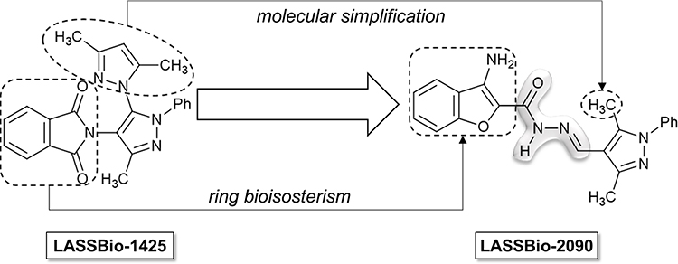 |
Figure 1 Design of LASSBio-2090 ((E)-3-amino-N’-((3,5-dimethyl-1-phenyl-1H-pyrazol-4-yl)methylene)benzofuran-2-carbohydrazide). |
The present study aimed to investigate whether treatment with LASSBio-2090 would improve metabolic and cardiovascular functions in a well-known experimental model of obesity-associated T2DM, the Zucker diabetic fatty (ZDF) rat.22
Methods
Drugs and Reagents
LASSBio-2090 was synthesized by Laboratório de Avaliação e Síntese de Substâncias Bioativas at Universidade Federal do Rio de Janeiro, Brazil, and was dissolved in dimethylsulfoxide which was purchased from Merck (Darmstadt, Germany). Phenylephrine (Phe) and acetylcholine (ACh) were dissolved in distilled water and purchased from Sigma-Aldrich (St. Louis, MO, USA). Antibodies rabbit anti-c-fos (ab7963) and rabbit anti-receptor for advanced glycation endproducts (RAGE, ab3611) were purchased from Abcam (Cambridge, UK). Antibodies rabbit anti-inducible nitric oxide synthase (iNOS, #2982), rabbit anti-TNF-α (#3707) and mouse anti-glyceraldehyde-3-phosphate dehydrogenase (GAPDH, #5174) were purchased from Cell Signaling (Danvers, MA, USA). Secondary antibodies goat anti-rabbit (#1,706,515) and goat anti-mouse (#1,706,516) conjugated to horseradish peroxidase were purchased from Bio-rad (Hercules, CA, USA).
Animals and Experimental Design
All experimental protocols were approved by the Ethics and Animal Care and Use Committee of Federal University of Rio de Janeiro (license number 005/18). Male ZDF (fa/fa, 30 weeks old, n = 12) and Zucker lean (ZL) (+/?, 30 weeks old, n = 6) rats were obtained from Centro de Desenvolvimento de Modelos Experimentais Para Biologia e Medicina (São Paulo, SP, Brazil), and kept on the animal facility of Graduate Program in Pharmacology and Medicinal Chemistry (Rio de Janeiro, RJ, Brazil). Animals were kept in accordance with Brazilian Guide of Production, Maintenance and Utilization of Animals for Teaching or Scientific Research Activities (1st edition, 2016) approved by the National Council for Control of Animal Experimentation. Rats were fed with standard chow and water ad libitum and housed under controlled conditions (22–24ºC; 12/12 h dark/light cycle). Non-diabetic littermates were used as controls and male obese ZDF rats were divided randomly into two groups: ZDF treated with vehicle (dimethylsulfoxide) intraperitoneally (ip) and ZDF treated with LASSBio-2090 at 100 µg/kg/day ip, for 2 weeks. Rats were weighed daily, and individual doses were appropriately adjusted.
Biochemical Analysis
Plasma levels of glucose, insulin, total cholesterol and triglycerides were measured before treatment (baseline) and at the end of the protocol (endpoint). Animals were fasted overnight for 12 hours and tail blood samples were taken the following morning. Glycemia and insulin were determined using an Accu-Chek monitoring system (Roche, Germany) and a commercial enzyme-linked immunosorbent assay (ELISA) kit (Rat Insulin ELISA; Merck, Darmstadt, Germany), respectively. Lipid profile was assessed using a commercial diagnostic kit (Bioclin, Belo Horizonte, Brazil). Surrogate indexes of insulin sensitivity were calculated from plasma biochemistry parameters to minimize stressful manipulation of obese animals. Quantitative insulin sensitivity check index (QUICKI) and triglyceride-glucose index (TyG) were calculated as originally described.23,24
Echocardiography
Experimental groups were anesthetized by a 3% isoflurane/oxygen mixture through a nose cone during spontaneous ventilation. Anesthetized spontaneously breathing animals were placed in a shallow left lateral decubitus position. The left hemithorax was shaved and prepared with acoustic coupling gel to increase probe contact. Room temperature was maintained at approximately 25°C to avoid hypothermia. Cardiac function and structure were assessed both before (baseline) and after treatment (endpoint) as previously reported.17
Invasive Hemodynamics Measurement
At the end of the protocol, rats were anesthetized with ketamine (80 mg/kg, ip) and xylazine (15 mg/kg, ip). Anesthesia depth was verified by pinching the animal’s paw with forceps. Subsequently, a catheter (PE-50) was introduced into the right carotid artery, connected to a pressure transducer (MLT884; ADInstruments, Colorado Springs, CO, USA). Systolic (SBP) and diastolic (DBP) blood pressures were measured. Afterwards, the catheter was introduced into the left ventricle (LV) to record systolic (LVSP) and end diastolic (LVEDP) intracavitary pressures. The LV contraction and relaxation rates were assessed by positive and negative pressure derivatives (dP/dt), respectively. All hemodynamic measurements were digitized (Powerlab; ADInstruments, Sydney, Australia) and analyzed using LabChart software (Version 7.0; ADInstruments, Sydney, Australia). Immediately after measurements, animals were killed via exsanguination by cardiac puncture, and tissues were collected in order to perform histological and molecular evaluations.
Vascular Reactivity in Isolated Aortas
After euthanasia, the thoracic aorta was removed from rats, cleaned of connective tissue and prepared for isometric tension recording. Aortic rings of 3–4 mm length were placed in chambers filled with physiological solution composed of mM: NaCl 123, KCl 4.7, MgCl2 1.2, KH2PO4 1.2, glucose 11.5, NaHCO3 15.5, CaCl2 1.2; oxygenated with 95% O2/5% CO2 and maintained at 37°C in resting tension of 1.0 g. After a period of 2 h, preparations were exposed to increasing concentrations of Phe (1 nM–10 μM) followed by the addition of increasing concentrations of ACh (1 nM–10 μM) to determine the endothelial vasodilator response.
Histomorphometric Analysis
For histological analysis, heart apex was fixed in zinc formalin, embedded in paraffin and tissue sections (5 µm) were stained and examined at 400x magnification under a Axiostar microscope (Zeiss, Jena, Germany). Interstitial cell number and collagen content were analyzed in hematoxylin and eosin and picro-Sirius red stained sections, respectively, in 15–20 fields of LV using Fiji software.17,25
Membrane Preparations and Western Blot Analysis
Heart homogenates were prepared for protein expression analysis by Western blot as previously published.17 Briefly, tissues were harvested, stored in lysis buffer with protease inhibitors, and frozen in liquid nitrogen. Heart samples were homogenized and centrifuged at 1000×g. Supernatants were collected and total protein concentrations were determined by spectrophotometry.26 Equal amounts of protein (50 μg) were separated in a 10% sodium dodecyl sulphate polyacrylamide gel electrophoresis (SDS-PAGE) gel and transferred to a nitrocellulose membrane using a semi-dry system (Bio-Rad, Hercules, CA, USA). Membranes were blocked with 5% nonfat dry milk in phospahte-buffered saline (PBS) containing 0.1% Tween and incubated with primary antibodies to c-fos (1:200), iNOS (1:1000), RAGE (1:200), TNF-α (1:1000), and GAPDH (1:1000) as loading control. Detection of specific bands was performed using secondary antibodies anti-rabbit (1:10,000) or anti-mouse (1:10,000) and ECL Prime (RPN2232; GE Healthcare, Chicago, IL, USA) as the substrate for horseradish peroxidase. Images were acquired by chemiluminescence using Image Quant LAS4000 (GE Healthcare, Chicago, IL, USA) and quantitation of protein content was analyzed by densitometry using ImageJ (version 1.6.0) and normalized to GAPDH.
Data Analysis and Statistics
Data were expressed as means ± standard errors of the mean (SEM) and were analyzed using the GraphPrism software (version 6.0; GraphPad, San Diego, CA, USA). The experimental groups were compared using one-way analysis of variance (ANOVA) with a significance level of p < 0.05, followed by a post-hoc Dunnett’s test.
Results
LASSBio-2090 Normalizes Blood Glucose, Insulin and Reduces Body Weight Gain and Lipid Levels in ZDF Rats
Body weights as well as levels of blood glucose, insulin, cholesterol and triglycerides of ZDF rats are shown in supplementary Table 1. No significant difference between baseline and endpoint measurements was detected in vehicle-treated groups (Table 1). After administration of vehicle for 14 days, body weight remained significantly higher in ZDF group when compared to ZL littermates (p < 0.05). However, ZDF rats treated with LASSBio-2090 showed reduced body weight when compared to vehicle-treated counterparts (p < 0.05).
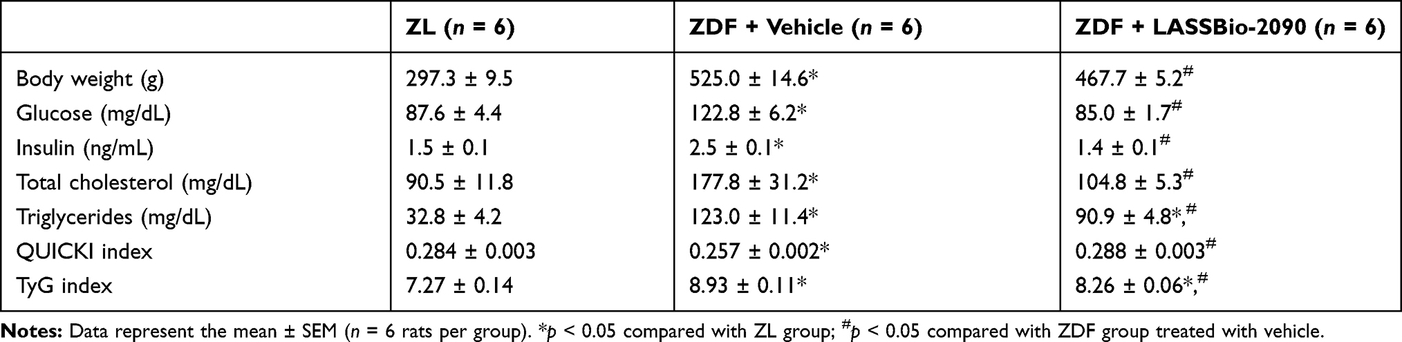 |
Table 1 Biochemical and Physical Parameters |
T2DM obese rats presented impaired glucose handling, with increased blood glucose and insulin levels when compared to ZL group (p < 0.05). Analysis of surrogate indexes of insulin resistance (QUICKI and TyG) showed a significant deterioration of tissue insulin sensitivity in ZDF animals (p < 0.05). Daily ip injections of LASSBio-2090 for 2 weeks normalized both glycemia and insulinemia and improved insulin sensitivity compared to vehicle-treated ZDF group (p < 0.05). Besides, LASSBio-2090 also reduced the elevated blood lipid content seen in ZDF rats compared to the lean group (p < 0.05). Lower plasma concentrations of total cholesterol and triglycerides were detected in ZDF + LASSBio-2090 animals than in ZDF + vehicle group (p < 0.05).
Hemodynamic Alterations and Endothelial Dysfunction in ZDF Rats Were Normalized After Treatment with LASSBio-2090
The effects of vehicle or LASSBio-2090 administration on arterial and LV hemodynamics on ZDF rats are shown in Table 2. Obese T2DM animals treated with vehicle exhibited higher SBP, and DBP in comparison with the ZL group (p < 0.05). After 2 weeks of treatment with LASSBio-2090, ZDF rats presented a significant reduction in those parameters (p < 0.05). Additionally, a significant increase in the LVSP was observed in vehicle-treated ZDF diabetic rats when compared with non-diabetic animals (p < 0.05). In contrast, normal intraventricular pressure was detected in obese T2DM animals treated with LASSBio-2090 (p < 0.05). The LVEDP was significantly higher in the ZDF + vehicle group than in control rats (p < 0.05), and LASSBio-2090 reduced this hemodynamic parameter (p < 0.05).
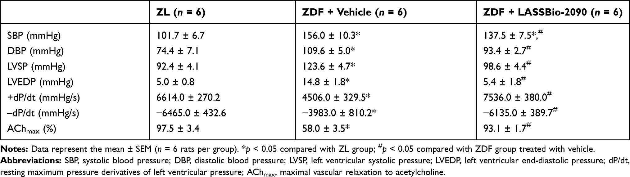 |
Table 2 Hemodynamic Parameters and Endothelial Function |
Positive and negative dP/dt were altered by obesity-associated T2DM compared to non-diabetic counterparts (p < 0.05). Daily injections of LASSBio-2090 for 2 weeks in ZDF group significantly normalized those data which represent myocardial contractility (+dP/dt) and relaxation (–dP/dt), respectively. Furthermore, T2DM impaired the vascular reactivity in ZDF rats as depicted by lower ACh-induced relaxation in the aorta than in ZL non-diabetic rats (p < 0.05). This endothelial dysfunction was totally reversed after treatment with LASSBio-2090 in diabetic rats compared to vehicle-treated ZDF animals (p < 0.05).
LASSBio-2090 Treatment Normalized Heart Dysfunction in Obese T2DM Rats
The effects of LASSBio-2090 administration on LV diastolic and systolic function from ZL and ZDF diabetic rats are shown in Figure 2. Doppler-derived filling pressure, or the ratio of early filling-to-mitral annular descent (E/e′), was higher in rats with T2DM compared to their controls at baseline (Supplementary Table 2) and treatment with DMSO did not induce any further change (Figure 2A; p < 0.05). Moreover, a moderate reduction of ejection fraction (EF) was observed in ZDF rats when compared to controls both before and after treatment, although still ranging between 40% and 50% (Supplementary Table 2 and Figure 2B; p < 0.05). Treatment with LASSBio-2090 for 2 weeks reversed these impairments in LV diastolic and systolic function in ZDF diabetic rats when compared to their vehicle-treated counterparts (p < 0.05).
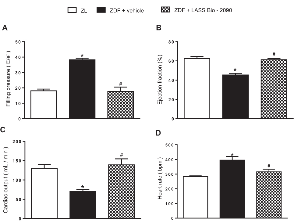 |
Figure 2 Effects of LASSBio-2090 or vehicle administration for 2 weeks on LV filling pressure (A), ejection fraction (B), cardiac output (C) and heart rate (D). Data represent the mean ± SEM (n = 6 rats per group). *p < 0.05 compared with ZL group; #p < 0.05 compared with ZDF group treated with vehicle. Ordinary one-way ANOVA with Dunnett’s multiple comparisons test. |
Additionally, a lower LV cardiac output (CO) was also observed (Figure 2C) in association with an increase of heart rate (HR; Figure 2D) from ZDF group than that observed in ZL animals, as already displayed in baseline measurements (Supplementary Table 2). Two weeks of treatment with LASSBio-2090 normalized these indexes of cardiac performance compared to vehicle-treated ZDF rats (Figure 2C,D; p < 0.05).
Treatment of Obese T2DM Rats with LASSBio-2090 Reduced LV Wall Hypertrophy
Figure 3A shows M-mode echocardiographic images of LVs from all experimental groups. Both anterior wall thickness (AWTd) and posterior wall thickness (PWTd) measured at end diastole were significantly greater in ZDF rats when compared with ZL group (Figure 3B and C, respectively; p < 0.05). Consequently, relative wall thickness (RWT) was significantly higher in diabetic animals than in non-diabetic rats (Figure 3D; p < 0.05), as shown before treatment (Supplementary Table 3). Importantly, treatment of ZDF rats with LASSBio-2090 also reversed these deleterious effects of diabetes-induced LV wall hypertrophy (Figure 3B–D; p < 0.05). No differences in end-diastolic internal LV diameter was observed either before or after treatment (Supplementary Table 3 and Figure 3E, respectively), as well as in end-systolic measures (data not shown).
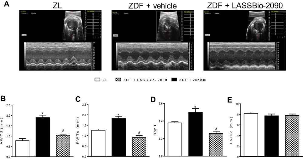 |
Figure 3 Effects of the intraperitoneal treatment with vehicle or LASSBio-2090 (100 µmol/kg/day) on LV wall thickness. Figure (A), representative images obtained by M-mode echocardiography. Figures (B and C), anterior (B) and posterior (C) wall thickness at diastole. Figure (D), relative left ventricle wall thickness. Figure (E), left ventricle internal diameter at diastole. Data represent the mean ± SEM (n = 6 rats per group). *p < 0.05 compared with ZL group; #p < 0.05 compared with ZDF group treated with vehicle. Ordinary one-way ANOVA with Dunnett’s multiple comparisons test. |
LASSBio-2090 Reversed LV Tissue Remodeling and Fibrosis in ZDF Rats
Representative images of LV tissue from all experimental groups showing cardiac interstitial cells and collagen content are shown in Figure 4A and B, respectively. It was observed that the quantitative histological analysis of interstitial cells (Figure 4C) and collagen fractional area (Figure 4D) in hearts from ZDF diabetic rats had an increase compared to ZL animals (p < 0.05). Importantly, LASSBio-2090 treatment significantly reduced the LV tissue remodeling and fibrosis due to the reduced cell number and collagen content in hearts from ZDF animals compared to T2DM obese rats treated with vehicle (p < 0.05).
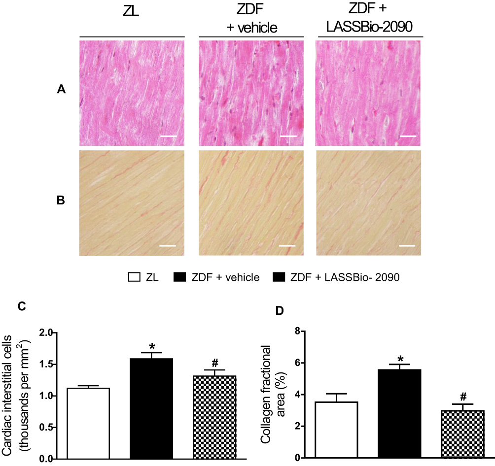 |
Figure 4 Effects of LASSBio-2090 or vehicle administration on interstitial cell number and collagen deposition in the heart tissue. (A) Hematoxylin & eosin and (B) picro-Sirius red stained sections. (C) Cardiac interstitial cell density. (D) Collagen fractional area. Data represent the mean ± SEM (n = 6 rats per group). *p < 0.05 compared with ZL group; #p < 0.05 compared with ZDF group treated with vehicle. Ordinary one-way ANOVA with Dunnett’s multiple comparisons test. Scale bar: 20 μm. |
Treatment with LASSBio-2090 Reversed LV Expression of Cardiac Stress and Inflammation Associated to T2DM
Administration of LASSBio-2090 in diabetic rats normalized the cardiac expression of markers involved with glycative and oxidative stress and inflammation. Figure 5A shows representative Western blot images demonstrating the expression of TNF-α, iNOS, c-fos and RAGE in LV tissue from all groups. T2DM increased the tissue content of these proteins (Figure 5B–E; p < 0.05 versus control group). LASSBio-2090 promoted return to control levels of RAGE, TNF-α, iNOS, and c-fos expression in hearts from T2DM rats compared to diabetic animals treated with vehicle (p < 0.05).
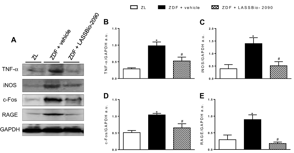 |
Figure 5 Effects of T2DM model on LV protein expression and treatment with vehicle or LASSBio-2090. Figure (A), representative Western blot images of TNF-α, iNOS, c-fos, RAGE and GAPDH (loading control). Figure (B–E), densitometric ratio of TNF-α (B), iNOS (C), c-fos (D) and RAGE (E). Data represent the mean ± SEM (n = 6 rats per group). *p < 0.05 compared with ZL group; #p < 0.05 compared with ZDF group treated with vehicle. Ordinary one-way ANOVA with Dunnett’s multiple comparisons test. |
Discussion
The present study outlines the protective effects of LASSBio-2090 against hypertension and cardiomyopathy in a well-characterized animal model of obesity-associated T2DM. As shown, LASSBio-2090 normalized glucose handling, lowered blood pressure, rescued cardiovascular function and attenuated LV wall remodeling. Evidence suggests that LASSBio-2090 acts mainly by TNF-α blockade, as previously described for its predecessor, LASSBio-1425.
As a consequence of a recessive loss-of-function mutation in the leptin receptor, ZDF rats present hyperphagia, become obese and develop dyslipidemia and insulin resistance at an early age.22,27 In this setting, LASSBio-2090 treatment not only reduced total body weight of ZDF rats by 5%–10% but also significantly decreased plasma cholesterol and triglyceride levels. Moderate weight loss and reduction of serum lipid concentrations are important goals of treatment of obese T2DM subjects because both are related to improved glycemic control and lower risk of cardiovascular disease.2–4,28–30 It is worth noting that LASSBio-2090 retains the lipid-lowering effect observed for LASSBio-1425,16 indicating a common mechanism of action.
Reduction of hyperglycemia and insulin resistance are imperative to T2DM treatment becaue both factors are responsible for the development of diabetes comorbidities such as DC.8,31 LASSBio-2090 showed anti-diabetic activity by normalizing fasting blood glucose in ZDF-treated rats. Besides, insulin resistance was significantly diminished by treatment with LASSBio-2090, as demonstrated by an improved QUICKI index and reduced plasma insulin in ZDF animals when compared to their vehicle-treated counterparts. The significant decrease in TyG index of obese rats treated with LASSBio-2090 indicates that the effect of this new NAH is a direct consequence of a reduction in gluco- and lipotoxic effects on insulin sensitivity. Elevated plasma lipid concentration occurs soon after weaning in ZDF rats and is a consequence of an imbalance between lipogenesis and lipolysis in adipocytes.27 Metabolic stressed adipose tissue shows an increased secretion of inflammatory cytokines, such as TNF-α, which negatively impacts insulin receptor signaling.12,32 Therefore, given the mild reduction in lipid dysmetabolism by LASSBio-2090, the effect of treatment on insulin resistance seems to be related to decreased circulating TNF-α levels, as previously reported for LASSBio-1425.16
LASSBio-2090's effects on glycemia and serum lipids may also contribute to vascular endothelial function, as these factors are directly related to the formation of advanced glycation endproducts (AGE) and oxidized low-density lipoprotein (oxLDL), which trigger pro-inflammatory responses on the vessel wall.3 Impaired vasodilator response to ACh is a direct consequence of endothelial cell activation, with increased expression of inflammatory cytokines and adhesion molecules and lower production of nitric oxide.4,6 In obese individuals and ZDF animals, increased TNF-α levels are strongly related to lower endothelium-dependent vasodilator response.12,33 LASSBio-2090 reversed the endothelial dysfunction in obese animals in a similar way as LASSBio-1425,16 suggesting once more a shared mechanism of action by TNF-α inhibition.
Insufficient control of vascular tone by endothelial cells is associated to increased blood pressure and vascular stiffening found in obese hypertense individuals.4,6 Metabolic disturbances and endothelial dysfunction led to development of hypertension in ZDF rats, which presented systemic hypertension, as demonstrated by increased SBP and DBP. Given the multifactorial nature of blood pressure regulation and arterial remodeling in obese hypertension, the improvement in glucose homeostasis and total reversal of endothelial dysfunction proved insufficient to normalize arterial hemodynamics and treatment with LASSBio-2090 for 2 weeks produced only a mild anti-hypertensive effect.
Cardiac structure and function not only are affected by hypertension but also by metabolic alterations developed during T2DM progression.3 Cardiac hemodynamics was directly affected by hypertension and diabetes in obese rats, leading to a marked reduction in CO. As result of higher afterload in vehicle-treated ZDF group, increased RWT was found by echocardiography, indicating myocardial wall hypertrophy. Despite the mild reduction in blood pressure, LASSBio-2090 normalized CO and wall thickness-to-chamber diameter ratio, indicating further effects on cardiac muscle other than afterload decrease.
In late-stage DC innumerous changes in metabolism, neurohumoral hyperactivation and cardiac fibrosis impair coronary circulation and systolic function.10 Obese rats show elevated HR, probably as a compensatory mechanism to severely reduced CO. Sympathetic overstimulation may also account for this alteration as well as to persistent elevated blood pressure.34–37 However, a positive inotropic response to adrenergic agonism was not observed in ZDF rats because of their reduced myocardial contractility (EF and +dP/dt). Similarly, positive lusitropism was attenuated by diastolic dysfunction, with reduced relaxation rate (–dP/dt) and higher filling pressure (E/e’) and LVEDP. Impaired diastolic filling is associated with obesity and T2DM as both generate metabolic and structural disturbances which lead to reduction in ventricular compliance and subsequent damage to LV passive filling.8 Two weeks of treatment with LASSBio-2090 ip normalized both diastolic and systolic functions and HR. This last finding indicates a reduction in sympathetic cardiac stimulation and suggests a distinct mechanism of enhanced cardiomyocyte function, apart from altered sensitivity to adrenoceptor activation.
Myocardial inflammation is an important feature in the development of DC.8,10 Cardiomyocyte stress by chronic hyperglycemia, dyslipidemia and increased workload is marked by impaired calcium handling and contractility and increased oxidative stress and cytokine secretion, leading to recruitment and activation of interstitial cells.8,13 Cytokine-stimulated cardiac fibroblasts show enhanced collagen secretion and exacerbated collagen deposition is found in hearts from T2DM subjects.9,13 Additionally, non-enzymatic formation of glycated collagen and AGE crosslinks are thought to contribute to myocardial stiffness in DC.8 Our findings are in agreement with previous data, as LV histological examination in ZDF diabetic rats presented increased interstitial cellularity and myocardial collagen content. In this context, besides reversing macroscopic remodeling (RWT), LASSBio-2090 normalized LV cell density and collagen deposition, indicating that its effects are mediated by a reduction in stress-induced signaling pathways involved both in myocyte hypertrophy and activation of interstitial cells.
TNF-α is one of the main pro-inflammatory cytokines responsible for the reduction of cardiomyocyte contractility and stimulation of heart fibrosis.13 It was reported that increased levels of TNF-α in hearts from diabetic rodents is significantly correlated with development of LV dysfunction and is negatively correlated with degree of contractility.38–41 This cytokine is also responsible for perturbations in cardiomyocyte metabolism given its negative effect on the intracellular cascade triggered by activation of insulin receptor.32 In the current study, LASSBio-2090 normalized the upregulation of TNF-α in the heart tissue from ZDF rats with DC and this was reflected in improved LV function in vivo as depicted by the normalization of contractility and compliance and reduction in cardiac remodeling.
Downstream effectors of TNF-α signaling activate the transcription of many pro-inflammatory and pro-remodeling genes, as targets of nuclear factor κB (NFκB) and activator protein-1 (AP-1) (Figure 6).42,43 An example is the expression of the inducible isoform of nitric oxide synthase (iNOS) by NFκB activation. In opposition to its endothelial and neuronal counterparts, iNOS is constitutively active and is responsible for an over-production of nitric oxide, thereby contributing to increased oxidative stress and depressed heart function in DC.44 ZDF rats treated with LASSBio-2090 had a decrease in iNOS protein content in LV tissue to ZL level, supposedly by reduced TNF-α-stimulated NFκB activation, further contributing to cardiac efficiency normalization.
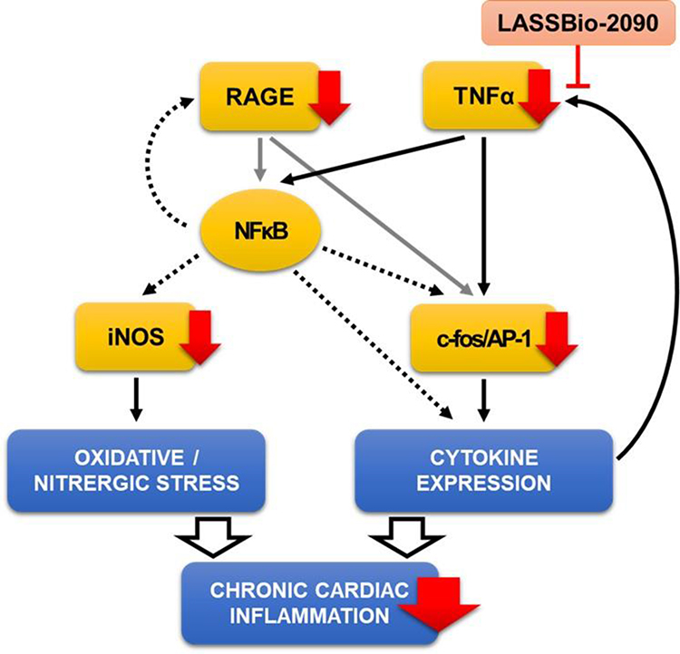 |
Figure 6 Proposed mechanism of action of LASSBio-2090. By reducing TNF-α levels, LASSBio-2090 hampers the transcription of proteins associated with sustained tissue inflammation (red arrows). |
Activation of AP-1 transcription factor is regulated by expression of its constituents, like the c-fos protein. Upregulation of c-fos is associated with inflammation and tissue remodeling, as this early response gene product is highly expressed after metabolic stress, cell injury or cytokine stimulation.45–47 This finding is in accordance with our data, as a higher level of c-fos was detected in LV tissue from T2DM obese rats. Two weeks of LASSBio-2090 treatment reduced LV wall hypertrophy and fibrosis and this action of the new NAH derivative appears to be related to reduction in c-fos expression in hearts from ZDF diabetic animals.
High glucose levels also can dramatically deregulate the structure and function of cardiomyocytes, as increased levels of glycated proteins and lipids (as AGE) activate the pattern-recognition receptor RAGE. The complex AGE–RAGE supports the inflammatory process by promoting oxidative stress and further activation of NFκB and AP-1,48–50 contributing to cardiac cell damage, leukocyte infiltration and fibroblast activation.51–53 Furthermore, RAGE and TNF-α protein contents are integrated in a positive feedback loop (Figure 6), further collaborating to sustained tissue inflammation.50,54-56 Treatment with LASSBio-2090 normalized RAGE expression in the myocardium of ZDF rats concomitantly to the remaining proteins, thereby contributing to enhanced cardiac function and LV wall histology in obese animals.
Finally, although the pharmacotherapy of diabetes has greatly evolved since the discovery of insulin, the development of anti-hyperglycemic drugs with associated anti-obesity effects has received attention only recently.29 Despite initially being designed to fit a pharmacophore model of DPP4 inhibitors, LASSBio-2090 caused a significant reduction in body weight, in contrast with the approved gliptins which are deemed “weight neutral”31 Moreover, LASSBio-2090 also improved cardiac systolic function, a further disparity with current knowledge about DDP4 inhibitors, which do not present such effects in rodents or in T2DM patients presenting EF below 60%.57–59 Therefore, although the contribution of DPP4 inhibition cannot be totally ruled out for the beneficial effects observed, it does not seem to be the main mechanism of action of LASSBio-2090.
Because its design was also based on modification of a thalidomide analogue, the new NAH demonstrated anti-TNF-α effects of the same magnitude and at an equimolar dose as its archetype, LASSBio-1425. The insertion of the NAH group between the phthalimide-like moiety and the aryl group preserved both the anti-inflammatory and anti-dyslipidemic effects of LASSBio-1425, and demonstrates the versatility of this privileged structure to the design of new bioactive molecules. LASSBio-2090 improved glucose and lipid metabolism and enhanced cardiac performance in T2DM obese rats, possibly by inhibition of TNF-α synthesis. Moreover, the impact of this new compound on body weight reduction shows a promising therapeutic potential to obese T2DM subjects, in which body adiposity is significantly associated with risk of developing cardiovascular disease. Although the limitation of the lack of similarity of the present rat model to a less severe clinical condition, LASSBio-2090 will certainly be of benefit to determine that the TNF-α synthesis pathway could interfere with the obesity-associated DC.
Conclusion
Despite the increased prevalence, no single therapy is currently used on treatment of obesity-associated T2DM and its cardiovascular comorbidities. The cardiac injuries induced by T2DM in ZDF rats were ameliorated by TNF-α inhibition with LASSBio-2090 treatment, a new approach that may constitute an innovative therapeutic strategy for treating these conditions in the clinical field in future. Data here presented suggests the potential pharmacological profile of this new NAH to prevent the progression of hypertension and DC in obese subjects. LASSBio-2090 treatment offered remarkable benefits as improved insulin sensitivity, significant weight reduction and preservation of cardiac and vascular function in ZDF obese rats, standing out as a new lead compound with anti-obesity and anti-diabetic effects.
Abbreviations
ACh, acetylcholine; AChmax, maximal vascular relaxation to acetylcholine; AGE, advanced glycation endproducts; ANOVA, analysis of variance; AP-1, activator protein-1; AWTd, anterior wall thickness at diastole; CO, cardiac output; DBP, diastolic blood pressure; DC, diabetic cardiomyopathy; DPP4, dipeptidyl peptidase 4; dP/dt, rate of change of intraventricular pressure; EF, ejection fraction; GAPDH, glyceraldehyde-3-phosphate dehydrogenase; HR, heart rate; iNOS, inducible nitric oxide synthase; ip, intraperitoneal; LV, left ventricle; LVEDP, left ventricle end diastolic pressure; LVIDd, left ventricle internal diameter at diastole; LVSP, left ventricle systolic pressure; NAH, N-acylhydrazone; NFκB, nuclear factor κB; oxLDL, oxidized low-density lipoprotein; Phe, phenylephrine; PWTd, posterior wall thickness at diastole; QUICKI, quantitative insulin sensitivity check index; RAGE, receptor for advanced glycation endproducts; RWT, relative wall thickness; SBP, systolic blood pressure; SEM, standard error of the mean; T2DM, type 2 diabetes mellitus; TNF, tumor necrosis factor; TyG, triglyceride–glucose index; ZDF, Zucker diabetic fatty; ZL, Zucker lean.
Acknowledgments
This work was supported by Conselho Nacional de Desenvolvimento Científico e Tecnológico (CNPq), Coordenação de Aperfeiçoamento de Pessoal de Nível Superior (CAPES), Fundação Carlos Chagas Filho de Amparo à Pesquisa do Estado do Rio de Janeiro (FAPERJ), Instituto Nacional de Ciência e Tecnologia de Fármacose Medicamentos (INCT-INOFAR), Centro Nacional de Biologia Estrutural e Bioimagem (CENABIO).
Author Contributions
All authors contributed to data analysis, drafting or revising the article, agree to the journal in which the paper was submitted, gave final approval of the version to be published, and agree to be accountable for all aspects of the work.
Disclosure
Marina MC Silva reports grants from CAPES, during the conduct of the study. Gisele Zapata-Sudo reports grants from Conselho Nacional de Desenvolvimento Científico e Tecnológico - CNPq, Fundação de Amparo à Pesquisa do Estado do Rio de Janeiro - FAPERJ, and Instituto Nacional de Ciência e Tecnologia em Fármacos e Medicamentos (INCT-INOFAR), during the conduct of the study. The authors declare no other possible conflicts of interest in this work.
References
1. Cho NH, Shaw JE, Karuranga S, et al. IDF diabetes atlas: global estimates of diabetes prevalence for 2017 and projections for 2045. Diabetes Res Clin Pract. 2018;138:271–281. doi:10.1016/j.diabres.2018.02.023
2. Apovian CM, Okemah J, O’Neil PM. Body weight considerations in the management of type 2 diabetes. Adv Ther. 2019;36(1):44–58. doi:10.1007/s12325-018-0824-8
3. Henning RJ. Type-2 diabetes mellitus and cardiovascular disease. Future Cardiol. 2018;14(6):491–509. doi:10.2217/fca-2018-0045
4. Cohen JB. Hypertension in obesity and the impact of weight loss. Curr Cardiol Rep. 2017;19(10):98. doi:10.1007/s11886-017-0912-4
5. Baena-Diez JM, Penafiel J, Subirana I, et al. Risk of cause-specific death in individuals with diabetes: a competing risks analysis. Diabetes Care. 2016;39(11):1987–1995. doi:10.2337/dc16-0614
6. Seravalle G, Grassi G. Obesity and hypertension. Pharmacol Res. 2017;122:1–7. doi:10.1016/j.phrs.2017.05.013
7. Cohen JB, Stephens-Shields AJ, Denburg MR, Anderson AH, Townsend RR, Reese PP. Obesity, renin-angiotensin system blockade and risk of adverse renal outcomes: a population-based cohort study. Am J Nephrol. 2016;43(6):431–440. doi:10.1159/000446862
8. Jia G, Whaley-Connell A, Sowers JR. Diabetic cardiomyopathy: a hyperglycaemia- and insulin-resistance-induced heart disease. Diabetologia. 2018;61(1):21–28. doi:10.1007/s00125-017-4390-4
9. Aneja A, Tang WHW, Bansilal S, Garcia MJ, Farkouh ME. Diabetic cardiomyopathy: insights into pathogenesis, diagnostic challenges, and therapeutic options. Am J Med. 2008;121(9):748–757. doi:10.1016/j.amjmed.2008.03.046
10. Battiprolu PK, Gillette TG, Wang ZV, Lavandero S, Hill JA. Diabetic cardiomyopathy: mechanisms and therapeutic targets. Drug Discov Today Dis Mech. 2010;7(2):e135–e143. doi:10.1016/j.ddmec.2010.08.001
11. Pollack RM, Donath MY, LeRoith D, Leibowitz G. Anti-inflammatory agents in the treatment of diabetes and its vascular complications. Diabetes Care. 2016;39(Supplement 2):S244–S252. doi:10.2337/dcS15-3015
12. Fuster JJ, Ouchi N, Gokce N, Walsh K. Obesity-induced changes in adipose tissue microenvironment and their impact on cardiovascular disease. Circ Res. 2016;118(11):1786–1807. doi:10.1161/CIRCRESAHA.115.306885
13. Mann DL. Stress-activated cytokines and the heart: from adaptation to maladaptation. Annu Rev Physiol. 2003;65:81–101. doi:10.1146/annurev.physiol.65.092101.142249
14. Singh S, Facciorusso A, Singh AG, et al. Obesity and response to anti-tumor necrosis factor-α agents in patients with select immune-mediated inflammatory diseases: a systematic review and meta-analysis. PLoS One. 2018;13(5):e0195123. doi:10.1371/journal.pone.0195123
15. Barbosa ML, de C, Fumian MM, Miranda ALP, de, Barreiro EJ, Lima LM. Therapeutic approaches for tumor necrosis factor inhibition. Braz J Pharm Sci. 2011;47(3):427–446. doi:10.1590/S1984-82502011000300002
16. Fumian MM, da Motta NAV, Maia R, Fraga CAM, Barreiro EJ, Ferreira de Brito FC. LASSBio-1425, an analog of thalidomide, decreases triglyceride and increases HDL cholesterol levels by inhibition of TNF-α production. Int J Cardiol. 2016;202:497–499. doi:10.1016/j.ijcard.2015.09.071
17. Alves BEO, de Alencar AKN, Gamba LER, et al. Reduction of cardiac and renal dysfunction by new inhibitor of DPP4 in diabetic rats. Pharmacol Rep. 2019;71(6):1190–1200. doi:10.1016/j.pharep.2019.07.005
18. Thota S, Rodrigues DA, Pinheiro P, et al. N-Acylhydrazones as drugs. Bioorg Med Chem Lett. 2018;28(17):2797–2806. doi:10.1016/j.bmcl.2018.07.015
19. Fraga CAM, Barreiro EJ. Medicinal chemistry of N-acylhydrazones: new lead-compounds of analgesic, antiinflammatory and antithrombotic drugs. Curr Med Chem. 2006;13(2):167–198. doi:10.2174/092986706775197881
20. Costante R, Stefanucci A, Carradori S, Novellino E, Mollica A. DPP-4 inhibitors: a patent review (2012 – 2014). Expert Opin Ther Pat. 2015;25(2):209–236. doi:10.1517/13543776.2014.991309
21. Lima L, Barreiro E. Bioisosterism: a useful strategy for molecular modification and drug design. Curr Med Chem. 2005;12(1):23–49. doi:10.2174/0929867053363540
22. Peterson RG. The Zucker diabetic fatty (ZDF) rat. In: Sima AAF, Shafrir E, editors. Animal Models in Diabetes: A Primer. Amsterdam: Harwood Academic Publishers; 2000:95–112.
23. Guerrero-Romero F, Simental-Mendía LE, González-Ortiz M, et al. The product of triglycerides and glucose, a simple measure of insulin sensitivity. comparison with the euglycemic-hyperinsulinemic clamp. J Clin Endocrinol Metab. 2010;95(7):3347–3351. doi:10.1210/jc.2010-0288
24. Katz A, Nambi SS, Mather K, et al. Quantitative insulin sensitivity check index: a simple, accurate method for assessing insulin sensitivity in humans. J Clin Endocrinol Metab. 2000;85(7):2402–2410. doi:10.1210/jcem.85.7.6661
25. Schindelin J, Arganda-Carreras I, Frise E, et al. Fiji: an open-source platform for biological-image analysis. Nat Methods. 2012;9(7):676–682. doi:10.1038/nmeth.2019
26. Bradford MM. A rapid and sensitive method for the quantitation of microgram quantities of protein utilizing the principle of protein-dye binding. Anal Biochem. 1976;72:248–254. doi:10.1016/0003-2697(76)90527-3
27. Liu RH, Mizuta M, Kurose T, Matsukura S. Early events involved in the development of insulin resistance in Zucker fatty rat. Int J Obes Relat Metab Disord. 2002;26(3):318–326. doi:10.1038/sj.ijo.0801924
28. American Diabetes Association. Obesity management for the treatment of type 2 diabetes: standards of medical care in diabetes—2020. Diabetes Care. 2020;43(Supplement1):S89–S97. doi:10.2337/dc20-S008.
29. Bessesen DH, Van Gaal LF. Progress and challenges in anti-obesity pharmacotherapy. Lancet Diabetes Endocrinol. 2018;6(3):237–248. doi:10.1016/S2213-8587(17)30236-X
30. Chukir T, Shukla AP, Saunders KH, Aronne LJ. Pharmacotherapy for obesity in individuals with type 2 diabetes. Expert Opin Pharmacother. 2018;19(3):223–231. doi:10.1080/14656566.2018.1428558
31. American Diabetes Association. Pharmacologic approaches to glycemic treatment: standards of medical care in diabetes—2020. Diabetes Care. 2020;43(Supplement 1):S98–S110. doi:10.2337/dc20-S009.
32. Zatterale F, Longo M, Naderi J, et al. Chronic adipose tissue inflammation linking obesity to insulin resistance and type 2 diabetes. Front Physiol. 2020;10. doi:10.3389/fphys.2019.01607
33. Gao X, Picchi A, Zhang C. Upregulation of TNF-alpha and receptors contribute to endothelial dysfunction in Zucker diabetic rats. Am J Biomed Sci. 2010;2(1):1–12. doi:10.5099/aj100100001
34. Cook RF, Bussey CT, Mellor KM, Cragg PA, Lamberts RR. β 1 -adrenoceptor, but not β 2 -adrenoceptor, subtype regulates heart rate in type 2 diabetic rats in vivo. Exp Physiol. 2017;102(8):911–923. doi:10.1113/EP086293
35. Jiang C, Carillion A, Na N, et al. Modification of the β-adrenoceptor stimulation pathway in Zucker obese and obese diabetic rat myocardium*. Crit Care Med. 2015;43(7):e241–e249. doi:10.1097/CCM.0000000000000999
36. Thaung HPA, Baldi JC, Wang H-Y, et al. Increased efferent cardiac sympathetic nerve activity and defective intrinsic heart rate regulation in type 2 diabetes. Diabetes. 2015;64(8):2944–2956. doi:10.2337/db14-0955
37. Lohmeier TE, Iliescu R. The sympathetic nervous system in obesity hypertension. Curr Hypertens Rep. 2013;15(4):409–416. doi:10.1007/s11906-013-0356-1
38. Parish RC, Todman S, Jain SK. Resting heart rate variability, inflammation, and insulin resistance in overweight and obese adolescents. Metab Syndr Relat Disord. 2016;14(6):291–297. doi:10.1089/met.2015.0140
39. Crendal E, Walther G, Vinet A, et al. Myocardial deformation and twist mechanics in adults with metabolic syndrome: impact of cumulative metabolic burden. Obesity. 2013;21(12):E679–E686. doi:10.1002/oby.20537
40. Tiwari S, Mishra M, Jadhav A, et al. The risk of heart failure and cardiometabolic complications in obesity may be masked by an apparent healthy status of normal blood glucose. Oxid Med Cell Longev. 2013;2013:1–16. doi:10.1155/2013/253657
41. Westermann D, Van Linthout S, Dhayat S, et al. Tumor necrosis factor-alpha antagonism protects from myocardial inflammation and fibrosis in experimental diabetic cardiomyopathy. Basic Res Cardiol. 2007;102(6):500–507. doi:10.1007/s00395-007-0673-0
42. Brenner D, Blaser H, Mak TW. Regulation of tumour necrosis factor signalling: live or let die. Nat Rev Immunol. 2015;15(6):362–374. doi:10.1038/nri3834
43. Kyriakis JM. Activation of the AP-1 transcription factor by inflammatory cytokines of the TNF family. Gene Expr. 1999;7(4–6):217–231.
44. Song D, Kuo K-H, Yao R, Hutchings SR, Pang CCY. Inducible nitric oxide synthase depresses cardiac contractile function in Zucker diabetic fatty rats. Eur J Pharmacol. 2008;579(1–3):253–259. doi:10.1016/j.ejphar.2007.09.043
45. Ma C, Fu Z, Guo H, Wei H, Zhao X, Li Y. The effects of Radix Angelica Sinensis and Radix Hedysari ultrafiltration extract on X-irradiation-induced myocardial fibrosis in rats. Biomed Pharmacother. 2019;112:108596. doi:10.1016/j.biopha.2019.01.057
46. Min W, Bin ZW, Quan Z, Bin HZJ, Sheng FG. The signal transduction pathway of PKC/NF-kappa B/c-fos may be involved in the influence of high glucose on the cardiomyocytes of neonatal rats. Cardiovasc Diabetol. 2009;8:8. doi:10.1186/1475-2840-8-8
47. Goetze S, Kintscher U, Kaneshiro K, et al. TNFα induces expression of transcription factors c-fos, Egr-1, and Ets-1 in vascular lesions through extracellular signal-regulated kinases 1/2. Atherosclerosis. 2001;159(1):93–101. doi:10.1016/S0021-9150(01)00497-X
48. Liu Q, Hua B, Su W, et al. AGEs impair Kv channel-mediated vasodilation of coronary arteries by activating the NF-κB signaling pathway in ZDF rats. Biomed Pharmacother. 2019;120:109527. doi:10.1016/j.biopha.2019.109527
49. Adamopoulos C, Piperi C, Gargalionis AN, et al. Advanced glycation end products upregulate lysyl oxidase and endothelin-1 in human aortic endothelial cells via parallel activation of ERK1/2–NF-κB and JNK–AP-1 signaling pathways. Cell Mol Life Sci. 2016;73(8):1685–1698. doi:10.1007/s00018-015-2091-z
50. Yao D, Brownlee M. Hyperglycemia-induced reactive oxygen species increase expression of the receptor for advanced glycation end products (RAGE) and RAGE ligands. Diabetes. 2010;59(1):249–255. doi:10.2337/db09-0801
51. Lim S, Lee ME, Jeong J, et al. sRAGE attenuates angiotensin II-induced cardiomyocyte hypertrophy by inhibiting RAGE-NFκB-NLRP3 activation. Inflamm Res. 2018;67(8):691–701. doi:10.1007/s00011-018-1160-9
52. Hegab Z, Mohamed TMA, Stafford N, Mamas M, Cartwright EJ, Oceandy D. Advanced glycation end products reduce the calcium transient in cardiomyocytes by increasing production of reactive oxygen species and nitric oxide. FEBS Open Bio. 2017;7(11):1672–1685. doi:10.1002/2211-5463.12284
53. Tsoporis JN, Izhar S, Proteau G, Slaughter G, Parker TG. S100B-RAGE dependent VEGF secretion by cardiac myocytes induces myofibroblast proliferation. J Mol Cell Cardiol. 2012;52(2):464–473. doi:10.1016/j.yjmcc.2011.08.015
54. Anuranjani BM. Concerted action of Nrf2-ARE pathway, MRN complex, HMGB1 and inflammatory cytokines - implication in modification of radiation damage. Redox Biol. 2014;2:832–846. doi:10.1016/j.redox.2014.02.008
55. Volz HC, Seidel C, Laohachewin D, et al. HMGB1: the missing link between diabetes mellitus and heart failure. Basic Res Cardiol. 2010;105(6):805–820. doi:10.1007/s00395-010-0114-3
56. Tanaka N, Yonekura H, Yamagishi S, Fujimori H, Yamamoto Y, Yamamoto H. The receptor for advanced glycation end products is induced by the glycation products themselves and tumor necrosis factor-α through nuclear factor-κB, and by 17β-estradiol through Sp-1 in human vascular endothelial cells. J Biol Chem. 2000;275(33):25781–25790. doi:10.1074/jbc.M001235200
57. Yamamoto M, Ishizu T, Seo Y, et al. Teneligliptin prevents cardiomyocyte hypertrophy, fibrosis, and development of hypertensive heart failure in dahl salt-sensitive rats. J Card Fail. 2018;24(1):53–60. doi:10.1016/j.cardfail.2017.09.001
58. McMurray JJV, Ponikowski P, Bolli GB, et al. Effects of vildagliptin on ventricular function in patients with type 2 diabetes mellitus and heart failure. JACC Hear Fail. 2018;6(1):8–17. doi:10.1016/j.jchf.2017.08.004
59. Arturi F, Succurro E, Miceli S, et al. Liraglutide improves cardiac function in patients with type 2 diabetes and chronic heart failure. Endocrine. 2017;57(3):464–473. doi:10.1007/s12020-016-1166-4
 © 2020 The Author(s). This work is published and licensed by Dove Medical Press Limited. The
full terms of this license are available at https://www.dovepress.com/terms
and incorporate the Creative Commons Attribution
- Non Commercial (unported, 3.0) License.
By accessing the work you hereby accept the Terms. Non-commercial uses of the work are permitted
without any further permission from Dove Medical Press Limited, provided the work is properly
attributed. For permission for commercial use of this work, please see paragraphs 4.2 and 5 of our Terms.
© 2020 The Author(s). This work is published and licensed by Dove Medical Press Limited. The
full terms of this license are available at https://www.dovepress.com/terms
and incorporate the Creative Commons Attribution
- Non Commercial (unported, 3.0) License.
By accessing the work you hereby accept the Terms. Non-commercial uses of the work are permitted
without any further permission from Dove Medical Press Limited, provided the work is properly
attributed. For permission for commercial use of this work, please see paragraphs 4.2 and 5 of our Terms.