Back to Journals » Drug Design, Development and Therapy » Volume 13
Network pharmacology study on the active components of Pterocypsela elata and the mechanism of their effect against cerebral ischemia
Authors Niu B, Zhang H, Li C, Yan F, Song Y, Hai G, Jiao Y, Feng Y
Received 7 March 2019
Accepted for publication 18 June 2019
Published 5 September 2019 Volume 2019:13 Pages 3009—3019
DOI https://doi.org/10.2147/DDDT.S207955
Checked for plagiarism Yes
Review by Single anonymous peer review
Peer reviewer comments 2
Editor who approved publication: Dr Sukesh Voruganti
Bingxuan Niu,1 Hui Zhang,1 Chunyan Li,2 Fulin Yan,1,2 Yu Song,1 Guangfan Hai,1 Yunjuan Jiao,3 Yansheng Feng3
1College of Pharmacy, Xinxiang Medical University, Xingxiang, Henan Province 453003, People’s Republic of China; 2Sanquan College of Xinxiang Medical University, Xinxiang, Henan Province 453002, People’s Republic of China; 3Basic Medical College, Xinxiang Medical University, Xinxiang, Henan Province 453003, People’s Republic of China
Correspondence: Fulin Yan
College of Pharmacy, Xinxiang Medical University, 601 Jinsui Avenue, Hongqi District, Xingxiang, Henan Province 453003, People’s Republic of China
Tel +1 378 251 3326
Email [email protected]
Objective: The aim of this study was to identify the active anti-ischemic components of Pterocypsela elata (P. elata) using a network pharmacology approach to construct an effective component anti-cerebral ischemic target network and systematically analyze this medicinal material.
Methods: Pharmacological studies have shown that P. elata has an obvious effect against cerebral ischemia. To identify the potential targets, 14 components of P. elata were docked to each structural element of the targets in the DRAR-CPI database by reverse docking technology. We then compared the identified potential targets with FDA-approved targets for stroke/cerebral infarction treatment in the DrugBank database and identified the active components of P. elata and their potential targets for stroke/cerebral infarction treatment. The active component-target networks were constructed using Cytoscape 3.5.1 software. The target protein-protein interactions were analyzed using the STRING database. KEGG pathway analysis and gene ontology (GO) enrichment analysis were performed through the Database for Annotation, Visualization and Integrated Discovery (DAVID).
Results: There were 14 active components identified from P. elata and 21 potential targets identified for cerebral ischemia treatment, including carbonic anhydrase 2, ribosyldihydronicotinamide dehydrogenase, cholinesterase, and glutathione S-transferase P. The main involved pathways include metabolic pathways, complement and coagulation cascades and steroid hormone biosynthesis.
Conclusion: Through a network pharmacology approach, we predicted the active components of P. elata and their potential targets for cerebral ischemia treatment. Our results provide new perspectives and clues for further studies on the anti-cerebral ischemia mechanism of P. elata.
Keywords: network pharmacology, Pterocypsela elata, cerebral ischemia, molecular docking
Introduction
Traditional Chinese medicine (TCM) originated in China and has evolved over thousands of years. To counteract diseases, Chinese people have gradually accumulated a profound knowledge of medicine and rich experience through thousands of years of clinical practice, medical development and innovation, and have formed a comprehensive and systematic TCM theory and unique medical system. Due to the synergistic effect of multi-component, multi-pathway and multi-target systems, the material basis, mechanisms and preparation of TCM active compounds remain unclear, and the quality of TCM is difficult to control. Additionally, TCM lacks scientific, logical and effective evaluation systems for assessing efficacy and safety, which makes it difficult to conduct comprehensive and systematic research at the tissue, organ, cell and molecular levels.1 Therefore, to eliminate obstacles to the modernization of TCM, it is important to clarify the material basis and mechanisms of TCM active compounds.
Network pharmacology approaches are based on the new “multi-target, multidrug, multi-pathway” concept. This approach can integrate the various databases of Chinese medicines, proteins, genes and others for analysis to construct a drug-target-disease network using bioinformatics to predict the mechanism of a medicine. The holistic and systematic features of the network pharmacology approach are consistent with the synergistic effect of multiple components in TCM. Through network pharmacology analysis, we can investigate TCM systematically, identify the active components, clarify the potential targets and mechanisms, and contribute to the modernization of TCM.2–5
With the aging of the population and the development of the economy, ischemic stroke has become a disease with a high morbidity and mortality that seriously affects health. Ischemia/reperfusion injury following ischemic stroke is a complex pathophysiological process. Therefore, the treatment and prevention of cerebral ischemia has long been the focus of medical research. Pterocypsela elata (P. elata), also known as Indian lettuce or bitter lettuce, is an annual or biennial herb belonging to the genus Pterocypsela Shih of the Compositae family, which is distributed throughout China. It has a long history of being used as a vegetable, animal feed and medicine. Both the whole herb and the root can be used in TCM, and it has the functions of clearing away heat and toxic materials, promoting blood circulation, and is an anti-oxidant and anti-diabetic.6 Studies have shown that it also has anti-ischemic effects. As a potential anti-ischemic drug, it deserves to be further studied. In this study, we screened the active anti-ischemic components of P. elata using a network pharmacology approach and constructed an active component anti-ischemic target network for analysis.
Materials and methods
Compound information
Based on previous studies in our group,7–10 a total of 14 molecular entities in P. elata were obtained: lactuside B (C21H32O9), 11β,13-dihydrolactucin acetate (C17H20O6), 11β, 13-dihydrolactucin (C15H18O5), 9α-hydroxyleucodin-9-O-β-xylopyranoside (C20H26O8), Guaianolide I (C23H30O11), Guaianolide II (xFF08;C15H12O3), (24R)-5α-stigrnast-7, 22(E)-dien-3α-ol (C29H48O), oleanolic acid (C30H48O3), β-amyrin (C30H50O), β-sitosterol (C29H50O), daucosterol (C35H60O6), 3, 3′, 4-trimethoxylellagic acid (C17 H12O8), stearic acid (C18H36O2).
Software and network database
ChemDraw 15.0 software, Open Babel GUI 2.4.1 software, Cytoscape 3.5 software, Batman-TCM database (http://bionet.ncpsb.org/batman-tcm/index.php/Home/Index/index), http://cpi.bio-x.cn/drar/DrugBank 5.1.0 software (https://www.drugbank.ca/, version 5.1.1, released 2018-07-03), String 10.5 software (http://www.string-db.org/), KEGG software (http://www.kegg.jp/).
Experimental methods
The chemical structures of 14 compounds in P. elata were drawn using ChemDraw software and then converted to files in sdf-MDL format using Open Bable GUI 2.4.1 software. All files of chemical structures were saved and uploaded to the Drug Repositioning and Adverse Reaction via Chemical-Protein Interactome (DRAR-CPI) server.
The FDA-approved targets for stroke/cerebral infarction treatment were identified using keywords such as “stroke”, “cerebral infarction” and “cerebral ischemia” to search the DrugBank database. Comparing the targets identified by the 14 compounds in P. elata with the FDA-approved targets, we found potential active components in P. elata and their targets for stroke/cerebral infarction treatment.
The potential targets were then analyzed via the Search Tool for the Retrieval of Interacting Genes/Proteins (STRING) database using single-target and multi-target analysis. For each target, gene ontology (GO) enrichment analysis was performed for the aspects of biological process, molecular function, cellular component and Kyoto Encyclopedia of Genes and Genomes (KEGG) pathways to explore the relationships between potential targets.
Finally, the active component-target-pathway network was visualized and analyzed using the Cytoscape software. Bioinformatic tools were employed to study the “multi-component, multi-target, and multi-pathway” mechanism.
Cell experiments
Oxygen-glucose deprivation (OGD) and recovery (OGDR)11
OGD model: Glucose-containing complete medium was replaced by Hank’s balanced salt solution (glucose-free; Invitrogen, 88284). Human neuroblastoma cell (SH-SY5Y cells) were incubated in hypoxic conditions (5% CO2, 0.3% O2 and 94.7% N2) for 4 hrs. Brain injury following cerebral ischemia was modeled in vitro.
OGDR: Four hours after OGD, the glucose-free medium was replaced by complete medium containing glucose and 10% (v/v) fetal bovine serum. Cells were placed in an incubator containing 95% air and 5% CO2 at 37 °C for 48 hrs. Cerebral ischemia/reperfusion injury was modeled in vitro. The cells of the control group were always incubated in complete medium and placed in an incubator containing 95% air and 5% CO2 at 37 °C.
Intervention group: Sixty minutes before OGD and 24 h after OGD, a sterilized water extraction of P. elata (ie, the crude drug) at concentrations of 1 mg/mL, 2 mg/mL, and 4 mg/mL was added into the medium. For the control group, the same volume of sterile distilled water was added.
MTT assay
Cell viability was detected by 3-(4,5-dimethylthiazol-2-yl)-2,5-Diphenylte-trazolium bromide (MTT) assay. Cells were seeded into 96-well culture plates at a density of 2×10³ cells/well. After OGDR, 20 μL of MTT (5 g/L) was added to each well, and the cells were incubated for 4 h. After discarding the supernatant, 150 μL of dimethyl sulfoxide (DMSO) was added to each well, and the plates were placed in an incubation shaker for 10 min. The absorbance at 490 nm was measured using a spectrophotometer.
Animal experiments
Experimental animals
A total of sixty male Sprague-Dawley rats (weight: 260 to 300 g) were obtained from the Experimental Animal Center, the Academy of Medical Sciences of Zhengzhou University. The animal certificate number is SYXK (Yu) 2005–0012.
Rat model of cerebral ischemia
The rats were randomly divided into 5 groups, 12 rats per group, named as the sham surgery group, model group, low-concentration group(10 g/kg), moderate concentration group(20 g/kg), and high concentration group(40 g/kg). Each group was pre-administered with a water extraction of P. elata (ie, the crude drug) for two weeks, twice per day, at concentrations of 10, 20, and 40 g/kg. Arterial occlusion lines were prepared using a No. 1.5 fishing line (produced by DaDong Yang). Each line was cut to a length of 5–6 cm. One end of the line was heated to form a spherical shape, and then the line was soaked in a 1% heparin solution for storage. The rats were anesthetized using 10% chloral hydrate. The rat model for cerebral ischemia and reperfusion injury was prepared using Longa’s suture method.13 It was established by modified middle cerebral artery occlusion (MCAO). The supine position was fixed on the hand table, and the common carotid artery (CCA) was separated from the median incision of the neck. The internal carotid artery (ICA) and external carotid artery (ECA) were separated from the right side in turn. The branches of ECA were electrocoagulated with a coagulator, and the distal part of the main trunk of ECA was separated and ligated. After clipping CCA and ICA with non-invasive artery, a loose joint was made and ECA was cut off near the bifurcation of CCA. The prepared occlusion line was inserted from the broken end of ECA. After the bifurcation of CCA, ICA non-invasive artery clamp was loosened. The occlusion line was inserted into the middle cerebral artery through ICA. When resistance was felt, the occlusion line was stopped, fixed, the non-invasive artery clamp on CCA was loosened, and the depth of occlusion line was inserted. 1.8–2.0 cm. All animals were occluded for 2 hrs and then pulled the occluded spherical end back to ECA for reperfusion. Neurological function assessment scores were derived 48 h after ischemia-reperfusion. The active animals were sacrificed, and specimens were obtained. Animal welfare and experimental procedures were carried out in accordance with the Guide for the Care and Use of Laboratory Animals (Ministry of Science and Technology of China, 2006), and were approved by the ethics committee of Xinxiang Medical University.
Rat neurological deficit scores
The neurological status was assessed after the animal was awake and 24 h after the injury according to the Zea-longa scoring system.13 A score of 0 was given to normal animals with no symptoms and no signs of neurological deficit. A score of 1 was given to the animals with mild neurological deficit, including the adduction and flexion of the left anterior limb after lifted by the tail. A score of 2 was given to animals whose body turned to the left while crawling (tail-catching phenomenon). A score of 3 was given to animals that fell to the left side while standing. A score of 4 was given to animals that could not walk on their own and had lost consciousness. Animals with scores of 1–3 were included in the experiment; those with a score of 0 or 4 were excluded.
Measurement of cerebral infarction volume
After neurological function assessment, 5 rats were randomly selected from each group. The rats were quickly decapitated on ice and placed at −20 °C for 10 min. The cerebellum, lower brain stem, olfactory bulb and frontal poles were excised. A series of 2 mm-thick continuous coronal slices, from anterior to posterior, were cut from the rest of the brain and incubated in 2% triphenyl tetrazolium chloride (TTC) solution for 30 min at 37 °C. The operation process should be protected from light, and the solution should cover the brain tissue. The principle of TTC staining relies on the oxidation of TTC by dehydrogenase. Tissue with normal levels of dehydrogenase is stained red, whereas the ischemic infarction tissue lacks dehydrogenase and is white. After staining, the brain slices were fixed with paraformaldehyde and stored at 4 °C. The infarction volume of each slice was calculated by image analysis software according to the following formula: total infarction volume = sum of infraction areas x slice thickness. To compensate for brain tissue swelling, the estimated infarction volume was corrected using the following formula: percentage of infarction volume in total volume = (measured infarction volume ×(1-(ipsilateral hemisphere volume-contralateral hemisphere volume)/ipsilateral hemisphere volume)/total brain volume ×100%.
Results
The effect of P. elata on cell viability
As shown in Figure 1, cell viability was significantly decreased in the model group with oxygen-glucose deprivation/recovery (P<0.01). Different concentrations of P. elata could increase the cell viability to various degrees. Compared with the model group, medium and high concentrations of P. elata showed stronger effects (P<0.05–0.01). Photomicrographs of each group are shown in Figure 2.
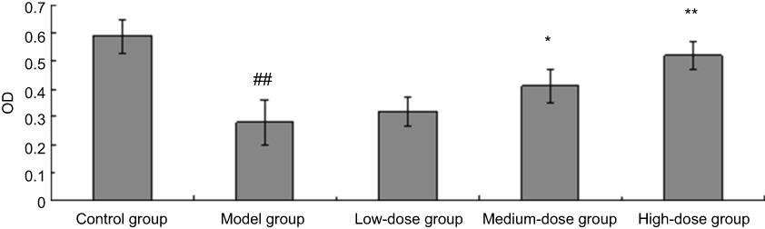 |
Figure 1 The effect of Pterocypsela elata (P. elata) on cell viability after oxygen-glucose deprivation/recovery (x̅ ± s, n=3). Notes: ##P<0.01 vs sham surgery group; *P<0.05, **P<0.01 vs model group. |
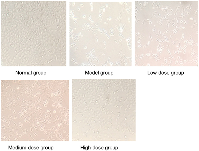 |
Figure 2 Photomicrographs of each group (100×). |
The effect of P. elata on neurological deficit scores of rats
As shown in Table 1, twenty-four hours after ischemia-reperfusion injury, the score of the model group was significantly higher than that of the sham surgery group (P<0.01). The groups with different concentrations of P. elata had lower behavioral scores in the injured rats, and the differences were significant between the behavioral scores of these groups and that of the model group (P<0.01 or P<0.05). Among them, the medium- and high-concentration groups showed stronger effects (P<0.01).
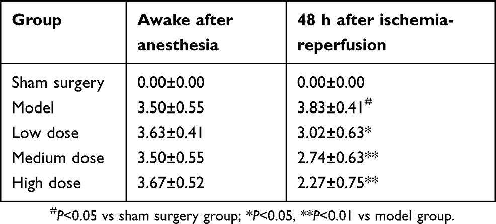 |
Table 1 The effect of Pterocypsela elata (P. elata) on rat neurological deficit scores (x̅ ± s, n=8) |
Effect of P. elata on infarction volume after ischemia-reperfusion injury
Cerebral infarction was assessed by TTC staining 48 h after reperfusion, and the white part was the infarction area. In the model group, the infarction area was large. In the sham surgery group, no infarction area was found. After P. elata treatment, the infarction area of the model group was reduced, and the reduction was most pronounced in the high-concentration group (Figure 3).
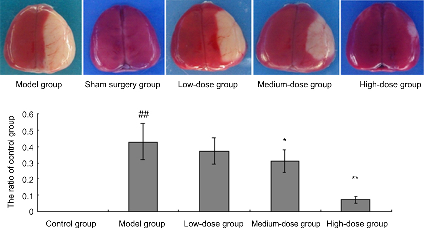 |
Figure 3 Effect of Pterocypsela elata (P. elata) on infarction volume and the ratio of control group after ischemia-reperfusion injury (x̅ ± s, n=8). Notes: ##P<0.01 vs sham surgery group; *P<0.05, **P<0.01 vs model group. |
Main active components and their protein target information
The interactions between potential drug molecules and proteins were analyzed by reverse docking based on their three-dimensional structures via the DRAR-CPI server. A scoring function was employed to score and rank the binding affinities between protein and ligand, and potential targets were predicted. The Z-score represents the strength of the interaction between a drug molecule and its protein target. A target with a Z-score value less than −0.5 is generally considered to be a potential target. The 14 components from P. elata resulted in 1,747 targets. After analyzing the target information, 312 non-repetitive potential targets were identified.
The abovementioned targets were compared with the FDA-approved targets for stroke/cerebral infarction treatment in the DrugBank database. A total of 14 potential active components in P. elata (Table 2) and 21 potential targets for stroke/cerebral infarction treatment were identified (Table 3).
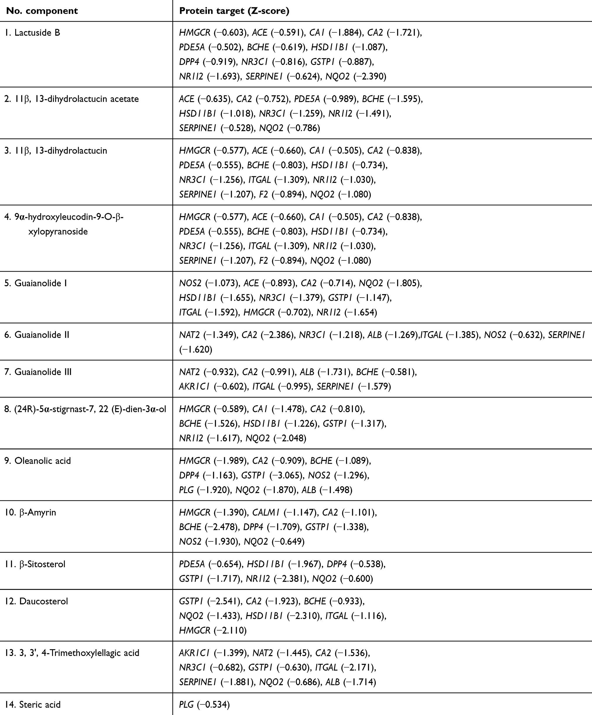 |
Table 2 Pterocypsela elata active components and Z-scores |
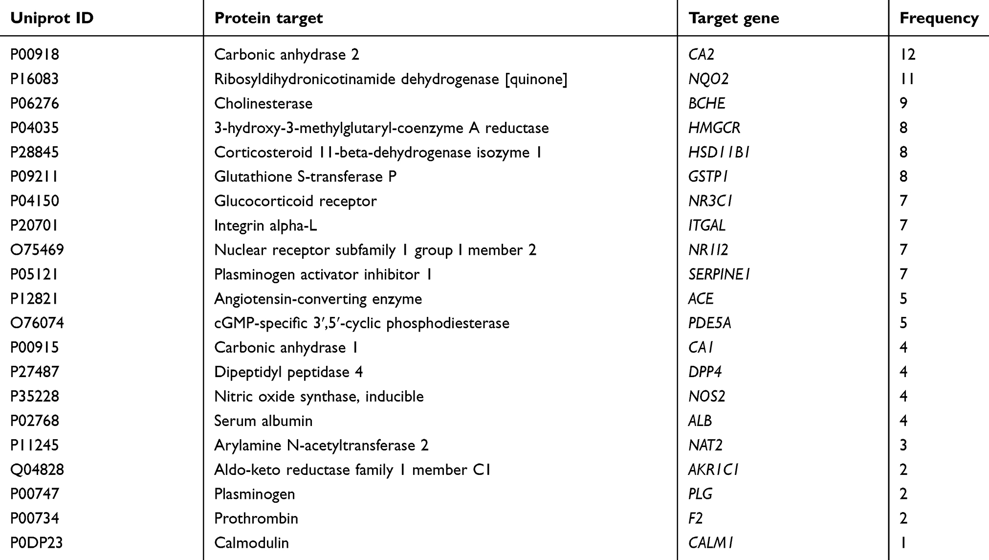 |
Table 3 Potential targets of Pterocypsela elata (P. elata) |
GO enrichment analysis of targets
The abovementioned potential targets of P. elata were assessed for single-target or multi-target GO enrichment analysis in the STRING database. There were 798 genes related to biological processes, and these appeared 1,372 times. The 26 biological processes with an occurrence frequency greater than or equal to 5 are shown in Figure 4A. There were 149 genes related to molecular functions that appeared 221 times. Sixteen molecular functions with an occurrence frequency greater than or equal to 3 are shown in Figure 4B. There were 86 genes related to cellular components that appeared 164 times. Sixteen cellular components with an occurrence frequency greater than or equal to 3 are shown in Figure 4C.
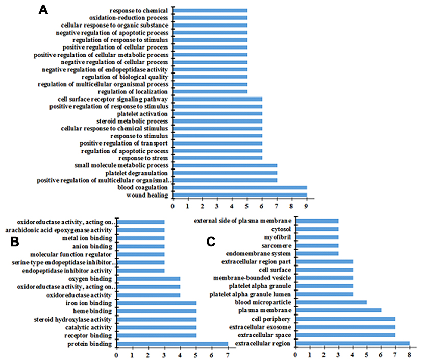 |
Figure 4 Results of GO enrichment analysis of potential targets. (A) Biological processes, (B) cellular components, (C) molecular functions. |
Pathway annotation and construction of a component-target-pathway network
The potential targets were analyzed using KEGG pathways. The results showed that targets were involved in 133 annotated KEGG pathways and appeared 264 times. Thirty-five pathways with an occurrence frequency greater than 3 are listed in Figure 5, including metabolic pathways, complement and coagulation cascades, steroid hormone biosynthesis, metabolism of xenobiotics by cytochrome P450, drug metabolism-cytochrome P450, and the oxytocin signaling pathway. The active component-target-metabolic pathway network is shown in Figure 6.
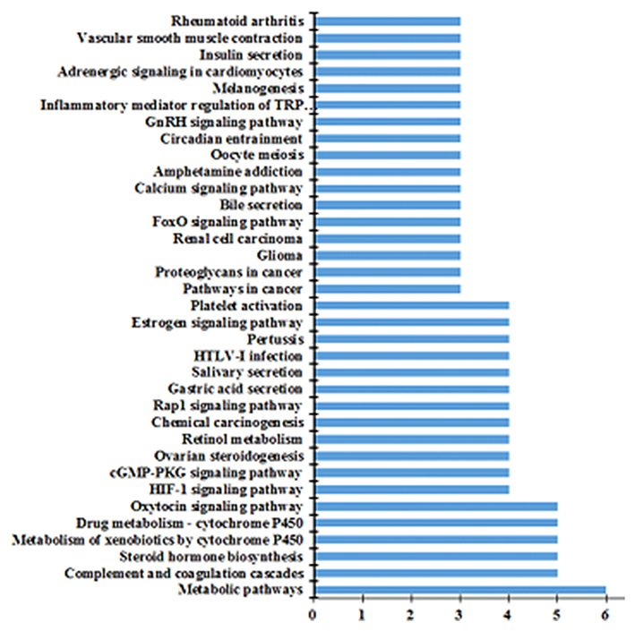 |
Figure 5 KEGG pathway analysis for Pterocypsela elata (P. elata). |
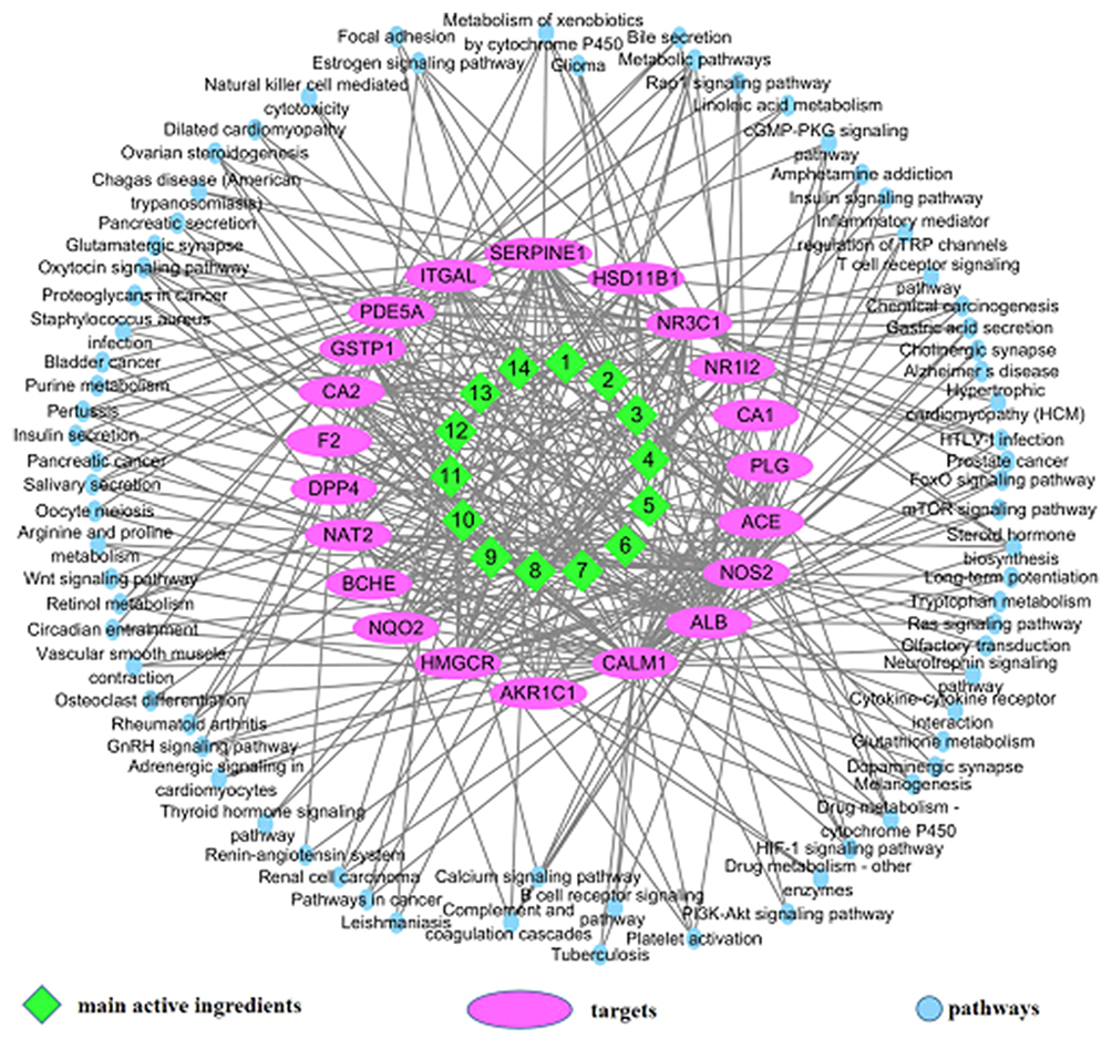 |
Figure 6 Active component-target-pathway network for Pterocypsela elata (P. elata). |
Discussion
Cell experiments showed that different concentrations of P. elata could increase cell viability to various degrees after oxygen-glucose deprivation/recovery. The groups with medium and high concentrations of P. elata showed stronger effects than the model group (P<0.05–0.01). A rat model for cerebral ischemia and reperfusion injury was prepared using Longa’s suture method.12 Through the neurological deficit score of the rats awake after anesthesia and at 48 h after drug administration, we learned that the three concentrations of P. elata (low, medium and high) could all reduce the neurological deficit score of the rats (P<0.05–0.01). TTC staining also showed that the low, medium and high concentrations of P. elata could all reduce the infarction volume at 48 hrs after reperfusion compared to the model group. The high-concentration group had the most obvious effect. The results demonstrated that P. elata has anti-ischemic effects.
Pharmacological experiments showed that P. elata has obvious anti-ischemic effects. Subsequently, the potential targets of each active compound in P. elata were obtained via the DRAR-CPI database by using reverse docking technology. Through a comparison to FDA-approved targets for stroke/cerebral infarction treatment in the DrugBank database, a total of 14 active components with potential activity against stroke/cerebral infarction and 21 potential targets were identified. Active components include lactuside B, 11β, 13-dihydrolactucin acetate, 11β, 13-dihydrolactucin, 9α-hydroxyleucodin-9-O-β-xylopyranoside, guaianolide I, guaianolide II, guaianolide III, (24R)-5α-stigrnast-7, 22 (E)-dien-3α-ol, oleanolic acid, β-amyrin, β-sitosterol, daucosterol, 3, 3′, 4-trimethoxylellagic acid, and steric acid. Among these, lactuside B has been reported to have anti-cerebral ischemia effects.7 The genes of 14 potential targets were CA2, NQO2, BCHE, HMGCR, HSD11B1, GSTP1, NR3C1, and ITGAL.
STRING is a database used for analyzing protein-protein associations. GO enrichment analysis can elucidate which GO terms are over-represented or under-represented in the aspect of biological processes, molecular functions, and cellular components.13,14 The obtained potential targets of P. elata were analyzed via GO enrichment analysis, and target-pathway networks were constructed via the STRING database. There were 26 biological processes with an occurrence frequency greater than or equal to 5. Among them, wound healing and blood coagulation appeared most frequently, followed by the positive regulation of multicellular organismal and platelet degradation. There were 16 molecular functions with an occurrence frequency greater than or equal to 3. Among them, protein binding occurred most frequently, followed by receptor binding, catalytic activity, and steroid hydroxylase activity. There were 16 cellular components with an occurrence frequency greater than or equal to 3. Among them, extracellular region was the most frequent, followed by extracellular space, extracellular exosome, and cell periphery.
KEGG pathway enrichment analysis was performed in the KEGG pathway database to generate hypotheses about potential targets and relevant pathways. The results showed 133 pathways associated with potential targets, and these appeared 264 times. Thirty-five pathways with an occurrence frequency greater than or equal to 3 are listed in Figure 3, including metabolic pathways, complement and coagulation cascades, steroid hormone biosynthesis, metabolism of xenobiotics by cytochrome P450, drug metabolism-cytochrome P450, and the oxytocin signaling pathway. Among them, amino acid metabolism, the coagulation cascade pathway, steroid hormone biosynthesis, and neuroactive ligand-receptor interaction are the pathways that are involved in cerebral ischemia-reperfusion injury, and these pathways are associated and interact with each other. Upon cerebral ischemia, a large amount of excitatory amino acids, including amino acids and aspartic acid, accumulate in the synaptic cleft, which can activate AMPA and NMDA receptors (glutamate receptors). Furthermore, cerebral ischemia induces significant Ca2+ influx and activates downstream Ca2+-dependent cell death pathways, which causes necrosis or apoptosis of neuronal cells and glial cells15 and further aggravates the cerebral ischemic injury. Steroid hormones can protect against cerebral ischemia injury by reducing oxidative stress and inhibiting the NMDA receptor.16,17
An active component-target-metabolic pathway network was constructed using the merge function of Cytoscape software. In the network diagram, “nodes” represent active components, potential targets and pathways; “edges” represent the interactions of the component-target and target-pathway. An analysis of the target protein-protein interaction (PPI) networks found that ACE, NOS2, NR3C1, CA1, PLG, ALB, CALM1, AKRIC1, HMGCR, NQO2 were the main target proteins in the interaction network and have anti-ischemic effects. Among them, ACE (angiotensin-converting enzyme) is a key component of the classical renin-angiotensin system (RAS), which can produce a series of angiotensins, such as angiotensin-I (Ang I), Ang II, and Ang-(1–7), and further plays an important role in the regulation of blood pressure, fluid-electrolyte balance, and protection against cerebral ischemia-reperfusion injury.18 Nitric oxide (NO) is the product of L-arginine catalyzed with nitric oxide synthase (NOS). NOS is mainly distributed in macrophages, inflammatory neutrophils, microglia and astrocytes and induces NOS mRNA expression under ischemic and hypoxic conditions.19 The large amount of NO produced by NOS during cerebral ischemia and reperfusion is toxic to nerve cells.20
In summary, our studies proved that P. elata has good anti-ischemic effects. Further analysis by network pharmacology showed that the main active components of P. elata include lactuside B, 11β, 13-dihydrolactucin acetate, and 11β, 13-dihydrolactucin, which regulate potential targets (NR3C1, CA1, PLG, ACE, NOS2, ALB, CALM1, AKRIC1, HMGCR, and NQO2) and intervene in metabolic pathways, complement and coagulation cascades, steroid hormone biosynthesis and other pathways to achieve a synergistic protection against cerebral ischemic injury. However, the main active components of P. elata and their regulation mechanisms require further verification. Our study provides a new perspective and clues for further studies on the anti-ischemic mechanism of the main active components of P. elata.
Acknowledgments
This work was supported by grants from National Natural Science Foundation of China (No. 81172953) and Natural Science Foundation of Henan Province (No. 182300410356).
Disclosure
The authors confirm that there are no conflicts of interest in this work.
References
1. Liu Z, Guo F, Wang Y, et al. BATMAN-TCM: a bioinformatics analysis tool for molecular mechANism of traditional Chinese medicine. Sci Rep. 2016;6:21146. doi:10.1038/srep21146
2. Yang K, Zeng L, Ge J. Exploring the pharmacological mechanism of danzhi xiaoyao powder on ER-positive breast cancer by a network pharmacology approach. Evid Based Complement Alternat Med. 2018;2018:5059743. doi:10.1155/2018/9567061
3. Zheng S, Jiang Y, Lu M, et al. Network pharmacological screening of herbal monomers that regulate apoptosis-associated genes in acute pancreatitis. Pancreas. 2016;46(1):89–96.
4. Anastasio TJ. Computational identification of potential multi-drug combinations for reduction of microglial inflammation in Alzheimer disease. Front Pharmacol. 2015;6:116. doi:10.3389/fphar.2015.00116
5. Nüesch E, Häuser W, Bernardy K, Barth J, Jüni P. Comparative efficacy of pharmacological and non-pharmacological interventions in fibromyalgia syndrome: network meta-analysis. Ann Rheum Dis. 2013;72:955–962. doi:10.1136/annrheumdis-2011-201249
6. Choi CI, Eom HJ, Kim KH. Antioxidant and α-glucosidase inhibitory phenolic constituents of Lactuca indica L. Russ J Bioorg Chem. 2016;42:310–315. doi:10.1134/S1068162016030079
7. Li SY, Sun J, Niu BX, Yan FL, Zhan HQ. [Effect of lactuside B on the expression of bcl-2 and bax mRNA and their protein in rats’ cerebral cortex after cerebral ischemia-reperfusion injury]. Yao Xue Xue Bao. 2011;46:1314–1320.
8. Bai Y-X, Tan J, Yan F-L, Ding -M-M, Wang X. A new guaianolide from the roots of Pterocypsela elata. Chin Chem Lett. 2013;24:55–56. doi:10.1016/j.cclet.2012.12.015
9. Bai YX, Liang HJ, Yan FL, Wang X, Yang YX. ChemInform abstract: a new guaianolide (I) and other constituents from Pterocypsela elata. Cheminform. 2014;45. doi:10.1002/chin.201413217
10. Zhan H-Q, Li P-F, Li S-Y, et al. Influence of lactuside B on the expression of AQP4 and TRPM7 mRNAs in the cerebral cortex after cerebral ischemia injury. Eur Rev Med Pharmacol Sci. 2014;18:1151–1157.
11. Darshit BS, Ramanathan M. Activation of AKT1/GSK-3 β/β -Catenin–TRIM11/Survivin pathway by novel GSK-3 β inhibitor promotes neuron cell survival: study in differentiated SH-SY5Y cells in OGD model. Mol Neurobiol. 2016;53(10):6716–6729.
12. Longa EZ, Weinstein PR, Carlson S, Cummins R. Reversible middle cerebral artery occlusion without craniectomy in rats. Stroke. 1989;20:84–91.
13. PANGXiao-Cong. Network pharmacology-based analysis of Chinese herbal Naodes heng formula for application to Alzheimer'sdisease. Chin J Nat Med. 2018;16:90.
14. Mao Y, Hao J, Jin ZQ, et al. Network pharmacology-based and clinically relevant prediction of the active ingredients and potential targets of Chinese herbs in metastatic breast cancer patients. Oncotarget. 2017;8(16):27007–27021.
15. Luo Y, Tang H, Li H, Zhao R, Huang Q, Liu J. Recent advances in the development of neuroprotective agents and therapeutic targets in the treatment of cerebral ischemia. Eur J Med Chem. 2018;162:132–146. doi:10.1016/j.ejmech.2018.11.014
16. Bastianetto S, Ramassamy C, Poirier J, Quirion R. Dehydroepiandrosterone (DHEA) protects hippocampal cells from oxidative stress-induced damage. Brain Res Mol Brain Res. 1999;66:35–41.
17. Karishma KK, Herbert J. Dehydroepiandrosterone (DHEA) stimulates neurogenesis in the hippocampus of the rat, promotes survival of newly formed neurons and prevents corticosterone-induced suppression. Eur J Neurosci. 2015;16:445–453. doi:10.1046/j.1460-9568.2002.02099.x
18. Petkow-Dimitrow P. [New therapeutic targets for ACE inhibitors and angiotensin receptor blockers]. Pol Arch Med Wewn. 2007;117:44–50.
19. Haga KK, Gregory LJ, Hicks CA, et al. The neuronal nitric oxide synthase inhibitor, TRIM, as a neuroprotective agent: effects in models of cerebral ischaemia using histological and magnetic resonance imaging techniques. Brain Res. 2003;993:42–53. doi:10.1016/j.brainres.2003.08.063
20. Kim JH, Yenari MA, Giffard RG, Cho SW, Park KA, Lee JE. Agmatine reduces infarct area in a mouse model of transient focal cerebral ischemia and protects cultured neurons from ischemia-like injury. Exp Neurol. 2004;189:122–130. doi:10.1016/j.expneurol.2004.05.029
 © 2019 The Author(s). This work is published and licensed by Dove Medical Press Limited. The
full terms of this license are available at https://www.dovepress.com/terms
and incorporate the Creative Commons Attribution
- Non Commercial (unported, 3.0) License.
By accessing the work you hereby accept the Terms. Non-commercial uses of the work are permitted
without any further permission from Dove Medical Press Limited, provided the work is properly
attributed. For permission for commercial use of this work, please see paragraphs 4.2 and 5 of our Terms.
© 2019 The Author(s). This work is published and licensed by Dove Medical Press Limited. The
full terms of this license are available at https://www.dovepress.com/terms
and incorporate the Creative Commons Attribution
- Non Commercial (unported, 3.0) License.
By accessing the work you hereby accept the Terms. Non-commercial uses of the work are permitted
without any further permission from Dove Medical Press Limited, provided the work is properly
attributed. For permission for commercial use of this work, please see paragraphs 4.2 and 5 of our Terms.