Back to Journals » International Journal of Nanomedicine » Volume 20
Natural, Engineered, and Hybrid Platelet Membrane–Based Nanotherapeutics for Inflammatory Diseases
Authors Liu B ![]() , Wang Y, Gong W, Han S, Lv Z, Zhang Z, Qi J, Song A, Yang Z, Duan L, Zhang T, Wang Z
, Wang Y, Gong W, Han S, Lv Z, Zhang Z, Qi J, Song A, Yang Z, Duan L, Zhang T, Wang Z ![]()
Received 6 August 2025
Accepted for publication 13 November 2025
Published 26 November 2025 Volume 2025:20 Pages 14149—14184
DOI https://doi.org/10.2147/IJN.S558928
Checked for plagiarism Yes
Review by Single anonymous peer review
Peer reviewer comments 2
Editor who approved publication: Professor Jie Huang
Boyuan Liu,1,* Yongjie Wang,1– 3,* Weiquan Gong,1 Song Han,1 Zhenshan Lv,1 Zilin Zhang,1 Jinwei Qi,1 Aijun Song,1 Zongyuan Yang,1 Longfei Duan,1 Tianhui Zhang,1 Zhenyu Wang1
1Department of Spine Surgery, The First Hospital of Jilin University, Changchun, Jilin, People’s Republic of China; 2Department of Orthopedics, Taizhou Central Hospital (Taizhou University Hospital), Taizhou, Zhejiang, People’s Republic of China; 3Department of Orthopaedics, Wenzhou Medical University Affiliated Taizhou Central Hospital, Taizhou, Zhejiang, People’s Republic of China
*These authors contributed equally to this work
Correspondence: Zhenyu Wang, Department of Spine Surgery, The First Hospital of Jilin University, Changchun, Jilin, People’s Republic of China, Email [email protected]
Abstract: Nanotherapeutics based on platelet membranes represent a new and advanced biomimetic approach in nanomedicine. By covering synthetic nanoparticle cores with natural platelet membranes, these platforms ingeniously combine the multifaceted biointerfacing abilities of platelets, such as long circulation, immune evasion, and targeting of inflamed tissues, with the many functions of engineered cores. This review systematically summarizes recent advances in the design and application of nanotherapeutics, categorizing them into three platforms: those derived from natural platelet membranes, those utilizing engineered platelet membranes for enhanced targeting or drug loading, and those employing hybrid membranes fused with other cell types to combine complementary functionalities. We emphasize their therapeutic efficacy in various inflammatory diseases such as atherosclerosis, ischemic injury (stroke and myocardial infarction), rheumatoid arthritis, microbial infections, and the tumor inflammatory microenvironment. Finally, we discuss the translational potential and current challenges of this technology and provide a critical perspective on its future development in precision medicine.
Keywords: platelet membrane, engineered platelet, hybrid membrane, inflammation, nanotherapy
Introduction
Inflammation is a defensive reaction that involves immune cells, blood vessels, and molecular mediators to counteract harmful stimuli such as pathogens, damaged cells, or irritants. It is considered a mechanism of innate immunity that aims to eliminate the initial cause of cell injury, remove necrotic cells and damaged tissues, and initiate tissue repair.1 During the inflammation process, immune cells such as neutrophils, monocytes, and lymphocytes migrate into the damaged areas to varying extents. Acute inflammation is an essential biological reaction that safeguards against infection, removes harmful substances, and aids in the recovery of injured tissue.2 Chronic inflammation can increase the probability of inflammatory harm, as well as infections and other systemic disorders,3 such as arthritis, atherosclerosis, and cancer.4,5 There is growing recognition that chronic or uncontrolled inflammation plays a significant role in the onset and advancement of several severe illnesses, including cancer, obesity, type 2 diabetes, atherosclerosis, ischemic disease, systemic inflammatory response syndrome, and even autoimmune disease.6 Various pharmacological groups, such as antibiotics, cytotoxic medicines, and hormones, can effectively combat inflammation. However, these treatments have systemic effects, and prolonged usage can lead to significant negative effects.7,8 Nanoparticle (NP)-mediated medication-delivery systems incorporating anti-inflammatory medicines are potentially more secure and efficient alternatives.
NP-based drug-delivery systems have shown significant promise in biomedical applications owing to their desirable and adjustable characteristics, such as loading capacity and sufficient protection for drugs, as well as their ability to control drug release, among other benefits.9–12 Nevertheless, many challenges remain. Plasma proteins readily opsonize synthetic NPs, leading to their efficient clearance by the immune system.13 Under certain circumstances, the NPs can stimulate the immune system to produce antibodies, which in turn hasten their removal from the body.14–17 Furthermore, NPs typically lack the ability to aggressively target the disease environment, unlike real cells. As a result, the overall accumulation of NPs is limited. Therefore, utilizing the cell membrane to disguise NPs for active distribution offers a novel opportunity for targeted therapy.18
The use of cell membrane–based nanotherapeutics has emerged as a highly promising approach for enhancing the transport of therapeutic drugs. This is achieved by combining the biomimetic characteristics of cell membranes with the functional adaptability of nanomaterials.19,20 This method involves enveloping artificial nanocarriers with a natural cell membrane, which addresses certain limitations of NPs created using traditional self-assembly techniques, such as poor colloidal stability and nonspecific tissue accumulation.21 Cell membrane–coated NPs have the ability to replicate the biological characteristics of the cells from which they are derived. This allows them to persist longer in the circulatory system and target specific diseases.22
Platelets are tiny, disc-shaped blood cells that lack a nucleus and range in size from 2 to 4 μm. Produced from megakaryocytes in the bone marrow,23 platelets play a vital role in the process of blood clotting, as they can specifically locate areas of blood vessel damage and ensure proper blood flow.24 Furthermore, platelets participating in the activities and interactions between cells are crucial in inflammatory, immunological, and tumor-development processes.25 Previous studies have shown that platelets, a type of immune cell, are naturally attracted to inflammation and tumors. This characteristic can be harnessed to deliver drugs with anti-inflammatory and anti-cancer properties.26–28 Therefore, platelet membranes and PEVs may be reliable sources of cell membranes for membrane-based nanotherapies.
This paper reviews the use of natural platelet membranes, engineered platelet membranes, or platelet membranes hybridized with other cell membranes as nanotherapies for the treatment of inflammatory illnesses. Herein, we provide a concise overview of the methodologies employed to extract and coat platelet membranes. Next, we discuss the various applications of platelets and their membranes in the treatment of inflammatory illnesses. Finally, we discuss the potential future prospects of this approach.
Preparation and Coating Methods of Platelet Membrane
The commonly employed techniques for producing biomembrane-derived NPs involve the following three stages: extracting the membranes, preparing NP cores, and covering the NP cores with vesicles produced from the membranes (Figures 1 and 2).29 Hu et al fabricated platelet membrane–cloaked nanoparticles (PNPs) by utilizing a cell membrane cloaking technique.18,30,31 These PNPs are composed of a biodegradable polymeric NP core that is fully protected by the plasma membrane of human platelets.32
 |
Figure 1 Schematic illustrations of five membrane engineering methods and introduced additional functions. (A) Different types of membrane materials used for engineering. (B) Membrane engineering methods: membrane hybridization, postinsertion method, chemical method, metabolic engineering and gene engineering. (C) Engineering biomembrane-derived nanoparticles: hollow or core-shell types. (D) Additional functions introduced by membrane engineering. Reprinted with permission from Wu Z, Zhang H, Yan J, Wei Y, Su J. Engineered biomembrane-derived nanoparticles for nanoscale theranostics. Theranostics. 2023;13(1):20–39. Copyright 2023 Theranostics.33 |
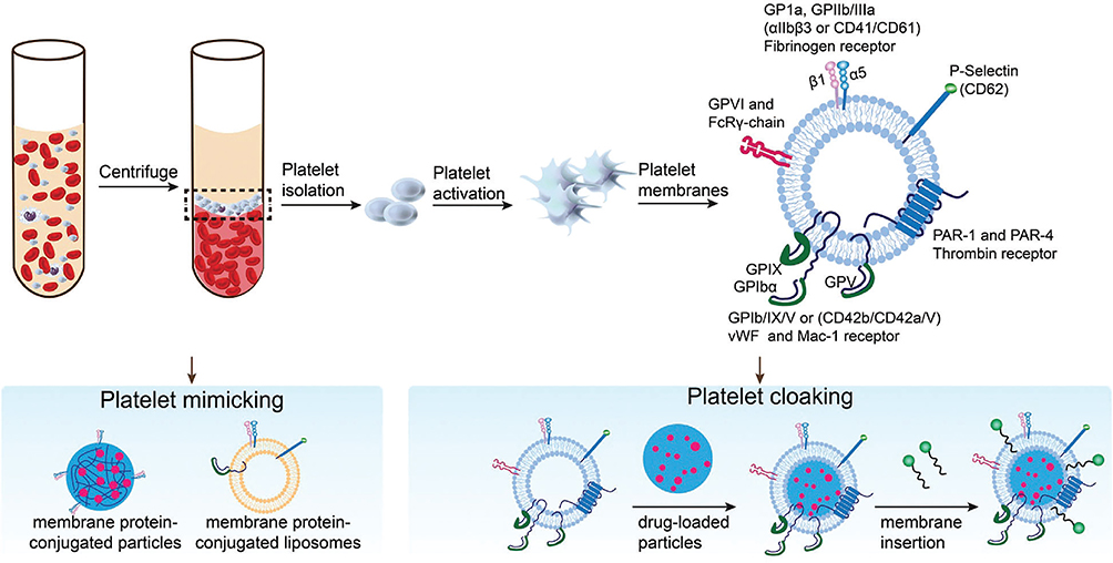 |
Figure 2 The fabrication of platelet-like nanoparticles for disease treatment. Platelets were collected by centrifugation and activated, and the membranes of the platelets were prepared for further usage. The area within the black dashed box indicates the platelet layer after centrifugation. Reprinted with permission from Li Z, Hu S, Cheng K. Platelets and their biomimetics for regenerative medicine and cancer therapies. J Mater Chem B. 2018;6(45):7354–7365. Copyright 2018 Journal of Materials Chemistry B.34 |
Methods of Platelet Membrane Extraction
Platelets were extracted approximately 30 minutes after blood collection.32 Before collecting the platelets, the blood and plasma samples were combined with ethylenediaminetetraacetic acid (EDTA), which inhibits platelet aggregation by deactivating the fibrinogen-binding integrin αIIbβ3.35 To separate the red and white blood cells, the blood and plasma samples were centrifuged at 100 × g for 20 min at room temperature, resulting in the isolation of platelets. Platelet-rich plasma (PRP) was centrifuged at 100 × g for 20 min to eliminate any residual blood cells. A phosphate buffer solution (PBS) containing EDTA and prostaglandin E1 was introduced into pure PRP to inhibit platelet activation. The platelets were subsequently separated via centrifugation at 800 × g for 20 min at room temperature. The supernatant was then removed, and the platelets were suspended in a solution of PBS containing EDTA. They were then combined with tablets to inhibit the protease enzyme activity. The platelet membrane was obtained using a repetitive freeze–thaw method. The platelet suspensions were initially frozen at –80 °C, defrosted at ambient temperature, and then separated by centrifugation at a speed of 4000 × g for a duration of 3 min. After three consecutive washes using a PBS solution containing protease inhibitor tablets, the collected platelet membranes were immersed in water and sonicated in a sealed glass vial for 5 min (Figure 3).32
 |
Figure 3 Schematic depicting the process of preparing PNPs. Reprinted with permission from Hu CM, Fang RH, Wang KC, et al. Nanoparticle biointerfacing by platelet membrane cloaking. Nature. 2015;526(7571):118–21. Copyright 2015 Nature.32 |
Methods for Coating Platelet Membranes onto Nanoparticles
Biomembrane-derived NPs can be created using many techniques, including co-extrusion, sonication, microfluidic electroporation, and cell membrane–templated polymerization. Table 1 summarizes the advantages and disadvantages of different methods for coating platelet membranes with NPs.
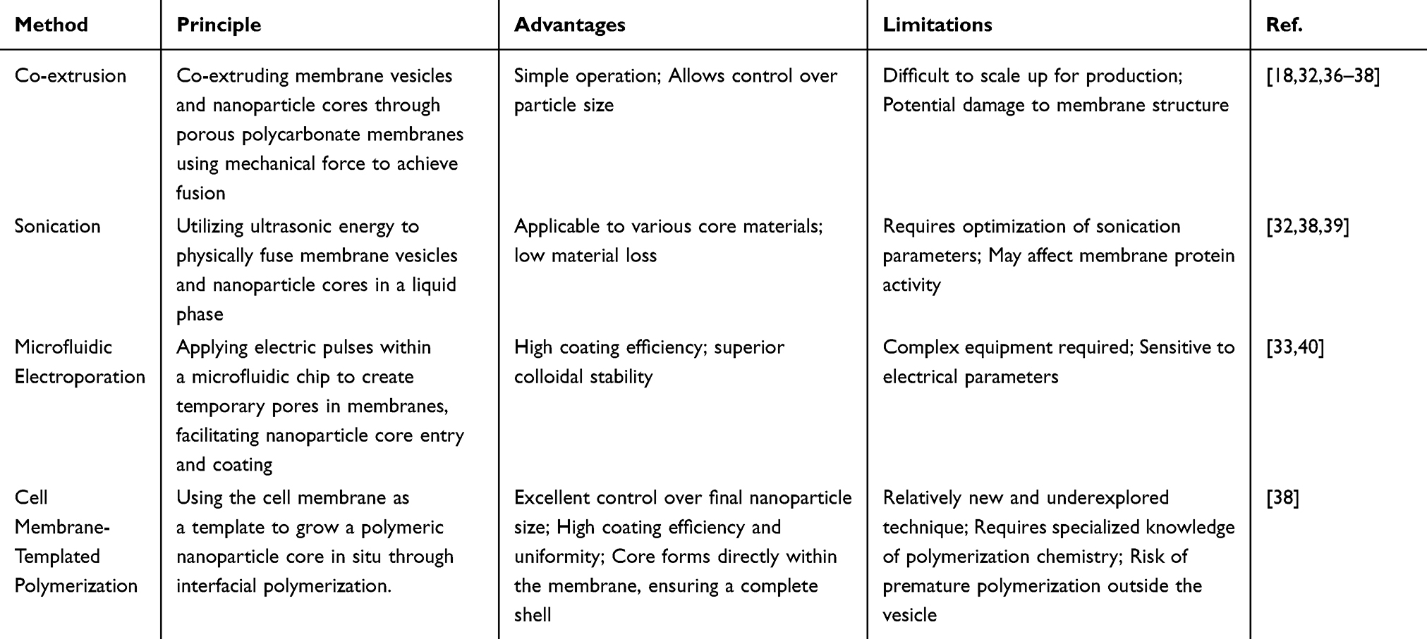 |
Table 1 Comparison of Different Coating Methods for Platelet Membrane–Based Nanoparticles |
- Co-extrusion: Co-extrusion involves passing the NP solution and vesicles through polycarbonate porous membranes of varying diameters while simultaneously sonicating them for a few minutes. This process results in the formation of NPs with a consistent cross-sectional profile.32 Extrusion applies a powerful force that can disrupt the structural integrity of the membrane, allowing it to rebuild around the inner core NP and effectively coat the NP cores.18,36,37 Although the extrusion technique shows promise, its use in large-scale production presents obstacles. Therefore, sonication has been demonstrated to be an effective method for fusing NP cores and membrane vesicles.38
- Sonication: Sonication employs ultrasonic radiation at certain frequencies to merge membranes and nano vehicles39 into core–shell structures, resulting in lower material loss than that caused by co-extrusion.32 The frequency, duration, and amplitude of sonication are the primary factors that determine the effectiveness of the fusion process. During sonication, powerful forces are applied to the NP core and membrane vesicles that are incubated together, resulting in the spontaneous formation of a core–shell structure.38
- Microfluidic electroporation: Microfluidic electroporation is a new method that applies an electric pulse between two electrodes to facilitate the coating of nanocores with biomembrane vesicles.33 Biomembrane-derived NPs, produced using microfluidic procedures, have superior colloidal stability and higher coating effectiveness than those made using standard extrusion methods.40 This strategy is gaining popularity because it allows NPs to remain stable while preventing them from clumping together.40
- Cell membrane–templated polymerization: This technology is new and thus has not yet been extensively studied. It relies on interactions between the cores and cell membranes. The polymeric core is synthesized in situ within the cores to enable effective coating and convenient modification of the NP size. By contrast, the typical process employs prefabricated polymers, which do not allow control over the already produced NPs (Figure 4).38
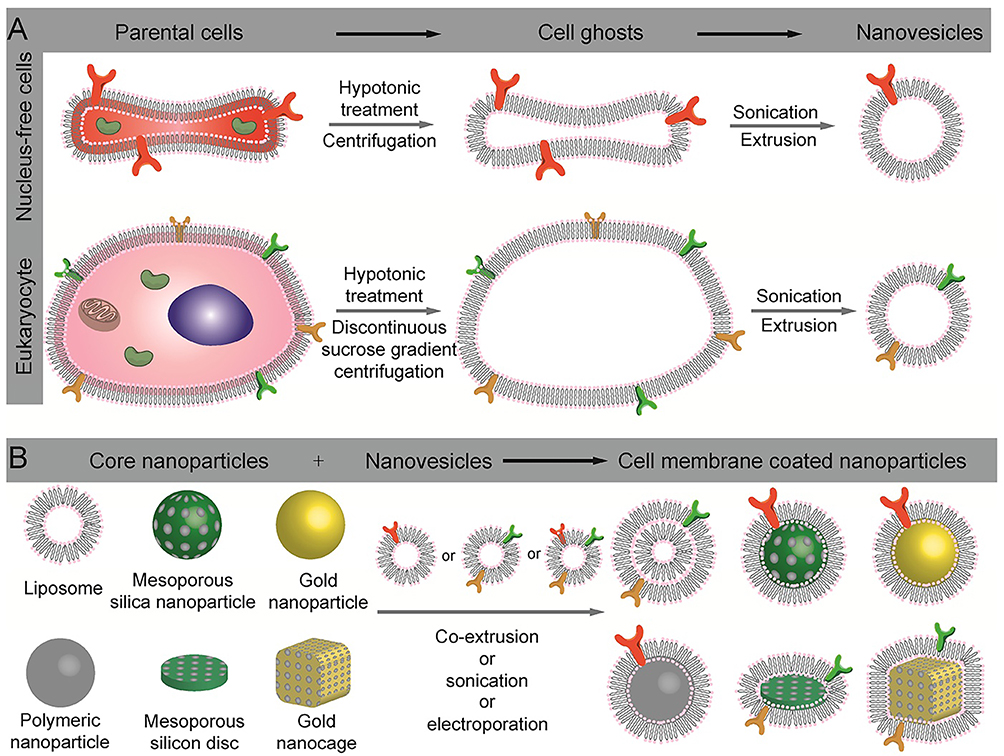 |
Figure 4 Schematic illustration of the preparation of cell-membrane coated nanoparticles. (A) Extract of intact cell ghost from parent cells and further processing into nanovesicles. (B) Different types of nanoparticles that have been used as inner cores, and their fusion with nanovesicles to construct cell membrane coated nanoparticles. Reprinted with permission from Zhai Y, Su J, Ran W, et al. Preparation and Application of Cell Membrane-Camouflaged Nanoparticles for Cancer Therapy. Theranostics. 2017;7(10):2575–2592. Copyright 2017 Theranostics.36 |
Natural Platelet Membrane–Based Nanotherapeutics for Inflammatory Diseases
Atherosclerosis
Atherosclerosis is a cardiovascular condition that poses a major hazard to human health. It is characterized by persistent thickening of the arterial walls, an inflammatory reaction, and a disruption in lipid metabolism.26,41–44 Inflammatory macrophages have been identified as a crucial element in atherosclerosis development. These macrophages are capable of releasing pro-inflammatory cytokines, which enhance the localized inflammatory reaction at the location of the atherosclerotic plaque, ultimately resulting in the creation of a plaque.43,45–47 Biomimetic NPs, created by enveloping a synthetic nanoparticulate core with a platelet membrane, can interact with activated endothelium, foam cells, and collagen. These effects were demonstrated to be specific to platelet membrane–coated NPs. These biomimetic nanocarriers can effectively target both well-established atherosclerotic plaques and subclinical sections of arteries that are prone to plaque formation.48 Huang et al effectively created a highly biocompatible Janus mesoporous silicon nanomotor with a drug-loaded platelet membrane bionic structure (MJAMS/PTX/aV). This nanomotor can not only enter the plaque but also be effectively absorbed by stimulated endothelial cells and inflammation-related macrophages upon exposure to near-infrared (NIR) radiation (Figure 5).49 Inflammation in the vicinity of atherosclerosis activates endothelial cells, in turn triggering the attachment and clustering of platelets.50,51 Wei et al have shown that using platelet membranes as functional coverings for nanocarriers allows for precise targeting of imaging payloads to atherosclerotic sites.48 Using rapamycin (RAP) as the model drug and based on platelets’ inherent affinity to plaques, Song et al created a platelet-like delivery system to specifically target atherosclerotic plaques. This was achieved by combining platelet membranes with poly (DL-lactide-co-glycolide) NPs (Figure 6). The resulting platelet membrane–coated NPs successfully targeted atherosclerotic plaques, slowed the advancement of atherosclerosis, and stabilized plaques when the NPs were loaded with rapamycin.28 Photodynamic therapy (PDT) triggers cell death through autophagy and controls lipid metabolism in foam cells produced from macrophages.52 Ma et al effectively developed a delivery system that mimics platelets. This technology allows for the precise targeting and noninvasive PDT of atherosclerotic plaques. In addition, SPECT/CT-guided PDT effectively mitigates the advancement of atherosclerosis by modulating lipid metabolism and diminishing inflammation.53 Zhang et al developed a bionic nano-delivery platform that used platelet membranes (PMs) to cover black phosphorus nanosheets (BPNSs) and specifically target macrophages in atherosclerotic plaques. The NPs were used to insert small interfering RNA into macrophages that inhibit Ca2+/calmodulin-dependent protein kinase γ (CaMKII γ). Moreover, macrophage efferocytosis was reestablished by inhibiting CaMKII γ and enhancing the expression of MerTK, a cytosolic receptor, thereby facilitating the removal of apoptotic cells from plaques.54 Chen et al created a biomimetic platelet membrane–coated polyphenol–cerium dioxide nanozyme complex that worked as a broad-spectrum antioxidant by eliminating superoxide anions, H2O2, and intracellular reactive oxygen species (ROS) while also boosting the activities of hepatic superoxide dismutase (SOD) and glutathione (GSH). This complex specifically targets atherosclerotic plaques and is highly effective at reducing oxidative stress, preventing lipid uptake and accumulation, controlling cholesterol efflux, and stopping the release of inflammatory factors.55
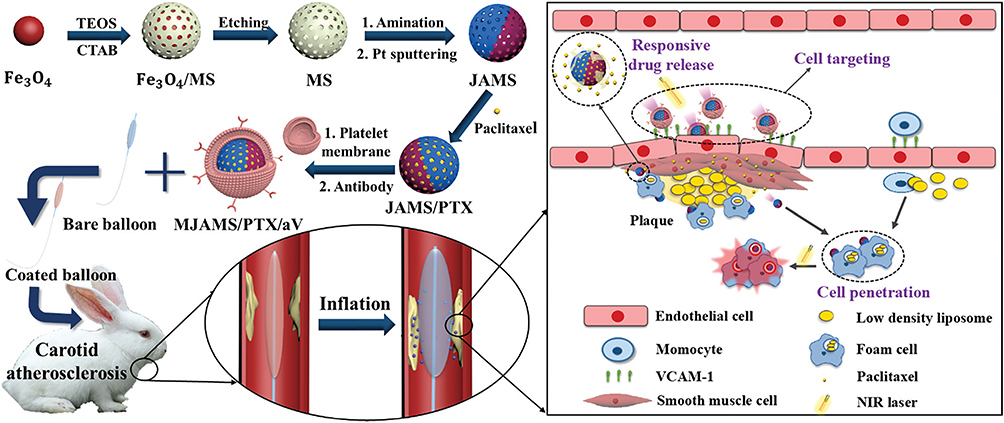 |
Figure 5 Schematic illustration of the synthesis process of MJAMS/PTX/aV and the mechanism of treatment of atherosclerosis using the MJAMS/PTX/aV coated balloon. Reprinted with permission from Huang Y, Li T, Gao W, et al. Platelet-derived nanomotor coated balloon for atherosclerosis combination therapy. J Mater Chem B. 2020;8(26):5765–5775. Copyright 2020 Journal of Materials Chemistry B.49 |
 |
Figure 6 Schematic design of RAP-loaded PNP. Reprinted with permission from Song Y, Huang Z, Liu X, et al. Platelet membrane-coated nanoparticle-mediated targeting delivery of Rapamycin blocks atherosclerotic plaque development and stabilizes plaque in apolipoprotein E-deficient (ApoE (-/-)) mice. Nanomedicine. 2019;15(1):13–24. Reproduced with permission from. Copyright 2019 Nanomedicine.28 |
Restenosis
Postoperative restenosis has emerged as a significant limitation of interventional endovascular therapy for cardiovascular conditions and significantly affects the long-term prognosis and quality of life of patients.56,57 The onset and advancement of this phenomenon are highly intricate, and a sequence of reactions, such as an inflammatory response, thrombus, and intimal hyperplasia (IH), typically occurs following initial damage to endothelial cells. These processes pose significant challenges for the development of effective anti-restenosis strategies.58,59 Hao et al developed a pH-responsive NP (RM/JQ1/PM) to encapsulate JQ1 (a multi-effect endothelium-protective epigenetic inhibitor) for targeted drug delivery with controlled release. The NPs were camouflaged using PMs. This system demonstrated a notable anti-inflammatory effect by impeding the nuclear factor-κB (NF-κB) signaling pathway and the release of pro-inflammatory cytokines from macrophages (Figure 7).60
 |
Figure 7 Process of RM/JQ1/PM preparation (1), targeting homing to injured vascular endothelium site (2), selectivity for different cells including VECs, VSMCs and activated macrophages (3) and cellular drug delivery in VSMCs as well as pH-responsive JQ1 release (4). Reprinted with permission from Hao X, Gai W, Ji F, et al. Biomimetic and responsive nanoparticles loading JQ1 for dual-targeting treatment of vascular restenosis via multiple actions. Chemical Engineering Journal. 2022; 431:133452. Copyright 2022 Chemical Engineering Journal.60 |
Ischemic Disease
Thrombi
Vascular diseases such as myocardial infarction, stroke, and peripheral artery disease are significant contributors to both morbidity and mortality worldwide.61 A common clinical manifestation in these diseases is the development of occlusive clots (thrombi) in blood vessels, which impede blood flow to vital organs.62 ROS, originating from activated platelets and disrupted endothelial cells, are widely recognized for their significant contributions to the formation of blood clots. They achieve this by facilitating platelet–endothelium interactions and causing malfunction in the endothelium.63–66 Surplus ROS also provokes the development of pro-inflammatory cytokines on the endothelium and promotes vascular blockage.67,68 Pawlowski et al created platelet microparticle (PMP)-inspired nanovesicles (PMINs) that can safeguard thrombolytic medications by enclosing them, preventing unintended absorption and effects. These nanovesicles can attach to blood clots using PMP-relevant molecular mechanisms and release the drugs when triggered by enzymes relevant to blood clots, which could also be an efficient delivery platform for other therapeutic payloads in the vascular compartment.69 Zhao et al produced H2O2-responsive platelet membrane–cloaked argatroban-loaded polymeric NPs (PNPArg). These NPs were designed for thrombus therapy, taking advantage of the abundance of H2O2 and the thrombus-targeting properties of platelets. PNPArg efficiently and specifically targeted blocked blood arteries and effectively inhibited the formation of blood clots. Additionally, they reduced the levels of H2O2 and inflammatory cytokines produced by ferric chloride in a mouse model of carotid artery thrombosis.70
Ischemic Stroke
Ischemic stroke, which accounts for 80% of all stroke cases,71,72 is recognized as a leading cause of death worldwide.73,74 At present, the first-line treatments for stroke patients are thrombolysis75 and antiplatelet therapy76 to improve cerebral circulation. The FDA approved the early use of recombinant tissue plasminogen activator (rtPA) as an exclusive and efficacious thrombolytic treatment for acute ischemic stroke (AIS).77–79 Enclosing melanin nanoparticles (MNPs) and tissue plasminogen activator (tPA) with vesicles derived from platelet membranes (PMs), Yu et al created a biomimetic nanovesicle called tPA/MNP@PM. These nanovesicles combine the thrombus-targeting ability of PM, photothermal conversion capabilities, and the ability to scavenge the free radicals of natural melanin. Site-specifically released MNPs, when combined with hemoperfusion, can cross the blood-brain barrier (BBB) and gather at the site of cerebral ischemia. It acts by removing different types of harmful free radicals and reduces damage caused by inflammation and immunological responses, thus providing neuroprotection following thrombolysis.80
Neutrophils (NEs) are the first and most prevalent inflammatory cells that emerge during microvascular reactions to ischemic stroke. By contrast, monocytes/macrophages move to the cerebral ischemic areas throughout the acute phase of inflammation.81–83 The excessive infiltration of NEs is linked to increased levels of ROS and inflammatory mediators, which exacerbate the damage to ischemic tissue.84–87 Tang et al presented platelet-mimetic nanoparticles (PTNPs) that can accurately detect inflammatory NEs in cerebral ischemic regions, enter cells by exploiting the specific attraction between platelets and polarized NEs, and deliver drugs to intervene with NEs, resulting in a significant decrease in infiltrating NEs in cerebral ischemic regions. This study showed that the combination of PTNPs, piceatannol (a selective inhibitor of splenic tyrosine kinase), and superparamagnetic iron oxide (a contrast agent for T2 imaging) effectively identified adherent NEs by coating the platelet membranes. The loaded piceatannol may be transported to attached NEs and released into the circulatory system, thereby diminishing NE infiltration and decreasing infarct size (Figure 8).88
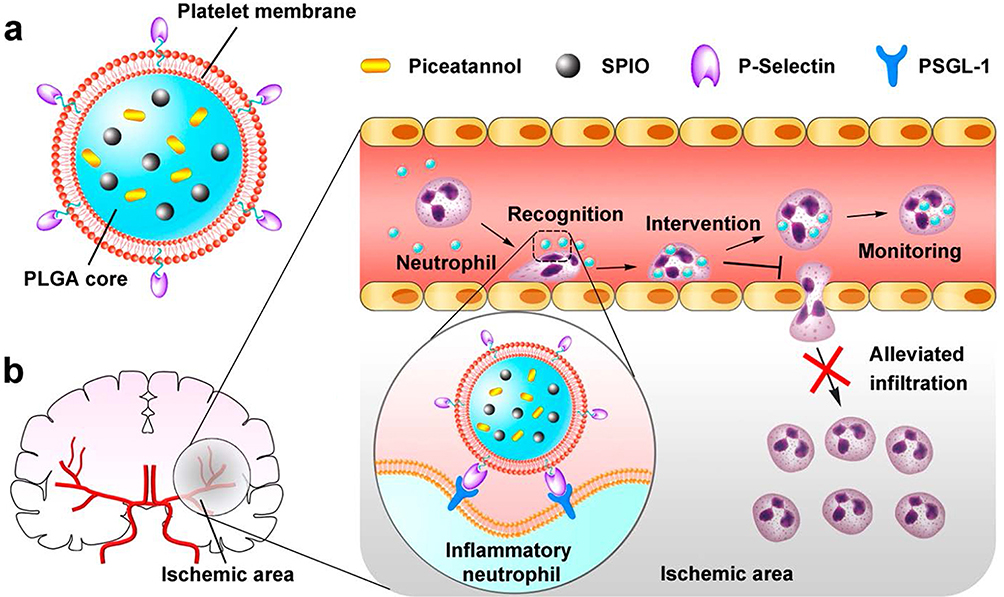 |
Figure 8 Effective recognition, intervention, and monitoring of inflammatory neutrophils by PTNPs for the treatment of AIS. (a) A diagram of the main components of PTNPs, including the SPIO and piceatannol co-loaded PLGA core and the platelet membrane coating shell. (b) The recognition, intervention, and monitoring of inflammatory neutrophils by PTNPs, along with the reduction in neutrophil infiltration. The red cross indicates the alleviated effect of PTNPs on neutrophil infiltration. Reprinted with permission from Tang C, Wang C, Zhang Y, et al. Recognition, Intervention, and Monitoring of Neutrophils in Acute Ischemic Stroke. Nano Lett. 2019;19(7):4470–4477. Copyright 2019 Nano Letters.88 |
Cerebral ischemic damage significantly affects the prognosis of AIS.89–92 During cerebral ischemic injury, neuronal death occurs in the vicinity of the affected area. The injury induces a low-pH environment, a strong neuroinflammatory reaction, and alterations in the activity of immune cells, resulting in the disruption of the BBB.93,94 During ischemic injury, microglia are activated into two different polarized phenotypes: the M1 phenotype, characterized by its pro-inflammatory properties, and the M2 phenotype, which is anti-inflammatory.95–97 The transition of microglia from M1 to M2 has the potential to protect neurons and decrease apoptosis. MiRNA-Let-7c mitigated ischemia injury by modulating neuronal survival and altering microglial phenotypes. Zhao et al created NPs that camouflaged with platelet membranes and had multiple functions. These NPs were specifically designed to target the brain and treat cerebral ischemic injuries. Their main purpose was to transport MiRNA-Let-7c, which aids in reducing brain cell apoptosis and controlling the transformation of microglial cells from the M1 to the M2 phenotype. This regulation was achieved through the activation of the Notch1 cell signaling pathway (Figure 9).98 Li et al described a thrombo-inflammatory cascade–targeted biomimetic nanobubble created by gas–liquid interfacial modular assembly of platelet membranes and the immunomodulator fingolimod (FTY720). Molecular dynamics simulations revealed a gas–liquid interfacial self-organization mechanism that governs the ordered spatial arrangement of platelet membrane lipid rafts and amphiphilic FTY720, facilitating the formation of platelet membrane-coated FTY720 nanobubbles. This type of modularly assembled nanobubble can improve the function of targeted proteins and drug molecules at the biointerface. This facilitates the targeting of lesions in a precise order, BBB crossing, and the drug uptake of microglial cells, greatly improving drug-delivery efficiency and reducing damage to brain tissue.99
 |
Figure 9 Schematic of the delivery of miRNA through platelet membrane-camouflaged nanoparticles to the site of the brain lesion to reduce apoptosis of neurons and regulate the phenotype of microglia in ischemia/reperfusion (I/R) injury. Schematic illustration of the constructed miRNA/TPHL@PM. In I/R injury, the miRNA/TPHL@PM is bound to neutrophils that can cross the blood-brain barrier via chemotaxis and deliver the miRNA to the site of the lesion. The brown and blue upward arrows indicate upregulation of the corresponding elements. Reprinted with permission from Zhao C, Chen Q, Li W, Zhang J, Yang C, Chen D. Multi-functional platelet membrane-camouflaged nanoparticles reduce neuronal apoptosis and regulate microglial phenotype during ischemic injury. Applied Materials Today. 2022; 27:101412. Copyright 2022 Applied Materials Today.98 |
Myocardial Infarction
A significant number of patients develop congestive heart failure (CHF) after myocardial infarction (MI). Survivors of an initial ischemic attack experience post-infarction inflammation and fibrosis, leading to chronic weakening of the heart.100,101 Importantly, after infarction, splenic monocytes contribute to the demise of cardiomyocytes that have survived the ischemia phase by generating ROS and secreting matrix metalloproteinases, exacerbating cardiac remodeling.4 In postinfarction, abundant monocytes from the spleen are attracted to the heart and engage with platelets while circulating. Cheng et al developed platelet-like proteoliposomes (PLPs) that exploit the interactions between platelets and circulating monocytes. Mimicking the typical behavior of platelets, PLPs exhibit a high affinity for monocytes but do not attach strongly to endothelial cells in vitro. Notably, PLPs augment the anti-inflammatory cobalt protoporphyrin to specifically target the heart in a manner independent of enhanced permeability and retention (EPR). This has led to improved therapeutic results.102 Yang et al fabricated ROS-responsive biomimetic NPs by placing niobium carbide MXenes (Nb2C) on mesoporous silicon nanoparticles (MSNs) that were already covered in the platelet membrane. During the acute phase of MI, these NPs are injected into the infarcted heart intravenously. The MSN mesoporous structure improves the ability of Nb2C to eliminate ROS from the infarction and mitigates oxidative stress. Silicon ions released from MSN further stimulate angiogenesis in the infarcted area.103
Rheumatoid Arthritis
Rheumatoid arthritis (RA) is a prevalent, long-lasting autoimmune inflammatory condition that leads to ongoing inflammation and damage to joints, cartilage, and bones. It can lead to the loss of joint function and significant disability.104 Research has demonstrated that the submicron “gaps” between endothelial cells are also present in the vasculature of the synovium in RA. This results in increased permeability of the affected joint.105 NPs have the potential to be extensively utilized for drug delivery in RA owing to their ability to efficiently penetrate the “gaps” and target the inflamed joints.106 Platelets can enhance inflammation in RA by producing pro-inflammatory microparticles. By promoting the production of interleukin-1, these microparticles can stimulate fibroblast-like synoviocytes (FLS) to produce cytokines.107 In addition to passive leaking, the intermolecular interactions between activated platelets and inflammatory synovial tissues contribute significantly to the buildup of platelet components in the inflamed joint in RA.72 He et al created poly lactic-co-glycolic acid (PLGA) NPs coated with platelet membranes to deliver drugs specifically to RA, taking inspiration from the natural connection between platelets and RA (Figure 10).106 The NPs were stabilized and their circulation improved by coating with platelet membranes, resulting in a passive targeting advantage.106 The study presents a novel biomimetic targeted technique that has significant promise for RA treatment.106
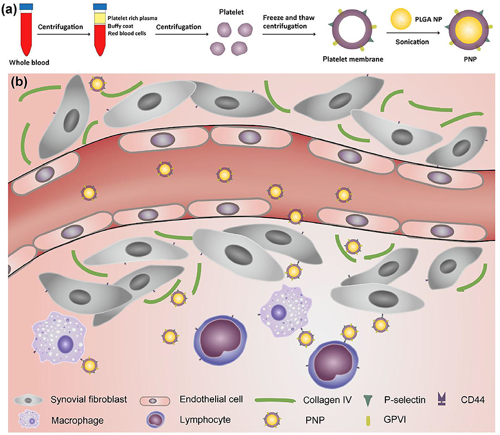 |
Figure 10 Schematic illustration of the targeted delivery of PNPs to the inflamed joint in RA. (a) Process used for the preparation of platelet membrane-coated PLGA nanoparticles. (b) Targeted delivery of PNPs in the inflamed joint via enhanced permeability and specific binding of PNPs to collagen and overexpressed CD44 on some cells of the inflamed joint tissue. Reprinted with permission from He Y, Li R, Liang J, et al. Drug targeting through platelet membrane-coated nanoparticles for the treatment of rheumatoid arthritis. Nano Research. 2018;11(11):6086–6101. Copyright 2018 Nano Research.106 |
Microbial Infection
The emergence of antimicrobial resistance (AMR) and multidrug resistance (MDR) has become a significant health problem and a major challenge in recent years.108 The prevalence of MDR bacteria has led to an increase in the morbidity and mortality rates associated with bacterial infections.109,110 Consequently, it is imperative to develop efficient techniques for quickly diagnosing bacteria111 and treatments that can bypass bacterial resistance mechanisms,112,113 and enhance the effectiveness of current antibiotics. Nanotechnology is a valuable tool for achieving these goals.114 Recent data have shown that platelets play a crucial role as sentinel effector cells in infectious illnesses.115–117 The innate immune response to invading pathogens is significantly affected by the crosstalk with platelets. Platelets have the ability to detect and respond to danger signals and can direct leukocytes to locations where injury, inflammation, or pathogen invasion has occurred.118–121 Kim et al investigated the application of human platelet membrane–cloaked nanoparticles (PNPs) as a biomimetic decoy technique to counteract S. aureus toxins and maintain the defensive activities of host cells.122 The PNPs prevented platelet damage caused by toxins released by S. aureus, thereby promoting platelet activation and bactericidal action.122 In addition, the PNPs prevented damage to macrophages caused by toxins generated by S. aureus. This helped enhance the macrophage oxidative burst, production of nitric oxide, and killing of bacteria. Furthermore, it reduced the release of neutrophil extracellular traps triggered by methicillin-resistant strains.122 Using ultrasound, Wu et al developed a potent adjuvant-like biomimetic nanovesicle to store meropenem-loaded polyamidoamine (PAMAM) in platelet membrane vesicles. This nanovesicle has the ability to effectively overcome carbapenem resistance in New Delhi metalloβ-lactamase–producing clinical isolates and treat severe bacterial infections.123 The nanovesicles effectively target bacterial infection sites and deliver PAMAM and meropenem drugs. They also rapidly release inflammatory microenvironment–powered drugs that help reduce the development of resistance to pathogens. This approach can be used to successfully treat various bacterial infections without systemic toxicity.123
Tumor Inflammatory Microenvironment
Over the past decade, cancer has been one of the primary causes of mortality worldwide.124 Chemotherapy, a widely used approach for cancer treatment, is hindered by its limited capacity to specifically target cancer cells and its significant side effects.125,126 To address these issues, extensive research and development has focused on targeted drug-delivery systems (TDDS), particularly those based on NPs.127 NPs offer several benefits, including a large capacity for drug loading, tunable physiochemical properties, and flexible modification. These properties make NPs suitable for encapsulating anti-cancer medications, which may then be modified in terms of solubility, stability, and in vivo behavior.128 Furthermore, the surface modification of NPs could prolong their presence in the bloodstream and enable precise targeting, thereby enhancing their effectiveness while reducing adverse effects.129,130 Recent studies have demonstrated that platelets play a critical role in promoting the metastatic progression of cancer through various mechanisms. These include facilitating tumor angiogenesis, aiding tumor survival in the bloodstream, and promoting the interaction between tumor cells and blood vessels.131 Researchers have created bionic methods for targeted drug delivery, considering the intimate interplay between platelets and tumor metastasis. Mai et al created platelet membranes (PMs) to serve as nanocarriers for the simultaneous encapsulation of metformin and IR780.132 These nanocarriers successfully corrected tumor hypoxia by inhibiting mitochondrial respiration. The attachment between the PM and tumor cells causes a prolonged circulation lifetime and higher accumulation of IR780 and Met in tumors.132 Shang et al developed a nanoplatform consisting of black phosphorus quantum dots (BPQDs) disguised with a platelet (PLT) membrane containing hederagenin (HED), named PLT@BPQDs-HED.133 PLTm vesicles act as a protective covering to encapsulate many drug-loaded nanocores. These nanocores can specifically target tumor locations and greatly enhance antitumor activity.133 The platform markedly decreased the survival of tumor cells and the mitochondrial membrane potential (MMP), while simultaneously enhancing the generation of intracellular ROS.133 Li et al developed NPs called MSN@PM-C-A by coating mesoporous silicon nanoparticles (MSNs) with platelet membranes. These NPs were designed to simultaneously deliver a vascular disruption agent, combretastatin A4 (CA4), and an anti-angiogenic drug, apatinib (Apa), for the combination treatment of tumors (Figure 11).134 The customized NPs gathered in tumor tissues by specifically adhering to damaged vessel sites through the platelet membrane surface. This leads to a notable disruption of blood vessels and the effective prevention of new blood vessel formation in animal models.134
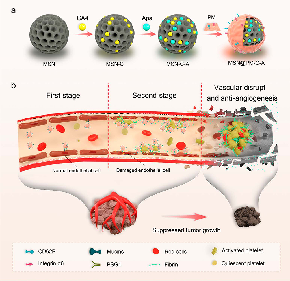 |
Figure 11 Illustration of the design principles and proposed action mechanism of MSN@PM-C-A in tumor vessels. (a) The preparation of MSN@ PM-C-A containing blank MSN (mesoporous silica nanoparticle) synthesis, drug loading, and platelet membrane coating. (b) Proposed antitumor mechanism of MSN@PM-C-A in the tumor-bearing mouse model. The platelet membrane camouflage endows nanoparticles with the ability to escape early systemic clearance as well as tumor targeting realized by receptor−ligand interactions between platelet membranes and vasculature endothelial cells (first-stage); the more platelet-membrane-camouflaged nanoparticles have a tropism for damaged vascular endothelium (disrupted vessels) and secondary microthrombosis induced by CA4, with release of Apa drugs (second stage). Therefore, in the tumor sites, the nanoparticles locally impaired the vascular endothelium and the more nanoparticles are persistently recruited to the sites of vasculature damages, engendering a mutually facilitated intratumoral vascular disruption and anti-angiogenesis, consequently exerting their antitumor effects by shutting off tumor blood supply. Reprinted with permission from Li B, Chu T, Wei J, et al. Platelet-Membrane-Coated Nanoparticles Enable Vascular Disrupting Agent Combining Anti-Angiogenic Drug for Improved Tumor Vessel Impairment. Nano Lett. 2021;21(6):2588–2595. Copyright 2021 Nano Letters.134 |
Engineering Platelet Membrane–Based Nanotherapeutics for Inflammatory Diseases
Atherosclerosis
Cardiovascular diseases (CVDs) are the primary cause of mortality worldwide.135,136 Atherosclerosis is the primary aetiology of CVDs, characterized by the gradual accumulation of plaque in the arteries, resulting in the constriction of the artery and ultimately leading to life-threatening complications.137,138 Recent findings indicate that the approach to treating atherosclerosis has changed to focus more on anti-inflammatory medication.139,140 There is strong evidence that vascular inflammation is intimately linked to the worsening of problems related to atherosclerosis.141,142 Platelets, a type of immune cell, have an innate affinity to sites of inflammation, such as atherosclerotic plaques, making them suitable for delivering anti-inflammatory medicines.26–28 MCC950, an NLRP3-inflammasome inhibitor that prevents the production of Interleukin-1β (IL-1β) caused by NLRP3 activator,143 has been shown to decrease the formation of atherosclerotic lesions and alleviate inflammation.144–146 Ma et al produced nanosized MCC950 loaded platelet-derived extracellular vesicles (MCC950-PEVs) by introducing MCC950 onto platelet extracellular vesicles (PEVs) and subsequently activating them.147 Administering MCC950-PEVs intravenously can effectively decrease the development of atherosclerotic plaques, decrease inflammation in the affected area, and hinder the proliferation of macrophages and T cells at the location of the plaque.147
Ischemic Disease
Thrombi
Thrombotic events, specifically cardiovascular-related illnesses,62 ischemic stroke,148 and pulmonary embolism (PE),149 are the primary causes of illness and death globally.150 Thrombus formation at specific locations can cause a sudden blockage of blood vessels, resulting in significant harm to tissues and potentially leading to organ failure, which can be life-threatening.151 Platelets preserve the structural integrity of blood vessels under normal circumstances and participate in the development of blood clots in abnormal situations. Platelets and platelet-like bionanoparticles have the potential for stroke detection, atherosclerosis treatment, and targeted thrombolysis owing to their roles in thrombus targeting and immune evasion.32,48,152 Xu et al created polymeric NPs coated with platelet membranes and attached rtPA to the surface. This design, called PNPPA, aims to deliver clot-targeted thrombolytic therapy.153 The engineered nanoplatelets effectively gather at the blood clots in mice, resulting in a notable enhancement in thrombolytic activity when compared with free rt-PA.153 Wang et al developed a platelet-based delivery system that combined urokinase (uPA) and arginine (Arg) to specifically dissolve blood clots and prevent their reformation. This approach involves attaching thrombolytic medicines to the surface of platelets using specialized engineering techniques.154 This engineered platelet-based drug-delivery system precisely delivers uPA to blood clots, extends the circulation of uPA in the bloodstream, and prevents the recurrence of blood clots while minimizing the risk of bleeding.154
Myocardial Infarction
Acute myocardial infarction (AMI), primarily resulting from the blockage of a coronary artery, is a significant global cause of mortality and impairment.155 An AMI triggers a sterile inflammatory reaction that promotes additional damage to the heart and contributes to negative changes in cardiac structure and function.156 IL-1β is a key factor in the sterile inflammatory response caused by AMI.156 Li et al created platelet microparticles (PMs) equipped with anti-IL-1β to counteract the effects of IL-1β during AMI and hinder unfavorable cardiac remodeling.156 Their findings indicate that the infarct-targeting PMs have the ability to attach to the damaged heart, enhancing the concentration of anti-IL-1β antibodies in that area. The anti-IL-1β platelet microparticles (IL1-PMs) protect the cardiomyocytes against apoptosis by neutralizing IL-1β and reducing the activity of caspase-3, which is driven by IL-1β.156
Rheumatoid Arthritis
Rheumatoid arthritis (RA) is a chronic inflammatory condition characterized by inflammation of the synovial membrane and damage to the joints.157,158 Biological products and kinase inhibition drugs have significantly enhanced the effectiveness of RA treatment.159 However, their efficacy in treating chronic synovial inflammation and joint deterioration must be improved.160,161 Synovial fibroblasts, which are stromal cells located in the synovium, play a crucial role in controlling the advancement of RA by generating several disease-causing proteins.162,163 Platelet microparticles (PMPs) are generated by activated platelets and have a significant impact on inflammatory disorders.164 Joints affected by arthritis and blood samples of patients with RA have revealed abundant PMPs.165 PMPs enhance inflammation of the synovial tissue by releasing interleukin-1 (IL-1) after attaching to RA synovial fibroblasts (RASFs).107 The results indicate that PMPs have a strong attraction to areas of inflammation and synovial fibroblasts in individuals with RA. All-trans retinoic acid (ATRA) is a vitamin A derivative that has been demonstrated to have anti-inflammatory effects in many animal models of RA.166 Furthermore, ATRA has the capability to disrupt the Golgi apparatus.167 Research has shown that specifically targeting the Golgi organelles of target cells with ATRA can successfully damage this organelle, leading to a decrease in protein secretion and expression.168–170 A peptide that targets the Golgi apparatus and contains the SXYQRL sequence can precisely localize to the Golgi apparatus.171 Deng et al hypothesized that modifying the Golgi-targeting peptide could result in the localization of PMP membrane (PMM)-camouflaged NPs in the Golgi of RASFs.172 The researchers developed Golgi-targeting PMM-camouflaged nanoparticles (Gol-PMMNPs) to specifically deliver ATRA to the Golgi organelles of RASFs in arthritic joints (Figure 12).172 The ATRA-Gol-PMMNPs significantly decreased the accumulation of harmful proteins in RASFs by damaging the Golgi organelles. This reduced the level of inflammation in synovial tissue and prevented damage to arthritic joint cartilage in rats with collagen-induced arthritis (CIA).172
 |
Figure 12 Schematic representation of ATRA-Gol-PMMNPs targeting the multiple pathogenic mediators produced by RASFs via integrin α2β1-mediated endocytosis and retrograde transport from endosomes to the Golgi apparatus. The white cross signifies the blockade of pathogenic proteins, chemokines, and cytokines expression in RASFs by ATRA-Gol-PMMNPs, consequently alleviating synovial inflammation, bone destruction and inflammatory migration in the arthritic joints of CIA rats. Reprinted with permission from Deng C, Zhao X, Chen Y, et al. Engineered Platelet Microparticle-Membrane Camouflaged Nanoparticles for Targeting the Golgi Apparatus of Synovial Fibroblasts to Attenuate Rheumatoid Arthritis. ACS Nano. 2022;16(11):18430–18447. Copyright 2022 ACS Nano.172 |
Pneumonia
Acute pneumonia is a severe respiratory tract infection caused by viruses, bacteria, fungi, or other pathogens. It is a leading cause of death globally.173 Mounting data indicates that individuals with severe pneumonia experience cytokine storm syndrome, a condition in which the immune system’s response results in unregulated inflammation of the lung tissue.174 Previous studies and clinical findings have demonstrated that despite the total eradication of the germs responsible for pneumonia using antibiotics or antiviral medications, individuals with severe pneumonia are still at high risk of succumbing to a cytokine storm in the lungs.175 Suppressing the cytokine storm could potentially be pivotal in preserving the lives of individuals suffering from severe pneumonia caused by a highly virulent strain such as SARS-Cov-2.176–179 Cellular delivery technologies have garnered significant attention owing to their exceptional biocompatibility and distinctive delivery characteristics.180–184 Platelets are inherently attracted to the site of inflammation.26–28 They have the ability to attach to the activated or inflamed vascular walls by interacting with several receptor patterns, such as CD40L, glycoproteins Iba, aIIb, and VI, as well as P-selectin.26–28 5-(p-Fluorophenyl)-2-ureido thiophene-3-carboxamide (TPCA-1) is a powerful and specific inhibitor of IkBkinases (IKK) in the NF-κB pathway. It has the ability to prevent the generation of TNF-a, IL-6, and IL-8 from human monocytes.185,186 Ma et al developed PEVs specifically designed for delivering target drugs to treat pneumonia.187 The TPCA-1 was loaded onto the platelets, which was followed by platelet activation in order to produce the TPCA-1-PEVs.187 The engineered PEVs have the ability to selectively target the inflamed lung tissue. Through injection, the nanosized PEVs are able to cross the vasculature and target the damaged area. This is achieved by exploiting the transient dilation and increased permeability of blood vessels generated by histamine in response to injury.188 In comparison to the free-drug-treated group, the PEVs significantly enhance treatment efficacy by inhibiting the infiltration of inflammatory cells in the lungs and mitigating the cytokine storm through the delivery of anti-inflammatory medications such as TPCA-1, which can decrease the generation of inflammatory components.187 Dexamethasone (DEX), a corticosteroid with anti-inflammatory properties, is commonly employed in clinical settings to mitigate inflammatory reactions.189 Nevertheless, the conventional method of administering DEX through direct injection is linked to numerous adverse effects, including hepatotoxicity and immunosuppression.190 Ma et al created a platform that specifically targets acute pneumonia using PEVs to deliver DEX.191 DEX can be readily loaded onto PEVs through hydrophobic interactions, forming DEX-loaded PEVs (DEX-PEVs).191 DEX-PEVs have the potential to significantly enhance the therapeutic effects of DEX by boosting the amount of the drug delivered to the inflammation site.191 The method reduced not only the occurrence of adverse effects but also the required dosage of DEX to a significant extent.191
Tumor Inflammatory Microenvironment
Surgery is the primary therapeutic approach for most solid tumors. Although surgical procedures have been consistently improving, the presence of persistent microtumors and/or circulating tumor cells (CTCs) following tumor removal continues to be a difficult problem.192–194 Cancer immunotherapy seeks to activate the immune system to fight cancer cells. This approach has garnered significant interest and has yielded remarkable results in clinical treatment.195,196 Tumor immunotherapy shows considerable potential in reducing the growth of primary tumors and tumor metastasis by utilizing the abilities of activated immune cells to recognize and eliminate cancer cells.197–199 Scientists are using platelets as effective delivery platforms in tumor immunotherapy.200 Engineered platelets possess significant promise to enhance the antitumor effects of immunotherapy and broaden their therapeutic uses.200 Wang et al attached anti-PDL1 (engineered monoclonal antibodies that target programmed-death ligand 1) to platelets to provide a preventive therapy for post-surgical cancer recurrence.201 They proposed that platelets can assist in transporting anti-PDL1 to remaining microtumors at the surgical site, as well as to CTCs in the bloodstream.201 Platelet activation resulted in the formation of a T-cell–inflamed tumor microenvironment, in turn leading to an upregulation of PDL1 expression at the tumor site.201 The release of Anti-PDL1 was successful following platelet activation, resulting in a blocking of PDL1 on both cancer cells and antigen-presenting cells (APCs).201 Fan et al developed an engineered platelet loaded with anti-CD47 to serve as a natural carrier for delivering anti-CD47 to tumors for macrophage immunotherapy. These NPs have a specific affinity for bacteria-treated tumors.202 Anti-CD47 were attached to purified blood platelets via a click reaction.203 Following the induction of rapid and extensive blood coagulation and inflammation in tumor tissues by Escherichia coli TOP10, the macrophages that are recruited are mostly polarized towards the antitumor M1-like phenotype by lipopolysaccharide in the bacterial wall.202 Anti-CD47 inhibits the interaction of CD47 and signal regulatory protein-α (SIRP-α) in M1-like phenotype macrophages, enhancing the digestion of polarized M1-like phenotype macrophages for tumor cells.202
Hybrid Platelet Membrane–Based Nanotherapeutics for Inflammatory Diseases
Atherosclerosis
Atherosclerosis, a persistent inflammatory condition characterized by the narrowing of blood arteries and the consequent limitation of blood circulation, is the primary cause of cardiovascular disorders and accounts for approximately one-third of global mortality.136 It is characterized by damage to endothelial cells, accumulation of lipids, and aggregation of inflammatory cells.204 Macrophages are the predominant leukocyte subset seen in plaques and have crucial functions in both the onset and advancement of atherosclerosis.205 During the initial stage of atherosclerosis, monocytes are drawn towards the damaged vascular cells and migrate to the arterial wall. Once there, they differentiate into pro-inflammatory macrophages.206 The macrophages with inflammatory properties secrete significant amounts of inflammatory-related cytokines, chemokines, and ROS, intensifying oxidative stress within the plaques.207 Macrophages constantly consume copious amounts of cholesterol derived from low-density lipoprotein (LDL), resulting in their transformation into foam cells. This transformation initiates a chronic inflammatory state associated with atherosclerosis.208 Under normal physiological conditions, platelets primarily circulate in close proximity to the walls of blood vessels without interacting with endothelial cells (ECs). Platelets quickly adhere to the damaged endothelium when the integrity of the endothelial layer is compromised.209,210 Mounting research has demonstrated that mesenchymal stem cells (MSCs) provide protection throughout all phases of atherosclerosis.211 MSCs have the ability to minimize endothelial dysfunction,212 polarize macrophages into anti-inflammatory phenotypes to decrease inflammation, and reduce the generation of foam cells.213,214 These actions help prevent the development of atherosclerosis and improve the stability of advanced atherosclerotic lesions.215 Li et al created platelet mimetic MSC-EVs (P-EVs) by adorning extracellular vesicles with platelet membranes to specifically treat atherosclerosis. P-EVs possess the inherent potential to target plaques, similar to platelets, and also exhibit the therapeutic benefits of MSC-EVs.216 Following this, the P-EVs conveyed miRNAs to the cytoplasm of macrophages by escaping from lysosomes. This process ultimately slows the development of arterial plaque buildup by transforming the macrophages into an anti-inflammatory phenotype, removing excessive ROS and decreasing lipid accumulation in the lesional macrophages (Figure 13).216 Song et al created biomimetic liposomes (P-Lipo) by hybridizing artificial liposomes with platelet membranes. This design is influenced by the manner in which platelet membrane components engage with atherosclerotic plaques, and the goal is to target atherosclerosis.217 The extrusion method is used to fuse natural platelet membranes with artificial lipid membranes, resulting in the formation of hybrid nanovesicles. These nanovesicles are believed to possess the targeted abilities of platelets as well as the physicochemical and pharmaceutical attributes of liposomes.217 Because the lipid bilayer of liposomes is easily fused with other bilayers, such as cell membranes, and owing to their highly fluid and dynamic features,218 P-Lipo can be scaled up and effectively maximize the utilization efficiency of platelet membranes.217 P-Lipo offers a secure and effective alternative for treating atherosclerosis and various other disorders associated with platelet involvement.217
 |
Figure 13 Schematic diagram of targeted delivery of P-EVs to plaques for the treatment on atherosclerosis. The green upward arrow indicates upregulation of the relevant process or factor expression, while the green downward arrow represents downregulation. Reprinted with permission from Li Q, Huang Z, Pang Z, et al. Targeted delivery of platelet membrane modified extracellular vesicles into atherosclerotic plaque to regress atherosclerosis. Chemical Engineering Journal. 2023; 452:138992. Copyright 2023 Chemical Engineering Journal.216 |
Ischemic Disease
Thrombi
Blood vessels can be suddenly blocked owing to thrombus formation at certain locations. This can result in significant tissue damage and even organ failure, a potentially fatal condition.151 The pathological interaction between oxidative stress, inflammation, and thrombosis induced by endothelial injury establishes a self-reinforcing cycle that compromises standard thrombolytic treatments. Jiang et al developed near-infrared (NIR) light-responsive nanomotors by combining thylakoid and platelet membranes with strontium-doped mesoporous polydopamine NPs. This allowed for the simultaneous dissolution of thrombi and restoration of the vascular microenvironment. After intravenous administration, the nanomotors preferentially gathered in the thrombi and were taken up by the damaged endothelial cells. Simultaneously, the thylakoid-embedded catalase converted hydrogen peroxide into oxygen. This not only prevents oxidative damage and platelet-endothelium interactions but also causes self-propulsive forces through gas propulsion, which helps the nanomotors penetrate the thrombus. Local NIR irradiation caused a small amount of photothermal conversion in the nanomotors, which softened the fibrin networks, facilitating intrathrombus infiltration, and accelerated localized thrombolysis.219
Ischemic Stroke
Stroke ranks as the second most prevalent cause of mortality on a global scale.220,221 Vascular occlusion leads to neuronal apoptosis, breakdown of the BBB, and necrosis of cerebral tissue, causing significant functional impairment in patients.222 Microglia is a form of neuroglia found throughout the central nervous system (CNS).223 As the first cells to be activated upon CNS damage, microglia are crucial for host defense, inflammatory modulation, neurodegeneration, and tissue repair.224 During ischemia/reperfusion injury, two distinct polarized phenotypes of microglia are prevalent: the classical M1 (pro-inflammatory) and the alternative M2 (anti-inflammatory).225 These phenotypes have specific roles in the injury process. M1 microglia increase the expression of pro-inflammatory cytokines, which worsen neuronal damage and hinder cellular repair. On the other hand, the M2 phenotype provides neuroprotection and supports brain recovery by producing anti-inflammatory cytokines.226 Li et al created a platelet hybrid microglia platform that can specifically polarize microglia into an anti-inflammatory phenotype using ultrasound irradiation. This platform is designed for targeted healing of the cerebrum following a stroke (Figure 14).227 The engineered microglia exhibit robust adhesion to damaged cerebral blood arteries through fusion with platelet membranes and achieve targeted anti-inflammatory polarization with ultrasound-responsive IL-4 liposome decoration.227 The microglial platform, when injected intravenously and subjected to insonation, exhibits anti-inflammatory polarization at the site of stroke. This platform has the ability to specifically polarize towards the desired phenotype for brain tissue regeneration and to accelerate M2 polarization of the endogenous microglia. This polarization process promotes the formation of vascular and neuronal structures, leading to improved behavior in mice affected by stroke.227
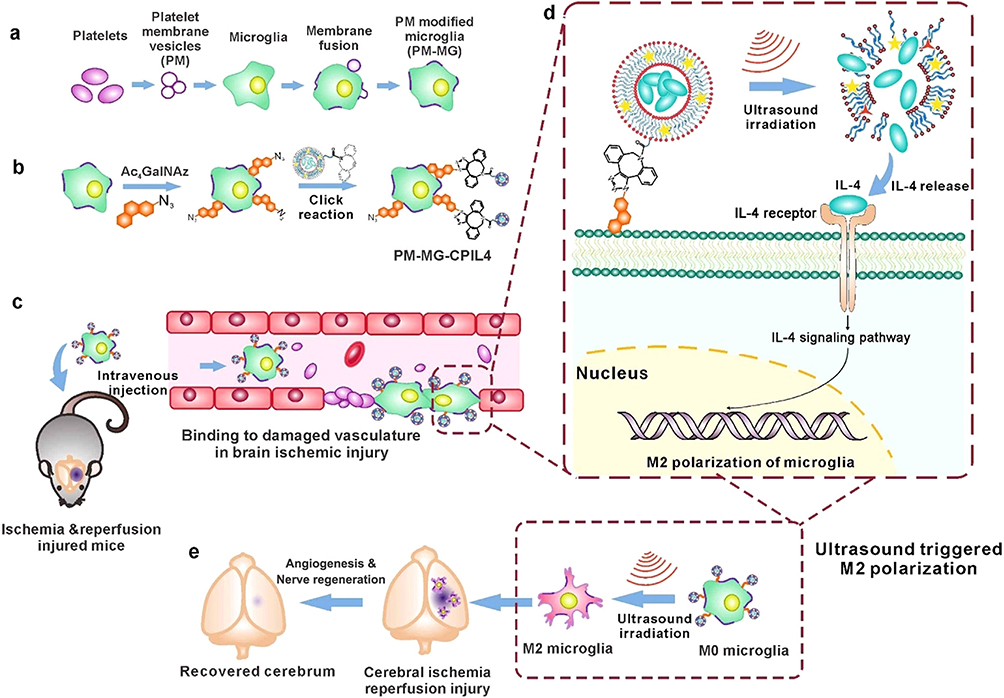 |
Figure 14 Schematic illustration of ultrasound triggered polarization of platelet fused microglia for targeted treatment of ischemia-reperfusion injury post stroke. (a) Platelet membrane vesicle generation and decoration on M0 microglia (MG) to form PM decorated MG (PM-MG) by membrane fusion. (b) Ultrasound responsive liposome coated IL-4 (CPIL4) is conjugated to PM-MG via click reaction to form PM-MG-CPIL4. (c) Tail vein injected PM-MG-CPIL4 can anchor to the injured cerebrovascular for targeted repair. (d) Upon ultrasound irradiation, IL-4 directly releases from the CPIL4 and subsequently binds to the IL-4 receptor on the cell surface to initiate M2 polarization of microglia through IL-4 signaling pathway. (e) M2 polarized microglia contribute to the angiogenesis and nerve regeneration of the injured brain post ischemic stroke and reperfusion injury. Reprinted with permission from Li Y, Teng X, Yang C, et al. Ultrasound Controlled Anti-Inflammatory Polarization of Platelet Decorated Microglia for Targeted Ischemic Stroke Therapy. Angew Chem Int Ed Engl. 2021;60(10):5083–5090. Copyright 2021 Angew Chem Int Ed Engl.227 |
Myocardial Ischemia Reperfusion
Ischemic cardiocerebrovascular disease, which includes MI and stroke, has significantly contributed to mortality rates over the last two decades. Even though prompt blood-flow restoration has greatly decreased immediate mortality, reperfusion does not seem to be risk-free, and some patients with MI and stroke either do not improve or worsen after reperfusion owing to reperfusion injury.228,229 Microcirculation in the body’s tissues is particularly vulnerable to the consequences of reperfusion, resulting in microvascular dysfunction. This dysfunction includes the breakdown of the endothelial barrier with hemorrhage, as well as impaired capillary perfusion (no-flow).230 Immediately following ischemia reperfusion, VE-cadherin, a highly important intercellular connection, rapidly disassembles and degrades. Meanwhile, endothelial cells experience significant damage as a result of the increased accumulation of ROS.231 These two factors work together shortly after the beginning of ischemia reperfusion, leading to impaired barrier function, which causes increased infiltration of proteins and cells and triggers severe bleeding and inflammation.210 Gao et al developed biomimetic NPs fused with the platelet membrane, which combine angiopoietin-like 4 (ANGPTL4) with liposomes encapsulating a thrombin-responsive peptide and Fe3O4. These NPs were designed to exploit the natural tendency of platelets to migrate to the injured endothelium and locally activate thrombin. Its purpose was to deliver both ANGPTL4 and Fe3O4, which act as ROS scavengers, to the damaged endothelium in the heart, which is affected by myocardial ischemia-reperfusion (MI/R).210 The NP effectively reached the endothelial cells as a result of platelet-mimetic adhesion, which was created by merging platelet membranes and lipid membranes.210 After the damaged endothelium causes thrombin-responsive peptide cleavage, ANGPTL4 attaches to endothelial cell receptors to prevent VE-cadherin junction rupture. In addition, endo-lysosomal escape of Fe3O4 to cytosol effectively scavenged ROS and prevented apoptosis in endothelial cells.210 The preservation of VE-Cadherin and the rescue of cells effectively maintained the integrity of the endothelial barrier, resulting in a reduction in leukocyte extravasation and intracardial hemorrhage.210 This action also reduced cardiomyocyte apoptosis and ultimately improved cardiac function.210 Zhou et al produced biomimetic nanocomplexes based on inflammation-sheddable cloaking using a platelet–macrophage hybrid membrane in order to effectively transfer Sav1 siRNA into cardiomyocytes and block the Hippo pathway while promoting cardiomyocyte regeneration.232 To mitigate MI/R injury after coronary recanalization in MI-affected hearts, He et al engineered NPs inspired by stromal-platelet membranes. These NPs feature a poly (lactic-co-glycolic acid) (PLGA) core, adorned with a dual membrane coating: a platelet membrane for precise adhesion to the compromised endothelial region and a stromal cell membrane to augment receptor–ligand interactions and confer immune evasion. This unique dual-membrane configuration reduces fibrosis and inflammation while encouraging angiomyogenesis.233
The strong inflammatory cascade that follows MI/R injury is a crucial step in the process of cardiac remodeling.234 Early after injury, inflammatory macrophages (M1) penetrate the damaged area and release a variety of cytokines along with phagocyte debris.235,236 Multiple investigations have shown that the timely transition to a reparative phenotype (M2) is essential for resolving inflammation.236–239 Tan et al created platelet-like fusogenic liposomes (PLPs) by drawing inspiration from the increase in circulating platelet–monocyte aggregates in patients after MI/R. They chose fusogenic liposomes for their ability to effectively carry their contents to the cytoplasms of target cells. Mesoporous silica nanospheres containing miR-21, an anti-inflammatory drug, can be targeted to inflammatory monocytes in the bloodstream of MI/R-induced mice by means of coating with PLPs.240 Subsequently, they undergo membrane fusion to enter the cytoplasms of monocytes, leading to reparative reprogramming of the inflammatory macrophages formed from it.240
Sepsis-Induced Acute Kidney Injury
Sepsis-induced acute kidney injury (SAKI) is a prevalent complication observed in critically ill patients and is characterized by rapid deterioration in renal function. It accounts for 60% of severe sepsis cases.241,242 Patients with SAKI have a considerably greater mortality risk than those with acute kidney injury (AKI) alone.243,244 Although the precise mechanisms of SAKI remain incompletely understood, they mostly contribute to microcirculation disturbances, immunological reactions, and cellular malfunctions.245 Microcirculatory disfunction can be caused by various mechanisms, including endothelial injury, autonomic nervous system reaction, shedding of the glycocalyx, and activation of the coagulation cascade.246 During sepsis, toxins in the bloodstream affect the vascular endothelium, causing a decrease in microcirculatory blood flow and the release of several inflammatory substances. This leads to damage to the microvascular endothelial barrier and loss of the glycocalyx.247 Yang et al developed a new form of hybrid microbubble by coating platelet membranes (Pla-MBs), considering the characteristics of endothelial injury in the early stage of SAKI and the quick adhesion of platelets to the damaged endothelium layer (Figure 15).247 Pla-MBs were created using ultrasound to recombine liposomes and platelets, which contained the natural platelet membrane obtained from isolated platelets. Pla-MBs were given different adhesive receptors, including integrin αIIbβ3, by applying a platelet membrane coating. This allows them to selectively adhere to the injured endothelium in SAKI.247 By utilizing the inherent properties of the platelet membrane, molecular ultrasound imaging, and quantitative analysis, Pla-MBs can specifically identify microbubble signals that bind to damaged blood vessels, enabling the early detection of SAKI in rats. Bedside sonographic devices may offer a sensitive and specific technique for the early detection of SAKI, which can help prevent and control SAKI and reduce mortality in septic patients.247 Zhang et al created a PM hybrid rapamycin liposome that not only facilitates rapamycin delivery but also harnesses the ability of PM to make long-lasting repairs. The intravenous administration of the NPs demonstrated their dual properties, including swift anti-inflammatory effects and enduring tissue-repair capabilities. Mechanistic studies have demonstrated that the NPs significantly contribute to the activation of autophagy and polarization of macrophages, consequently decreasing renal cell apoptosis and lowering pro-inflammatory cytokine levels after ischemia-reperfusion injury (IRI). Hybridization with PM seems to help reshape damaged nephron units by encouraging the recruitment and differentiation of CD34+ cells.248
 |
Figure 15 Schematic diagram of the preparation of Pla-MBs and kidney-targeted molecular ultrasound imaging in vivo. (a) Preparation of Pla-MBs. (b) Molecular ultrasound imaging of sepsis-induced acute kidney injury via Pla-MBs. Reprinted with permission from Yang J, Miao X, Guan Y, et al. Microbubble Functionalization with Platelet Membrane Enables Targeting and Early Detection of Sepsis-Induced Acute Kidney Injury. Adv Healthc Mater. 2021;10(23): e2101628. Copyright 2021 Adv. Healthcare Mater.247 |
Nonalcoholic Steatohepatitis
Nonalcoholic fatty liver disease (NAFLD) is a prevalent liver condition that affects approximately 30% of adults globally and 80% of individuals with obesity or diabetes.249,250 NAFLD encompasses a spectrum of conditions ranging from mild steatosis to more severe nonalcoholic steatohepatitis (NASH), which can progress to varying levels of liver fibrosis. While individuals with mild steatosis may not always acquire a more severe condition, NASH has the potential to advance to cirrhosis or even liver cancer.251 NASH is distinguished by the presence of insulin resistance, steatosis, and necroinflammation, which may or may not be accompanied by centrilobular fibrosis.252 Xie et al developed a novel drug delivery system utilizing a hybrid membrane composed of platelet membranes and neutrophil membranes that were coated with gelatin NPs containing vitamin E and pioglitazone (PNM-G).253 Pioglitazone effectively enhances insulin resistance and liver histology in patients diagnosed with NASH.254 Vitamin E can interact with free radicals, eliminating a significant amount of ROS in the liver. It also hinders the oxidation of unsaturated fatty acids by blocking this process. Additionally, vitamin E can enhance the mitochondrial membrane potential, which in turn inhibits the generation of ROS in the mitochondria. This protective action effectively safeguards liver cells and reduces the harmful effects of the second hit. Vitamin E alleviates steatosis, inflammation, ballooning, and steatohepatitis in individuals diagnosed with NASH.255–259 Functionalizing the biomimetic surfaces of gelatin NPs with PNM-G can improve their ability to target inflammatory areas and enrich the liver tissue.253 PNM-G possesses immune-escape capabilities and dual NASH-targeting functionality.253 PNM-G presents a promising therapeutic alternative to NASH by utilizing an innovative method of administering medication.
Microbial Infection
The use of NPs for treating infections is intriguing owing to treatment failures resulting from antimicrobial resistance and the dearth of new medications currently being developed. Furthermore, access to drugs is hampered by the fact that many infections occur inside cells in either active or latent form.260–262 NPs provide a viable solution for addressing these issues. Currently, there is a strong focus on their use as antimicrobial agents to combat various diseases, including bacteria, viruses, fungi, and parasites.110 Jin et al created a biomimetic phage-platelet hybrid nanoparticle (PPHN) by physically attaching the BCP1-BGL virus to platelet membrane NPs. BCP1-BGL phages and PPHNs were subjected to a range of trials to investigate their prolonged circulation and antibacterial abilities.263 Esteban-Fernández de Ávila et al published a study on the development of biomimetic nanorobots propelled by ultrasound. These nanorobots are composed of gold nanowires coated with a mixture of red blood cell (RBC) membranes and platelet (PLT) membranes. The purpose of these nanorobots is to perform multiple tasks, including biodetoxification and concurrent elimination of pathogenic bacteria and toxins.264
Tumor Inflammatory Microenvironment
Cancer therapies have long faced challenges in effectively targeting tumors and penetrating them at the intratumoral level.265 In order to optimize the accuracy of drug accumulation specifically at tumor locations, numerous delivery systems have been created to take advantage of the enhanced permeability and retention (EPR) effect, with a particular focus on nanoscale vectors.266,267 Biomimetic particles obtained from biological sources have garnered growing interest owing to their distinct benefits, such as enhanced biocompatibility, the ability to reduce immunogenicity, and the capability for active targeting.268,269 Platelets play a crucial role in the advancement and spread of cancer, particularly by recognizing and interacting with circulating tumor cells (CTCs) in the bloodstream.270,271
Platelets have the ability to specifically target cancers by attaching specific cognate signaling molecules on the surface of tumor cells and accumulating in them.272–274 These inherent physiological characteristics support the utilization of platelets as a feasible approach for designing targeted drug delivery to tumors and improving the ability of drugs to penetrate the tumor.275 Kim et al fabricated gold nanostars coated with red blood cells (RBC) and platelet membranes loaded with curcumin (named R/P-cGNS). This study showed that R/P-cGNS effectively transported medicines to their intended location, thereby improving anti-cancer efficacy. Additionally, R/P-cGNSs prevented macrophage phagocytosis and exhibited anti-inflammatory effects on macrophages.276 Hao et al initially synthesized gold nanocages (AuNCs) co-loaded with DOX and indocyanine green (ICG). Subsequently, the AuNC surface was decorated with a hybrid cell membrane derived from platelets and neutrophils, resulting in the formation of nanokillers (NSKs).277 NSKs exhibited enhanced cellular uptake, deeper infiltration into tumors, and increased toxicity towards tumor cells. They captured and eliminated circulating tumor cells (CTCs) and exosomes through high-affinity membrane adhesion receptors. This process effectively severed communication between immune cells and exosomes.277 In their study, Liu et al demonstrated that polypyrrole nanoparticles (PPy NPs) coated with fused RBC and PLT membranes could eliminate tumor cells when exposed directly to NIR irradiation. The resulting PPy NPs possessed the combined characteristics of RBC and PLT. They could circulate throughout the body for a lengthy period and demonstrated self-targeting properties.278 Zhang et al developed a biomimetic nanoplatform using a combination of a leukocyte/platelet hybrid membrane (LPHM) and dendritic large-pore mesoporous silicon nanoparticles (DLMSNs). This approach successfully combined photothermal therapy (PTT), photodynamic therapy (PDT), and chemotherapy to target triple-negative breast cancer (TNBC) and achieved synergistic effects.279 The large pores of the DLMSNs were used to co-load a NIR fluorescent dye, IR780, with DOX. This resulted in the formation of DLMSN@DOX/IR780 (DDI) NPs. These NPs were then coated with LPHM to create LPHM@DDI NPs (Figure 16).279 When exposed to a NIR laser, the LPHM@DDI NPs demonstrated a combined impact of synergistic cytotoxicity and apoptosis-inducing activity in TNBC cells. Additionally, they significantly inhibited tumor development and recurrence in TNBC mice by destroying the tumor and preventing the formation of new blood vessels.279
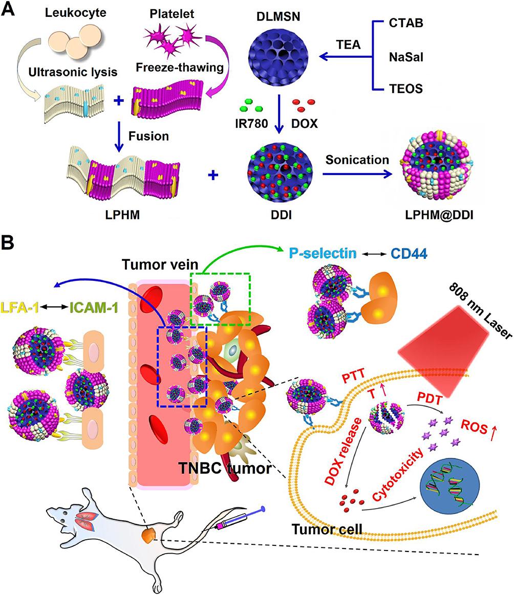 |
Figure 16 Schematic illustrations for preparation method of LPHM@DDI NPs (A) and their synergistic effects in TNBC treatment via combining PTT/PDT with chemotherapy (B). Blue dashed box: NPs target tumor vascular and enhance vascular permeability through activation of the LFA-1/ICAM-1 adhesion pathway. Green dashed box: Following extravasation, NPs specifically target to tumor cells via P-selectin/CD44 interactions. The red text “T” stands for temperature. The red upward arrow indicates upregulation of the corresponding elements. Reprinted with permission from Zhang T, Liu H, Li L, et al. Leukocyte/platelet hybrid membrane-camouflaged dendritic large pore mesoporous silica nanoparticles co-loaded with photo/chemotherapeutic agents for triple negative breast cancer combination treatment. Bioact Mater. 2021;6(11):3865–3878. Copyright 2021 Bioactive Materials.279 |
Conclusions and Prospects
This paper reviews the applications of nanotherapy based on natural platelet membranes, engineered platelet membranes, and platelet membranes hybridized with other cell membranes in the treatment of different inflammatory diseases. Bionic cell-membrane strategies have made significant progress in the field of nanomedicine. Disguised by a cell membrane, biomimetic NPs exhibit the characteristics of the cell membrane and the functional adaptability of the core nanomaterial. Unlike other carrier systems, natural cell membranes do not elicit immune responses. Moreover, surface constituents can engage with receptors on specific cells, enabling the accurate targeting and regulation of cargo release at the site of inflammation. By tailoring the origin of the membrane and the composition of the NP core, bionic NPs with distinct capabilities can be generated.
However, to turn this promising technology into a clinical therapy, we still need to define and overcome a series of challenges.
First, we face challenges related to the scale of production and associated costs. Achieving large-scale, low-cost production of platelet membrane–based nanomaterials that meet commercial and regulatory standards is a major bottleneck. This solution requires the development of simpler, more economical, and process-robust manufacturing techniques to ensure consistency across product batches.
Secondly, biocompatibility and long-term safety issues arise. Before introducing this membrane-coated system containing artificial and biological components into the human body, biocompatibility and safety must be ensured. This will involve a comprehensive assessment of the immunogenicity, potential toxicity, and metabolic pathways in the body.
Thirdly, issues related to membrane stability and functional integrity exist. The inherently low stability and degradability of platelets and their derived biological membranes pose challenges for maintaining their functions during transportation and storage. Therefore, preserving these biological materials for a long time without compromising membrane protein activity and function is crucial.
Finally, the targeted precision of platelets and their potential risks should be considered. Platelets and their biological membranes tend to aggregate in blood vessels, which presents a dual challenge. This tendency not only forms the basis of its targeted nature but may also limit its application in nonvascular-related inflammation and increase the risk of accidental accumulation at non-targeted vascular sites.
In the future, development in this field should focus on the following directions:
Precise engineering of membrane functions: In the future, we should not be satisfied with a simple membrane coating. Instead, through methods such as genetic engineering, chemical modification, or metabolic labeling, we should refine the engineering of membranes to enhance their targeting ability and loading capacity, or introduce environmentally responsive release mechanisms.
Developing intelligent hybrid membrane systems: Hybridizing the membranes of platelets with those of other cells (such as stem cells, immune cells, and red blood cells) is an effective strategy for enhancing the circulation time, active targeting ability, and additional therapeutic functions (such as immunomodulation and tissue repair) of biomimetic NPs. The key lies in optimizing the fusion process and preserving the functional integrity of each component of the cell membrane as much as possible.
Deepening translational medical research: To accelerate clinical translation, prioritizing research that conforms to translational standards is necessary. This includes systematically evaluating the pharmacokinetics, safety, and efficacy of drugs in larger animal models that are closer to clinical settings and actively collaborating with regulatory agencies to establish quality standards for such complex products.
Expanding application boundaries: In addition to their role as drug-delivery carriers, we should actively explore the use of platelet membrane NPs in diagnostic imaging (eg, as an imaging agent targeting inflammation) and immunomodulation (eg, as an “attractant” to neutralize inflammatory factors), among other applications.
In conclusion, nanotherapeutic technology based on platelet membranes has enormous potential for the treatment of inflammatory diseases. By focusing on addressing the challenges it faces in terms of large-scale production, safety, stability, and targeting, and by conducting in-depth studies on the aforementioned future directions, we can reasonably anticipate that platelet-based delivery systems are likely to evolve into next-generation intelligent drug carriers that meet various medical needs.
Abbreviations
AIS, acute ischemic stroke; AKI, acute kidney injury; AMI, Acute myocardial infarction; AMR, antimicrobial resistance; ANGPTL4, angiopoietin-like 4; Apa, apatinib; APC, antigen-presenting cell; ApoE(−/−) mice, Apolipoprotein E-deficient mice; Arg, arginine; ATRA, All-trans retinoic acid; AuNC, gold nanocage; BBB, blood-brain barrier; BPNS, black phosphorus nanosheet; BPQD, black phosphorus quantum dot; CA4, combretastatin A4; CaMKIIγ, Ca2+/calmodulin-dependent protein kinase γ; CHF, congestive heart failure; CIA, collagen-induced arthritis; CNS, central nervous system; CPIL4, liposome coated IL-4; CTAB, cetyl trimethyl ammonium bromide; CTC, circulating tumor cell; CVD, Cardiovascular disease; DDI, DLMSN@DOX/IR780; DEX, Dexamethasone; Dex-PEV, DEX-Loaded-PEV; DLMSN, dendritic large pore mesoporous silicon nanoparticle; DOX, doxorubicin; EC, endothelial cell; EDTA, Ethylenediaminetetraacetic acid; EPR, enhanced permeability and retention; EV, extracellular vesicle; FLS, fibroblast-like synoviocyte; FTY720, fingolimod; Gol-PMM, Golgi-targeting peptide modified PMM; Gol-PMMNP, Golgi-targeting PMM-camouflaged nanoparticle; GSH, glutathione; HED, hederagenin; ICAM-1, intercellular adhesion molecule-1; ICG, indocyanine green; IH, intimal hyperplasia; IKK, IkBkinases; IL-1, interleukin-1; IL1-PM, IL-1β platelet microparticle; IL-1β, interleukin-1β; I/R, ischemia/reperfusion; IRI, ischemia-reperfusion injury; i.v., intravenous injection; JAMS, Janus aminated mesoporous silica; LDL, low density lipoprotein; LFA-1, Integrin lymphocyte function-associated antigen-1; LPHM, leukocyte/platelet hybrid membrane; MCC950-PEV, MCC950 loaded platelet-derived extracellular vesicle; MDR, multidrug resistance; MG, microglia; MI, myocardial infarction; MI/R, myocardial ischemia-reperfusion; MMP, mitochondrial membrane potential; MNP, melanin nanoparticles; MSC, mesenchymal stem cell; MSN, mesoporous silicon nanoparticle; NAFLD, Nonalcoholic fatty liver disease; NaSal, sodium salicylate; NASH, nonalcoholic steatohepatitis; Nb2C, niobium carbide MXenes; NE, Neutrophil; NF-κB, Nuclear factor-κB; NIR, near-infrared radiation; NP, nanoparticle; NSK, nanokiller; PAMAM, polyamidoamine; PBS, phosphate buffer solution; PDT, Photodynamic therapy; PE, pulmonary embolism; PEV, platelets extracellular vesicle; Pla-MB, hybrid microbubble by coating platelet membrane; PLGA, poly lactic-co-glycolic acid; PLP, splatelet-like proteoliposomes; PLP, platelet-like fusogenic liposome; PLT, platelet; PM, platelet membranes; PMIN, PMP -inspired nanovesicle; PMM, PMPs membrane; PMP, platelet microparticle; PM-MG, PM decorated MG; PNPArg, platelet membrane-cloaked argatroban-loaded polymeric nanoparticle; PNP, platelet membrane-cloaked nanoparticle; PPHN, phage-platelet hybrid nanoparticle; PPy NP, polypyrrole nanoparticle; PRP, platelet-rich plasma; PTNP, platelet-mimetic nanoparticle; PTT, photothermal therapy; P-EV, platelet mimetic MSC-EV; P-Lipo, biomimetic liposomes; RA, Rheumatoid arthritis; RAP, Rapamycin; RASF, RA synovial fibroblast; RBC, red blood cell; ROS, Reactive oxygen species; rtPA, recombinant tissue plasminogen activator; SAKI, Sepsis-induced acute kidney injury; SIRP-α, signal regulatory protein-α; SOD, superoxide dismutase; SPIO, Small particle of iron oxide; TDDS, targeted drug delivery system; TEA, triethyl amine; TEOS, tetraethyl orthosilicate; TNBC, Triple-negative breast cancer; tPA, tissue plasminogen activator; TPCA-1, thiophene-3-carboxamide; TPHL, T7-PEG-PHis-PLys; uPA, urokinase; VEC, vascular endothelial cell; VSMC, vascular smooth muscle cell.
Data Sharing Statement
No datasets were generated or analyzed during the current study.
Acknowledgment
Boyuan Liu and Yongjie Wang are co-first authors for this study. Graphical Abstract is created in BioRender. Xiao, C. (2025) https://BioRender.com/wxfsdgh. We would like to thank Editage (www.editage.cn) for English language editing.
Author Contributions
All authors contributed to the study conception and design, reviewed the manuscript, and approved the final version. All authors made a significant contribution to the work reported, whether that is in the conception, study design, execution, acquisition of data, analysis and interpretation, or in all these areas; took part in drafting, revising or critically reviewing the article; gave final approval of the version to be published; have agreed on the journal to which the article has been submitted; and agree to be accountable for all aspects of the work.
Funding
We acknowledge the financial support from the National Natural Science Foundation of China (52103163), Graduate Innovation Fund of Jilin University (2024CX130 and 2024CX261), and Natural Science Foundation of Jilin province (YDZJ202401232ZYTS).
Disclosure
The authors declare that they have no conflicts of interest in this work.
References
1. Karin M, Clevers H. Reparative inflammation takes charge of tissue regeneration. Nature. 2016;529(7586):307–315. doi:10.1038/nature17039
2. Sonnenberg GF, Artis D. Innate lymphoid cells in the initiation, regulation and resolution of inflammation. Nat Med. 2015;21(7):698–708. doi:10.1038/nm.3892
3. Nasef NA, Mehta S, Ferguson LR. Susceptibility to chronic inflammation: an update. Arch Toxicol. 2017;91(3):1131–1141. doi:10.1007/s00204-016-1914-5
4. Tabas I, Glass CK. Anti-inflammatory therapy in chronic disease: challenges and opportunities. Science. 2013;339(6116):166–172. doi:10.1126/science.1230720
5. Dong X, Chu D, Wang Z. Leukocyte-mediated delivery of nanotherapeutics in inflammatory and tumor sites. Theranostics. 2017;7(3):751–763. doi:10.7150/thno.18069
6. Pawelec G, Goldeck D, Derhovanessian E. Inflammation, ageing and chronic disease. Curr Opin Immunol. 2014;29:23–28. doi:10.1016/j.coi.2014.03.007
7. Hou XL, Tong Q, Wang WQ, et al. Suppression of inflammatory responses by dihydromyricetin, a flavonoid from ampelopsis grossedentata, via inhibiting the activation of NF-κB and MAPK signaling pathways. J Nat Prod. 2015;78(7):1689–1696. doi:10.1021/acs.jnatprod.5b00275
8. Arulselvan P, Tan WS, Gothai S, et al. Anti-inflammatory potential of ethyl acetate fraction of moringa oleifera in downregulating the NF-κB signaling pathway in lipopolysaccharide-stimulated macrophages. Molecules. 2016;21(11). doi:10.3390/molecules21111452
9. Yang G, Phua SZF, Lim WQ, et al. A hypoxia-responsive albumin-based nanosystem for deep tumor penetration and excellent therapeutic efficacy. Adv Mater. 2019;31(25):e1901513. doi:10.1002/adma.201901513
10. Sauvage F, Fraire JC, Remaut K, et al. Photoablation of human vitreous opacities by light-induced vapor nanobubbles. ACS Nano. 2019;13(7):8401–8416. doi:10.1021/acsnano.9b04050
11. Gong N, Ma X, Ye X, et al. Carbon-dot-supported atomically dispersed gold as a mitochondrial oxidative stress amplifier for cancer treatment. Nat Nanotechnol. 2019;14(4):379–387. doi:10.1038/s41565-019-0373-6
12. Qin SY, Zhang AQ, Cheng SX, Rong L, Zhang XZ. Drug self-delivery systems for cancer therapy. Biomaterials. 2017;112:234–247. doi:10.1016/j.biomaterials.2016.10.016
13. Chen F, Wang G, Griffin JI, et al. Complement proteins bind to nanoparticle protein corona and undergo dynamic exchange in vivo. Nat Nanotechnol. 2017;12(4):387–393. doi:10.1038/nnano.2016.269
14. Wang G, Griffin JI, Inturi S, et al. In vitro and in vivo differences in murine third complement component (C3) opsonization and macrophage/leukocyte responses to antibody-functionalized iron oxide nanoworms. Front Immunol. 2017;8:151. doi:10.3389/fimmu.2017.00151
15. Simpson CA, Agrawal AC, Balinski A, Harkness KM, Cliffel DE. Short-chain PEG mixed monolayer protected gold clusters increase clearance and red blood cell counts. ACS Nano. 2011;5(5):3577–3584. doi:10.1021/nn103148x
16. Yang Q, Lai SK. Anti-PEG immunity: emergence, characteristics, and unaddressed questions. Wiley Interdiscip Rev Nanomed Nanobiotechnol. 2015;7(5):655–677. doi:10.1002/wnan.1339
17. Ando H, Abu Lila AS, Kawanishi M, et al. Reactivity of IgM antibodies elicited by PEGylated liposomes or PEGylated lipoplexes against auto and foreign antigens. J Control Release. 2018;270:114–119. doi:10.1016/j.jconrel.2017.12.002
18. Hu CM, Zhang L, Aryal S, Cheung C, Fang RH, Zhang L. Erythrocyte membrane-camouflaged polymeric nanoparticles as a biomimetic delivery platform. Proc Natl Acad Sci U S A. 2011;108(27):10980–10985. doi:10.1073/pnas.1106634108
19. Fang RH, Jiang Y, Fang JC, Zhang L. Cell membrane-derived nanomaterials for biomedical applications. Biomaterials. 2017;128:69–83. doi:10.1016/j.biomaterials.2017.02.041
20. Zhang P, Liu G, Chen X. Nanobiotechnology: cell membrane-based delivery systems. Nano Today. 2017;13:7–9. doi:10.1016/j.nantod.2016.10.008
21. Fang RH, Kroll AV, Gao W, Zhang L. Cell membrane coating nanotechnology. Adv Mater. 2018;30(23):e1706759. doi:10.1002/adma.201706759
22. Parodi A, Molinaro R, Sushnitha M, et al. Bio-inspired engineering of cell- and virus-like nanoparticles for drug delivery. Biomaterials. 2017;147:155–168. doi:10.1016/j.biomaterials.2017.09.020
23. Thushara RM, Hemshekhar M, Sunitha K, et al. Sesamol induces apoptosis in human platelets via reactive oxygen species-mediated mitochondrial damage. Biochimie. 2013;95(11):2060–2068. doi:10.1016/j.biochi.2013.07.032
24. Holinstat M. Normal platelet function. Cancer Metastasis Rev. 2017;36(2):195–198. doi:10.1007/s10555-017-9677-x
25. Vieira-de-Abreu A, Campbell RA, Weyrich AS, Zimmerman GA. Platelets: versatile effector cells in hemostasis, inflammation, and the immune continuum. Semin Immunopathol. 2012;34(1):5–30. doi:10.1007/s00281-011-0286-4
26. Hansson GK, Libby P, Tabas I. Inflammation and plaque vulnerability. J Intern Med. 2015;278(5):483–493. doi:10.1111/joim.12406
27. Zaldivia MTK, McFadyen JD, Lim B, Wang X, Peter K. Platelet-derived microvesicles in cardiovascular diseases. Front Cardiovasc Med. 2017;4:74. doi:10.3389/fcvm.2017.00074
28. Song Y, Huang Z, Liu X, et al. Platelet membrane-coated nanoparticle-mediated targeting delivery of Rapamycin blocks atherosclerotic plaque development and stabilizes plaque in apolipoprotein E-deficient (ApoE(-/-)) mice. Nanomedicine. 2019;15(1):13–24. doi:10.1016/j.nano.2018.08.002
29. Chen HY, Deng J, Wang Y, Wu CQ, Li X, Dai HW. Hybrid cell membrane-coated nanoparticles: a multifunctional biomimetic platform for cancer diagnosis and therapy. Acta Biomater. 2020;112:1–13. doi:10.1016/j.actbio.2020.05.028
30. Hu CM, Fang RH, Copp J, Luk BT, Zhang L. A biomimetic nanosponge that absorbs pore-forming toxins. Nat Nanotechnol. 2013;8(5):336–340. doi:10.1038/nnano.2013.54
31. Hu CM, Fang RH, Luk BT, Zhang L. Nanoparticle-detained toxins for safe and effective vaccination. Nat Nanotechnol. 2013;8(12):933–938. doi:10.1038/nnano.2013.254
32. Hu CM, Fang RH, Wang KC, et al. Nanoparticle biointerfacing by platelet membrane cloaking. Nature. 2015;526(7571):118–121. doi:10.1038/nature15373
33. Wu Z, Zhang H, Yan J, Wei Y, Su J. Engineered biomembrane-derived nanoparticles for nanoscale theranostics. Theranostics. 2023;13(1):20–39. doi:10.7150/thno.76894
34. Li Z, Hu S, Cheng K. Platelets and their biomimetics for regenerative medicine and cancer therapies. J Mater Chem B. 2018;6(45):7354–7365. doi:10.1039/c8tb02301h
35. Gachet C, Hanau D, Spehner D, et al. Alpha IIb beta 3 integrin dissociation induced by EDTA results in morphological changes of the platelet surface-connected canalicular system with differential location of the two separate subunits. J Cell Biol. 1993;120(4):1021–1030. doi:10.1083/jcb.120.4.1021
36. Zhai Y, Su J, Ran W, et al. Preparation and application of cell membrane-camouflaged nanoparticles for cancer therapy. Theranostics. 2017;7(10):2575–2592. doi:10.7150/thno.20118
37. Zhang D, Ye Z, Liu H, et al. Cell membrane coated smart two-dimensional supraparticle for in vivo homotypic cancer targeting and enhanced combinational theranostics. Nanotheranostics. 2021;5(3):275–287. doi:10.7150/ntno.57657
38. Dash P, Piras AM, Dash M. Cell membrane coated nanocarriers - an efficient biomimetic platform for targeted therapy. J Control Release. 2020;327:546–570. doi:10.1016/j.jconrel.2020.09.012
39. Copp JA, Fang RH, Luk BT, et al. Clearance of pathological antibodies using biomimetic nanoparticles. Proc Natl Acad Sci U S A. 2014;111(37):13481–13486. doi:10.1073/pnas.1412420111
40. Rao L, Cai B, Bu LL, et al. Microfluidic electroporation-facilitated synthesis of erythrocyte membrane-coated magnetic nanoparticles for enhanced imaging-guided cancer therapy. ACS Nano. 2017;11(4):3496–3505. doi:10.1021/acsnano.7b00133
41. Go AS, Mozaffarian D, Roger VL, et al. Heart disease and stroke statistics--2014 update: a report from the American Heart Association. Circulation. 2014;129(3):e28–e292. doi:10.1161/01.cir.0000441139.02102.80
42. Chung EJ, Tirrell M. Recent advances in targeted, self-assembling nanoparticles to address vascular damage due to atherosclerosis. Adv Healthc Mater. 2015;4(16):2408–2422. doi:10.1002/adhm.201500126
43. Libby P. Inflammation in atherosclerosis. Nature. 2002;420(6917):868–874. doi:10.1038/nature01323
44. Zhang Y, Koradia A, Kamato D, Popat A, Little PJ, Ta HT. Treatment of atherosclerotic plaque: perspectives on theranostics. J Pharm Pharmacol. 2019;71(7):1029–1043. doi:10.1111/jphp.13092
45. Cervadoro A, Palomba R, Vergaro G, et al. Targeting inflammation with nanosized drug delivery platforms in cardiovascular diseases: immune cell modulation in atherosclerosis. Front Bioeng Biotechnol. 2018;6:177. doi:10.3389/fbioe.2018.00177
46. Chinetti-Gbaguidi G, Colin S, Staels B. Macrophage subsets in atherosclerosis. Nat Rev Cardiol. 2015;12(1):10–17. doi:10.1038/nrcardio.2014.173
47. AJ L. Atherosclerosis. Nature. 2000;407(6801):233–241. doi:10.1038/35025203
48. Wei X, Ying M, Dehaini D, et al. Nanoparticle functionalization with platelet membrane enables multifactored biological targeting and detection of atherosclerosis. ACS Nano. 2018;12(1):109–116. doi:10.1021/acsnano.7b07720
49. Huang Y, Li T, Gao W, et al. Platelet-derived nanomotor coated balloon for atherosclerosis combination therapy. J Mater Chem B. 2020;8(26):5765–5775. doi:10.1039/d0tb00789g
50. Langer HF, Gawaz M. Platelet-vessel wall interactions in atherosclerotic disease. Thromb Haemost. 2008;99(3):480–486. doi:10.1160/th07-11-0685
51. Massberg S, Brand K, Grüner S, et al. A critical role of platelet adhesion in the initiation of atherosclerotic lesion formation. J Exp Med. 2002;196(7):887–896. doi:10.1084/jem.20012044
52. Sun X, Guo S, Yao J, et al. Rapid inhibition of atherosclerotic plaque progression by sonodynamic therapy. Cardiovasc Res. 2019;115(1):190–203. doi:10.1093/cvr/cvy139
53. Ma Y, Ma Y, Gao M, et al. Platelet-mimicking therapeutic system for noninvasive mitigation of the progression of atherosclerotic plaques. Adv Sci. 2021;8(8):2004128. doi:10.1002/advs.202004128
54. Zhang J, Wang Z, Liao Y, et al. Black phosphorus nanoplatform coated with platelet membrane improves inhibition of atherosclerosis progression through macrophage targeting and efferocytosis. Acta Biomater. 2025;192:377–393. doi:10.1016/j.actbio.2024.11.041
55. Chen Y, Tang Q, Zhu Q, et al. Biomimetic platelet membrane-based polyphenol–ceo2 nanozyme complex: a broad-spectrum antioxidative system for comprehensive atherosclerosis treatment. ACS Appl Mater Interfaces. 2025;17(30):43536–43552. doi:10.1021/acsami.5c06637
56. Giacoppo D, Alfonso F, Xu B, et al. Paclitaxel-coated balloon angioplasty vs. drug-eluting stenting for the treatment of coronary in-stent restenosis: a comprehensive, collaborative, individual patient data meta-analysis of 10 randomized clinical trials (DAEDALUS study). Eur Heart J. 2020;41(38):3715–3728. doi:10.1093/eurheartj/ehz594
57. Feng L, Dou C, Xia Y, et al. Enhancement of nanozyme permeation by endovascular interventional treatment to prevent vascular restenosis via macrophage polarization modulation. Adv Funct Mater. 2020;30(52):2006581. doi:10.1002/adfm.202006581
58. Feng S, Hu Y, Peng S, et al. Nanoparticles responsive to the inflammatory microenvironment for targeted treatment of arterial restenosis. Biomaterials. 2016;105:167–184. doi:10.1016/j.biomaterials.2016.08.003
59. Zhu B, Gong Y, Shen L, et al. Total panax notoginseng saponin inhibits vascular smooth muscle cell proliferation and migration and intimal hyperplasia by regulating WTAP/p16 signals via m(6)A modulation. Biomed Pharmacother. 2020;124:109935. doi:10.1016/j.biopha.2020.109935
60. Hao X, Gai W, Ji F, et al. Biomimetic and responsive nanoparticles loading JQ1 for dual-targeting treatment of vascular restenosis via multiple actions. Chem Eng J. 2022;431:133452. doi:10.1016/j.cej.2021.133452
61. Mozaffarian D, Benjamin EJ, Go AS, et al. Heart disease and stroke statistics-2016 update: a report from the American Heart Association. Circulation. 2016;133(4):e38–360. doi:10.1161/cir.0000000000000350
62. Lippi G, Franchini M, Targher G. Arterial thrombus formation in cardiovascular disease. Nat Rev Cardiol. 2011;8(9):502–512. doi:10.1038/nrcardio.2011.91
63. Krötz F, Sohn HY, Pohl U. Reactive oxygen species: players in the platelet game. Arterioscler Thromb Vasc Biol. 2004;24(11):1988–1996. doi:10.1161/01.ATV.0000145574.90840.7d
64. Freedman JE. Oxidative stress and platelets. Arterioscler Thromb Vasc Biol. 2008;28(3):s11–6. doi:10.1161/atvbaha.107.159178
65. Leopold JA, Loscalzo J. Oxidative risk for atherothrombotic cardiovascular disease. Free Radic Biol Med. 2009;47(12):1673–1706. doi:10.1016/j.freeradbiomed.2009.09.009
66. Dayal S, Wilson KM, Motto DG, Miller FJ Jr, Chauhan AK, Lentz SR. Hydrogen peroxide promotes aging-related platelet hyperactivation and thrombosis. Circulation. 2013;127(12):1308–1316. doi:10.1161/circulationaha.112.000966
67. Cai H. Hydrogen peroxide regulation of endothelial function: origins, mechanisms, and consequences. Cardiovasc Res. 2005;68(1):26–36. doi:10.1016/j.cardiores.2005.06.021
68. Vara D, Pula G. Reactive oxygen species: physiological roles in the regulation of vascular cells. Curr Mol Med. 2014;14(9):1103–1125. doi:10.2174/1566524014666140603114010
69. Pawlowski CL, Li W, Sun M, et al. Platelet microparticle-inspired clot-responsive nanomedicine for targeted fibrinolysis. Biomaterials. 2017;128:94–108. doi:10.1016/j.biomaterials.2017.03.012
70. Zhao Y, Xie R, Yodsanit N, et al. Hydrogen peroxide-responsive platelet membrane-coated nanoparticles for thrombus therapy. Biomater Sci. 2021;9(7):2696–2708. doi:10.1039/d0bm02125c
71. Kim HY, Kim TJ, Kang L, et al. Mesenchymal stem cell-derived magnetic extracellular nanovesicles for targeting and treatment of ischemic stroke. Biomaterials. 2020;243:119942. doi:10.1016/j.biomaterials.2020.119942
72. Zhang T, Li F, Xu Q, et al. Ferrimagnetic nanochains-based mesenchymal stem cell engineering for highly efficient post-stroke recovery. Adv Funct Mater. 2019;29(24):1900603. doi:10.1002/adfm.201900603
73. Shi J, Yu W, Xu L, et al. Bioinspired nanosponge for salvaging ischemic stroke via free radical scavenging and self-adapted oxygen regulating. Nano Lett. 2020;20(1):780–789. doi:10.1021/acs.nanolett.9b04974
74. Xu W, Lin J, Gao M, et al. Rapid computer-aided diagnosis of stroke by serum metabolic fingerprint based multi-modal recognition. Adv Sci. 2020;7(21):2002021. doi:10.1002/advs.202002021
75. Wang H, Xu X, Guan X, et al. Liposomal 9-aminoacridine for treatment of ischemic stroke: from drug discovery to drug delivery. Nano Lett. 2020;20(3):1542–1551. doi:10.1021/acs.nanolett.9b04018
76. Lu Y, Li C, Chen Q, et al. Microthrombus-targeting micelles for neurovascular remodeling and enhanced microcirculatory perfusion in acute ischemic stroke. Adv Mater. 2019;31(21):e1808361. doi:10.1002/adma.201808361
77. Hay SI, Abajobir AA, Abate KH, et al. Global, regional, and national disability-adjusted life-years (DALYs) for 333 diseases and injuries and healthy life expectancy (HALE) for 195 countries and territories, 1990-2016: a systematic analysis for the Global Burden of Disease Study 2016. Lancet. 2017;390(10100):1260–1344. doi:10.1016/s0140-6736(17)32130-x
78. Brott T, Bogousslavsky J. Treatment of acute ischemic stroke. N Engl J Med. 2000;343(10):710–722. doi:10.1056/nejm200009073431007
79. Krishnamurthi RV, Feigin VL, Forouzanfar MH, et al. Global and regional burden of first-ever ischaemic and haemorrhagic stroke during 1990-2010: findings from the global burden of disease study 2010. Lancet Glob Health. 2013;1(5):e259–81. doi:10.1016/s2214-109x(13)70089-5
80. Yu W, Yin N, Yang Y, et al. Rescuing ischemic stroke by biomimetic nanovesicles through accelerated thrombolysis and sequential ischemia-reperfusion protection. Acta Biomater. 2022;140:625–640. doi:10.1016/j.actbio.2021.12.009
81. Ross AM, Hurn P, Perrin N, Wood L, Carlini W, Potempa K. Evidence of the peripheral inflammatory response in patients with transient ischemic attack. J Stroke Cerebrovasc Dis. 2007;16(5):203–207. doi:10.1016/j.jstrokecerebrovasdis.2007.05.002
82. Jin R, Yang G, Li G. Inflammatory mechanisms in ischemic stroke: role of inflammatory cells. J Leukoc Biol. 2010;87(5):779–789. doi:10.1189/jlb.1109766
83. Shichita T, Sakaguchi R, Suzuki M, Yoshimura A. Post-ischemic inflammation in the brain. Front Immunol. 2012;3:132. doi:10.3389/fimmu.2012.00132
84. Buck BH, Liebeskind DS, Saver JL, et al. Early neutrophilia is associated with volume of ischemic tissue in acute stroke. Stroke. 2008;39(2):355–360. doi:10.1161/strokeaha.107.490128
85. Kumar AD, Boehme AK, Siegler JE, Gillette M, Albright KC, Martin-Schild S. Leukocytosis in patients with neurologic deterioration after acute ischemic stroke is associated with poor outcomes. J Stroke Cerebrovasc Dis. 2013;22(7):e111–e117. doi:10.1016/j.jstrokecerebrovasdis.2012.08.008
86. Lakhan SE, Kirchgessner A, Hofer M. Inflammatory mechanisms in ischemic stroke: therapeutic approaches. J Transl Med. 2009;7:97. doi:10.1186/1479-5876-7-97
87. Zhang K, Tu M, Gao W, et al. Hollow prussian blue nanozymes drive neuroprotection against ischemic stroke via attenuating oxidative stress, counteracting inflammation, and suppressing cell apoptosis. Nano Lett. 2019;19(5):2812–2823. doi:10.1021/acs.nanolett.8b04729
88. Tang C, Wang C, Zhang Y, et al. Recognition, intervention, and monitoring of neutrophils in acute ischemic stroke. Nano Lett. 2019;19(7):4470–4477. doi:10.1021/acs.nanolett.9b01282
89. Phuyal S, Poudel R, Shrestha GS, et al. Endovascular management of acute ischaemic stroke in Nepal. Lancet Glob Health. 2020;8(5):e635–e636. doi:10.1016/s2214-109x(20)30071-1
90. Joy MT, Carmichael ST. Encouraging an excitable brain state: mechanisms of brain repair in stroke. Nat Rev Neurosci. 2021;22(1):38–53. doi:10.1038/s41583-020-00396-7
91. Campbell BCV, De Silva DA, Macleod MR, et al. Ischaemic stroke. Nat Rev Dis Primers. 2019;5(1):70. doi:10.1038/s41572-019-0118-8
92. Jiang XC, Xiang JJ, Wu HH, et al. Neural stem cells transfected with reactive oxygen species-responsive polyplexes for effective treatment of ischemic stroke. Adv Mater. 2019;31(10):e1807591. doi:10.1002/adma.201807591
93. Wang Y, Luo J, Li SY. Nano-curcumin simultaneously protects the blood-brain barrier and reduces M1 microglial activation during cerebral ischemia-reperfusion injury. ACS Appl Mater Interfaces. 2019;11(4):3763–3770. doi:10.1021/acsami.8b20594
94. Lipsman N, Meng Y, Bethune AJ, et al. Blood-brain barrier opening in Alzheimer’s disease using MR-guided focused ultrasound. Nat Commun. 2018;9(1):2336. doi:10.1038/s41467-018-04529-6
95. Ma Y, Wang J, Wang Y, Yang GY. The biphasic function of microglia in ischemic stroke. Prog Neurobiol. 2017;157:247–272. doi:10.1016/j.pneurobio.2016.01.005
96. Masuda T, Prinz M. Microglia: a unique versatile cell in the central nervous system. ACS Chem Neurosci. 2016;7(4):428–434. doi:10.1021/acschemneuro.5b00317
97. Subedi L, Gaire BP. Phytochemicals as regulators of microglia/macrophages activation in cerebral ischemia. Pharmacol Res. 2021;165:105419. doi:10.1016/j.phrs.2021.105419
98. Zhao C, Chen Q, Li W, Zhang J, Yang C, Chen D. Multi-functional platelet membrane-camouflaged nanoparticles reduce neuronal apoptosis and regulate microglial phenotype during ischemic injury. Appl. Mater. Today. 2022;27:101412. doi:10.1016/j.apmt.2022.101412
99. Li M, Xu X, Tang G, et al. Modular assembled biomimetic nanobubbles for synergistic therapy of ischemic stroke via cascade modulation thrombo-inflammatory network. Bioact Mater. 2025;52:753–772. doi:10.1016/j.bioactmat.2025.06.054
100. Hausenloy DJ, Yellon DM. Targeting myocardial reperfusion injury--the search continues. N Engl J Med. 2015;373(11):1073–1075. doi:10.1056/NEJMe1509718
101. Heusch G. Cardioprotection: chances and challenges of its translation to the clinic. Lancet. 2013;381(9861):166–175. doi:10.1016/s0140-6736(12)60916-7
102. Cheng B, Toh EK, Chen KH, et al. Biomimicking platelet-monocyte interactions as a novel targeting strategy for heart healing. Adv Healthc Mater. 2016;5(20):2686–2697. doi:10.1002/adhm.201600724
103. Yang W, Li X, Lei J, et al. Targeted anti-inflammatory nanozymes with pro-angiogenic activity for myocardial infarction therapy. Adv. Healthcare Mater. 2025;14(14):2404979. doi:10.1002/adhm.202404979
104. Mitragotri S, Yoo JW. Designing micro- and nano-particles for treating rheumatoid arthritis. Arch Pharm Res. 2011;34(11):1887–1897. doi:10.1007/s12272-011-1109-9
105. Cloutier N, Paré A, Farndale RW, et al. Platelets can enhance vascular permeability. Blood. 2012;120(6):1334–1343. doi:10.1182/blood-2012-02-413047
106. He Y, Li R, Liang J, et al. Drug targeting through platelet membrane-coated nanoparticles for the treatment of rheumatoid arthritis. Nano Res. 2018;11(11):6086–6101. doi:10.1007/s12274-018-2126-5
107. Boilard E, Nigrovic PA, Larabee K, et al. Platelets amplify inflammation in arthritis via collagen-dependent microparticle production. Science. 2010;327(5965):5803. doi:10.1126/science.1181928
108. Bilal M, Rasheed T, Iqbal HMN, Hu H, Wang W, Zhang X. Macromolecular agents with antimicrobial potentialities: a drive to combat antimicrobial resistance. Int J Biol Macromol. 2017;103:554–574. doi:10.1016/j.ijbiomac.2017.05.071
109. Siddiqi KS, Husen A, Rao RAK. A review on biosynthesis of silver nanoparticles and their biocidal properties. J Nanobiotechnology. 2018;16(1):14. doi:10.1186/s12951-018-0334-5
110. Zazo H, Colino CI, Lanao JM. Current applications of nanoparticles in infectious diseases. J Control Release. 2016;224:86–102. doi:10.1016/j.jconrel.2016.01.008
111. Park KS. Nucleic acid aptamer-based methods for diagnosis of infections. Biosens Bioelectron. 2018;102:179–188. doi:10.1016/j.bios.2017.11.028
112. Khameneh B, Diab R, Ghazvini K, Fazly Bazzaz BS. Breakthroughs in bacterial resistance mechanisms and the potential ways to combat them. Microb Pathog. 2016;95:32–42. doi:10.1016/j.micpath.2016.02.009
113. Ventola CL. The antibiotic resistance crisis: part 1: causes and threats. P T. 2015;40(4):277–283.
114. Parisi OI, Scrivano L, Sinicropi MS, Puoci F. Polymeric nanoparticle constructs as devices for antibacterial therapy. Curr Opin Pharmacol. 2017;36:72–77. doi:10.1016/j.coph.2017.08.004
115. Wong CH, Jenne CN, Petri B, Chrobok NL, Kubes P. Nucleation of platelets with blood-borne pathogens on Kupffer cells precedes other innate immunity and contributes to bacterial clearance. Nat Immunol. 2013;14(8):785–792. doi:10.1038/ni.2631
116. Mantovani A, Garlanda C. Platelet-macrophage partnership in innate immunity and inflammation. Nat Immunol. 2013;14(8):768–770. doi:10.1038/ni.2666
117. Garraud O, Cognasse F. Are platelets cells? And if yes, are they immune cells? Front Immunol. 2015;6:70. doi:10.3389/fimmu.2015.00070
118. McDonald B, Dunbar M. Platelets and intravascular immunity: guardians of the vascular space during bloodstream infections and sepsis. Front Immunol. 2019;10:2400. doi:10.3389/fimmu.2019.02400
119. Gaertner F, Massberg S. Patrolling the vascular borders: platelets in immunity to infection and cancer. Nat Rev Immunol. 2019;19(12):747–760. doi:10.1038/s41577-019-0202-z
120. Kerris EWJ, Hoptay C, Calderon T, Freishtat RJ. Platelets and platelet extracellular vesicles in hemostasis and sepsis. J Investig Med. 2020;68(4):813–820. doi:10.1136/jim-2019-001195
121. Deppermann C, Kubes P. Start a fire, kill the bug: the role of platelets in inflammation and infection. Innate Immun. 2018;24(6):335–348. doi:10.1177/1753425918789255
122. Kim J-K, Uchiyama S, Gong H, Stream A, Zhang L, Nizet V. Engineered biomimetic platelet membrane-coated nanoparticles block staphylococcus aureus cytotoxicity and protect against lethal systemic infection. Engineering. 2021;7(8):1149–1156. doi:10.1016/j.eng.2020.09.013
123. Wu S, Yu T, Zhou R, et al. Adjuvant-like biomimetic nanovesicles combat New Delhi metallo-β-lactamases (NDMs) producing superbugs infections. Nano Today. 2021;38:101185. doi:10.1016/j.nantod.2021.101185
124. Siegel R, Naishadham D, Jemal A. Cancer statistics, 2013. CA Cancer J Clin. 2013;63(1):11–30. doi:10.3322/caac.21166
125. Chen W, Zheng R, Zhang S, et al. Annual report on status of cancer in China, 2010. Chin J Cancer Res. 2014;26(1):48–58. doi:10.3978/j.issn.1000-9604.2014.01.08
126. Chen W, Zheng R, Zeng H, Zhang S, He J. Annual report on status of cancer in China, 2011. Chin J Cancer Res. 2015;27(1):2–12. doi:10.3978/j.issn.1000-9604.2015.01.06
127. Peer D, Karp JM, Hong S, Farokhzad OC, Margalit R, Langer R. Nanocarriers as an emerging platform for cancer therapy. Nat Nanotechnol. 2007;2(12):751–760. doi:10.1038/nnano.2007.387
128. Mitragotri S, Burke PA, Langer R. Overcoming the challenges in administering biopharmaceuticals: formulation and delivery strategies. Nat Rev Drug Discov. 2014;13(9):655–672. doi:10.1038/nrd4363
129. Owens DE 3rd, Peppas NA. Opsonization, biodistribution, and pharmacokinetics of polymeric nanoparticles. Int J Pharm. 2006;307(1):93–102. doi:10.1016/j.ijpharm.2005.10.010
130. Danhier F, Feron O, Préat V. To exploit the tumor microenvironment: passive and active tumor targeting of nanocarriers for anti-cancer drug delivery. J Control Release. 2010;148(2):135–146. doi:10.1016/j.jconrel.2010.08.027
131. Gay LJ, Felding-Habermann B. Contribution of platelets to tumor metastasis. Nat Rev Cancer. 2011;11(2):123–134. doi:10.1038/nrc3004
132. Mai X, Zhang Y, Fan H, et al. Integration of immunogenic activation and immunosuppressive reversion using mitochondrial-respiration-inhibited platelet-mimicking nanoparticles. Biomaterials. 2020;232:119699. doi:10.1016/j.biomaterials.2019.119699
133. Shang Y, Wang Q, Wu B, et al. Platelet-membrane-camouflaged black phosphorus quantum dots enhance anticancer effect mediated by apoptosis and autophagy. ACS Appl Mater Interfaces. 2019;11(31):28254–28266. doi:10.1021/acsami.9b04735
134. Li B, Chu T, Wei J, et al. Platelet-membrane-coated nanoparticles enable vascular disrupting agent combining anti-angiogenic drug for improved tumor vessel impairment. Nano Lett. 2021;21(6):2588–2595. doi:10.1021/acs.nanolett.1c00168
135. Roth GA, Huffman MD, Moran AE, et al. Global and regional patterns in cardiovascular mortality from 1990 to 2013. Circulation. 2015;132(17):1667–1678. doi:10.1161/circulationaha.114.008720
136. Roth GA, Forouzanfar MH, Moran AE, et al. Demographic and epidemiologic drivers of global cardiovascular mortality. N Engl J Med. 2015;372(14):1333–1341. doi:10.1056/NEJMoa1406656
137. Stocker R, Keaney JF Jr. Role of oxidative modifications in atherosclerosis. Physiol Rev. 2004;84(4):1381–1478. doi:10.1152/physrev.00047.2003
138. Sabatine MS, Giugliano RP, Keech AC, et al. Evolocumab and clinical outcomes in patients with cardiovascular disease. N Engl J Med. 2017;376(18):1713–1722. doi:10.1056/NEJMoa1615664
139. Bäck M, Hansson GK. Anti-inflammatory therapies for atherosclerosis. Nat Rev Cardiol. 2015;12(4):199–211. doi:10.1038/nrcardio.2015.5
140. Tousoulis D, Oikonomou E, Economou EK, Crea F, Kaski JC. Inflammatory cytokines in atherosclerosis: current therapeutic approaches. Eur Heart J. 2016;37(22):1723–1732. doi:10.1093/eurheartj/ehv759
141. Lozhkin A, Vendrov AE, Pan H, Wickline SA, Madamanchi NR, Runge MS. NADPH oxidase 4 regulates vascular inflammation in aging and atherosclerosis. J Mol Cell Cardiol. 2017;102:10–21. doi:10.1016/j.yjmcc.2016.12.004
142. Husain K, Hernandez W, Ansari RA, Ferder L. Inflammation, oxidative stress and renin angiotensin system in atherosclerosis. World J Biol Chem. 2015;6(3):209–217. doi:10.4331/wjbc.v6.i3.209
143. Coll RC, Hill JR, Day CJ, et al. MCC950 directly targets the NLRP3 ATP-hydrolysis motif for inflammasome inhibition. Nat Chem Biol. 2019;15(6):556–559. doi:10.1038/s41589-019-0277-7
144. van der Heijden T, Kritikou E, Venema W, et al. NLRP3 inflammasome inhibition by MCC950 reduces atherosclerotic lesion development in apolipoprotein E-deficient mice-brief report. Arterioscler Thromb Vasc Biol. 2017;37(8):1457–1461. doi:10.1161/atvbaha.117.309575
145. Coll RC, Robertson AA, Chae JJ, et al. A small-molecule inhibitor of the NLRP3 inflammasome for the treatment of inflammatory diseases. Nat Med. 2015;21(3):248–255. doi:10.1038/nm.3806
146. Viana-Huete V, Fuster JJ. Potential therapeutic value of interleukin 1b-targeted strategies in atherosclerotic cardiovascular disease. Rev Esp Cardiol. 2019;72(9):760–766. doi:10.1016/j.rec.2019.03.006
147. Ma Q, Fan Q, Han X, et al. Platelet-derived extracellular vesicles to target plaque inflammation for effective anti-atherosclerotic therapy. J Control Release. 2021;329:445–453. doi:10.1016/j.jconrel.2020.11.064
148. Powers WJ, Rabinstein AA, Ackerson T, et al. 2018 guidelines for the early management of patients with acute ischemic stroke: a guideline for healthcare professionals from the American Heart Association/American Stroke Association. Stroke. 2018;49(3):e46–e110. doi:10.1161/str.0000000000000158
149. Kearon C, Akl EA, Ornelas J, et al. Antithrombotic therapy for VTE disease: CHEST guideline and expert panel report. Chest. 2016;149(2):315–352. doi:10.1016/j.chest.2015.11.026
150. Benjamin EJ, Blaha MJ, Chiuve SE, et al. Heart disease and stroke statistics—2017 update: a report from the American Heart Association. Circulation. 2017;135(10):e146–e603. doi:10.1161/CIR.0000000000000485
151. Furie B, Furie BC. Mechanisms of thrombus formation. N Engl J Med. 2008;359(9):938–949. doi:10.1056/NEJMra0801082
152. Wang S, Wang R, Meng N, et al. Platelet membrane-functionalized nanoparticles with improved targeting ability and lower hemorrhagic risk for thrombolysis therapy. J Control Release. 2020;328:78–86. doi:10.1016/j.jconrel.2020.08.030
153. Xu J, Zhang Y, Xu J, et al. Engineered nanoplatelets for targeted delivery of plasminogen activators to reverse thrombus in multiple mouse thrombosis models. Adv Mater. 2020;32(4):e1905145. doi:10.1002/adma.201905145
154. Wang S, Wang R, Meng N, et al. Engineered platelets-based drug delivery platform for targeted thrombolysis. Acta Pharm Sin B. 2022;12(4):2000–2013. doi:10.1016/j.apsb.2022.01.004
155. Kolodgie FD, Virmani R, Finn AV, Romero ME. Embolic myocardial infarction as a consequence of atrial fibrillation: a prevailing disease of the future. Circulation. 2015;132(4):223–226. doi:10.1161/circulationaha.115.017534
156. Li Z, Hu S, Huang K, Su T, Cores J, Cheng K. Targeted anti-IL-1β platelet microparticles for cardiac detoxing and repair. Sci Adv. 2020;6(6):eaay0589. doi:10.1126/sciadv.aay0589
157. McInnes IB, Schett G. The pathogenesis of rheumatoid arthritis. N Engl J Med. 2011;365(23):2205–2219. doi:10.1056/NEJMra1004965
158. Scott DL, Wolfe F, Huizinga TW. Rheumatoid arthritis. Lancet. 2010;376(9746):1094–1108. doi:10.1016/s0140-6736(10)60826-4
159. Burmester GR, Pope JE. Novel treatment strategies in rheumatoid arthritis. Lancet. 2017;389(10086):2338–2348. doi:10.1016/s0140-6736(17)31491-5
160. Chen Z, Bozec A, Ramming A, Schett G. Anti-inflammatory and immune-regulatory cytokines in rheumatoid arthritis. Nat Rev Rheumatol. 2019;15(1):9–17. doi:10.1038/s41584-018-0109-2
161. Pratt AG, Siebert S, Cole M, et al. Targeting synovial fibroblast proliferation in rheumatoid arthritis (TRAFIC): an open-label, dose-finding, phase 1b trial. Lancet Rheumatol. 2021;3(5):e337–e346. doi:10.1016/s2665-9913(21)00061-8
162. Croft AP, Campos J, Jansen K, et al. Distinct fibroblast subsets drive inflammation and damage in arthritis. Nature. 2019;570(7760):246–251. doi:10.1038/s41586-019-1263-7
163. Filer A. The fibroblast as a therapeutic target in rheumatoid arthritis. Curr Opin Pharmacol. 2013;13(3):413–419. doi:10.1016/j.coph.2013.02.006
164. Boilard E, Duchez AC, Brisson A. The diversity of platelet microparticles. Curr Opin Hematol. 2015;22(5):437–444. doi:10.1097/moh.0000000000000166
165. Knijff-Dutmer EA, Koerts J, Nieuwland R, Kalsbeek-Batenburg EM, van de Laar MA. Elevated levels of platelet microparticles are associated with disease activity in rheumatoid arthritis. Arthritis Rheum. 2002;46(6):1498–1503. doi:10.1002/art.10312
166. Kirchmeyer M, Koufany M, Sebillaud S, Netter P, Jouzeau JY, Bianchi A. All-trans retinoic acid suppresses interleukin-6 expression in interleukin-1-stimulated synovial fibroblasts by inhibition of ERK1/2 pathway independently of RAR activation. Arthritis Res Ther. 2008;10(6):R141. doi:10.1186/ar2569
167. Wu YN, Gadina M, Tao-Cheng JH, Youle RJ. Retinoic acid disrupts the Golgi apparatus and increases the cytosolic routing of specific protein toxins. J Cell Biol. 1994;125(4):743–753. doi:10.1083/jcb.125.4.743
168. Li H, Deng C, Tan Y, et al. Chondroitin sulfate-based prodrug nanoparticles enhance photodynamic immunotherapy via Golgi apparatus targeting. Acta Biomater. 2022;146:357–369. doi:10.1016/j.actbio.2022.05.014
169. Li H, Zhang P, Luo J, et al. Chondroitin sulfate-linked prodrug nanoparticles target the golgi apparatus for cancer metastasis treatment. ACS Nano. 2019;13(8):9386–9396. doi:10.1021/acsnano.9b04166
170. Luo J, Zhang P, Zhao T, et al. Golgi apparatus-targeted chondroitin-modified nanomicelles suppress hepatic stellate cell activation for the management of liver fibrosis. ACS Nano. 2019;13(4):3910–3923. doi:10.1021/acsnano.8b06924
171. Wong SH, Hong W. The SXYQRL sequence in the cytoplasmic domain of TGN38 plays a major role in trans-Golgi network localization. J Biol Chem. 1993;268(30):22853–22862.
172. Deng C, Zhao X, Chen Y, et al. Engineered platelet microparticle-membrane camouflaged nanoparticles for targeting the golgi apparatus of synovial fibroblasts to attenuate rheumatoid arthritis. ACS Nano. 2022;16(11):18430–18447. doi:10.1021/acsnano.2c06584
173. Chen N, Zhou M, Dong X, et al. Epidemiological and clinical characteristics of 99 cases of 2019 novel coronavirus pneumonia in Wuhan, China: a descriptive study. Lancet. 2020;395(10223):507–513. doi:10.1016/s0140-6736(20)30211-7
174. Mehta P, McAuley DF, Brown M, Sanchez E, Tattersall RS, Manson JJ. COVID-19: consider cytokine storm syndromes and immunosuppression. Lancet. 2020;395(10229):1033–1034. doi:10.1016/s0140-6736(20)30628-0
175. Vaninov N. In the eye of the COVID-19 cytokine storm. Nat Rev Immunol. 2020;20(5):277. doi:10.1038/s41577-020-0305-6
176. Moore JB, June CH. Cytokine release syndrome in severe COVID-19. Science. 2020;368(6490):473–474. doi:10.1126/science.abb8925
177. Li H, Liu L, Zhang D, et al. SARS-CoV-2 and viral sepsis: observations and hypotheses. Lancet. 2020;395(10235):1517–1520. doi:10.1016/s0140-6736(20)30920-x
178. Cao X. COVID-19: immunopathology and its implications for therapy. Nat Rev Immunol. 2020;20(5):269–270. doi:10.1038/s41577-020-0308-3
179. Schett G, Sticherling M, Neurath MF. COVID-19: risk for cytokine targeting in chronic inflammatory diseases? Nat Rev Immunol. 2020;20(5):271–272. doi:10.1038/s41577-020-0312-7
180. Han X, Wang C, Liu Z. Red blood cells as smart delivery systems. Bioconjug Chem. 2018;29(4):852–860. doi:10.1021/acs.bioconjchem.7b00758
181. Lu Y, Hu Q, Jiang C, Gu Z. Platelet for drug delivery. Curr Opin Biotechnol. 2019;58:81–91. doi:10.1016/j.copbio.2018.11.010
182. Han X, Chen J, Chu J, et al. Platelets as platforms for inhibition of tumor recurrence post-physical therapy by delivery of anti-PD-L1 checkpoint antibody. J Control Release. 2019;304:233–241. doi:10.1016/j.jconrel.2019.05.008
183. Wu G, Zhang J, Zhao Q, et al. Molecularly engineered macrophage-derived exosomes with inflammation tropism and intrinsic heme biosynthesis for atherosclerosis treatment. Angew Chem Int Ed Engl. 2020;59(10):4068–4074. doi:10.1002/anie.201913700
184. Wen D, Wang J, Van Den Driessche G, et al. Adipocytes as anticancer drug delivery depot. Matter. 2019;1(5):1203–1214. doi:10.1016/j.matt.2019.08.007
185. Podolin PL, Callahan JF, Bolognese BJ, et al. Attenuation of murine collagen-induced arthritis by a novel, potent, selective small molecule inhibitor of IkappaB Kinase 2, TPCA-1 (2-[(aminocarbonyl)amino]-5-(4-fluorophenyl)-3-thiophenecarboxamide), occurs via reduction of proinflammatory cytokines and antigen-induced T cell Proliferation. J Pharmacol Exp Ther. 2005;312(1):373–381. doi:10.1124/jpet.104.074484
186. Sachse F, Becker K, Basel TJ, Weiss D, Rudack C. IKK-2 inhibitor TPCA-1 represses nasal epithelial inflammation in vitro. Rhinology. 2011;49(2):168–173. doi:10.4193/Rhino10.099
187. Ma Q, Fan Q, Xu J, et al. Calming cytokine storm in pneumonia by targeted delivery of TPCA-1 using platelet-derived extracellular vesicles. Matter. 2020;3(1):287–301. doi:10.1016/j.matt.2020.05.017
188. Liu Y, Sun D, Fan Q, et al. The enhanced permeability and retention effect based nanomedicine at the site of injury. Nano Res. 2020;13(2):564–569. doi:10.1007/s12274-020-2655-6
189. Petta I, Peene I, Elewaut D, Vereecke L, De Bosscher K. Risks and benefits of corticosteroids in arthritic diseases in the clinic. Biochem Pharmacol. 2019;165:112–125. doi:10.1016/j.bcp.2019.04.009
190. Ramamoorthy S, Cidlowski JA. Corticosteroids: mechanisms of action in health and disease. Rheum Dis Clin North Am. 2016;42(1):15–31,vii. doi:10.1016/j.rdc.2015.08.002
191. Ma Q, Yao C, Shi H, et al. Targeted delivery of dexamethasone in acute pneumonia. Biomater Sci. 2021;9(16):5569–5576. doi:10.1039/d1bm00924a
192. Baker DG, Masterson TM, Pace R, Constable WC, Wanebo H. The influence of the surgical wound on local tumor recurrence. Surgery. 1989;106(3):525–532.
193. Lukianova-Hleb EY, Kim YS, Belatsarkouski I, Gillenwater AM, O’Neill BE, Lapotko DO. Intraoperative diagnostics and elimination of residual microtumors with plasmonic nanobubbles. Nat Nanotechnol. 2016;11(6):525–532. doi:10.1038/nnano.2015.343
194. Stephan SB, Taber AM, Jileaeva I, Pegues EP, Sentman CL, Stephan MT. Biopolymer implants enhance the efficacy of adoptive T-cell therapy. Nat Biotechnol. 2015;33(1):97–101. doi:10.1038/nbt.3104
195. Fesnak AD, June CH, Levine BL. Engineered T cells: the promise and challenges of cancer immunotherapy. Nat Rev Cancer. 2016;16(9):566–581. doi:10.1038/nrc.2016.97
196. Xin Yu J, Hubbard-Lucey VM, Tang J. Immuno-oncology drug development goes global. Nat Rev Drug Discov. 2019;18(12):899–900. doi:10.1038/d41573-019-00167-9
197. Robert C. A decade of immune-checkpoint inhibitors in cancer therapy. Nat Commun. 2020;11(1):3801. doi:10.1038/s41467-020-17670-y
198. Zacharakis N, Chinnasamy H, Black M, et al. Immune recognition of somatic mutations leading to complete durable regression in metastatic breast cancer. Nat Med. 2018;24(6):724–730. doi:10.1038/s41591-018-0040-8
199. Koerner J, Horvath D, Herrmann VL, et al. PLGA-particle vaccine carrying TLR3/RIG-I ligand Riboxxim synergizes with immune checkpoint blockade for effective anti-cancer immunotherapy. Nat Commun. 2021;12(1):2935. doi:10.1038/s41467-021-23244-3
200. Wang Y, Li Z, Mo F, Gu Z, Hu Q. Engineered platelets: advocates for tumor immunotherapy. Nano Today. 2021;40:101281. doi:10.1016/j.nantod.2021.101281
201. Wang C, Sun W, Ye Y, Hu Q, Bomba HN, Gu Z. In situ activation of platelets with checkpoint inhibitors for post-surgical cancer immunotherapy. Nat Biomed Eng. 2017;1(2):0011. doi:10.1038/s41551-016-0011
202. Fan J-X, Liu X-H, Wang X-N, et al. Antibody engineered platelets attracted by bacteria-induced tumor-specific blood coagulation for checkpoint inhibitor immunotherapy. Adv. Funct. Mater. 2021;31(22):2009744. doi:10.1002/adfm.202009744
203. Kong F, Gao F, Li H, et al. CD47: a potential immunotherapy target for eliminating cancer cells. Clin Transl Oncol. 2016;18(11):1051–1055. doi:10.1007/s12094-016-1489-x
204. Libby P, Ridker PM, Hansson GK. Progress and challenges in translating the biology of atherosclerosis. Nature. 2011;473(7347):317–325. doi:10.1038/nature10146
205. Barrett TJ. Macrophages in atherosclerosis regression. Arterioscler Thromb Vasc Biol. 2020;40(1):20–33. doi:10.1161/atvbaha.119.312802
206. Moore KJ, Sheedy FJ, Fisher EA. Macrophages in atherosclerosis: a dynamic balance. Nat Rev Immunol. 2013;13(10):709–721. doi:10.1038/nri3520
207. Adamson S, Leitinger N. Phenotypic modulation of macrophages in response to plaque lipids. Curr Opin Lipidol. 2011;22(5):335–342. doi:10.1097/MOL.0b013e32834a97e4
208. Susser LI, Rayner KJ. Through the layers: how macrophages drive atherosclerosis across the vessel wall. J Clin Invest. 2022;132(9). doi:10.1172/jci157011
209. Ruggeri ZM, Mendolicchio GL. Adhesion mechanisms in platelet function. Circ Res. 2007;100(12):1673–1685. doi:10.1161/01.RES.0000267878.97021.ab
210. Gao J, Song Y, Wang Q, et al. Precisely co-delivery of protein and ROS scavenger with platesomes for enhanced endothelial barrier preservation against myocardial ischemia reperfusion injury. Chem Eng J. 2022;446:136960. doi:10.1016/j.cej.2022.136960
211. Li F, Guo X, Chen SY. Function and Therapeutic Potential of Mesenchymal Stem Cells in Atherosclerosis. Front Cardiovasc Med. 2017;4:32. doi:10.3389/fcvm.2017.00032
212. Lin YL, Yet SF, Hsu YT, Wang GJ, Hung SC. Mesenchymal stem cells ameliorate atherosclerotic lesions via restoring endothelial function. Stem Cells Transl Med. 2015;4(1):44–55. doi:10.5966/sctm.2014-0091
213. Wang ZX, Wang CQ, Li XY, et al. Mesenchymal stem cells alleviate atherosclerosis by elevating number and function of CD4(+)CD25 (+)FOXP3 (+) regulatory T-cells and inhibiting macrophage foam cell formation. Mol Cell Biochem. 2015;400(1–2):163–172. doi:10.1007/s11010-014-2272-3
214. Lee DK, Song SU. Immunomodulatory mechanisms of mesenchymal stem cells and their therapeutic applications. Cell Immunol. 2018;326:68–76. doi:10.1016/j.cellimm.2017.08.009
215. Kirwin T, Gomes A, Amin R, Sufi A, Goswami S, Wang B. Mechanisms underlying the therapeutic potential of mesenchymal stem cells in atherosclerosis. Regener Med. 2021;16(7):669–682. doi:10.2217/rme-2021-0024
216. Li Q, Huang Z, Pang Z, et al. Targeted delivery of platelet membrane modified extracellular vesicles into atherosclerotic plaque to regress atherosclerosis. Chem Eng J. 2023;452:138992. doi:10.1016/j.cej.2022.138992
217. Song Y, Zhang N, Li Q, et al. Biomimetic liposomes hybrid with platelet membranes for targeted therapy of atherosclerosis. Chem Eng J. 2021;408:127296. doi:10.1016/j.cej.2020.127296
218. Kraft JC, Freeling JP, Wang Z, Ho RJ. Emerging research and clinical development trends of liposome and lipid nanoparticle drug delivery systems. J Pharm Sci. 2014;103(1):29–52. doi:10.1002/jps.23773
219. Jiang Y, Li Y, Wang K, et al. NIR-driven nanomotors integrating with platelet-thylakoid hybrid membranes for synchronized thrombolysis and vascular remodeling. Adv Mater. 2025;3e11733. doi:10.1002/adma.202511733
220. Feigin VL, Lawes CM, Bennett DA, Barker-Collo SL, Parag V. Worldwide stroke incidence and early case fatality reported in 56 population-based studies: a systematic review. Lancet Neurol. 2009;8(4):355–369. doi:10.1016/s1474-4422(09)70025-0
221. Katan M, Luft A. Global burden of stroke. Semin Neurol. 2018;38(2):208–211. doi:10.1055/s-0038-1649503
222. Hayakawa K, Esposito E, Wang X, et al. Transfer of mitochondria from astrocytes to neurons after stroke. Nature. 2016;535(7613):551–555. doi:10.1038/nature18928
223. Kettenmann H, Hanisch UK, Noda M, Verkhratsky A. Physiology of microglia. Physiol Rev. 2011;91(2):461–553. doi:10.1152/physrev.00011.2010
224. Salter MW, Stevens B. Microglia emerge as central players in brain disease. Nat Med. 2017;23(9):1018–1027. doi:10.1038/nm.4397
225. Hu X, Li P, Guo Y, et al. Microglia/macrophage polarization dynamics reveal novel mechanism of injury expansion after focal cerebral ischemia. Stroke. 2012;43(11):3063–3070. doi:10.1161/strokeaha.112.659656
226. Hu X, Leak RK, Shi Y, et al. Microglial and macrophage polarization—new prospects for brain repair. Nat Rev Neurol. 2015;11(1):56–64. doi:10.1038/nrneurol.2014.207
227. Li Y, Teng X, Yang C, et al. Ultrasound controlled anti-inflammatory polarization of platelet decorated microglia for targeted ischemic stroke therapy. Angew Chem Int Ed Engl. 2021;60(10):5083–5090. doi:10.1002/anie.202010391
228. Heusch G. Myocardial ischaemia-reperfusion injury and cardioprotection in perspective. Nat Rev Cardiol. 2020;17(12):773–789. doi:10.1038/s41569-020-0403-y
229. Curzen N, Gurbel PA, Myat A, Bhatt DL, Redwood SR. What is the optimum adjunctive reperfusion strategy for primary percutaneous coronary intervention? Lancet. 2013;382(9892):633–643. doi:10.1016/s0140-6736(13)61453-1
230. Kaski JC, Crea F, Gersh BJ, Camici PG. Reappraisal of ischemic heart disease. Circulation. 2018;138(14):1463–1480. doi:10.1161/circulationaha.118.031373
231. Zille M, Ikhsan M, Jiang Y, Lampe J, Wenzel J, Schwaninger M. The impact of endothelial cell death in the brain and its role after stroke: a systematic review. Cell Stress. 2019;3(11):330–347. doi:10.15698/cst2019.11.203
232. Zhou Y, Liang Q, Wu X, et al. siRNA delivery against myocardial ischemia reperfusion injury mediated by reversibly camouflaged biomimetic nanocomplexes. Adv Mater. 2023;35(23):e2210691. doi:10.1002/adma.202210691
233. He M, Li Y, Zhu D, et al. Stromal-platelet membrane-inspired nanoparticles (SPIN) for targeted heart repair. Bioact Mater. 2025;53:45–57. doi:10.1016/j.bioactmat.2025.06.055
234. Frangogiannis NG. The inflammatory response in myocardial injury, repair, and remodelling. Nat Rev Cardiol. 2014;11(5):255–265. doi:10.1038/nrcardio.2014.28
235. Nahrendorf M, Swirski FK, Aikawa E, et al. The healing myocardium sequentially mobilizes two monocyte subsets with divergent and complementary functions. J Exp Med. 2007;204(12):3037–3047. doi:10.1084/jem.20070885
236. Yan X, Anzai A, Katsumata Y, et al. Temporal dynamics of cardiac immune cell accumulation following acute myocardial infarction. J Mol Cell Cardiol. 2013;62:24–35. doi:10.1016/j.yjmcc.2013.04.023
237. Cao DJ, Schiattarella GG, Villalobos E, et al. Cytosolic DNA sensing promotes macrophage transformation and governs myocardial ischemic injury. Circulation. 2018;137(24):2613–2634. doi:10.1161/circulationaha.117.031046
238. Yan X, Zhang H, Fan Q, et al. Dectin-2 deficiency modulates th1 differentiation and improves wound healing after myocardial infarction. Circ Res. 2017;120(7):1116–1129. doi:10.1161/circresaha.116.310260
239. Fan Q, Tao R, Zhang H, et al. Dectin-1 contributes to myocardial ischemia/reperfusion injury by regulating macrophage polarization and neutrophil infiltration. Circulation. 2019;139(5):663–678. doi:10.1161/circulationaha.118.036044
240. Tan H, Song Y, Chen J, et al. Platelet-like fusogenic liposome-mediated targeting delivery of miR-21 improves myocardial remodeling by reprogramming macrophages post myocardial ischemia-reperfusion injury. Adv Sci. 2021;8(15):e2100787. doi:10.1002/advs.202100787
241. Assinger A, Schrottmaier WC, Salzmann M, Rayes J. Platelets in sepsis: an update on experimental models and clinical data. Front Immunol. 2019;10:1687. doi:10.3389/fimmu.2019.01687
242. Poston JT, Koyner JL. Sepsis associated acute kidney injury. BMJ. 2019;364:k4891. doi:10.1136/bmj.k4891
243. Bouchard J, Acharya A, Cerda J, et al. A prospective international multicenter study of AKI in the intensive care unit. Clin J Am Soc Nephrol. 2015;10(8):1324–1331. doi:10.2215/cjn.04360514
244. Uchino S, Bellomo R, Kellum JA, et al. Patient and kidney survival by dialysis modality in critically ill patients with acute kidney injury. Int J Artif Organs. 2007;30(4):281–292. doi:10.1177/039139880703000402
245. Lelubre C, Vincent JL. Mechanisms and treatment of organ failure in sepsis. Nat Rev Nephrol. 2018;14(7):417–427. doi:10.1038/s41581-018-0005-7
246. Peerapornratana S, Manrique-Caballero CL, Gómez H, Kellum JA. Acute kidney injury from sepsis: current concepts, epidemiology, pathophysiology, prevention and treatment. Kidney Int. 2019;96(5):1083–1099. doi:10.1016/j.kint.2019.05.026
247. Yang J, Miao X, Guan Y, et al. Microbubble functionalization with platelet membrane enables targeting and early detection of sepsis-induced acute kidney injury. Adv Healthc Mater. 2021;10(23):e2101628. doi:10.1002/adhm.202101628
248. Zhang M, Li Y, Xu T, et al. A rapamycin-loading platelet membrane hybrid liposome with anti-inflammation effect and long-lasting repair capability for acute kidney injury. J Control Release. 2025;380:927–942. doi:10.1016/j.jconrel.2025.02.012
249. Chalasani N, Younossi Z, Lavine JE, et al. The diagnosis and management of proinflammatory fatty liver disease: practice guideline by the American Association for the Study of Liver Diseases, American College of Gastroenterology, and the American Gastroenterological Association. Hepatology. 2012;55(6):2005–2023. doi:10.1002/hep.25762
250. Musso G, Cassader M, Gambino R. Non-alcoholic steatohepatitis: emerging molecular targets and therapeutic strategies. Nat Rev Drug Discov. 2016;15(4):249–274. doi:10.1038/nrd.2015.3
251. Wong RJ, Aguilar M, Cheung R, et al. Nonalcoholic steatohepatitis is the second leading etiology of liver disease among adults awaiting liver transplantation in the United States. Gastroenterology. 2015;148(3):547–555. doi:10.1053/j.gastro.2014.11.039
252. Musso G, Cassader M, Rosina F, Gambino R. Impact of current treatments on liver disease, glucose metabolism and cardiovascular risk in non-alcoholic fatty liver disease (NAFLD): a systematic review and meta-analysis of randomised trials. Diabetologia. 2012;55(4):885–904. doi:10.1007/s00125-011-2446-4
253. Xie W, Liu P, Gao F, et al. Platelet-neutrophil hybrid membrane-coated gelatin nanoparticles for enhanced targeting ability and intelligent release in the treatment of non-alcoholic steatohepatitis. Nanomedicine. 2022;42:102538. doi:10.1016/j.nano.2022.102538
254. Geach T. NASH: pioglitazone safe and effective for treating T2DM in patients with NASH. Nat Rev Endocrinol. 2016;12(9):498. doi:10.1038/nrendo.2016.113
255. Vilar-Gomez E, Vuppalanchi R, Gawrieh S, et al. Vitamin E improves transplant-free survival and hepatic decompensation among patients with nonalcoholic steatohepatitis and advanced fibrosis. Hepatology. 2020;71(2):495–509. doi:10.1002/hep.30368
256. Lavine JE, Schwimmer JB, Van Natta ML, et al. Effect of vitamin E or metformin for treatment of nonalcoholic fatty liver disease in children and adolescents: the TONIC randomized controlled trial. JAMA. 2011;305(16):1659–1668. doi:10.1001/jama.2011.520
257. Sanyal AJ, Chalasani N, Kowdley KV, et al. Pioglitazone, vitamin E, or placebo for nonalcoholic steatohepatitis. N Engl J Med. 2010;362(18):1675–1685. doi:10.1056/NEJMoa0907929
258. Erhardt A, Stahl W, Sies H, Lirussi F, Donner A, Häussinger D. Plasma levels of vitamin E and carotenoids are decreased in patients with Nonalcoholic Steatohepatitis (NASH). Eur J Med Res. 2011;16(2):76–78. doi:10.1186/2047-783x-16-2-76
259. Daniel KE, Tekeste T, Ditah IC. Low vitamin E levels in NAFLD patients are associated with increased all-cause mortality: a 23-year prospective population-based study. Gastroenterology. 2017;3:152.
260. Hillaireau H, Couvreur P. Nanocarriers’ entry into the cell: relevance to drug delivery. Cell Mol Life Sci. 2009;66(17):2873–2896. doi:10.1007/s00018-009-0053-z
261. Panyam J, Labhasetwar V. Sustained cytoplasmic delivery of drugs with intracellular receptors using biodegradable nanoparticles. Mol Pharm. 2004;1(1):77–84. doi:10.1021/mp034002c
262. Sendi P, Proctor RA. Staphylococcus aureus as an intracellular pathogen: the role of small colony variants. Trends Microbiol. 2009;17(2):54–58. doi:10.1016/j.tim.2008.11.004
263. Jin P, Wang L, Sha R, et al. A blood circulation-prolonging peptide anchored biomimetic phage-platelet hybrid nanoparticle system for prolonged blood circulation and optimized anti-bacterial performance. Theranostics. 2021;11(5):2278–2296. doi:10.7150/thno.49781
264. Esteban-Fernández de Ávila B, Angsantikul P, Ramírez-Herrera DE, et al. Hybrid biomembrane-functionalized nanorobots for concurrent removal of pathogenic bacteria and toxins. Sci Rob. 2018;3(18). doi:10.1126/scirobotics.aat0485
265. Lv Y, Li F, Wang S, et al. Near-infrared light-triggered platelet arsenal for combined photothermal-immunotherapy against cancer. Sci Adv. 2021;7(13). doi:10.1126/sciadv.abd7614
266. Bao W, Liu X, Lv Y, et al. Nanolongan with multiple on-demand conversions for ferroptosis-apoptosis combined anticancer therapy. ACS Nano. 2019;13(1):260–273. doi:10.1021/acsnano.8b05602
267. Lu G, Li F, Zhang F, et al. Amplifying nanoparticle targeting performance to tumor via diels–alder cycloaddition. Adv. Funct. Mater. 2018;28(30):1707596. doi:10.1002/adfm.201707596
268. Ding H, Lv Y, Ni D, et al. Erythrocyte membrane-coated NIR-triggered biomimetic nanovectors with programmed delivery for photodynamic therapy of cancer. Nanoscale. 2015;7(21):9806–9815. doi:10.1039/c5nr02470f
269. Lv Y, Liu M, Zhang Y, et al. Cancer cell membrane-biomimetic nanoprobes with two-photon excitation and near-infrared emission for intravital tumor fluorescence imaging. ACS Nano. 2018;12(2):1350–1358. doi:10.1021/acsnano.7b07716
270. Varki A. Trousseau’s syndrome: multiple definitions and multiple mechanisms. Blood. 2007;110(6):1723–1729. doi:10.1182/blood-2006-10-053736
271. Ortiz-Otero N, Mohamed Z, King MR. Platelet-based drug delivery for cancer applications. Adv Exp Med Biol. 2018;1092:235–251. doi:10.1007/978-3-319-95294-9_12
272. Buergy D, Wenz F, Groden C, Brockmann MA. Tumor-platelet interaction in solid tumors. Int J Cancer. 2012;130(12):2747–2760. doi:10.1002/ijc.27441
273. Ho-Tin-Noé B, Goerge T, Cifuni SM, Duerschmied D, Wagner DD. Platelet granule secretion continuously prevents intratumor hemorrhage. Cancer Res. 2008;68(16):6851–6858. doi:10.1158/0008-5472.Can-08-0718
274. Franco AT, Corken A, Ware J. Platelets at the interface of thrombosis, inflammation, and cancer. Blood. 2015;126(5):582–588. doi:10.1182/blood-2014-08-531582
275. Lv Y, Wei W, Ma G. Recent advances in platelet engineering for anti-cancer therapies. Particuology. 2022;64:2–13. doi:10.1016/j.partic.2021.09.006
276. Kim MW, Lee G, Niidome T, Komohara Y, Lee R, Park YI. Platelet-like gold nanostars for cancer therapy: the ability to treat cancer and evade immune reactions. Front Bioeng Biotechnol. 2020;8:133. doi:10.3389/fbioe.2020.00133
277. Ye H, Wang K, Lu Q, et al. Nanosponges of circulating tumor-derived exosomes for breast cancer metastasis inhibition. Biomaterials. 2020;242:119932. doi:10.1016/j.biomaterials.2020.119932
278. Liu Y, Wang X, Ouyang B, et al. Erythrocyte-platelet hybrid membranes coating polypyrrol nanoparticles for enhanced delivery and photothermal therapy. J Mater Chem B. 2018;6(43):7033–7041. doi:10.1039/c8tb02143k
279. Zhang T, Liu H, Li L, et al. Leukocyte/platelet hybrid membrane-camouflaged dendritic large pore mesoporous silica nanoparticles co-loaded with photo/chemotherapeutic agents for triple negative breast cancer combination treatment. Bioact Mater. 2021;6(11):3865–3878. doi:10.1016/j.bioactmat.2021.04.004
 © 2025 The Author(s). This work is published and licensed by Dove Medical Press Limited. The
full terms of this license are available at https://www.dovepress.com/terms
and incorporate the Creative Commons Attribution
- Non Commercial (unported, 4.0) License.
By accessing the work you hereby accept the Terms. Non-commercial uses of the work are permitted
without any further permission from Dove Medical Press Limited, provided the work is properly
attributed. For permission for commercial use of this work, please see paragraphs 4.2 and 5 of our Terms.
© 2025 The Author(s). This work is published and licensed by Dove Medical Press Limited. The
full terms of this license are available at https://www.dovepress.com/terms
and incorporate the Creative Commons Attribution
- Non Commercial (unported, 4.0) License.
By accessing the work you hereby accept the Terms. Non-commercial uses of the work are permitted
without any further permission from Dove Medical Press Limited, provided the work is properly
attributed. For permission for commercial use of this work, please see paragraphs 4.2 and 5 of our Terms.