Back to Journals » International Journal of Nanomedicine » Volume 17
Nanovesicles-Mediated Drug Delivery for Oral Bioavailability Enhancement
Authors Ren Y, Nie L, Zhu S, Zhang X ![]()
Received 13 July 2022
Accepted for publication 3 October 2022
Published 17 October 2022 Volume 2022:17 Pages 4861—4877
DOI https://doi.org/10.2147/IJN.S382192
Checked for plagiarism Yes
Review by Single anonymous peer review
Peer reviewer comments 3
Editor who approved publication: Prof. Dr. Anderson Oliveira Lobo
Yuehong Ren,1,* Linghui Nie,2,* Shiping Zhu,3 Xingwang Zhang1
1Department of Pharmaceutics, College of Pharmacy, Jinan University, Guangzhou, People’s Republic of China; 2ASD Medical Rehabilitation Center, the Second People’s Hospital of Guangdong Province, Guangzhou, People’s Republic of China; 3Department of Chinese Traditional Medicine, The First Affiliated Hospital of Jinan University, Guangzhou, People’s Republic of China
*These authors contributed equally to this work
Correspondence: Shiping Zhu, Department of Chinese Traditional Medicine, The First Affiliated Hospital of Jinan University, 613 West Huangpu Avenue, Guangzhou, 513630, People’s Republic of China, Email [email protected] Xingwang Zhang, Department of Pharmaceutics, College of Pharmacy, Jinan University, No. 855 East Xingye Avenue, Guangzhou, 511443, People’s Republic of China, Email [email protected]
Abstract: Bioavailability is an eternal topic that cannot be circumvented by peroral drug delivery. Adequate blood drug exposure after oral administration is a prerequisite for effective treatment. Nanovesicles as pleiotropic oral vehicles can solubilize, encapsulate, stabilize an active ingredient and promote the payload absorption via various mechanisms. Vesicular systems with nanoscale size, such as liposomes, niosomes and polymersomes, provide a versatile platform for oral delivery of drugs with distinct nature. The amphiphilicity of vesicles in structure allows hydrophilic and lipophilic molecule(s) either or both to be loaded, being encapsulated in the aqueous cavity or the inner core, respectively. Depending on high oral transport efficiency based on their structural flexibility, gastrointestinal stability, biocompatibility, and/or intestinal epithelial affinity, nanovesicles can markedly augment the oral bioavailability of various poorly absorbed drugs. Vesicular drug delivery systems (VDDSs) demonstrate a lot of preferences and are becoming more prominent of late years in biomedical applications. Equally, these systems can potentiate a drug’s therapeutic index by ameliorating the oral absorption. This review devotes to comment on various VDDSs with special emphasis on the peroral drug delivery. The classification of nanovesicles, preparative processes, intestinal transport mechanisms, in vivo fate, and design rationale were expounded. Knowledge on vesicles-mediated oral drug delivery for bioavailability enhancement has been properly provided. It can be concluded that VDDSs with many merits will step into an energetic arena in oral drug delivery.
Keywords: vesicles, oral drug delivery, bioavailability, liposomes, niosomes, exosomes
Introduction
Oral administration is a preferred option for the treatment of most types of chronic diseases, besides the formulation of insoluble drugs.1 Many medications are taken orally since they are intended to produce a systemic effect after absorption, arriving to different parts of the body via the bloodstream. However, the unique and diverse physiology of the gastrointestinal (GI) tract, eg, polytropic pH, mucus turnover, sophisticated cell lines, immunologic defense and excretory elimination, constitutes a formidable barrier to oral drug delivery.2 There is no universal drug delivery system as yet, behaving perfectly to achieve all the lofty goals, though plenty of attempts have been made to idealize the efficiency of oral delivery. In recent years, a number of novel drug delivery systems have emerged in an attempt to cater the urgency of oral administration. Among these, vesicles have drawn much interest owing to the outstanding ability of addressing sundry problems encountered in oral drug delivery.3–5
 |
Figure 1 Vesicular drug delivery systems available for oral administration. |
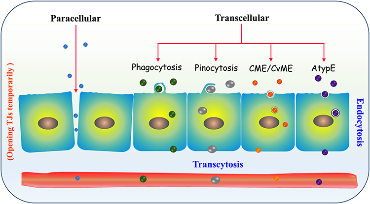 |
Figure 2 Transport route and mechanisms of vesicles across the absorptive epithelia. Abbreviations: CME, clathrin-mediated endocytosis; CvME, caveolin-mediated endocytosis; AtypE, clathrin-/caveolin-independent atypical endocytosis. |
 |
Figure 3 Mechanisms of vesicles-mediated oral drug delivery: (A) conventional VDDSs utilizing pinocytosis or unspecific endocytosis on the basis of supersaturation, and (B) targeted VDDSs via targeting to specialized intestinal epithelial cells for the promotion of absorption. |
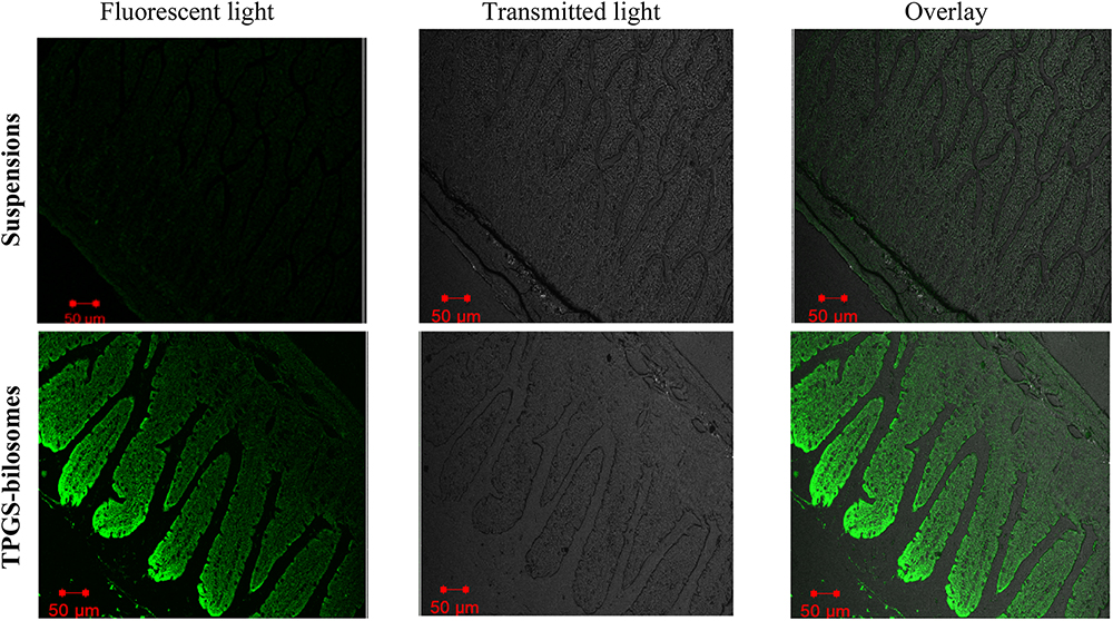 |
Figure 4 Confocal laser scanning microscopy imaging of curcumin-loaded TPGS-bilosomes and suspensions in the duodenum following oral administration. Reproduced from Hegazy H, Amin MM, Fayad W, et al. TPGS surface modified bilosomes as boosting cytotoxic oral delivery systems of curcumin against doxorubicin resistant MCF-7 breast cancer cells. Int J Pharm. 2022;619:121717. Copyright (2022), with permission from Elsevier.72 |
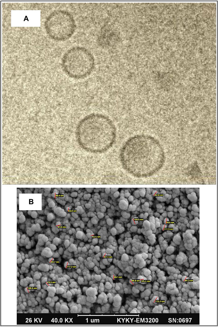 |
Figure 5 Morphology of niosomes loading paclitaxel characterized by cryogenic transmission electron microscopy (Cryo-TEM) (A) and scanning electron microscopy (SEM) (B). Reproduced from Alemi A, Zavar Reza J, Haghiralsadat F, et al. Paclitaxel and curcumin coadministration in novel cationic PEGylated niosomal formulations exhibit enhanced synergistic antitumor efficacy. J Nanobiotechnol ©. 2018;16(1):28. Creative Commons Attribution 4.0 International License (http://creativecommons.org/licenses/by/4.0/).74 |
 |
Figure 6 Prominent example of exosomes-mediated oral drug delivery for antitumor application. (A) Detection of orthotopic lung cancer using bioluminescent A549-Red-luc cells: image of live animals after 27 days of inoculation with 2×10⁶ cells (n = 3) (A1) and mean of bioluminescence signals (A2). (B) Inhibition of A549 orthotopic lung tumors in NOD Scid female mice by i.v. PTX, i.v. Abraxane® and fPTX-Exos, and P.O. fExos and fPTX-Exos (treatment thrice/week at 6 mg/kg, n = 10). (C) Inhibition of A549 orthotopic lung tumors in NOD Scid female mice by i.v. PTX, Abraxane®, fPTX-Exos (once weekly) and p.o. PTX-Exos and fPTX-Exos (thrice weekly) at 4 mg/kg until three weeks, then switched to 8 mg/kg (n = 10): representative images of animals at different time points in the indicated treatment groups (C1) and time-dependent tumor inhibition (C2). Student’s t-test. *p < 0.05; **p < 0.01; ***p < 0.001. Adapted from Kandimalla R, Aqil F, Alhakeem SS, et al. Targeted oral delivery of paclitaxel using colostrum-derived exosomes. Cancers. 2021;13(15):3700. Copyright © 2021 by the authors. Licensee MDPI, Basel, Switzerland. This article is an open access article distributed under the terms and conditions of the Creative Commons Attribution (CC BY) license (https://creativecommons.org/licenses/by/4.0/).100 |
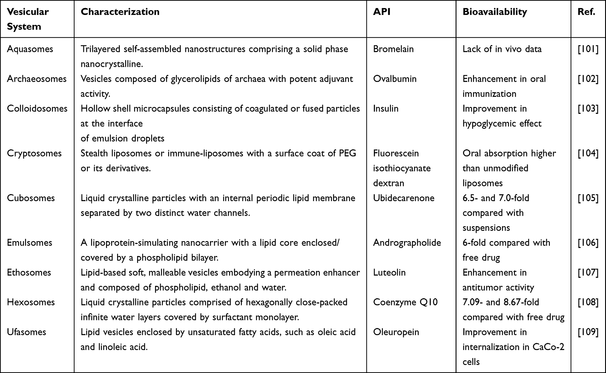 |
Table 1 Novel Vesicular Systems Available for Oral Drug Delivery |
Vesicles are waterbody-containing nanostructures enclosed by a lipid/polymer bilayer encapsulating an active ingredient or therapeutic molecule. Vesicular drug delivery systems (VDDSs) are the integrative term involving all sorts of vesicles, like liposomes, niosomes, polymersomes, etc. VDDSs bridge gap between hydrophilic and hydrophobic heterogeneity in one drug delivery system. Either hydrophilic drug or lipophilic one can be encapsulated in vesicles, even both of them simultaneously.6 In addition to proper colloidal stability, the existence of water compartments increases the membrane flexibility of vesicles, enabling easy transepithelial transport of multifarious cargos. The indwelling potential of vesicles-mediated oral delivery has been confirmed in many drugs,7–10 resulting in significant escalation of bioavailability. Nanovesicles can efficiently translocate into the blood stream across the intestinal epithelia via facilitated diffusion, membrane mobile transport and receptor/ligand-mediated transport. The transport process may involve the structural evolution and deformation of vesicles. Their superior physicochemical property and active interaction with the absorptive epithelium well facilitate the drug absorption. Although no oral vesicles-based medication has been successfully marketed, a hepatocyte directed vesicle (HDV), liposomes encapsulating insulin, has completed its Phase III clinical trial. Nevertheless, the clinical outcomes have not been released. Another vesicular nanomedicine, salbutamol sulphate-loaded niosomes, also fulfills the clinical trial as a pulmonary drug delivery system. Despite these advances, there has been no an all-round survey of vesicles-mediated oral drug delivery available as yet.
Encapsulation of a drug into vesicles can markedly modify its oral pharmacokinetics, hence the bioavailability. The present work aims to provide an overall insight into the potential applications of various vesicles in oral drug delivery with an emphasis on bioavailability enhancement. A variety of nanovesicles eligible for oral drug delivery have been introduced, including liposomes, bilosomes, niosomes, phytosomes, polymersomes, and exosomes, among others. The preparative processes, intestinal transport mechanisms, and in vivo fate of vesicles were discussed. The fundamentals of oral VDDSs design were schematically illustrated. At last, vesicles-mediated oral drug delivery was elaborated in categories with examples prominent in bioavailability enhancement.
Vesicular Drug Delivery Systems
The biologic origin of vesicles can date back to 1965 by Bingham when the concept of “Bingham bodies” was first clued.11 From then on, diverse vesicles have sprung up and then were vastly developed for various biomedical purposes. Vesicular drug delivery systems (VDDSs) are defined as drug vehicles with a vesicular structure comprising one or more concentric or continuous bilayers as a result of self-assembly of amphiphiles in aqueous medium. Nowadays, various vesicles such as liposomes, niosomes, phytosomes and transfersomes have become the important vehicle of choice in drug delivery.12 VDDSs are provided with several advantages over the conventional ones, including excellent cell membrane permeability, good biocompatibility and degradability, self-adapting deformation, and versatile capability of incorporating both hydrophilic and lipophilic drugs, sustaining drug release, overcoming problems of drug insolubility, instability and degradation, mediating targeted drug delivery, and ameliorating bioavailability.
The dimension of vesicles is highly tunable that can be as large as micrometers or as small as nanometers, depending on the preparative materials and techniques. Furthermore, the surface of vesicles can be customized or modified with functional materials when a specific delivery purpose is required.13,14 Of course, the vesicle-forming materials determine the physicochemical properties, in vivo fate, and drug delivery performance of vesicles. These determinants include the degradability, GI digestibility, and intestinal absorbability of biomaterials.15 The administration of vesicles is also flexible, which can be applied via oral, intravenous, or transdermal route. A wide variety of vesicles are available for drug delivery. According to the source of material used, they can be divided into natural vesicles, artificial vesicles and biological vesicles. Natural vesicles are prepared from naturally occurring materials such as phospholipids and phytoconstituents, artificial vesicles are fabricated by synthetic materials like man-made lipids and polymers, and biological vesicles are cell- or virus-derived nanocapsules. The commonly used vesicles in VDDSs include but not limited to liposomes, bilosomes, niosomes, phytosomes, polymersomes and exosomes as illustrated in Figure 1. Furthermore, some other novel vesicular systems have emerged in recent years, eg, ethosomes, cryptosomes, ufasomes, vesosome, enzymosomes, virosomes, archaeosomes, and genosomes. It should be noted that some “somes” may not have a vesicular structure. These VDDSs with sorts of structures and functions provide a complete set of solutions to formulation challenges encountered in modern medicine.
Approaches for Preparation of Vesicles
The general preparation of vesicles involves evaporating a mixture of surfactants to produce a thin film followed by hydration or homogenization. The preparation methods of various vesicles are largely identical to those of liposomes but with minor differences. Thin-film hydration, reverse-phase evaporation and microfluidic technology are the commonly used techniques other than ether/ethanol injection, single-pass process, supercritical fluid, and Handjani-Vila method.
Thin-Film Hydration
An appropriate proportion of amphiphile and cholesterol or other vesicle-forming helper is dissolved in an organic solvent (chloroform, diethyl ether, ethanol or their combination) and evaporated under reduced pressure to form a thin film on the wall of a round-bottomed flask. The resultant film is then hydrated with an aqueous solution at a temperature slightly above the phase transition temperature (Tt) of the materials for a specified time under fast rotation. The drug can be laden into vesicles by dissolving it in the organic phase upon preparation. This approach is suitable for the preparation of vesicles consisting of hydrophobic drug and low-Tt materials. However, the vesicles prepared by this method generally have large size and are not uniform in particle size distribution, which is not friendly to the encapsulation of water-soluble molecules. Homogenization is sometimes necessary to obtain small and uniform vesicles by shaking or sonication. Process variables to be validated include the ratio of amphiphile to helper, the ratio of drug to vesicular materials, and the hydration conditions (temperature, medium, and rotation speed). Through this approach, Tagrida et al16 developed betel leaf extract-loaded liposomes with better physical characteristics, higher encapsulation efficiency, and slower release than those prepared through ethanol injection method. Thin-film hydration was also utilized for preparation of niosomes to encapsulate methylene blue.17 The water-soluble molecule was efficiently encapsulated in the niosomal formulation containing Span 60, cholesterol, and Cremophor® ELP.
Reverse-Phase Evaporation
In this process, the amphiphile and its adjuvant are dissolved in a nonpolar organic solvent (chloroform, ether or a mixture of them), into which an aqueous phase containing a drug or nothing is added. The binary system is then emulsified followed by evaporation to remove the organic solvent under a reduced pressure. Vesicular dispersions are obtained combined with further homogenization or not. This process is either suitable for the preparation of lipophilic drug-loaded vesicles or hydrophilic drug-loaded vesicles. There is an emulsification process in this method, which tends to form coarse dispersions upon reverse-phase evaporation, eg, large unilamellar vesicles. If small vesicles are to be prepared, further homogenization is necessary to be undertaken. Assisted by ultrasound, spherical uniform lipid vesicles encapsulating uricase with improved stability were fabricated using this approach.18 Likewise, Demartis et al19 prepared deformable lipid nanovesicles using such technique for dermal delivery of a sono-photosensitizer and reported that the photosensitizer was intercalated within the phospholipid bilayer of unilamellar vesicles.
Microfluidization
Microfluidization represents an advanced homogenization or emulsification technique for continuous production of micro/nanoparticles, which is implemented through an equipment called a microfluidizer. This apparatus also allows the production of lipid vesicle in a convenient mode. Microfluidization stands for a unique high-pressure homogenization (HPH), typically manipulated by forcing a liquid through a narrow nozzle at high pressure whereby to rupture large particles into small ones. The main advantage of microfluidization over other HPH is that it is less prone to clogging as it works at a constant shear rate. This technique can be applied for the production of vesicles from lab to industry but is not suitable for vesiculation of heat-labile drugs. Microfluidization has been widely applied in the preparation of various drug vehicles.20 Thiele et al21 demonstrated that a rapid, continuous, well-reproducible and size-controlled preparation of unilamellar block copolymer vesicles could be enabled by microfluidization. The feasibility of scalable solvent-free production of liposomes was examined by Khadke et al22 using a high shear combined with microfluidic processing. Liposomes could be prepared within a clinically acceptable size (100 nm around) with low PDI and high drug loading. The process proved to be applicable for both aqueous core-loaded and bilayer-loaded drugs.
Intestinal Transport Mechanisms of Vesicles
The GI transport of substances is a sophisticated process but retains a high transport efficiency. The transport mechanisms of vesicles across the GI epithelium have not been well understood till now. Because of the diversity of intestinal epithelial cells and the interference of the mucous layer, a straightforward observation on the cellular uptake of vesicles becomes difficult. It is further hampered by lack of widely accepted markers or inhibitors to elucidate the transport pathways. Nevertheless, the use of ex vivo imaging and in vitro cell models, to a certain extent, depicts the transport mechanisms of particulates across the GI epithelium.23–26 The intestinal epithelial uptake of vesicles can be proceeded both through the paracellular and transcellular route where there are two transport mechanisms (endocytosis and transcytosis) responsible for the transepithelial translocation of vesicles as illustrated in Figure 2.
The paracellular transport requires the vesicular particulates as small as enough in size or transformable to enable them across the intercellular space. Vesicles below 5 nm are likely to be transported through this pathway into the basolateral blood capillary and lymphatic capillary beneath the GI epithelium along the concentration gradient, since the paracellular breadth between adjacent cells appears to be <5 nm.27 In general, the size distribution of vesicular carriers is uneven or unimodal. There is always a fraction of ultrafine particulates existent in the system. Only these small vesicles have the opportunity to infiltrate into the microcirculation via this route, but the amount transported is marginal due to a low proportion of small vesicles. In another case, those vesicles that can provisionally loosen the tight junctions (TJs) may get access to the underlying microcirculation via this pathway.28,29 It should bear in mind that the paracellular route is not the predominant transport pattern of vesicles owing to dimensional limitation.
The transcellular route plays an important role in intestinal transport of vesicles, which substantially involves two successive processes: endocytosis and transcytosis. The membrane mobile transport pertinent to uptake of particulates is referred as endocytosis, a cytosis by which a cell absorbs a molecule by engulfing.30 Transcytosis is a type of transepithelial translocation that transports cargos across the interior of a cell into adjacent cell.31 Generalized endocytosis includes phagocytosis, pinocytosis, carrier-mediated endocytosis, and carrier-independent endocytosis.32 One or more endocytic mechanisms may be involved in the transport process of vesicles, depending on the nature of VDDSs. Immuno-stimulating vesicles are principally phagocytized by M-cells and presented to the intestinal immune cells (eg, lymphocytes and macrophages) in the gut-associated lymphoid tissue (GALT).33 For large liquid or soft vesicles, pinocytosis is the first and foremost endocytotic pathway, since it can quickly assimilate a large quantity of droplets as large as 2 μm.34 Carrier-mediated endocytosis is a form of transport that takes advantage of special membrane proteins to internalize particles into cells. Carrier proteins that have been identified so far include clathrin, caveolin, RhoA, CDC42, flotillin, ARF6, etc,32 among which clathrin and caveolin are the best-characterized ones that widely participate in sorts of receptor-/ligand-mediated endocytosis. The particulates taken up by clathrin-mediated endocytosis are usually larger than that by caveolin-mediated endocytosis. Particulates below 200 nm can be efficiently ingested by clathrin-mediated endocytosis, while small ones (eg, <60 nm) tend to be assimilated by caveolin-mediated endocytosis. Apart from clathrin-/caveolin-mediated endocytosis, clathrin-/caveolin-independent endocytosis has been reported to be involved in the transport of a variety of vehicles.35,36
In vivo Fate of Vesicles
Drug vehicles experience a series of biological dispositions before access to the absorptive epithelia after oral administration. The in vivo fate of vesicles will affect the performance of VDDSs. The unwanted premature drug release and structural damage of vehicles may cause a frustrated absorption. It is crucial to well understand the in vivo fate of vesicles in the gut.37,38 The most significant factors that dictate oral delivery efficiency of vesicles are premature release and structural stability of VDDSs, which mostly depend on the properties of both payloads and vehicles used. The in vivo fate of oral vesicles is bound up with structural evolution along with unexpected drug release.
Drug transportation requires no or less premature drug release in order to allow more drug molecules to be transported by way of carriers. Burst release and drug leakage are detrimental to oral drug delivery. Hydrophilic molecules and immiscible ones with the carrier materials tend to release from the inner cavity of vesicles. The transepithelial transport of hydrophilic drugs is nullified if fast release emerges, and recrystallization may take place for hydrophobic drugs. In addition, burst release most likely occur at the moment of vesicle collapse, especially for lipid vesicles.39 In the GI lumen, multiple factors induce structural destruction of vesicles, including ions, enzymes, and intestinal flora. Lipid vesicles such as liposomes and phytosomes are vulnerable to enzymatic degradation.40 Structural evolution of vesicles in the GI tract alters the release, absorption and transport of cargos. A consideration of stability is necessitated for designing oral drug vesicles. For instance, bile salt-incorporated liposomes (bilosomes) show some resistance to digestive enzymes.41 By contrast, niosomes composed of nonionic surfactants and polymersomes made of block copolymers are more stable in biorelevant media and can be steadily transported across the GI tract.42,43 Surface coating can also be applied for modulating the properties of vesicles to increase their in vitro/vivo stability. Using a polymer or ligand to decorate the surface of vesicles, both the stability and permeability of vesicles can be greatly improved.3 Enhancive chemical stability and preventible aggregation as a result of coating have been verified in the case of liposomes.44
Oral Delivery Strategies
Oral administration is one of the most acceptable routes for medication, though it is always challenged by inadequate blood drug exposure and off-target effect. To overcome the biological barrier, it can formulate a drug into nanomedicine so as to solve the issues associated with solubility, stability and bioavailability.45 Nanomedicine refers to a nanoscale theranostic system that applies the knowledge and tools of nanotechnology to address medical challenges. VDDSs as a versatile platform of nanomedicine is now incurring much more interests in oral drug delivery. Various conventional and targeted oral VDDSs have been duly developed for unearthing the peroral potential based on different mechanisms (Figure 3).
The Use of Conventional VDDSs
Conventional nanocarriers have been widely proven useful for bioavailability enhancement.46–48 The underlying principles lie in improvement of apparent solubility, augment of absorption area, and co-transport with carriers. Like other micro/nano-drug delivery systems, VDDSs also belong to one of supersaturated drug delivery systems that can significantly increase the dispersity of drug in the GI lumen, hence the absorption area, even the GI residence time. When solubilized into nanoparticles, a supersaturated solution of poorly water-soluble drugs is presented in the GI tract after oral administration. Drug release is normally less or proceeds slowly, which provide opportunities for intestinal uptake of drug in the form of integral nanoparticles. Nanoparticles, including various vesicles, are likely assimilated by enterocytes via pinocytosis and unspecific endocytosis subsequently.49 Conventional VDDSs based on a supersaturation approach have been applied for oral delivery of various therapeuticals. For instance, a nonionic surfactant vesicular system consisting of cholesterol, Tween 80 and Span 80 was developed by Wang et al50 for oral delivery of Ginsenoside Rb1. The developed vesicles notably improved the pharmacokinetics of Ginsenoside Rb1, exhibiting a 1.82-fold increase in relative oral bioavailability. To overcome the drawbacks relating to poor solubility and low bioaccessibility, a food-grade lecithin-based phytosomal formulation was proposed for oral delivery of quercetin.51 The phytosomal vesicles significantly improved the solubility and oral absorption in terms of both exposure and maximum concentration in healthy volunteers in a clinical study compared to unformulated quercetin. These are epitomes of the use of conventional vesicles to facilitate oral absorption of drugs.
The Use of Targeted VDDSs
Targeted drug delivery can further enhance the oral absorption rate and extent on the base of the nanoscale effect.52 To this end, VDDSs’ design that utilizes the physiological functions of the intestinal epithelia becomes particularly important for achieving efficient oral drug delivery. Targeted delivery, to put it bluntly, is a biomimetic drug delivery strategy that mimics the transport process of nutrients or essential ingredients into the human body. The performance of targeted drug delivery systems is generally considered to be superior to that of non-targeted systems due to active involvement of specific transporter- or receptor-mediated endocytosis. Overall, oral targeted VDDSs can be designed in three ways: 1) targeting to a specific receptor; 2) targeting to a transporter; and 3) targeting to specialized enterocytes.
Receptors and transporters have different concepts and physiological functions. Receptor is an outer membrane protein that specifically combines with extracellular signaling molecules (ligands) to cause a certain cell activity. Molecular conformation begins to change when a receptor binds to a ligand, causing various cell responses, such as intercellular signal transduction, intercellular adhesion, and endocytosis. It acts as a “messenger”.53 A transporter is a membrane-spanning protein that mediates the transport of molecules or ions across the cell membrane. It is usually a carrier protein or enzyme, playing the role of “vehicle”.54 Targeted drug delivery systems mainly involve a variety of smart particulates that have the navigating capacity to a specific functional membrane protein in cells. The membrane protein can be a receptor or a transporter lining on or within various conventional or specialized cells. Receptor- or transporter-mediated influx greatly facilitates the cellular uptake of vehicle and its cargos. To improve the oral delivery of insulin, a biotinylated liposomal vehicle was constructed in our group.55 The liposomal vesicles targeting to the intestinal biotin receptor effectively promoted oral absorption of insulin and caused a prolonged hypoglycemic effect. Intestinal acetic acid transporter-mediated mechanism was also utilized for oral delivery of docetaxel with polymer-functionalized liposomes (DTX-ACSL-Lip).56 The enhanced uptake of DTX-ACSL-Lip by Caco-2 cells was achieved via the acetic acid transporter. The oral bioavailability of docetaxel was improved by 10.70-folds with a 3.45-fold increase in Cmax and a 1.19-fold prolongation in MRT through transporter-targeted liposomal vesicles. By dint of the interaction between lectins and glycans, mannosylated liposomes were applied to target model antigen to mannosyl receptors expressed in intestinal antigen-presenting cells (APCs).57 Potent immune responses were induced by this route. Therefore, vesicles-mediated oral drug delivery can be potentiated by conjugating a ligand with high affinity to intestinal specialized cells.
Optimizing Oral Bioavailability with Vesicles
Liposomes
Liposomes are spherical vesicles made of biodegradable natural or synthetic phospholipids, structurally having one or more concentric lipid bilayers. Since the initiation in 1966 by Bangham, liposomes have been evolving steadily as a versatile platform for drug delivery and theranostic purposes.58 Thanks to the capacity to encapsulate diverse entities, excellent biocompatibility, and pleasing interaction with biomembrane, liposomes are also seeking to shift from their original parenteral application to peroral delivery. Liposomal drug delivery for bioavailability enhancement has resurged with the rapid increased publications over the past decade.59 Both the prototype and updated version of liposomes are used for oral drug delivery. Ghassemi et al60 developed conventional and various surfactant-enriched liposomes by the thin-film hydration technique and investigated their performance in oral delivery of carvedilol. Compared to the suspension, conventional and Labrasol-containing liposomes significantly improved the oral bioavailability and Cmax of carvedilol. A chylomicron flow blocker study indicated contribution of lymphatic transport in absorption of nanovesicles. To improve the GI stability of liposomes, a coating approach was implemented to liposomes via layer-by-layer deposition of a pH-sensitive polymer and glycol chitosan.61 Enhanced oral absorption of sorafenib, approximately fourfold increase in AUC and Cmax, was attained through structurally stabilized liposomes. Likewise, N-trimethyl chitosan chloride-modified liposomes improved the oral delivery of curcumin.62 In order for stabilization, proliposomes were developed for cyclosporine A to increase its oral bioavailability.63 Proliposomes that were hydrated with distilled water for oral administration resulted in a 9.6-fold relative bioavailability. In our previous report, an attempt to fortify the structural stability of liposomes by selenization was made.64 Unfortunately, the modified liposomes with Se depositing onto the interior and exterior bilayers did not substantially facilitate absorption of model drug, though a reinforced structure of liposomes was established.
Bilosomes
Bilosomes are lipid vesicles structurally resembling liposomes formed by incorporating bile salts into the phospholipid bilayers. Bilosomes can be deemed as one of flexible liposomes, conceptually easy to confuse with transfersomes widely applied in transdermal drug delivery. Transfersomes are composed of phospholipids and edge activator such as Tween 80, Span 80 and sodium cholate that facilitate the ultra-deformable property of vesicles.65 Bilosomes are synonymous with transfersomes when the edge activator used is a bile salt and intended for oral administration. Bilosomes are more stable in the GI tract than liposomes and can be transported through the bile acid transporter.66,67 Accordingly, bilosomes largely meet the challenges of oral delivery of drugs and vaccines as a vesicular carrier. Bilosomes composed of soybean phosphatidylcholine and sodium deoxycholate were prepared aiming to enhance the oral bioavailability of fenofibrate.68 The pharmacokinetic results disclosed that a higher rate of fenofibrate was absorbed from bilosomes than liposomes composed of phosphatidylcholine and cholesterol, exhibiting a 1.57-fold bioavailability relative to liposomes. The superiority of bilosomes for absorption improvement was demonstrated in a model drug of piperine via ex vivo permeation and in vivo pharmacokinetic studies.69 Boosted oral bioavailability through bilosomes was gotten compared to the suspension formulation. In another study, bilosomes not only improved the GI stability but also the bioavailability of epigallocatechin gallate (EGCG).70 In addition, oral vaccination was designed for tetanus toxoid using mannosylated bilosomes.71 The bilosomes targeting to the mannosyl receptor of APCs not only significantly elicited high systemic immune response but also exhibited mucosal and cell-mediated immune responses. Recently, TPGS-modified bilosomes were developed for oral delivery of curcumin against doxorubicin-resistant MCF-7 breast cancer.72 The optimum formula exhibited 6.6- and 3.4-fold increase in an ex vivo duodenal permeation assay compared to the suspension (Figure 4), in addition to remarkably modified cellular uptake.
Niosomes
Niosomes are nonionic surfactant-based vesicles, generally unilamellar, in which an aqueous cavity is tightly enclosed by a lipophilic bilayer.73 As shown in Alemi et al’s study,74 niosomes appear a spherical, hollow, and surface-smooth morphology (Figure 5). Niosomes possess a number of advantages over liposomes, such as inexpensive excipients, high chemical stability, tolerant sterilization, and easy scale-up production. Nowadays, niosomes are identified as a preferable vesicular drug delivery system in virtue of multiple merits. Nonionic surfactants, the main component of niosomes, can solubilize some poorly soluble drugs and provide sustained release of drugs by entrapping them into the lipophilic bilayers. In addition, niosomal formulation can improve the oral bioavailability of encapsulated drugs and prolong their duration of action other than passable biocompatibility. Niosomes encapsulating paclitaxel (PTX) were prepared for enhancing its oral bioavailability.75 Niosomes fabricated from Span 40 and coated with carbopol showed better stability than uncoated ones in bile salt-containing biorelevant medium and resulted in higher drug plasma levels and bioavailability after oral administration. Afterward, a biocompatible creatinine-based niosomal delivery system was developed for oral delivery of clarithromycin.76 The niosomal formulation gave rise to a twofold enhancement of oral bioavailability compared with the commercial formulation. In a pertaining report, niosomal encapsulation was employed by Mohsen et al77 to ameliorate the oral bioavailability and achieve a sustained delivery of glimepiride. Using niosomes, the hypoglycemic effect of glimepiride was maintained for a longer duration at the therapeutic level, and a sevenfold enhancement in relative bioavailability was observed. To investigate the in situ–in vivo absorption correlation of niosomes, the effects of niosomal encapsulation on intestinal absorption and oral bioavailability of nateglinide were performed.78 The in situ intestinal absorption reflected no significant alteration in the transport parameters between niosomal and free drug. However, niosomal nateglinide showed significantly improved hypoglycemic effect. The discrepancy was assumed to be associated with different transport pathways shared by niosomal and free drug, where niosomes may undergo lymphatic transport that minimizes presystemic metabolism. Recently, tailored niosomes based on Span 40 and cholesterol were engineered for increasing bioavailability of rosuvastatin.79 The ex vivo intestinal permeability study showed significant improvement in drug permeation with niosomes, and the in vivo pharmacokinetic results also confirmed enhancement of oral bioavailability, approximately twofold accession.
Phytosomes
Phytosomes are colloidal, nanoscale, liposome-like vesicles concomitant with micellar or hexagonal nanostructure formed by phytomedicine-phospholipid complexes in an aqueous medium.80 In the arena of solubilization and controlled release, several problems are encountered, such as booming in amphiphobic entities, inadequate entrapment rate, and drug leakage and burst release. In the context of enabling formulation, a novel lipid drug delivery system, phytosomes, has evolved. In the pharmaceutical territory, phytosomes are sometimes called pharmacosomes. They work as a befitting carrier for oral delivery of drugs quite precisely owing to their unique properties like smaller size, amphiphilicity, high drug loading, and physical stability. Since drugs are covalently bound to phospholipid molecules, phytosomes hold many advantages, eg, sustaining drug release, reducing drug leakage and toxicity, and enhancing bioavailability of poorly permeable drugs. Apigenin-phospholipid phytosomes were synthesized and exploited for suitability of improving solubility, enhancing bioavailability, and potentiating antioxidant.81 Significantly enhanced bioavailability and antioxidant activity against liver injury were arisen from phytosomal apigenin. When formulated into phytosomes, the apparent solubility and oral bioavailability of celastrol, an anticancer phytomedicine, were markedly modified, causing fourfold and fivefold increase in AUC and Cmax, respectively.82 Another phytocomponent, silybin, was also formulated as phytosomes to improve the oral pharmacokinetics.83 Besides elevated drug exposure to the blood, the phytosomal nanosuspensions exhibited more potent hepatoprotective effects in terms of pharmacodynamics. An enhancement up to 10-fold in oral bioavailability of mangiferin was achieved using phytosomal nanoparticles based on evaluation on the main pharmacokinetic parameters, including Cmax, Tmax, and AUC.84 Interestingly, phytosomes comprising phospholipid and piperine have ever been designed as a bioenhancer to increase oral bioavailability of domperidone, a P-glycoprotein substrate.85 It suggested that piperine-loaded phytosomes have the capacity of inhibiting P-glycoprotein transporter, thus promoting absorption of the payload upon co-delivery.
Polymersomes
Polymersomes are polymeric vesicles self-assembled by amphiphilic copolymer.86 Compared with surfactants, polymers have excellent physicochemical stability and can be biodegraded in vivo. Due to the advancement of polymer chemistry, it becomes easier to synthesize a variety of functional block copolymers, which has aroused great interest of scientists to develop polymersomes as a drug delivery vehicle. Like surfactants, amphiphilic polymers can be assembled into a variety of aggregates. It depends mainly on the mass or volume fraction of the hydrophilic block in copolymer (ƒ) and the interaction parameter of its hydrophobic block with H2O (χ) whether they can assemble into vesicles.87 For PEG-based block copolymers with a high χ, vesicular structures tend to form when ƒ of PEG (ƒPEG) is 10–40%. At a ƒPEG of 45–55%, cylindrical micelles are generated, and at a ƒPEG of 55–70%, spherical micelles are predominantly formed. Polymersomes are widely used in drug delivery, including oral administration, owing to their high stability and versatility. They can encapsulate either hydrophilic or hydrophobic drug, or both at the same time to achieve the purpose of combined therapy. Polymersomes can also protect protein drugs from damage in the GI lumen. For example, Alibolandi et al88 synthesized a copolymer of dextran-b-poly(lactide-co-glycolide) for preparation of polymersomes to orally deliver insulin. Insulin was efficiently encapsulated in polymersomes (>90%) and was negligibly released in SGF except in SIF. Significant hypoglycemic effect was harvested in the diabetic rats. Doxorubicin hydrochloride, another example of water-soluble drugs, is highly challenging for oral chemotherapy. It was encapsulated into polymersomes formed by amphiphilic β-cyclodextrin-centered triarm star polymer in an attempt to enable oral delivery.89 The polymeric vesicles brought about a 7.32-fold increase in oral bioavailability and an 8.22-fold extension in half-life compared with free drug, resulting in a substantial in vivo anticancer efficacy. For hydrophobic drugs, polymersomes also exhibit superior absorption-promoting effects. Sorafenib, a hydrophobic anticancer drug, has been nanosized into polymersomes using poly (butadiene)-b-poly (ethylene oxide).90 A sustained release of sorafenib up to 144 h and high stability within 3 months were discovered for polymersomes. Oral administration of polymersomes demonstrated enhancive Cmax and AUC0-96 by 1.7 and 2.77-fold, respectively, compared to the suspension group. To elaborate the performance of polymersomes versus conventional liposomes as bioavailability enhancers, Youssef et al43 prepared flutamide-loaded polymersomes and liposomes and investigated their potentials for oral delivery. As a result, a significantly higher stability in SIF was presented by polymersomes compared with liposomes, along with great enhancement in oral bioavailability of flutamide. Beyond these, targeted polymersomes have also been attempted to orally deliver therapeutic molecules. As an example, folate-conjugated polymersomes was designed for oral delivery of PTX.91 The ligand-anchored polymersomes resulted in a higher oral bioavailability than conventional polymersomes. Moreover, the introduction of TPGS in folated polymersomes furthered the cytotoxic potency, cellular uptake and bioavailability.
Exosomes
Exosomes are extracellular vesicles with a diameter of 30–100 nm secreted by living cells that are involved in the processes of many biological and pathological events.92 Exosomes are heterogeneous lipid bilayer vesicles, as mediators for intercellular communication, which play an important role in various processes, eg, modulation of the immune response, homeostasis, coagulation, angiogenesis, cancer progression, and inflammation.93 They can not only serve as biomarkers for diagnosis but also exhibit tremendous potential as natural drug delivery vehicles since the discovery, based on their size and ability to transfer biological materials to recipient cells. Various therapeutic molecules can be loaded in exosomes, including small molecule compounds, proteins, and oligonucleotides. The unique biological compatibility, preferable stability, and high affinity to cells confer exosomes great expectations for biomedical applications. In recent years, the use of exosomes has been tentatively extended to the oral field from the arena of systemic drug delivery, especially edible food-/plant-derived exosomes.94,95 For example, milk-derived exosomes were developed for oral delivery of PTX.96 Paclitaxel-loaded exosomes exhibited excellent stability in the GI fluids and sustained release of drug. The exosomes significantly enhanced the antitumor efficacy of PTX while attenuated its systemic and immunologic toxicities, though the oral bioavailability was not reported. The potential of exosomes from cow milk for oral delivery was further verified by Betker et al.97 It has been shown that absorption of milk exosomes from the GI tract occurs via the “neonatal” Fc receptor as intact particles, and they can be ulteriorly modified with ligands to promote retention in target tissues. A comparative study on the capacity of cow milk- and intestinal epithelial cell-derived exosomes to enhance oral bioavailability was subsequently carried out with curcumin as a model drug.98 Epithelial cell-derived extracellular vesicles showed higher cell uptake compared to cow milk-derived extracellular vesicles, demonstrating a superior effect on promoting absorption.
What’s more, surface-engineered and targeted exosomes have been developed for oral applications with encouraging results. Warren et al99 reported bovine milk-derived exosomes (mExos) with tunable surface for oral delivery of small interfering RNA (siRNA). It was found that negatively charged hydrophobic mExos can penetrate multiple biological barriers. A hydrophilic PEG coating was introduced on mExo surface significantly reduced degradation of mExo in the acidic gastric environment and enhanced their permeability through the intestinal mucin. Both mExo and PEG-mExo demonstrated high uptake by enterocytes and mediated functional intracellular delivery of siRNA. For targeted delivery of PTX via the oral route, colostrum-derived exosomes were also prepared and modified with folate.100 Oral administration of folate-attached PTX-loaded exosomes (fPTX-Exos) produced significant inhibition (>50%) of subcutaneous xenograft tumor, while the same dose of PTX showed insignificant inhibition. In an orthotopic lung cancer model, oral dosing of fPTX-Exos resulted in greater tumor inhibiting efficacy (55%) than i.v. injected PTX (24–32%) and similar efficacy as i.v. Abraxane® (59%), commercially available PTX albumin nanoparticles (Figure 6). Taken together, these studies turn out that exosomes are promising biomaterials as oral delivery vehicles.
Other VDDSs
In addition to those VDDSs abovementioned, many other vesicular systems have been established for optimizing oral bioavailability. The vesicles occasionally used have structures similar to liposomes or niosomes, but differ in the vesicle-forming materials and adjuvants. They are specifically engineered to solve multifarious medical challenges based on different application scenarios. Surely, these exotic vesicles have also earned a place in the field of oral drug delivery. Some newly emerging vesicular systems already employed for bioavailability enhancement are presented in Table 1.
Conclusions
Vesicles are particularly important drug vehicles for oral delivery of various therapeuticals by virtue of structural diversity, great flexibility for drug loading, and prominent ability in absorption promotion. There are dozens of vesicles available for oral applications, among which some possess excellent biocompatibility and intestinal permeability, some present high in vitro/vivo stability, some are provided with self-adaptable structure, and some are qualified with targeting property. Vesicles are waterbody-containing micro/nanocarriers that enable them to encapsulate either hydrophobic or hydrophilic drug or both of them, showing a flexibility in drug loading. The absorption-promoting effects of various vesicles have been verified with diverse model drugs. Of course, the physicochemical stability, biosafety, and scale-up production are the practical problems to be solved in VDDSs. Although this work does not cover all the issues, cumulative evidences disclose that VDDSs as bioavailability enhancers can provide efficient solutions to formulating “problem” drugs with a significant absorption barrier.
Acknowledgment
This work was jointly supported by Basic and Applied Basic Research Project of Guangzhou Science and Technology Plan (202201010743) and the Fundamental Research Funds for the central Universities (No. 21622414).
Disclosure
The authors report no conflicts of interest in this study.
References
1. Kaur G, Arora M, Ravi Kumar MNV. Oral drug delivery technologies-a decade of developments. J Pharmacol Exp Ther. 2019;370(3):529–543. doi:10.1124/jpet.118.255828
2. Reinholz J, Landfester K, Mailänder V. The challenges of oral drug delivery via nanocarriers. Drug Deliv. 2018;25(1):1694–1705. doi:10.1080/10717544.2018.1501119
3. He H, Lu Y, Qi J, et al. Adapting liposomes for oral drug delivery. Acta Pharm Sin B. 2019;9(1):36–48. doi:10.1016/j.apsb.2018.06.005
4. Wang T, Fu Y, Sun S, et al. Exosome-based drug delivery systems in cancer therapy. Chin Chem Lett. 2022. doi:10.1016/j.cclet.2022.05.022
5. Manconi M, Caddeo C, Manca ML, et al. Oral delivery of natural compounds by phospholipid vesicles. Nanomedicine. 2020;15(18):1795–1803. doi:10.2217/nnm-2020-0085
6. Pramod PS, Takamura K, Chaphekar S, et al. Dextran vesicular carriers for dual encapsulation of hydrophilic and hydrophobic molecules and delivery into cells. Biomacromolecules. 2012;13(11):3627–3640. doi:10.1021/bm301583s
7. Imran M, Shah MR, Ullah F, et al. Sugar-based novel niosomal nanocarrier system for enhanced oral bioavailability of levofloxacin. Drug Deliv. 2016;23(9):3653–3664. doi:10.1080/10717544.2016.1214991
8. Imran M, Shah MR, Ullah F, et al. Double-tailed acyl glycoside niosomal nanocarrier for enhanced oral bioavailability of Cefixime. Artif Cells Nanomed Biotechnol. 2017;45(7):1440–1451. doi:10.1080/21691401.2016.1246451
9. Ullah S, Shah MR, Shoaib M, et al. Creatinine-based non-phospholipid vesicular carrier for improved oral bioavailability of azithromycin. Drug Dev Ind Pharm. 2017;43(6):1011–1022. doi:10.1080/03639045.2017.1291667
10. Joseph A, Kumar D, Balakrishnan A, et al. Surface-engineered liposomal particles of calcium ascorbate with fenugreek galactomannan enhanced the oral bioavailability of ascorbic acid: a randomized, double-blinded, 3-sequence, crossover study. RSC Adv. 2021;11(60):38161–38171. doi:10.1039/d1ra06483e
11. Mirtaleb MS, Shahraky MK, Ekrami E, et al. Advances in biological nano-phospholipid vesicles for transdermal delivery: a review on applications. J Drug Deliv Sci Technol. 2021;61:102331. doi:10.1016/j.jddst.2021.102331
12. Limongi T, Susa F, Marini M, et al. Lipid-based nanovesicular drug delivery systems. Nanomaterials. 2021;11(12):3391. doi:10.3390/nano11123391
13. Hardwick J, Taylor J, Mehta M, et al. Targeting cancer using curcumin encapsulated vesicular drug delivery systems. Curr Pharm Des. 2021;27(1):2–14. doi:10.2174/1381612826666200728151610
14. Goel H, Razdan K, Singla R, et al. Engineered site-specific vesicular systems for colonic delivery: trends and implications. Curr Pharm Des. 2020;26(42):5441–5455. doi:10.2174/1381612826666200813132301
15. Alqahtani MS, Kazi M, Alsenaidy MA, et al. Advances in oral drug delivery. Front Pharmacol. 2021;12:618411. doi:10.3389/fphar.2021.618411
16. Tagrida M, Prodpran T, Zhang B, et al. Liposomes loaded with betel leaf (Piper betle L.) ethanolic extract prepared by thin film hydration and ethanol injection methods: characteristics and antioxidant activities. J Food Biochem. 2021;45(12):e14012. doi:10.1111/jfbc.14012
17. Yeo LK, Chaw CS, Elkordy AA. The effects of hydration parameters and co-surfactants on methylene blue-loaded niosomes prepared by the thin film hydration method. Pharmaceuticals. 2019;12(2):46. doi:10.3390/ph12020046
18. Tan QY, Wang N, Yang H, et al. Preparation and characterization of lipid vesicles containing uricase. Drug Deliv. 2010;17(1):28–37. doi:10.3109/10717540903508953
19. Demartis S, Rassu G, Murgia S, et al. Improving dermal delivery of rose bengal by deformable lipid nanovesicles for topical treatment of melanoma. Mol Pharm. 2021;18(11):4046–4057. doi:10.1021/acs.molpharmaceut.1c00468
20. Ganesan P, Karthivashan G, Park SY, et al. Microfluidization trends in the development of nanodelivery systems and applications in chronic disease treatments. Int J Nanomedicine. 2018;13:6109–6121. doi:10.2147/ijn.S178077
21. Thiele J, Steinhauser D, Pfohl T, et al. Preparation of monodisperse block copolymer vesicles via flow focusing in microfluidics. Langmuir. 2010;26(9):6860–6863. doi:10.1021/la904163v
22. Khadke S, Roces CB, Donaghey R, et al. Scalable solvent-free production of liposomes. J Pharm Pharmacol. 2020;72(10):1328–1340. doi:10.1111/jphp.13329
23. Yang D, Liu D, Qin M, et al. Intestinal mucin induces more endocytosis but less transcytosis of nanoparticles across enterocytes by triggering nanoclustering and strengthening the retrograde pathway. ACS Appl Mater Interfaces. 2018;10(14):11443–11456. doi:10.1021/acsami.7b19153
24. Xia D, He Y, Li Q, et al. Transport mechanism of lipid covered saquinavir pure drug nanoparticles in intestinal epithelium. J Control Release. 2018;269:159–170. doi:10.1016/j.jconrel.2017.11.012
25. Akbari A, Lavasanifar A, Wu J. Interaction of cruciferin-based nanoparticles with Caco-2 cells and Caco-2/HT29-MTX co-cultures. Acta Biomater. 2017;64:249–258. doi:10.1016/j.actbio.2017.10.017
26. Soundararajan R, Sasaki K, Godfrey L, et al. Direct in vivo evidence on the mechanism by which nanoparticles facilitate the absorption of a water insoluble, P-gp substrate. Int J Pharm. 2016;514(1):121–132. doi:10.1016/j.ijpharm.2016.08.013
27. Melchior F, Gerace L. Mechanisms of nuclear protein import. Curr Opin Cell Biol. 1995;7(3):310–318. doi:10.1016/0955-0674(95)80084-0
28. Zhang J, Zhu X, Jin Y, et al. Mechanism study of cellular uptake and tight junction opening mediated by goblet cell-specific trimethyl chitosan nanoparticles. Mol Pharm. 2014;11(5):1520–1532. doi:10.1021/mp400685v
29. Sonaje K, Lin KJ, Tseng MT, et al. Effects of chitosan-nanoparticle-mediated tight junction opening on the oral absorption of endotoxins. Biomaterials. 2011;32(33):8712–8721. doi:10.1016/j.biomaterials.2011.07.086
30. Mukherjee S, Ghosh RN, Maxfield FR. Endocytosis. Physiol Rev. 1997;77(3):759–803. doi:10.1152/physrev.1997.77.3.759
31. Tuma P, Hubbard AL. Transcytosis: crossing cellular barriers. Physiol Rev. 2003;83(3):871–932. doi:10.1152/physrev.00001.2003
32. Canton I, Battaglia G. Endocytosis at the nanoscale. Chem Soc Rev. 2012;41(7):2718–2739. doi:10.1039/C2cs15309b
33. Islam MA, Firdous J, Badruddoza AZM, et al. M cell targeting engineered biomaterials for effective vaccination. Biomaterials. 2019;192:75–94. doi:10.1016/j.biomaterials.2018.10.041
34. Kerr MC, Teasdale RD. Defining macropinocytosis. Traffic. 2009;10(4):364–371. doi:10.1111/j.1600-0854.2009.00878.x
35. Costa Verdera H, Gitz-Francois JJ, Schiffelers RM, et al. Cellular uptake of extracellular vesicles is mediated by clathrin-independent endocytosis and macropinocytosis. J Control Release. 2017;266:100–108. doi:10.1016/j.jconrel.2017.09.019
36. Wang J, Li L, Du Y, et al. Improved oral absorption of doxorubicin by amphiphilic copolymer of lysine-linked ditocopherol polyethylene glycol 2000 succinate: in vitro characterization and in vivo evaluation. Mol Pharm. 2015;12(2):463–473. doi:10.1021/mp500833m
37. Wu W, Li T, Zheng Y. Editorial of special issue “the biological fate of drug nanocarriers”. Acta Pharm Sin B. 2021;11(4):850–851. doi:10.1016/j.apsb.2021.04.004
38. Dufort S, Sancey L, Coll JL. Physico-chemical parameters that govern nanoparticles fate also dictate rules for their molecular evolution. Adv Drug Deliv Rev. 2012;64(2):179–189. doi:10.1016/j.addr.2011.09.009
39. Yaroslavov PA, Panova DI, Sybachin DA, et al. Payload release by liposome burst: thermal collapse of microgels induces satellite destruction. Nanomed Nanotechnol Biol Med. 2017;13(4):1491–1494. doi:10.1016/j.nano.2017.02.001
40. Liu W, Ye A, Han F, et al. Advances and challenges in liposome digestion: surface interaction, biological fate, and GIT modeling. Adv Colloid Interface Sci. 2019;263:52–67. doi:10.1016/j.cis.2018.11.007
41. Hu S, Niu M, Hu F, et al. Integrity and stability of oral liposomes containing bile salts studied in simulated and ex vivo gastrointestinal media. Int J Pharm. 2013;441(1–2):693–700. doi:10.1016/j.ijpharm.2012.10.025
42. Yaghoobian M, Haeri A, Bolourchian N, et al. The impact of surfactant composition and surface charge of niosomes on the oral absorption of repaglinide as a BCS II model drug. Int J Nanomedicine. 2020;15:8767–8781. doi:10.2147/ijn.S261932
43. Youssef SF, Elnaggar YS, Abdallah OY. Elaboration of polymersomes versus conventional liposomes for improving oral bioavailability of the anticancer flutamide. Nanomedicine. 2018;13(23):3025–3036. doi:10.2217/nnm-2018-0238
44. Meland H-G, Røv-Johnsen A, Smistad G, et al. Studies on surface coating of phospholipid vesicles with a non-ionic polymer. Colloids Surf B Biointerfaces. 2014;114:45–52. doi:10.1016/j.colsurfb.2013.09.054
45. Parodi A, Buzaeva P, Nigovora D, et al. Nanomedicine for increasing the oral bioavailability of cancer treatments. J Nanobiotechnology. 2021;19(1):354. doi:10.1186/s12951-021-01100-2
46. Zhang L, Wang S, Zhang M, et al. Nanocarriers for oral drug delivery. J Drug Target. 2013;21(6):515–527. doi:10.3109/1061186x.2013.789033
47. Pathak K, Raghuvanshi S. Oral bioavailability: issues and solutions via nanoformulations. Clin Pharmacokinet. 2015;54(4):325–357. doi:10.1007/s40262-015-0242-x
48. Nabi B, Rehman S, Baboota S, et al. Insights on oral drug delivery of lipid nanocarriers: a win-win solution for augmenting bioavailability of antiretroviral drugs. AAPS Pharm Sci Tech. 2019;20(2):60. doi:10.1208/s12249-018-1284-9
49. Oh N, Park JH. Endocytosis and exocytosis of nanoparticles in mammalian cells. Int J Nanomed. 2014;9(Suppl 1):51–63. doi:10.2147/IJN.S26592
50. Wang Q, Wang Y, Xie Y, et al. Nonionic surfactant vesicles as a novel drug delivery system for increasing the oral bioavailability of Ginsenoside Rb1. Food Biosci. 2021;42:101064. doi:10.1016/j.fbio.2021.101064
51. Riva A, Ronchi M, Petrangolini G, et al. Improved oral absorption of quercetin from quercetin phytosome®, a new delivery system based on food grade lecithin. Eur J Drug Metab Pharmacokinet. 2019;44(2):169–177. doi:10.1007/s13318-018-0517-3
52. Date AA, Hanes J, Ensign LM. Nanoparticles for oral delivery: design, evaluation and state-of-The-art. J Control Release. 2016;240504–240526. doi:10.1016/j.jconrel.2016.06.016
53. Fletcher A. The cell membrane and receptors. Anaesth Intensive Care Med. 2017;18(6):316–320. doi:10.1016/j.mpaic.2017.03.005
54. Diallinas G, Martzoukou O. Transporter membrane traffic and function: lessons from a mould. FEBS J. 2019;286(24):4861–4875. doi:10.1111/febs.15078
55. Zhang X, Qi J, Lu Y, et al. Biotinylated liposomes as potential carriers for the oral delivery of insulin. Nanomedicine. 2014;10(1):167–176. doi:10.1016/j.nano.2013.07.011
56. Guo X, Zhang J, Cai Q, et al. Acetic acid transporter-mediated, oral, multifunctional polymer liposomes for oral delivery of docetaxel. Colloids Surf B Biointerfaces. 2021;198:111499. doi:10.1016/j.colsurfb.2020.111499
57. Wang N, Wang T, Zhang M, et al. Mannose derivative and lipid A dually decorated cationic liposomes as an effective cold chain free oral mucosal vaccine adjuvant-delivery system. Eur J Pharm Biopharm. 2014;88(1):194–206. doi:10.1016/j.ejpb.2014.04.007
58. Wu W, Lu Y, Qi J. Oral delivery of liposomes. Ther Deliv. 2015;6(11):1239–1241. doi:10.4155/tde.15.69
59. Lee MK. Liposomes for enhanced bioavailability of water-insoluble drugs: in vivo evidence and recent approaches. Pharmaceutics. 2020;12(3):264. doi:10.3390/pharmaceutics12030264
60. Ghassemi S, Haeri A, Shahhosseini S, et al. Labrasol-enriched nanoliposomal formulation: novel approach to improve oral absorption of water-insoluble drug, carvedilol. AAPS Pharm Sci Tech. 2018;19(7):2961–2970. doi:10.1208/s12249-018-1118-9
61. Zhao M, Lee SH, Song JG, et al. Enhanced oral absorption of sorafenib via the layer-by-layer deposition of a pH-sensitive polymer and glycol chitosan on the liposome. Int J Pharm. 2018;544(1):14–20. doi:10.1016/j.ijpharm.2018.04.020
62. Chen H, Wu J, Sun M, et al. N-trimethyl chitosan chloride-coated liposomes for the oral delivery of curcumin. J Liposome Res. 2012;22(2):100–109. doi:10.3109/08982104.2011.621127
63. Shah NM, Parikh J, Namdeo A, et al. Preparation, characterization and in vivo studies of proliposomes containing Cyclosporine A. J Nanosci Nanotechnol. 2006;6(9–10):2967–2973. doi:10.1166/jnn.2006.403
64. Zhu M, Zhu S, Liu Q, et al. Selenized liposomes with ameliorative stability that achieve sustained release of emodin but fail in bioavailability. Chin Chem Lett. 2022. doi:10.1016/j.cclet.2022.04.080
65. Opatha SAT, Titapiwatanakun V, Chutoprapat R. Transfersomes: a promising nanoencapsulation technique for transdermal drug delivery. Pharmaceutics. 2020;12(9):855. doi:10.3390/pharmaceutics12090855
66. Senior K. Bilosomes: the answer to oral vaccine delivery? Drug Discov Today. 2001;6(20):1031–1032. doi:10.1016/s1359-6446(01)02010-4
67. Deng F, Bae YH. Bile acid transporter-mediated oral drug delivery. J Control Release. 2020;327:100–116. doi:10.1016/j.jconrel.2020.07.034
68. Chen Y, Lu Y, Chen J, et al. Enhanced bioavailability of the poorly water-soluble drug fenofibrate by using liposomes containing a bile salt. Int J Pharm. 2009;376(1–2):153–160. doi:10.1016/j.ijpharm.2009.04.022
69. Zakaria MY, Fayad E, Althobaiti F, et al. Statistical optimization of bile salt deployed nanovesicles as a potential platform for oral delivery of piperine: accentuated antiviral and anti-inflammatory activity in MERS-CoV challenged mice. Drug Deliv. 2021;28(1):1150–1165. doi:10.1080/10717544.2021.1934190
70. Wang L, Huang X, Jing H, et al. Bilosomes as effective delivery systems to improve the gastrointestinal stability and bioavailability of epigallocatechin gallate (EGCG). Food Res Int. 2021;149:110631. doi:10.1016/j.foodres.2021.110631
71. Jain S, Harde H, Indulkar A, et al. Improved stability and immunological potential of tetanus toxoid containing surface engineered bilosomes following oral administration. Nanomedicine. 2014;10(2):431–440. doi:10.1016/j.nano.2013.08.012
72. Hegazy H, Amin MM, Fayad W, et al. TPGS surface modified bilosomes as boosting cytotoxic oral delivery systems of curcumin against doxorubicin resistant MCF-7 breast cancer cells. Int J Pharm. 2022;619:121717. doi:10.1016/j.ijpharm.2022.121717
73. Kumar GP, Rajeshwarrao P. Nonionic surfactant vesicular systems for effective drug delivery—an overview. Acta Pharm Sin B. 2011;1(4):208–219. doi:10.1016/j.apsb.2011.09.002
74. Alemi A, Zavar Reza J, Haghiralsadat F, et al. Paclitaxel and curcumin coadministration in novel cationic PEGylated niosomal formulations exhibit enhanced synergistic antitumor efficacy. J Nanobiotechnol. 2018;16(1):28. doi:10.1186/s12951-018-0351-4
75. Sezgin-Bayindir Z, Onay-Besikci A, Vural N, et al. Niosomes encapsulating paclitaxel for oral bioavailability enhancement: preparation, characterization, pharmacokinetics and biodistribution. J Microencapsul. 2013;30(8):796–804. doi:10.3109/02652048.2013.788088
76. Ullah S, Shah MR, Shoaib M, et al. Development of a biocompatible creatinine-based niosomal delivery system for enhanced oral bioavailability of clarithromycin. Drug Deliv. 2016;23(9):3480–3491. doi:10.1080/10717544.2016.1196768
77. Mohsen AM, AbouSamra MM, ElShebiney SA. Enhanced oral bioavailability and sustained delivery of glimepiride via niosomal encapsulation: in-vitro characterization and in-vivo evaluation. Drug Dev Ind Pharm. 2017;43(8):1254–1264. doi:10.1080/03639045.2017.1310224
78. Sultan AA, El-Gizawy SA, Osman MA, et al. Niosomes for oral delivery of nateglinide: in situ-in vivo correlation. J Liposome Res. 2018;28(3):209–217. doi:10.1080/08982104.2017.1343835
79. Liu Q, Xu J, Liao K, et al. Oral bioavailability improvement of tailored rosuvastatin loaded niosomal nanocarriers to manage ischemic heart disease: optimization, ex vivo and in vivo studies. AAPS Pharm Sci Tech. 2021;22(2):58. doi:10.1208/s12249-021-01934-x
80. Zhu S, Luo C, Feng W, et al. Selenium-deposited tripterine phytosomes ameliorate the antiarthritic efficacy of the phytomedicine via a synergistic sensitization. Int J Pharm. 2020;578:119104. doi:10.1016/j.ijpharm.2020.119104
81. Telange DR, Patil AT, Pethe AM, et al. Formulation and characterization of an apigenin-phospholipid phytosome (APLC) for improved solubility, in vivo bioavailability, and antioxidant potential. Eur J Pharm Sci. 2017;108:36–49. doi:10.1016/j.ejps.2016.12.009
82. Freag MS, Saleh WM, Abdallah OY. Self-assembled phospholipid-based phytosomal nanocarriers as promising platforms for improving oral bioavailability of the anticancer celastrol. Int J Pharm. 2018;535(1–2):18–26. doi:10.1016/j.ijpharm.2017.10.053
83. Chi C, Zhang C, Liu Y, et al. Phytosome-nanosuspensions for silybin-phospholipid complex with increased bioavailability and hepatoprotection efficacy. Eur J Pharm Sci. 2020;144:105212. doi:10.1016/j.ejps.2020.105212
84. Telange DR, Sohail NK, Hemke AT, et al. Phospholipid complex-loaded self-assembled phytosomal soft nanoparticles: evidence of enhanced solubility, dissolution rate, ex vivo permeability, oral bioavailability, and antioxidant potential of mangiferin. Drug Deliv Transl Res. 2021;11(3):1056–1083. doi:10.1007/s13346-020-00822-4
85. Islam N, Irfan M, Hussain T, et al. Piperine phytosomes for bioavailability enhancement of domperidone. J Liposome Res. 2022;32(2):172–180. doi:10.1080/08982104.2021.1918153
86. Zhang X-Y, Zhang P-Y. Polymersomes in nanomedicine - a review. Curr Med Chem. 2017;13(2):124–129. doi:10.2174/1573413712666161018144519
87. Lee JS, Feijen J. Polymersomes for drug delivery: design, formation and characterization. J Control Release. 2012;161(2):473–483. doi:10.1016/j.jconrel.2011.10.005
88. Alibolandi M, Alabdollah F, Sadeghi F, et al. Dextran-b-poly(lactide-co-glycolide) polymersome for oral delivery of insulin: in vitro and in vivo evaluation. J Control Release. 2016;227:58–70. doi:10.1016/j.jconrel.2016.02.031
89. Hu M, Shen Y, Zhang L, et al. Polymersomes via self-assembly of amphiphilic β-cyclodextrin-centered triarm star polymers for enhanced oral bioavailability of water-soluble chemotherapeutics. Biomacromolecules. 2016;17(3):1026–1039. doi:10.1021/acs.biomac.5b01676
90. Khan MA, Ali S, Venkatraman SS, et al. Fabrication of poly (butadiene-block-ethylene oxide) based amphiphilic polymersomes: an approach for improved oral pharmacokinetics of Sorafenib. Int J Pharm. 2018;542(1–2):196–204. doi:10.1016/j.ijpharm.2018.03.023
91. Pan XQ, Gong YC, Li ZL, et al. Folate-conjugated pluronic/polylactic acid polymersomes for oral delivery of paclitaxel. Int J Biol Macromol. 2019;139:377–386. doi:10.1016/j.ijbiomac.2019.07.224
92. Patil SM, Sawant SS, Kunda NK. Exosomes as drug delivery systems: a brief overview and progress update. Eur J Pharm Biopharm. 2020;154:259–269. doi:10.1016/j.ejpb.2020.07.026
93. Fuloria S, Subramaniyan V, Dahiya R, et al. Mesenchymal stem cell-derived extracellular vesicles: regenerative potential and challenges. Biology. 2021;10(3):172. doi:10.3390/biology10030172
94. Shi Y, Guo S, Liang Y, et al. Construction and evaluation of liraglutide delivery system based on milk exosomes: a new idea for oral peptide delivery. Curr Pharm Biotechnol. 2022;23(8):1072–1079. doi:10.2174/1389201022666210820114236
95. Umezu T, Takanashi M, Murakami Y, et al. Acerola exosome-like nanovesicles to systemically deliver nucleic acid medicine via oral administration. Mol Ther Methods Clin Dev. 2021;21:199–208. doi:10.1016/j.omtm.2021.03.006
96. Agrawal AK, Aqil F, Jeyabalan J, et al. Milk-derived exosomes for oral delivery of paclitaxel. Nanomedicine. 2017;13(5):1627–1636. doi:10.1016/j.nano.2017.03.001
97. Betker JL, Angle BM, Graner MW, et al. The potential of exosomes from cow milk for oral delivery. J Pharm Sci. 2019;108(4):1496–1505. doi:10.1016/j.xphs.2018.11.022
98. Carobolante G, Mantaj J, Ferrari E, et al. Cow milk and intestinal epithelial cell-derived extracellular vesicles as systems for enhancing oral drug delivery. Pharmaceutics. 2020;12(3):226. doi:10.3390/pharmaceutics12030226
99. Warren MR, Zhang C, Vedadghavami A, et al. Milk exosomes with enhanced mucus penetrability for oral delivery of siRNA. Biomater Sci. 2021;9(12):4260–4277. doi:10.1039/d0bm01497d
100. Kandimalla R, Aqil F, Alhakeem SS, et al. Targeted oral delivery of paclitaxel using colostrum-derived exosomes. Cancers. 2021;13(15):3700. doi:10.3390/cancers13153700
101. Kutlehria A, Kaushik P, Sharma S, et al. Aquasomes as a carrier system for oral delivery of bromelain. Int Res J Pharm. 2018;9:123–129. doi:10.7897/2230-8407.098177
102. Li Z, Zhang L, Sun W, et al. Archaeosomes with encapsulated antigens for oral vaccine delivery. Vaccine. 2011;29(32):5260–5266. doi:10.1016/j.vaccine.2011.05.015
103. Nan F, Wu J, Qi F, et al. Preparation of uniform-sized colloidosomes based on chitosan-coated alginate particles and its application for oral insulin delivery. J Mater Chem B. 2014;2(42):7403–7409. doi:10.1039/c4tb01259c
104. Yamazoe E, Fang JY, Tahara K. Oral mucus-penetrating PEGylated liposomes to improve drug absorption: differences in the interaction mechanisms of a mucoadhesive liposome. Int J Pharm. 2021;593:120148. doi:10.1016/j.ijpharm.2020.120148
105. Muheem A, Shakeel F, Warsi MH, et al. A combinatorial statistical design approach to optimize the nanostructured cubosomal carrier system for oral delivery of ubidecarenone for management of doxorubicin-induced cardiotoxicity: in vitro-in vivo investigations. J Pharm Sci. 2017;106(10):3050–3065. doi:10.1016/j.xphs.2017.05.026
106. Elsheikh MA, Rizk SA, Elnaggar YSR, et al. Nanoemulsomes for enhanced oral bioavailability of the anticancer phytochemical andrographolide: characterization and pharmacokinetics. AAPS Pharm Sci Tech. 2021;22(7):246. doi:10.1208/s12249-021-02112-9
107. Elsayed MMA, Okda TM, Atwa GMK, et al. Design and optimization of orally administered luteolin nanoethosomes to enhance its anti-tumor activity against hepatocellular carcinoma. Pharmaceutics. 2021;13(5):648. doi:10.3390/pharmaceutics13050648
108. Swarnakar NK, Thanki K, Jain S. Lyotropic liquid crystalline nanoparticles of CoQ10: implication of lipase digestibility on oral bioavailability, in vivo antioxidant activity, and in vitro-in vivo relationships. Mol Pharm. 2014;11(5):1435–1449. doi:10.1021/mp400601g
109. Cristiano MC, Froiio F, Mancuso A, et al. Oleuropein-laded ufasomes improve the nutraceutical efficacy. Nanomaterials. 2021;11(1):105. doi:10.3390/nano11010105
 © 2022 The Author(s). This work is published and licensed by Dove Medical Press Limited. The
full terms of this license are available at https://www.dovepress.com/terms
and incorporate the Creative Commons Attribution
- Non Commercial (unported, 3.0) License.
By accessing the work you hereby accept the Terms. Non-commercial uses of the work are permitted
without any further permission from Dove Medical Press Limited, provided the work is properly
attributed. For permission for commercial use of this work, please see paragraphs 4.2 and 5 of our Terms.
© 2022 The Author(s). This work is published and licensed by Dove Medical Press Limited. The
full terms of this license are available at https://www.dovepress.com/terms
and incorporate the Creative Commons Attribution
- Non Commercial (unported, 3.0) License.
By accessing the work you hereby accept the Terms. Non-commercial uses of the work are permitted
without any further permission from Dove Medical Press Limited, provided the work is properly
attributed. For permission for commercial use of this work, please see paragraphs 4.2 and 5 of our Terms.