Back to Journals » International Journal of Nanomedicine » Volume 20
Nanotechnology-Enhanced Extracellular Vesicles -Based Chipsets in Early Cancer Detection and Theranostics
Authors Maitra S, Sarkar S, Chandra M, Ballal S, Kalia R, Arya R, Zaki MEA ![]() , Uti DE
, Uti DE ![]()
Received 28 March 2025
Accepted for publication 18 July 2025
Published 14 August 2025 Volume 2025:20 Pages 9899—9929
DOI https://doi.org/10.2147/IJN.S529128
Checked for plagiarism Yes
Review by Single anonymous peer review
Peer reviewer comments 2
Editor who approved publication: Professor Xing Zhang
Swastika Maitra,1 Subham Sarkar,2 Muktesh Chandra,3 Suhas Ballal,4 Rishiv Kalia,5 Renu Arya,6 Magdi EA Zaki,7 Daniel Ejim Uti8,9
1Centre for Global Health Research, Saveetha Institute of Medical and Technical Sciences, Saveetha University, Chennai, Tamil Nadu, 600077, India; 2Department of Biotechnology, St. Xavier’s College (Autonomous), Kolkata, 700016, India; 3Marwadi University Research Center, Department of Bioinformatics, Faculty of Engineering and Technology, Marwadi University, Rajkot, Gujarat, 360003, India; 4Department of Chemistry and Biochemistry, School of Sciences, JAIN (Deemed to Be University), Bangalore, Karnataka, India; 5Centre for Research Impact & Outcome, Chitkara University Institute of Engineering and Technology, Chitkara University, Rajpura, Punjab, 140401, India; 6Department of Pharmacy, Chandigarh Pharmacy College, Chandigarh Group of Colleges-Jhanjeri, Mohali, Punjab, 140307, India; 7Chemistry Department, College of Science, Imam Mohammad ibn Saud Islamic University (IMSIU), Riyadh, Kingdom of Saudi Arabia; 8Department of Biochemistry/Research and Publications, Kampala International University, Kampala, Uganda; 9Department of Biochemistry, Faculty of Basic Medical Sciences, College of Medicine, Federal University of Health Sciences, Otukpo, Benue State, Nigeria
Correspondence: Daniel Ejim Uti, Department of Biochemistry/Research and Publications, Kampala International University Uganda, Email [email protected]; [email protected] Magdi EA Zaki, Chemistry Department, College of Science, Imam Mohammad ibn Saud Islamic University (IMSIU), Riyadh, Kingdom of Saudi Arabia, Email [email protected]
Abstract: Nanotechnology has revolutionized cancer diagnostics, particularly through exosome-based chipsets that offer early, non-invasive, and highly sensitive detection. These nanoscale platforms isolate and analyze extracellular vesicles (exosomes) carrying molecular signatures from cancer cells. Microfluidic and material science innovations enable detection from body fluids with high specificity, surpassing conventional diagnostic tools. Engineered exosomes also offer therapeutic potential, especially in targeting metastasis. This review explores exosome formation, roles in cancer, and the latest advancements in chipset technology, emphasizing their diagnostic and therapeutic potential. Despite challenges in standardization and clinical integration, ongoing research and trials indicate a transformative shift in cancer care driven by exosome-based technologies. Special emphasis is placed on chip-integrated nanotechnologies developed for exosome isolation and analysis, detailing recent innovations, device specifications, and diagnostic potential. The review aims to bridge the gap between fundamental exosome biology and the translational relevance of chip-based platforms in clinical cancer diagnostics.
Keywords: nanotechnology, exosomes, cancer detection, microfluidics, biomarkers
Graphical Abstract:

Introduction
Cancer remains a major global health threat. It is currently listed among the top leading causes of death globally.1 The number of new cases and death continues to rise to millions every year as suggested by the statistics. More than any other factor in cancer treatment and especially in the outcomes experienced by the patient, the stage of the disease plays a crucial role.2 Early diagnosis is important as much for the sake of increasing survivability as for the enrichment of treatment possibilities and the patients’ quality of life.3 However, ordinary diagnostic tools, like imaging, biopsies, etc. that are used as standards by oncologists today are usually ineffective at this stage, or are effective only at a very high cost for the patient, in terms of both money and physical condition.4 Nanotechnology is a progressing science that has a radical solution to this problem.5 Other emerging features in this field are the exosome-based chip sets, which use the diagnostic and messaging properties of exosomes.6 Exosomes are small extracellular vesicles (EVs) (30–150 nm in size) that emerges from the endosomal pathway, particularly by the inward budding of multivesicular bodies (MVBs) and are then released into the extracellular space when the MVBs fuse with the plasma membrane.7,8 They are involved in cell signalling and convey a load that is in some way or the other represents the functional and diseased status of the cell that has produced it – cancer included.9
Exosomes are the most important in oncology thus their use since they hold biomarkers of cancer that may not be present with symptoms or even imaging.10–12 The signatures and the content, therefore, is not only able to detect the presence of cancer but also details concerning the type of cancer present, its site, and possibly its genetic content.13,14 This leads to shortening of time between the assessment and the beginning of the highly specific and individualised nature of treatment.15 Exosomes in diagnostics, therefore, defy the traditional health care system’s reactive and generalized approach towards diseases.16
Due to the use of nanotechnology to study and control exosomes, available tools to capture and analyze these biomarkers have been advanced such as exosome-based chipsets.17–19 These chipsets incorporate features of material science, microfluidics and bio engineering enabling single step isolation and analysis of exosomes from body fluids including blood, urine or saliva.17,18 These chipsets take advantage of the particularity of exosome properties and compositions, which can help diagnose cancer in its very early stages, or before common diagnostic tools would show any signs of cancer, for that matter.20 Compared to other circulating analytes such as cell-free DNA (cfDNA), extracellular vesicles (exosomes) present several diagnostic advantages. cfDNA is primarily derived from apoptotic or necrotic cells and predominantly provides information limited to genomic mutations or epigenetic changes. In contrast, exosomes originate from active cellular processes, encapsulating not just DNA but also RNA species (such as microRNAs and long non-coding RNAs), proteins, lipids, and metabolites. This complex and multilayered molecular cargo reflects the dynamic physiological state of the originating cells and offers a more holistic view of tumor biology. Furthermore, the lipid bilayer of exosomes confers protection to their cargo against enzymatic degradation, improving stability in circulation and making them more suitable for biomarker discovery, especially in early-stage disease.
From a technical perspective, chip-based detection of exosomes allows real-time, multiplexed analysis with potential for point-of-care integration. However, exosome diagnostics currently face challenges related to heterogeneity in exosome subpopulations, lack of standardization in isolation protocols, and relatively lower throughput compared to established cfDNA assays. On the other hand, cfDNA enjoys better assay standardization, regulatory familiarity, and is already widely used in liquid biopsies for cancer. Therefore, while cfDNA remains a valuable analyte for detecting somatic mutations and tumor burden, exosomes hold promise for expanding the scope of cancer diagnostics to include functional profiling, treatment response prediction, and even therapeutic delivery.
In this article, we explore why the future is bright in diagnosing cancer through the use of nanotechnology in creating exosome-based chipsets. Thus, this section provides the rationale for early detection emphasizing the functions of the nanotechnological diagnostic technologies that leads to the subsequent detailed examination of the mechanisms, recent developments and future prospects in the field. The more we zoom into identifying the various aspects of exosome biology, technology advancement, and clinical use, one realises the revolution that these discoveries are going to bring to cancer diagnostics and therapy.
Exosomes: Biological Overview
Exosomes are the small vesicles approximately sized between 30–150 nm in diameter which are derived from the endosomal system of the eukaryotic cell.21,22 They are produced through the inward budding of a later endosomes to form the MVBs.23 When MVBs merge with plasma membrane, they release their contents, the exosomes, to the extracellular space and can be internalized by other cells in the vicinity or a different location and influence a range of biological processes.24–26 The contents of exosomes are proteins, lipids and nucleic acids, and these reflect the physiological or pathological status of the parent cells.27 Exosomes commonly contain tetraspanins such as CD63, CD81 and CD9, heat shock proteins and other molecules implicated in vesicle formation and release, although expression may vary depending on cell type and physiological conditions.28 It also contains cell specific proteins that can reveal pathological states such as cancer states.
Exosomes carry various types of Ribonucleic Acid (RNA) such as microRNA (miRNA) and messenger RNA (mRNA) fragments, which can contribute to intercellular communication.29 miRNAs are present here in very high concentrations and are known to be involved in post-transcriptional regulation of genes as well as being stabilized in different biological fluids.30,31 This attribute renders exosomal miRNAs diagnostic candidates especially in cancer because they reflect the molecular profile of the tumor cells of origin.
In cancer, exosomes initiate several tumor supportive functions such as angiogenesis, metastasis and modulating tumor environment.32 They also pave the way of other anatomically distant locations to allow metastatic invasion by regulating local cellularity and matrix deposition to form pre-metastatic niches.33 The potential of cancer-derived EVs to reflect both the molecular and pathological status of tumor cells and to contribute to tumor progression highlights their promise as candidates for early cancer diagnostics.34 This way, instead of diagnosing the cancer once the tumor or cell mass is developed and more difficult to treat, the exosomes can be caught and possibly allow clinicians to diagnose the disease at a much earlier point of time.35,36 Comprehensively identifying and effectively exploiting the biological characteristics of exosomes using nanotechnology-integrated exosome-based chipsets present a promising direction to approach a non-invasive, early-stage cancer diagnostics solution. Figure 1 represents the vesicle formation and its release, the main processes of microvesicle shedding and exosome secretion are presented. Exosome-mediated transport depends on membrane rearrangements in which specific proteins help oppose microvesicle liberation and generate MVBs that subsequently liberate the exosomes. They contain certain proteins such as integrins and tetraspanins to facilitate cell-cell signaling and communication.37,38
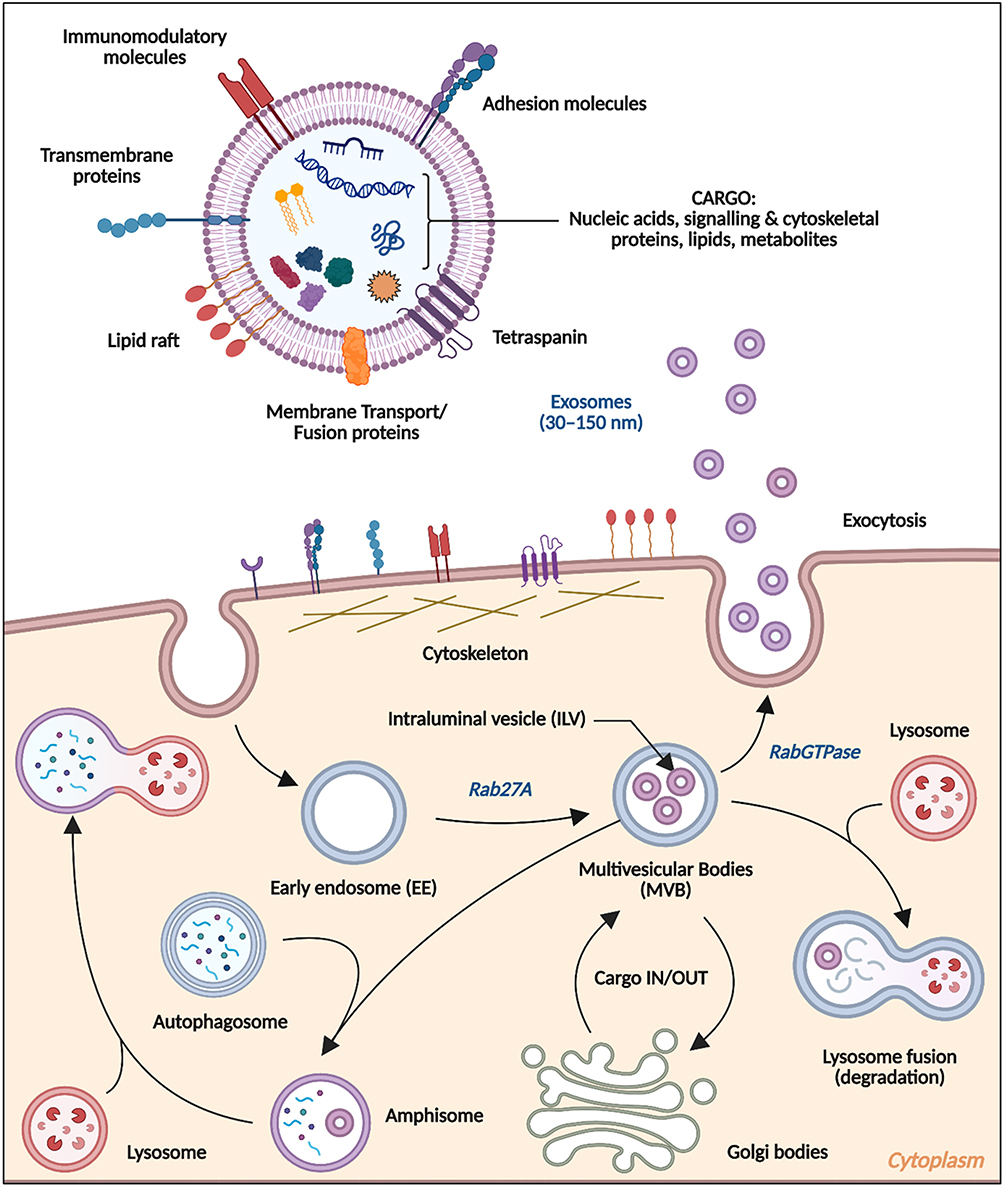 |
Figure 1 Biogenesis of Extracellular Vesicles (inset Exosomes). Created in BioRender. Dhara, B. (2025) https://BioRender.com/fu1t5oy. |
The different classes of EVs (microvesicles, exosomes, and apoptotic bodies) are shown in Figure 2. The method of separation of the various classes of EVs by centrifugation are also shown in Figure 2
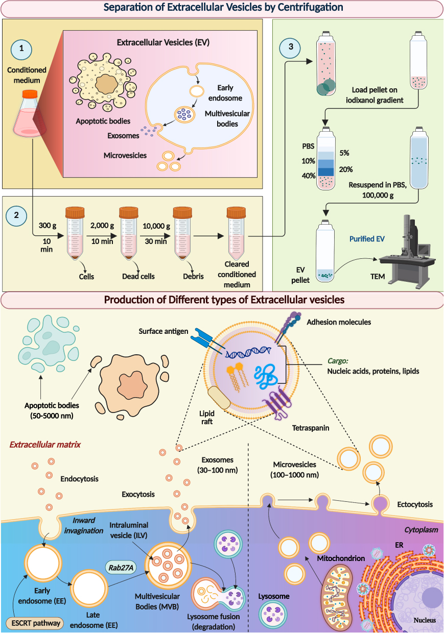 |
Figure 2 Different classes of EVs (Microvesicles, Exosomes, and Apoptotic bodies), and method to separate them by Centrifugation. Created in BioRender. Dhara, B. (2025) https://BioRender.com/c2u8njb. |
Mechanisms and Regulation of Exosome Biogenesis and Cargo Sorting
Exosome biogenesis is a critical process in cellular communication involving the formation of small EVs known as exosomes, which facilitate intercellular transfer of a wide range of molecular signals (Figure 3). This process begins with the inward budding of the membrane in parts of the cell called endosomes, which mature into MVBs containing numerous intraluminal vesicles (ILVs). These vesicles are loaded with diverse biomolecules, such as proteins, lipids, and RNAs, destined for various functions, including signalling and waste removal.39 The cargo sorting into ILVs is primarily orchestrated by the Endosomal Sorting Complex Required for Transport (ESCRT) machinery. This complex machinery comprises multiple components—ESCRT-0, ESCRT-I, ESCRT-II, and ESCRT-III—each playing a sequential role in the selection and sequestration of ubiquitinated cargo molecules into the developing ILVs.40 The ESCRT machinery also assists in the scission of ILVs from the endosomal membrane, a process facilitated by the ATPase VPS4, which disassembles the ESCRT-III complex, allowing the completion of vesicle formation.41 Alongside the ESCRT-dependent mechanisms, exosome biogenesis can occur via ESCRT-independent pathways. These pathways often involve lipids such as ceramide, which promotes membrane curvature and vesicle budding without the need for ESCRT components.42 Additionally, tetraspanins, including CD63 and CD81, contribute to the formation of tetraspanin-enriched microdomains that act as sites for selective cargo sorting into ILVs.43 The regulation of exosome biogenesis and cargo sorting is influenced by various cellular signals and conditions, which can alter the molecular composition of the exosomes and their functional properties.44 For instance, changes in cellular stress levels, such as hypoxia or oxidative stress, can modify the rate of exosome release and the nature of their cargo, potentially impacting processes like tumor metastasis or immune responses.45 This complex interplay of molecular mechanisms and pathways highlights the intricate nature of exosome biogenesis and emphasises its importance in mediating cellular communication across a range of physiological and pathological contexts.
 |
Figure 3 Biogenesis of Exosomes by (1) ESCRT independent pathway involving sphingomyelin, ceramide, and tetraspanins, and (2) ESCRT dependent pathway involving ESCRT, ALIX, TGS101, and tetraspanins. Created in BioRender. Dhara, B. (2025) https://BioRender.com/qec2nib. |
Strategies in Exosome Engineering: Active and Passive Cargo Loading
Exosome engineering encompasses sophisticated methodologies for the selective incorporation of specific cargoes, which can be broadly classified into active and passive loading processes. Active loading involves genetic modifications or the use of specific proteins to direct the sorting of targeted molecules into exosomes during their biogenesis (Figure 4). This method leverages the cell’s natural vesicular transport mechanisms, incorporating engineered RNA or proteins that bind to the exosome-associated molecules, ensuring their inclusion in the newly formed exosomes. For instance, researchers might introduce specific RNA sequences or binding proteins that interact with the exosome formation machinery to facilitate the selective incorporation of therapeutic agents like small interfering RNAs (siRNAs) or antigens.46
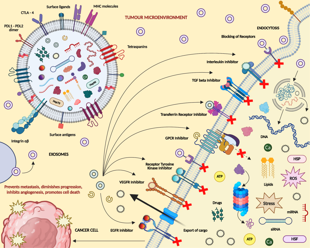 |
Figure 4 Engineering of Exosomes for inhibiting Cancer. Created in BioRender. Dhara, B. (2025) https://BioRender.com/w4vm9id. |
On the other hand, passive loading techniques involve the incorporation of desired cargoes into already formed exosomes through methods such as electroporation, sonication, or incubation.47 This technique is typically used for the encapsulation of bioactive molecules, such as drugs or nucleic acids, that do not naturally get packaged into exosomes. While this method offers the advantage of loading a wide variety of cargoes after exosome isolation, it can sometimes affect the structural integrity and functional properties of the exosomes (Figure 5). Exosome modification can be achieved by (a) Genetic Manipulation – the genetic makeup of the parent cell is modified to change the cargo, surface antigens, and receptors of exosomes, (b) Chemical Modification – cargo (such as anticancer drugs) can be introduced via photochemical reactions, lipofection, electroporation, or sonication. Surface functionalization of exosomes can also be performed with the help of click chemistry, (c) Membrane Fusion – fusion of exosomes with other cells (such as monocytes) can yield new exosomes with surfaces modified with receptors and transmembrane proteins, and containing special cargo. Both active and passive loading strategies are crucial in the field of drug delivery, particularly in the development of targeted therapies that exploit the natural homing abilities of exosomes. Each method has its advantages and is chosen based on the cargo’s nature, the desired specificity, and the efficiency of loading required for therapeutic efficacy.
 |
Figure 5 Modifications of Exosomes by genetic manipulation, membrane fusion, and chemical manipulation. Created in BioRender. Dhara, B. (2025) https://BioRender.com/17dl4h4. |
Vesicles or liposomes or transfersomes containing CRISPR-Cas9 edited plasmid (chimeric/Adenine or Cytosine edited DNA), or virus-like nanoparticles containing DNA, siRNA, RISC, microRNA, and/or desired mRNA can be used to transfect a cancer cell. siRNA, RISC and microRNA can prevent the expression of natural proteins involved in exosome biogenesis in cancer cells, thus significantly reducing the population of exosomes containing cargo which help in cancer cell communication and metastasis. Moreover, the transfected chimeric/edited DNA or desired mRNA can lead to expression of our desired proteins which will help us to manipulate the cancer cell behaviour and metabolism. These transfected nucleic acids can be loaded in the exosomes produced by the transfected cell, and these exosomes can carry them to different cells of the neighbourhood, initiating the aforementioned processes in those cells too, thus spreading the effect throughout the cancer microenvironment (Figure 6).
 |
Figure 6 Transfection of CRISPR-Cas9 edited plasmid and other nucleic acids via VLNP to manipulate exosome biogenesis in cancer cells and role of exosomes in tumour microenvironment. Created in BioRender. Dhara, B. (2025) https://BioRender.com/egaebxn. Abbreviations: ECM, extracellular membrane; VLNP, virus-like nanoparticle transfection. |
Therapeutic Targeting of Exosome Biogenesis in Cancer Treatment
The strategic inhibition of exosome biogenesis presents a promising avenue for cancer therapy. Research has highlighted several small-molecule compounds capable of disrupting the exosomal release pathways, which are critical for cancer cells to communicate and sustain their malignant behavior. For instance, sphingomyelinases, particularly neutral sphingomyelinase (nSMase), play a pivotal role in the biogenesis of exosomes by converting sphingomyelin to ceramide, which facilitates membrane curvature and vesicle formation. Small-molecule inhibitors like GW4869 directly target nSMase, effectively reducing exosome secretion and potentially limiting cancer progression.48 Furthermore, other compounds such as imipramine and cambinol have been studied for their capacity to inhibit exosome release, offering a dual therapeutic benefit by disrupting cancer cell communication and enhancing the efficacy of chemotherapeutic agents. These inhibitors operate by modulating the lipid composition of exosome membranes, underscoring the lipid-dependent pathways as crucial targets in exosome biogenesis. By focusing on these molecular pathways, researchers aim to develop targeted therapies that can mitigate the adverse effects of tumor-derived exosomes on tumor growth and metastasis, paving the way for novel interventions in oncology.
Nanotechnology in Exosome Isolation and Detection
The use of nanotechnology in the isolation and detection of exosomes has been made possible because of the small size of these biomolecules and the need to handle them in the most effective way49 (Figures 7 and 8D). Nanotechnology is thus applied in isolation and analysis of exosomes because of their small size and complexities which include difficulty in isolation.50 The advanced approaches including Nanoparticle Tracking Analysis (NTA) and Tunable Resistive Pulse Sensing (TRPS) have been improved for increased efficiency and specificity of exosome identification as described in Table 1 and Figure 7. NTA is used to calculate the size and concentration of nanoparticles in the motion induced by laser light, which delivers the quantitative data for diagnostics needed by NTA.51 TRPS provide for complementarity by detecting alterations in electrical response as the exosome translocates through a nanopore – providing a veritable measurement of size and concentration.52 However, microfluidic chip technology is a giant leap in this area as discussed in Table 1. These chips incorporate nanoscale sensing and microfluidic circuits that must allow for the off-chip meter of exosomes.53 These chips improve the detection sensitivity and specificity by embedding nanomaterial such as graphene oxide and gold nanoparticles thus making it easier to incorporate exosome diagnostics into clinical practice.
 |
Figure 7 Isolation, Characterization, and Cancer Theranostic of Exosomes. Reprinted Mukerjee N, Bhattacharya A, Maitra S, et al. Exosome isolation and characterization for advanced diagnostic and therapeutic applications. Materials Today Bio. 2025; 31(101613). Creative Commons.54 Timeline of Exosome based cancer therapy. Reprinted from Hussen BM, Faraj GS, Rasul MF, et al. Strategies to overcome the main challenges of the use of exosomes as drug carrier for cancer therapy. Can Cell Inter. 2022;22(1):323. Creative Commons.55 |
 |
Figure 8 (A) Purification and enrichment of exosomes; (B) Immunoaffinity enrichment of exosomes. Adapted from Mukerjee, N, Sarkar, S, Uti, DE, et al. Advancements in exosome-based cancer diagnosis: from chipsets to nano vaccine. Cancer Biology & Therapy. 2025;26(1). Creative Commons.56 Created in BioRender. Dhara, B. (2025) https://BioRender.com/t1akwpb. |
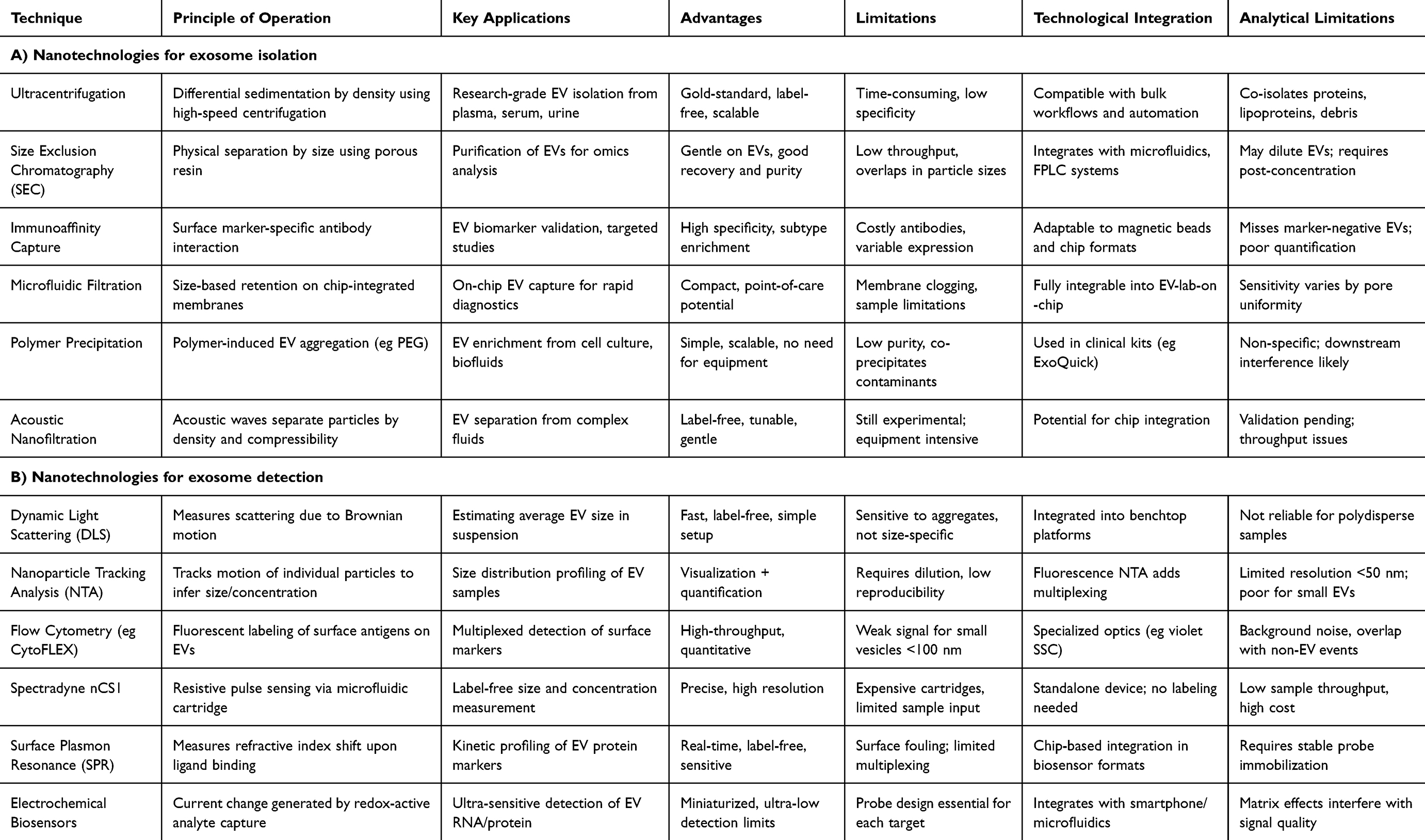 |
Table 1 Nanotechnological Approaches Towards Isolation and Detection of Exosomes.54,57 |
Additionally, newer exosome detection methods such as dynamic light scattering (DLS), spectradyne nCS1 analysis, flow cytometry platforms like CytoFLEX, and tunable resistive pulse sensing have expanded the scope of nanoparticle analysis. On the isolation side, ultracentrifugation, tangential flow filtration (TFF), and size-exclusion chromatography are now widely standardized.
Exosome Characterization
Chipsets, particularly microfluidic and biosensor-integrated devices, offer a miniaturized and automated platform for the isolation, detection, and analysis of extracellular vesicles (exosomes). Unlike conventional diagnostic tools that rely on multi-step procedures and require high sample volumes, exosome-based chipsets can combine capture, enrichment, and biomarker detection within a single device. These systems offer advantages in terms of speed, sensitivity, scalability, and point-of-care applicability. Their development represents a significant shift toward rapid and personalized diagnostics for cancer and other diseases. Conventional diagnostic approaches for cancer, including imaging (CT, MRI, PET) and biomarker-based assays (eg, CA-125, PSA), are often limited by poor sensitivity in early-stage detection, invasiveness, or delayed results. These methods can also suffer from variability and lack of disease specificity. In contrast, chip-based exosome diagnostic platforms offer rapid, minimally invasive, and potentially more sensitive alternatives. Their ability to process small biofluid volumes, integrate multiple detection modalities, and provide point-of-care usability positions them as next-generation diagnostic tools.
Microscopy-Based Methods
Microscopy-based methods are indispensable for the characterization of EVs, providing essential insights into their physical, structural, and molecular properties. These methods are crucial in both research and clinical applications, offering detailed visualization of EV size, morphology, and surface characteristics. Various advanced techniques, including Scanning Electron Microscopy (SEM), Transmission Electron Microscopy (TEM), Cryo-Electron Microscopy (Cryo-EM), and Atomic Force Microscopy (AFM), have been developed to address the challenges of characterizing these nanoscale vesicles.58
 |
Figure 9 Single EV analysis (SEA). (a) EVs are biotinylated and captured on a flat surface coated with neutravidin (Av). Following captivation, EVs are stained with fluorescent antibodies and images. Subsequently, fluorophores are quenched, and the staining process is repeated for a different set of markers. Reprinted with permission from Lee K, Fraser K, Ghaddar B, et al. Multiplexed Profiling of Single Extracellular Vesicles. ACS Nano. 2017;12(1). Copyright © 2017 American Chemical Society. (b) Example of a SEA image. EVs from the Glioblastoma Gli36-WT cell line were biotinylated and captured. Individual EV were detected through staining with fluorescent streptavidin StAv (top left). For molecular profiling, EVs were stained for pan-EV markers (CD9, CD63, CD81) as well as tumor markers (EGFR, EGFRvIII, IDH1, and IDH1R132). Spots with circles represent single EVs. (c) Two-dimensional t-distributed stochastic neighbour embedding (tSNE) mapping of individual EVs analyzed for 11 protein markers. The original map was redrafted for EVs from a single cell line. Data from other cell lines are shaded light gray. EVs from Gli36-WT and Gli36-IDH1R132 cell lines clustered similarly, whereas EVs from Gli36-EGFRvIII cells showed recognizable clusters. Microfluidic filtering methods. Reprinted with permission from Lee K, Fraser K, Ghaddar B, et al. Multiplexed Profiling of Single Extracellular Vesicles. ACS Nano. doi:10.1021/acsnano.7b07060.64 (d) A microfluidic device that uses membrane filters to isolate EVs from unprocessed blood samples. The device consists of size-selective filters (<1 μm) and capillary guide and is assembled by a magnetic sandwich. (e) Filtered blood sample revealed a single EV population with an average size of 167 nm. Reprinted with permission from Rho J, Chung J, Im H, et al. Magnetic nanosensor for detection and profiling of Erythrocyte-Derived Microvesicles. ACS Nano. 2013;7:11227–11233.65 (f)Schematic of the acoustic wave sorter. The device consists of a pair of interdigitated transducers to generate a standing ultrasound wave to exert differential forces on vesicles of different sizes. (g)During operation, vesicles in an acoustic region experience radiation pressure that is directly proportional to the vesicle size and move toward the pressure node. Larger vesicles move faster than smaller vesicles, thereby forming differential separation trajectories. (h) By in situ manipulation of cutoff size, vesicles could be segregated with versatile size selection and a good separation yield. Reprinted with permission from Lee K, Shao H, Weissleder R, Lee H. Acoustic purification of extracellular microvesicles. ACS Nano. 2015;9:2321–2327.66 |
SEM is widely used to produce high-resolution three-dimensional images of the surface topology of EVs. By scanning a focused beam of electrons across the sample, SEM generates signals that reveal the vesicle’s surface structure and composition. Despite requiring sample dehydration and fixation, SEM provides detailed images, often showing EVs as cup-shaped due to preparation artifacts. TEM, on the other hand, excels in its ability to produce ultra-high-resolution two-dimensional images by transmitting electrons through a thin sample. This method is particularly effective for visualizing the internal structures of EVs, such as their lipid bilayer, enclosed molecular cargo, and specific protein markers through immunogold labeling. TEM is considered a gold standard for morphological studies due to its capacity to achieve resolutions of less than 1 nm. Cryo-EM represents a breakthrough in EV imaging as it preserves their native state. By analyzing frozen-hydrated samples at extremely low temperatures, cryo-EM avoids the structural alterations caused by dehydration or chemical fixation, revealing vesicles in their natural round morphology. This method is particularly advantageous for studying EVs under near-physiological conditions, offering unparalleled structural fidelity. AFM provides an additional layer of detail by generating topographical maps and analyzing the mechanical properties of EVs. This technique measures surface stiffness, adhesion, and elasticity by scanning a sharp cantilever over the vesicle. Unlike SEM and TEM, AFM requires minimal sample preparation and can study EVs in air or liquid environments. It is especially valuable for assessing vesicle integrity and interactions with surrounding biological structures. Each of these microscopy techniques contributes unique strengths to EV research. Together, they enable a comprehensive understanding of EVs, from their surface characteristics and internal composition to their mechanical and physical properties, making them invaluable tools in the study of these versatile biological entities.58
Dynamic Light Scattering (DLS)
DLS is extensively used to analyze the size distribution of particles in suspension. This technique is based on the principle of light scattering from particles that are undergoing Brownian motion.58 In a DLS experiment, a laser beam is directed at the sample, and the scattered light is detected at various angles. The intensity of the scattered light fluctuates over time due to the random motion of the particles.58 These fluctuations are analyzed to determine the diffusion coefficients of the particles, which are then converted into particle size using the Stokes-Einstein equation.
DLS is favoured for its rapid assessment capabilities, allowing for the measurement of a broad size distribution within minutes.62 It requires minimal sample volume and preparation, which makes it a convenient choice for preliminary sample analysis. However, DLS has limitations, particularly its sensitivity to the presence of larger particles or aggregates, which can skew the size distribution results. The technique also assumes that particles are spherical, which might not be the case for all EVs, as they can exhibit various shapes depending on their biogenesis and the cellular environment.58
Nanoparticle Tracking Analysis (NTA)
NTA provides detailed insights into both the size distribution and concentration of particles by visually tracking their Brownian motion. In NTA, particles within a sample are illuminated by a laser, and their movement is captured in real time using a video camera. This technique allows for the direct observation and analysis of individual particles, which are tracked to determine their hydrodynamic size based on the Stokes-Einstein equation.58
One of the key advantages of NTA is its ability to provide single-particle analysis, which offers a more accurate representation of particle size distributions and concentrations. NTA is particularly useful when detailed information on each particle is necessary, as it can also differentiate particles based on fluorescence labeling. This capability makes NTA ideal for studying specific subpopulations of EVs.58 However, NTA can be more time-consuming than DLS due to the need to capture and analyze extensive video data, and it often requires dilution of samples to optimal concentrations for accurate tracking.
EV Enrichment Techniques
Single EV Analysis
Single EV Analysis (SEA) is a cutting-edge technique designed to analyze individual EVs at high resolution. This method involves isolating and immobilizing single EVs on a substrate, enabling the precise characterization of their protein and nucleic acid contents (Figure 9a–c). Typically, EVs are captured on microarray chips or within microfluidic channels, where they can be stained with specific antibodies or probes for detailed imaging and analysis.63 SEA is especially valuable for assessing the heterogeneity among EV populations, providing insights into their diverse functions and origins.58 This technique allows researchers to study the unique molecular signatures of individual vesicles, enhancing our understanding of their roles in disease processes and potential for use in targeted therapies.
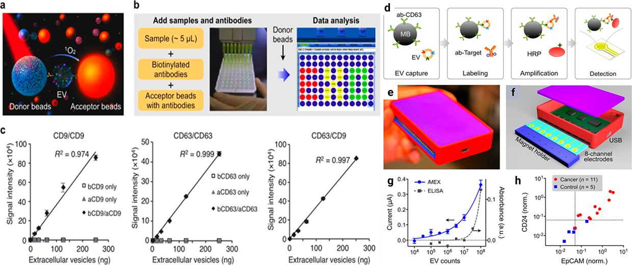 |
Figure 10 ExoScreen technology and iMEX platform: (a) Principle of ExoScreen assay; (b) Workflow of the assay; (c) Correlation between ExoScreen measurements for CD9 positive EVs, CD63 positive EVs, or CD63/CD9 double-positive EVs and EV protein concentration in a dilution series. Reprinted from Yoshioka Y, et al. Ultra-Sensitive Liquid Biopsy of Circulating Extracellular Vesicles Using Exoscreen. Nat Commun. 2014;5:3591. Creative Commons.67 Electrochemical detection: (d) Schematic representation of assay performed on the iMEX platform. EVs are captured on magnetic beads directly in plasma and labeled with horseradish peroxidase (HRP) enzymes for electrochemical detection; (e) Photograph of an established iMEX platform; (f) Schematic representation of the sensor; (g) Varying number of EVs were spiked into human plasma and assayed by ELISA and iMEX; (h) Plasma samples from ovarian cancer patients (n = 11) and healthy controls (n = 5) were analyzed with the iMEX assay. Reprinted with permission from Jeong S, Park J, Pathania D, Castro CM, Weissleder R, Lee H. Integrated magneto-electrochemical sensor for Exosome analysis. ACS Nano. 2016;10:1802–1809. Copyright 2016 American Chemical Society.68 |
Microfluidic Filtering
Microfluidic filtering harnesses the precision of microfabricated systems to isolate and sort EVs based on size and other physical properties. In this technique, EV-containing samples are processed through channels etched at the microscale, where structures can be designed to selectively trap or divert particles of specific dimensions (Figure 9d–e). This method is highly effective for purifying EVs from biological fluids, offering a high degree of control over flow dynamics and particle handling. Microfluidic filtering can be integrated with other detection or analysis technologies directly on the chip, facilitating rapid, on-site diagnostic assessments.58
Contact-Free Sorting
Contact-free sorting involves the manipulation of EVs without direct physical contact, typically using forces such as magnetic fields, acoustic waves, or optical traps (Figure 9f–h). This approach minimizes potential damage or contamination, preserving the integrity of the vesicles for subsequent analysis.58 Techniques like acoustic focusing use sound waves to sort particles based on density and compressibility, while optical tweezers employ lasers to trap and move individual vesicles. These methods are particularly useful for sorting EVs based on subtle physical differences, enabling the enrichment of specific subpopulations for detailed study.
Immunoaffinity Enrichment
Immunoaffinity enrichment leverages the specificity of antibody-antigen interactions to selectively isolate EVs expressing particular surface markers. In this technique, antibodies targeting known EV markers are immobilized on beads, plates, or within columns, and samples are passed through these prepared media. EVs with the target antigens bind to the antibodies, allowing other components to be washed away. This method is highly selective, enabling the purification of EV subsets associated with specific cell types or pathological conditions, making it invaluable for biomarker discovery and disease monitoring.58
EV Protein Analysis Techniques
Exosome proteins, particularly those expressed on the surface, serve as vital biomarkers for cancer detection and classification. Surface proteins such as tetraspanins, integrins, and disease-specific antigens provide a direct molecular fingerprint of the originating cells. These proteins are not only stable but also accessible for antibody-based detection methods. This section outlines key analytical techniques used for profiling these proteins, enabling high-throughput, label-free, and multiplexed evaluation of exosomes in clinical diagnostics. Exosome surface proteins such as CD63, CD81, and CD9 are pivotal for targeted detection, given their abundance on exosomal membranes. These markers are used in chip-based capture strategies where surfaces are functionalized with antibodies or aptamers for specific binding. Capturing exosomes via surface protein affinity not only improves purity but also enables subtype-specific isolation, which is essential for disease-specific biomarker discovery.
Micronuclear Magnetic Resonance (µnmr)
Micronuclear Magnetic Resonance (µNMR) is a highly sensitive analytical technique used for the characterization of proteins within EVs. µNMR utilizes magnetic nanoparticles that are functionalized with antibodies specific to target EV proteins. When these nanoparticles bind to their targets, they alter the local magnetic environment, which can be detected and quantified by NMR. This technique is capable of detecting very low concentrations of EVs and can provide quantitative information about the protein content of vesicles.58 µNMR is particularly useful for profiling protein expressions in EVs derived from small sample volumes, offering a powerful tool for clinical diagnostics and research applications.
Surface Plasmon Resonance (SPR)
Surface Plasmon Resonance (SPR) is a label-free technique used for real-time monitoring of molecular interactions on EV surfaces. SPR measures changes in the refractive index near the surface of a metal film when EVs bind to ligands that are immobilized on the film. This interaction causes a measurable change in the intensity of reflected light, directly correlating to the mass of the bound EVs. SPR can provide detailed kinetic data about binding events, such as association and dissociation rates, making it invaluable for studying the dynamics of EV protein interactions and for screening potential therapeutic targets.58
ExoScreen Technology
ExoScreen is a highly sensitive assay for detecting and quantifying proteins on the surface of EVs. It uses a dual-antibody sandwich approach, where two sets of antibodies are conjugated to photosensitizer beads (Figure 10a–c). When these beads come into proximity due to binding to a target protein on an EV, a chemiluminescent reaction is triggered. This reaction is only initiated when both antibodies have bound to their respective epitopes on the same vesicle, ensuring high specificity and sensitivity.58 ExoScreen is particularly useful for detecting low-abundance proteins and for screening EVs in complex biological fluids, such as blood or urine, without the need for prior purification.
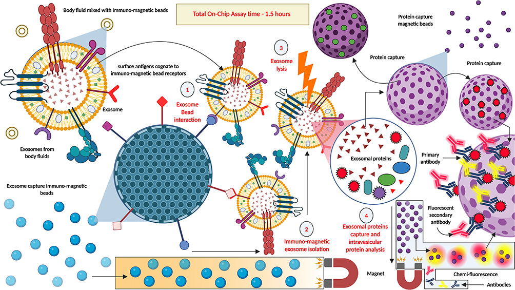 |
Figure 11 On-Chip Assay with body fluids containing exosomes. For analyzing the protein composition of these exosomes, body fluids are mixed with exosomes to capture immune-magnetic beads. Exosomes bind to these beads and can be isolated from the body fluids using a magnetic field. Exosomes are lysed, and the lysed contents are mixed with protein capture immune-magnetic beads and heterogenous population primary antibodies which bind to the captured proteins. Fluorescent secondary antibodies are then added, which bind to specific primary antibodies which are bound to specific captured proteins. By magnetic channelization and chemi-fluorescence intensity, the presence and relative abundance of the intravesicular proteins can be estimated. Created in BioRender. Dhara, B. (2025) https://BioRender.com/6kdxla5. |
Electrochemical Detection
Electrochemical detection methods are employed in EV research to quantify specific protein markers on the vesicle surfaces (Figure 10d–h). These techniques typically involve the use of electrodes coated with antibodies or other specific capture agents. When EVs bind to these coated electrodes, they produce a measurable electrical signal that can be amplified and quantified.58 Electrochemical sensors are known for their sensitivity, low cost, and rapid response times, making them well-suited for point-of-care testing and multiplexed analyses of EV proteins in clinical samples.
Exosome-Based Chipsets for the Detection of Cancer
Exosome based chip sets currently adopt the molecular profiling of exosomes for cancer diagnostics.69 These devices include microfluidics that work to analyze biological substances that may include blood or urine samples.70 These chipsets have specially designed surface capture agents incorporated in them which are usually antibodies or aptamers that target biomarkers that are unique to cancer cells and are present on or within exosomes.17,71 Such selective binding is important, since it separates the exosomes that contain relevant biomarkers of cancer.72 Figure 8 shows different strategies or methods of extracting exosomal proteins by microfluidic chip-based systems and immunoaffinity approaches. The continuous microfluidic platform allows the integration of flow-based mixing, separation and multiple marker analysis for the exosomes while the immuno-magnetic beads selectively bind onto the target exosomal proteins. Exosome studies can be performed in situ alongside an integrated microfluidic system, imaging, gene profiling, and particle analysis thereby providing better checks to isolate and characterize the exosomes with great accuracy.73 When a biological sample is loaded into this microfluidic device, these capture agents bind only to the exosomes with the proper biomarkers.74 After the capturing phase, the bound exosomes are identified through various techniques such as fluorescence where molecules with attached marker light up; changes in electrical signals or plasmonic sensing that detects any alteration in the surface plasmon resonance as a result of the exosome binding.75,76 Subsequently, the detection signals that have been obtained are quantized and characterised to give the diagnostic information of the exosomes.77 This includes the existence of cancer and possibly its stage depending on biomarker profiles of the exosomes obtained from the tumor.78,79 This integrated and highly efficient operation of exosome-based chipsets elucidates their potential to deliver sensitive and specific diagnosis of cancer, elevating the field of oncological diagnosis to a new level. To analyze the protein composition of the exosomes, on-chip assay can be performed, where the body fluids are mixed with immune-magnetic capture beads, and the exosomes bind to those immune-magnetic beads and are isolated using a magnetic field. Following this the exosomes are lysed and protein capture magnetic beads and primary antibodies are added. The exosomal proteins interact with these beads and primary antibodies, providing a landing bay for fluorescently labelled secondary antibodies which interact with specific primary antibodies bound to the captured proteins. By magnetic isolation and chemi-fluorescence, one can analyze the protein composition (relative abundance) of the exosomes (Figure 11). Recent developments in microfluidic chip platforms have enabled the simultaneous execution of exosome isolation, enrichment, and molecular analysis on a single chip. Technologies like digital microfluidics, electrochemical biosensors, and paper-based microfluidic platforms allow for multiplexed, real-time exosome detection. These chipsets are particularly suited for point-of-care applications and early diagnostics. Despite these advancements, challenges remain in terms of clinical standardization, reproducibility, and regulatory approval for large-scale deployment.
 |
Figure 12 Clinical trials on Exosomes.103 Created in BioRender. Dhara, B. (2025) https://BioRender.com/75qwt96. |
Advances in nanotechnology have significantly improved the effectiveness of the exosome-based chipsets especially in cancer detection systems.80 Among the leading technologies that have shaped these developments are the nanowire sensors, quantum dot labels and plasmonic nanoparticles which proved very useful not only in enhancing the sensitivity but also the specificity of these diagnostic methods.81–83
There are certain attributes such as ultra-sensitive electrical properties which nanowire sensors are designed to harness and they are especially useful in the detection of cancer markers.84 These sensors work based on the principle of subtle change in electrical resistance as and when exosomes, loaded with cancer biomarkers, come in contact with them.85–87 This makes it possible for the specific exosomes that are being targeted to bring about a detectable change in the electrical conductance at the nanowire surface. This change is so distinct that it can detect and count even low-abundance exosomes which are generally hidden from other standard profiling methods. This high sensitivity if provided by nanowire sensors makes it easy for early detection of cancers since even minimal disease markers may be vital in early diagnosis.88 At the same time, another innovation has taken place in the labeling technology contained in these chipsets and this is through the use of quantum dots.89 The quantum dots’ bright and stable fluorescence make them ideal to be conjugated with molecules bearing an affinity for exosomes.90 These fluorescent tags are very useful as once bound, exosomes could be easily visualized and counted under the microscope regardless of low concentration of the biomarkers on exosomes. A valuable quality of the quantum dots is their stability as compared to the traditional fluorescent dyes that tend to photo bleach quickly under illumination.91,92 Therefore, quantum dots help the researchers and clinicians to carry out the experiments or diagnoses for an extended period, without destabilising or compromising the strength of the quantum dot signal, thus providing a precise quantification, and characterization of the exosomal biomarkers. Additional advancements in the diagnostic realm are plasmonic nanoparticles that build upon their optical properties of the respective chipsets by applying the method of Surface enhanced Raman scattering (SERS).93 These nanoparticles are going to increase the local electromagnetic field values in their vicinity when excited, a factor that significantly increases the Raman-enhancement factor for closer situated molecules.94,95 This enhancement can be of particular importance to better understand the nature and the state of functional organization of the exosomes since the molecular composition of these carriers can be assessed.96,97 As a result, specific biomarkers of exosomes associated with cancer are better characterized by plasmonic nanoparticles, thus enhancing the level of diagnostics’ molecular analysis.98 Altogether, these discoveries provide the interdisciplinary synthesis of materials science and biotechnology to create exosome-based chipsets for demonstrated aims to perform at levels undreamt of in detecting and analyzing biomarkers of cancer. These technologies therefore have the potential not only to improve the diagnostic armamentarium but also increase the knowledge of cancer biology and thus potentially improve the therapeutic intervention as well.99
Clinical Trials
Over the past decade, several attempts to harness extracellular vesicles (exosomes) for liquid biopsy applications have encountered critical technological and analytical limitations. Early-generation exosome diagnostic platforms, such as label-free impedance biosensors and polymer-based precipitation kits (eg, ExoQuick), demonstrated ease of use but suffered from poor specificity, co-isolation of contaminants, and lack of reproducibility across clinical samples. These systems often failed to differentiate tumor-derived exosomes from those released by non-malignant cells, thereby undermining their diagnostic utility. In parallel, competing liquid biopsy approaches like circulating tumor DNA (ctDNA) and cell-free DNA (cfDNA) gained attention for their non-invasive potential. However, these analytes exhibit low stability in circulation, limited representation of protein-level information, and reduced sensitivity in early-stage cancers where nucleic acid fragments may be scarce or fragmented. In contrast, exosome-based chipsets offer a unique combination of biomolecular richness (nucleic acids, proteins, lipids), structural integrity, and real-time detectability using surface-enhanced, multiplexed nanotechnologies. These features enable chip-based exosome platforms to outperform traditional methods by supporting multi-omic profiling and preserving spatial-temporal information essential for early cancer detection.
Nanocarriers, especially exosome-based chipsets, are focused on their revolutionary functions in detecting cancer at initial stages. Exosome analyzer is nanotechnology-based devices to isolate and analyze exosomes, which are EVs that transport diagnostic biomarkers including proteins, DNA and RNA from cancer cells.100,101 The article reveals how chipsets utilising exosomes could transform diagnostics, for example by enabling non-invasive, highly sensitive and specific, and early-stage diagnostics for cancer that is critical for optimising the right patient outcomes. Figure 12 highlights clinical trials related to exosomes based across all types of cancer including blood, plasma, urine and other humoral fluids also as biomarkers. Pancreatic cancer, lung cancer, and colorectal cancers are some of the clinical trial areas today which depict the importance of exosome research. These trials assess exosome’s diagnostic and prognostic capability in cancer and its therapeutic as well as signaling research uses. For instance, the studies establish the approaches used in isolation of exosomes from blood and tissue in managing cancer progression and treatment outcomes. It also covers limitations: such as putting forth requirements in terms of the methods of exosome isolation; these correspond to clinical practice attempts to enhance and perfect such approaches to the use of exosomes for diagnostics. Detailed exosomes clinical trials are given in Table 2, while their comparison with other methods are given in Table 3. This article suggests that future research on exosomes is bright for cancer diagnostic and targeted therapy, even as clinical trials and technology breakthroughs offer the way forward for exosome research in cancer treatment. Despite their transformative potential, exosome-integrated chipsets face several translational bottlenecks that extend beyond mere cost or device integration challenges. Chief among these is reproducibility—a fundamental issue arising from the lack of harmonized protocols for exosome isolation, enrichment, and downstream analysis. Variations in fluidic chip design, sample input quality, and processing times can lead to inconsistent diagnostic outputs, complicating cross-laboratory validations. Sensitivity remains another concern, particularly in detecting tumor-specific exosomes amidst a vast background of vesicles from normal tissues. This issue is further aggravated in early-stage malignancies or low-volume biofluid samples. Additionally, standardization in the field is lacking across multiple dimensions: from pre-analytical sample handling (eg, centrifugation speeds, filtration cut-offs) to analytical quantification strategies (eg, nanoparticle tracking vs surface marker expression). The absence of universally accepted performance metrics impedes regulatory clearance and large-scale clinical deployment.
 |
Table 2 Detailed Overview of Exosome-Based Clinical Trials in Cancer.102 |
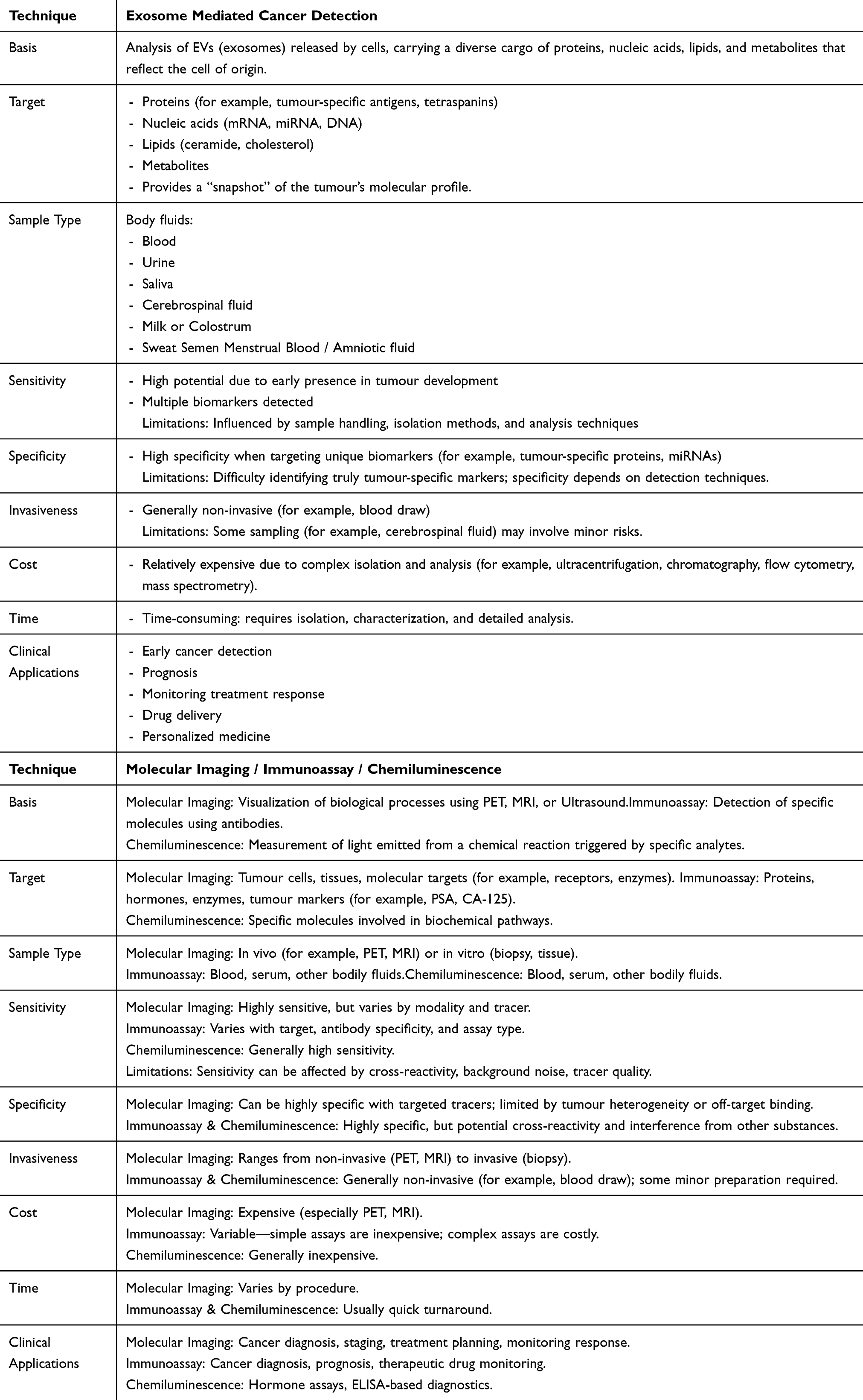 |
Table 3 Comparing Between Exosomes and Other Clinical Cancer Detection Methods |
Different biofluid sources present varying challenges and opportunities for exosome isolation and analysis. For example, plasma and serum are rich in proteins and lipids that can interfere with conventional ultracentrifugation or precipitation techniques. Chip-based microfluidic approaches, in contrast, allow selective enrichment and detection even in complex matrices like blood. Urine and saliva, though less invasive to collect, often yield lower exosome concentrations, requiring chip-based amplification strategies. These platforms are also being tailored to capture exosomes carrying specific protein or nucleic acid signatures, enhancing biomarker sensitivity. Thus, the selection of chip-based techniques can be optimized based on the physicochemical properties of the target biofluid.
Challenges and Future Directions
Based on the positive outcomes of the diagnostic potential of exosome-based chipsets for early-stage detection of the disease, researchers are faced with several disadvantages that prevent their wide application. One is non-homogeneity of samples due to variability in the method used in isolation of exosomes; this greatly reduces the reliability of the diagnostic test. Setting up benchmark measures is critical towards moving some of these solutions from the lab to practical application.103 The high cost incurred in both the development as well as manufacturing of the nanotechnology-based chipsets also consists of notable challenges. These factors include the use of gold nanoparticles and the generally complex nature of the fabrication techniques which lead to high cost of production.104 While improving the accuracy of the diagnostic tools is important, it is equally important to discover more affordable and efficient materials or streamline the process of creating these devices so that they can be produced cheaply in places with fewer resources. Moreover, the inclusion of these chipsets into currently running healthcare systems is an even bigger challenge. They are dependent on certain hardware to run the operations and analyze the data and the end users which are the healthcare practitioners must be in a position to understand the results. Operational implementation and integration with the current modes of medical practice and electronic medical records entails technology, as well as legal barriers that have to be surmounted.
These chipsets are still under active research as proven by the efforts to improve the surface properties of the chipsets and developing special detection mechanisms that make them even more specific and sensitive. It could mean the ability to detect cancer at an early stage through better tests with better technology and therefore significantly improve the quality of life of patients. Moreover, the idea of diagnostics of personalized medicine via exosome-based chipsets has much potential. Personalizing the diagnostic and therapeutic strategies according to molecular characteristics of tumors can prospectively change the management of cancer. This entails a solid understanding of the molecular characteristics of cancers as well as how they display themselves in exosomes. Reasons discovered for continued innovation together with cross-disciplinary study and cooperation are important in eradicating existing hurdles towards optimizing this effective technology in hospitals.
Exosome-based approaches are not developed to completely replace the established cancer management techniques, but rather to function as powerful complementary tools, strategically addressing significant unaddressed needs in the field. For instance, in diagnosis, while a traditional tissue biopsy remains one of the best standards for definitive pathological confirmation, it is inherently invasive, carries procedural risks, and represents a static snapshot of a tumour at a particular point of time. Exosome diagnostics, on the other hand, offer the promise of early, non-invasive detection through a liquid biopsy, where specific tumour-derived exosomal cargo could serve as highly sensitive biomarkers. This allows for frequent, real-time monitoring of disease progression and treatment response without repeated invasive procedures, a capability that current imaging techniques such as CT or MRI, while excellent for visualizing tumour size and location, often lack in terms of molecular detail and sensitivity for microscopic disease. Similarly, exosome-based therapies aim for targeted, less toxic drug delivery, a critical advancement over systemic chemotherapy, which indiscriminately attacks rapidly dividing cells, leading to severe side effects like hair loss, nausea, and immune suppression. By engineering exosomes to specifically home in on cancer cells, potentially delivering concentrated therapeutic payloads like anti-cancer agents or gene-editing tools, exosome therapies aspire to minimize damage to healthy tissues, a limitation that even some targeted therapies or antibody-drug conjugates (ADCs) can still face due to off-target binding or systemic drug release. Therefore, the ultimate success and widespread clinical adoption of exosome technologies will hinge on their ability to definitively demonstrate superior advantages in terms of sensitivity, specificity, safety, efficacy, and practical utility when compared to the existing and continually evolving landscape of traditional and emerging diagnostic and therapeutic alternatives.
While no exosome-based cancer diagnostic or therapeutic technologies have been definitively failed and completely abandoned after thorough clinical trials, the field has encountered significant challenges that represent substantial hurdles in achieving broad clinical implementation. For diagnostics, initial methods for isolating exosomes, such as ultracentrifugation, often yielded impure and damaged samples, proving ineffective for accurate and reproducible biomarker analysis. Furthermore, many early-identified exosomal biomarkers lacked the necessary specificity, leading to high rates of false positives or negatives in larger studies, thus failing to provide reliable diagnostic accuracy. A pervasive issue has been the lack of standardized protocols, causing unacceptably poor reproducibility of results across different laboratories, thereby hindering the validation required for clinical utility. In the realm of exosome-based therapies, initial attempts at loading therapeutic cargo into exosomes often resulted in very low encapsulation efficiency or damage to the exosomes, thus failing to deliver a therapeutically meaningful dose to cancer cells. A critical failure to date has also been the inability to develop robust, scalable, and cost-effective manufacturing processes to produce clinical-grade exosomes in sufficient quantities for widespread therapeutic application. Finally, early exosome formulations often exhibited poor stability and rapid clearance from the bloodstream, preventing effective accumulation at tumour sites and thereby failing to achieve the desired therapeutic effect in vivo. These challenges, while not leading to outright abandonment of the technology, represent significant limitations that researchers are actively working to overcome to realize the full potential of exosome-based approaches in cancer care.
Conclusion
The integration of exosome-based chipsets into the field of cancer diagnostics represents a pivotal advancement in the early detection and personalized management of cancer. By leveraging nanotechnology, these platforms offer a unique ability to isolate and analyze exosomes—nanoscale vesicles that serve as dynamic biomarkers of cellular states. This capability allows for unprecedented insights into the molecular and functional characteristics of cancer at its earliest stages, facilitating early intervention and improving treatment outcomes. The non-invasive nature of these technologies, combined with their high sensitivity and specificity, positions them as a significant improvement over traditional diagnostic methods such as imaging and biopsies, which are often limited in their ability to detect early-stage cancer. Moreover, the versatility of exosome-based chipsets extends beyond diagnostics to include therapeutic applications. Engineered exosomes provide an innovative vehicle for delivering targeted therapies, particularly in combating metastasis and complex tumor microenvironments. The integration of advanced technologies such as quantum dots, nanowire sensors, and plasmonic nanoparticles has further enhanced the precision and efficiency of these platforms, setting the stage for their broader adoption in clinical settings.
Despite their promise, challenges remain in standardizing exosome isolation methods, reducing manufacturing costs, and integrating these tools into existing healthcare frameworks. There are several limitations of exosome-based cancer diagnosis and therapy. Exosomes are secreted by virtually all cell types, and their cargo varies significantly depending on the cell of origin, its physiological state, and the surrounding microenvironment. This biological complexity means that a patient’s biofluid contains a highly diverse population of exosomes from both healthy and cancerous cells, as well as other conditions. Distinguishing cancer-specific exosomes from this heterogeneous background is a major analytical challenge. While research has identified some tumour-specific exosome biomarkers, a universal and highly specific set of biomarkers for early detection or precise disease monitoring across all cancer types is still lacking. A suspected cancer biomarker might be elevated in multiple conditions, leading to false positives.
Current isolation methods (for example, ultracentrifugation, size-exclusion chromatography, immunoaffinity capture, microfluidics, precipitation) struggle to achieve both high purity by removing contaminants like proteins, lipoproteins, and other EVs, and high yield simultaneously. Impurities can interfere with diagnostic assays, leading to inaccurate results, while low yield can limit the quantity of material available for analysis, especially for early-stage cancers where number of exosomes might be low. There’s a significant lack of standardized protocols for exosome isolation and characterization across different laboratories and studies. Minor variations in centrifugation times, storage temperatures, or reagent batches can drastically impact the quality, quantity, and cargo profile of isolated exosomes, compromising reproducibility and comparability of research findings, which hinder the development of definitive and dependable diagnostic assays for clinical purposes. Some isolation methods, particularly those involving severe physical forces like ultracentrifugation, can damage exosome integrity, altering their structure and potentially their cargo, which could affect diagnostic accuracy. In early stages of cancer, the concentration of tumour-derived exosomes in biofluids can be very low, making their detection challenging even with highly sensitive methods. The nanoscale size of exosomes and the complex matrix of biological fluids make their efficient isolation and sensitive detection difficult. Developing biosensors and analytical platforms with sufficient sensitivity and specificity for different exosome biomarkers is a happening area of research. The diagnostic reliability and sensitivity of exosome-based assays still need extensive validation through large-scale clinical studies across diverse patient populations to prove their utility in routine clinical settings.
Exosomes have a limited internal volume. This inherent constraint poses a significant challenge for loading large therapeutic molecules such as chromatin remodelling complexes or gene editing tools, or high payloads of smaller drugs that might be required for effective therapy. Current methods for loading therapeutic cargo into exosomes (for example, incubation, electroporation, sonication, transfection of donor cells) often suffer from low loading efficiency, variability, and potential damage to either the exosome or the cargo. Achieving consistent and high loading remains a major hurdle. While exosomes have a natural propensity to target certain cells, achieving precise and exclusive targeting of cancer cells while minimizing uptake by healthy cells is complex. Modifying exosomes for enhanced tumour-specific targeting is an important research area, but it adds layers of complexity and potential for off-target effects if not optimized. Cancer cells can also quickly adapt and change their surface receptor expression, making single-ligand targeting less effective over time. Although generally considered less immunogenic than synthetic nanoparticles, exosomes, especially those derived from allogeneic sources, can still potentially elicit an immune response in vivo, leading to their rapid clearance or reduced therapeutic efficacy. Exosomes can be susceptible to degradation in the body and are quickly cleared from circulation. This short half-life limits their systemic delivery and sustained therapeutic effect, requiring frequent administration or strategies to enhance their in vivo stability.
Producing clinical-grade exosomes in sufficient quantities for therapeutic applications is a significant challenge. Current methods are often labour-intensive, time-consuming, and have low yields, making large-scale manufacturing difficult and expensive. Maintaining consistent quality, purity, and cargo content across different production batches of exosomes is crucial for therapeutic reproducibility and safety, but it remains a substantial hurdle due to the inherent biological variability of exosome production and isolation. As a relatively new class of biological therapeutics, there is lack of clear, standardized guidelines for their manufacturing, quality control, and clinical testing creates uncertainty for scientists and pharmaceutical companies. Furthermore, demonstrating the long-term safety and consistent efficacy of exosome-based therapies in vigorous clinical trials is essential for regulatory approval and widespread implementation.
Addressing these barriers will require interdisciplinary collaboration, regulatory support, and continuous innovation to ensure that these technologies are both accessible and effective in diverse clinical environments. Additionally, efforts to educate healthcare providers and streamline the analysis of data generated by these chipsets will be crucial for their widespread implementation. Exosome-based chipsets can play a transformative role in the evolution of precision medicine. By enabling early, accurate, and personalized cancer diagnostics, these platforms have the potential to significantly improve patient prognoses and quality of life. Continued research and investment in this field will not only refine the technology but also expand its applications across a broader spectrum of diseases, solidifying its position as a cornerstone of future medical practice. Chip-based nanotechnology offers multiple advantages for exosome biomarker discovery and clinical diagnostics, including high sensitivity, miniaturization, and integration with multiplexed detection platforms. These devices enable rapid sample processing and minimal reagent use, making them ideal for resource-constrained settings. However, limitations include inconsistent exosome capture due to heterogeneity in surface markers, issues with long-term reproducibility, and lack of universal chip standards. Clinical translation also faces hurdles related to inter-laboratory variability, regulatory clearance, and the need for large-scale validation studies. Current research is now focused on improving chip biocompatibility, scalability, and automation for high-throughput use. Bridging these gaps will be essential for transitioning exosome-chip platforms from bench to bedside.
Abbreviations
EVs, Extracellular Vesicles; CD63, CD81, CD9, Cluster of Differentiation (CD); miRNA, MicroRNA; mRNA, Messenger RNA; MVB, Multivesicular bodies; ESCRT, Endosomal Sorting Complex Required for Transport; nSMase, Neutral sphingomyelinase; TRPS, Tunable Resistive Pulse Sensing; NTA, Nanoparticle Tracking Analysis; TEM, Transmission Electron Microscopy; SEM, Scanning Electron Microscopy; Cryo-EM, Cryo-Electron Microscopy; AFM, Atomic Force Microscopy; µNMR, Micronuclear Magnetic Resonance; SPR, Surface Plasmon Resonance; DLS, Dynamic Light Scattering; SEA, Single Extracellular Vesicle Analysis; EGFR, Epidermal Growth Factor Receptor; SERS, Surface-Enhanced Raman Scattering; VLNP, Virus-Like Nanoparticles; NSCLC, Non-Small Cell Lung Cancer; PEG, Polyethylene Glycol; CRISPR, Clustered Regularly Interspaced Short Palindromic Repeats; RISC, RNA-Induced Silencing Complex.
Data Sharing Statement
All used data is within the manuscript.
Acknowledgment
All authors would like to acknowledge their respective departments for the conduct of the study.
Author Contributions
All authors made a significant contribution to the work reported, whether that is in the conception, study design, execution, acquisition of data, analysis and interpretation, or in all these areas; took part in drafting, revising or critically reviewing the article; gave final approval of the version to be published; have agreed on the journal to which the article has been submitted; and agree to be accountable for all aspects of the work.
Funding
This review did not receive any funding from any individual or organization.
Disclosure
The authors declare that there are no competing interests for this work.
References
1. Yu Z, Bai X, Zhou R, et al. Differences in the incidence and mortality of digestive cancer between global cancer observatory 2020 and global burden of disease 2019. Int J Cancer. 2024;154(4):615–625. doi:10.1002/ijc.34740
2. Liu B, Zhou H, Tan L, Siu KT, Guan XY. Exploring treatment options in cancer: tumor treatment strategies. Signal Transduct Target Ther. 2024;9(1):175.
3. Alcorta A, López-Gómez L, Capasso R, Abalo R. Vitamins and fatty acids against chemotherapy-induced intestinal mucositis. Pharmacol Ther. 2024;5:108689. doi:10.1016/j.pharmthera.2024.108689
4. Ringborg U, von Braun J, Baumann M, et al. Strategies to decrease inequalities in cancer therapeutics, care and prevention: proceedings on a conference organized by the pontifical Academy of sciences and the European Academy of cancer sciences, Vatican city, February 23–24, 2023. Mol Oncol. 2024;18(2):245. doi:10.1002/1878-0261.13575
5. Huang X, Auffan M, Eckelman MJ, et al. Trends, risks and opportunities in environmental nanotechnology. Nat Rev Earth Environ. 2024;5(8):572–587. doi:10.1038/s43017-024-00567-5
6. Mojtaba Mousavi S, Alireza Hashemi S, Yari Kalashgrani M, et al. Recent progress in prompt molecular detection of exosomes using CRISPR/Cas and microfluidic‐assisted approaches toward smart cancer diagnosis and analysis. ChemMedChem. 2024;19(1):e202300359. doi:10.1002/cmdc.202300359
7. Kalra H, Drummen GP, Mathivanan S. Focus on extracellular vesicles: introducing the next small big thing. Int J Mol Sci. 2016;17(2):170. doi:10.3390/ijms17020170
8. Ratajczak MZ, Ratajczak J. Extracellular microvesicles/exosomes: discovery, disbelief, acceptance, and the future? Leukemia. 2020;34(12):3126–3135. doi:10.1038/s41375-020-01041-z
9. Hánělová K, Raudenská M, Masařík M, Balvan J. Protein cargo in extracellular vesicles as the key mediator in the progression of cancer. Cell Commun Signaling. 2024;22(1):25. doi:10.1186/s12964-023-01408-6
10. Ghosh S, Rajendran RL, Mahajan AA, et al. Harnessing exosomes as cancer biomarkers in clinical oncology. Can Cell Inter. 2024;24(1):278. doi:10.1186/s12935-024-03464-5
11. Rahimian S, Najafi H, Afzali B, Doroudian M. Extracellular vesicles and exosomes: novel insights and perspectives on lung cancer from early detection to targeted treatment. Biomedicines. 2024;12(1):123. doi:10.3390/biomedicines12010123
12. Wang Z, Wang Q, Qin F, Chen J. Exosomes: a promising avenue for cancer diagnosis beyond treatment. Front Cell Develop Biol. 2024;12:1344705. doi:10.3389/fcell.2024.1344705
13. Srivastava S, Jayaswal N, Kumar S, et al. Unveiling the potential of proteomic and genetic signatures for precision therapeutics in lung cancer management. Cell Signalling. 2024;113:110932. doi:10.1016/j.cellsig.2023.110932
14. Shi W, Wartmann T, Accuffi S, et al. Integrating a microRNA signature as a liquid biopsy-based tool for the early diagnosis and prediction of potential therapeutic targets in pancreatic cancer. Br J Cancer. 2024;130(1):125–134. doi:10.1038/s41416-023-02488-4
15. Jin X, Zhang J, Zhang Y, et al. Different origin-derived exosomes and their clinical advantages in cancer therapy. Front Immunol. 2024;15:1401852. doi:10.3389/fimmu.2024.1401852
16. Pagani A, Duscher D, Geis S, et al. The triple adipose-derived stem cell exosome technology as a potential tool for treating triple-negative breast cancer. Cells. 2024;13(7):614. doi:10.3390/cells13070614
17. Maitra S, Mukerjee N, Alharbi HM, Ghosh A, Thorat ND, Thorat ND. Targeted therapies for HPV‐associated cervical cancer: harnessing the potential of exosome‐based chipsets in combating leukemia and HPV‐mediated cervical cancer. J med Virol. 2024;96(4):e29596. doi:10.1002/jmv.29596
18. Mukerjee N, Maitra S, Ghosh A, et al. Synergizing proteolysis-targeting chimeras and nanoscale exosome-based delivery mechanisms for HIV and antiviral therapeutics. ACS Appl Nano Mater. 2024;7(4):3499–3514. doi:10.1021/acsanm.3c04537
19. Mukerjee N, Maitra S, Ghosh A, Thorat ND, Thorat ND. Exosome-mediated PROTAC delivery for treatment of RNA viral infections and zoonoses. Drug Discov Today. 2024;23(7):104044. doi:10.1016/j.drudis.2024.104044
20. Ramchandani M, Kumari P, Goyal AK. Aptamers as theranostics in cardiovascular diseases. J Nanotheranostics. 2023;4(3):408–428. doi:10.3390/jnt4030018
21. Maurya R, Sebastian P. Extracellular vesicle-associated microRNA in human parasitic diseases. In: MicroRNA in Human Infectious Diseases. Academic Press; 2024:293–306.
22. Ghosh D, Rudra DS, Pal U. Cell-Derived Exosome-Based Materials for Biomedical Applications. In: Handbook of the Extracellular Matrix: Biologically-Derived Materials. Cham: Springer International Publishing; 2024:1–26.
23. Parameshwar K, Mounika N, Parameshwar R, Narayana NA. Exosomes: biogenesis, Composition, and Synthesis. In: Exosomes Based Drug Delivery Strategies for Brain Disorders. Singapore: Springer Nature Singapore; 2024:37–53.
24. Zubkova E, Kalinin A, Beloglazova I, Menshikov M, Menshikov M. Autophagy-dependent secretion: crosstalk between autophagy and exosome biogenesis. Curr Issues Mol Biol. 2024;46(3):2209–2235. doi:10.3390/cimb46030142
25. Yang C, Xue Y, Mao C, Wan M, Wan M. Extracellular vesicles and their engineering strategies, delivery systems, and biomedical applications. J Control Release. 2024;365:1089–1123. doi:10.1016/j.jconrel.2023.11.057
26. Zhao W, Li K, Li L, et al. Mesenchymal stem cell-derived exosomes as drug delivery vehicles in disease therapy. Int J Mol Sci. 2024;25(14):7715. doi:10.3390/ijms25147715
27. Payandeh Z, Tangruksa B, Synnergren J, et al. Extracellular vesicles transport RNA between cells: unraveling their dual role in diagnostics and therapeutics. Mol Aspect Med. 2024;99:101302. doi:10.1016/j.mam.2024.101302
28. Hurwitz SN, Rider MA, Bundy JL, Liu X, Singh RK, Meckes DG Jr. Proteomic profiling of NCI-60 extracellular vesicles uncovers common protein cargo and cancer type-specific biomarkers. Oncotarget. 2016;7(52):86999. doi:10.18632/oncotarget.13569
29. Iqbal Z, Rehman K, Mahmood A, et al. Exosome for mRNA delivery: strategies and therapeutic applications. J Nanobiotechnol. 2024;22(1):395. doi:10.1186/s12951-024-02634-x
30. Mangiapane G, D’Agostino VG. Emerging roles of bases modifications and DNA repair proteins in onco-miRNA processing: novel insights in cancer biology. Cancer Genet Ther. 2024;25:1–8.
31. Ray A, Banerjee S, Biswas K, Biswas K. Non-canonical targets of microRNAs: role in transcriptional regulation, Disease pathogenesis and potential for therapeutic targets. MicroRNA. 2024;13(2):83–95. doi:10.2174/0122115366278651240105071533
32. Nedaeinia R, Najafgholian S, Salehi R, et al. The role of cancer-associated fibroblasts and exosomal miRNAs-mediated intercellular communication in the tumor microenvironment and the biology of carcinogenesis: a systematic review. Cell Death Discovery. 2024;10(1):380. doi:10.1038/s41420-024-02146-5
33. Bhatia R, Chang J, Munoz JL, Walker ND. Forging new therapeutic targets: efforts of tumor derived exosomes to prepare the pre-metastatic niche for cancer cell dissemination and dormancy. Biomedicines. 2023;11(6):1614. doi:10.3390/biomedicines11061614
34. Ghosh S, Mahajan AA, Dey A, et al. Exosomes in bone cancer: unveiling their vital role in diagnosis, prognosis, and therapeutic advancements. J Cancer. 2024;15(13):4128. doi:10.7150/jca.95709
35. Tang H, Yu D, Zhang J, et al. The new advance of exosome-based liquid biopsy for cancer diagnosis. J Nanobiotechnol. 2024;22(1):610. doi:10.1186/s12951-024-02863-0
36. De Giorgis V, Barberis E, Manfredi M. Extracellular vesicles proteins for early cancer diagnosis: from omics to biomarkers. In: Seminars in Cancer Biology. Academic Press; 2024.
37. Rana S, Yue S, Stadel D, Zöller M. Toward tailored exosomes: the exosomal tetraspanin web contributes to target cell selection. Int J Biochem Cell Biol. 2012;44(9):1574–1584. doi:10.1016/j.biocel.2012.06.018
38. Shen YQ, Sun L, Wang SM, Zheng XY, Xu R. Exosomal integrins in tumor progression, treatment and clinical prediction. Int J Oncol. 2024;65(6):118. doi:10.3892/ijo.2024.5706
39. Van Niel G, Angelo G, Raposo G. Shedding light on the cell biology of extracellular vesicles. Nat Rev Mol Cell Biol. 2018;19(4):213–228. doi:10.1038/nrm.2017.125
40. Raiborg C, Stenmark H. The ESCRT machinery in endosomal sorting of ubiquitylated membrane proteins. Nature. 2009;458(7237):445–452. doi:10.1038/nature07961
41. Henne WM, Buchkovich NJ, Emr SD. The ESCRT pathway. Dev Cell. 2011;21(1):77–91. doi:10.1016/j.devcel.2011.05.015
42. Trajkovic K, Hsu C, Chiantia S, et al. Ceramide triggers budding of exosome vesicles into multivesicular endosomes. Science. 2008;319(5867):1244–1247. doi:10.1126/science.1153124
43. Andreu Z, Yáñez-Mó M. Tetraspanins in extracellular vesicle formation and function. Front Immunol. 2014;5:442. doi:10.3389/fimmu.2014.00442
44. Lee YJ, Shin KJ, Chae YC. Regulation of cargo selection in exosome biogenesis and its biomedical applications in cancer. Exp Mol Med. 2024;56(4):877–889. doi:10.1038/s12276-024-01209-y
45. Kalluri R, LeBleu VS. The biology, function, and biomedical applications of exosomes. Science. 2020;367(6478):eaau6977. doi:10.1126/science.aau6977
46. Villarroya-Beltri C, Baixauli F, Gutiérrez-Vázquez C, Sánchez-Madrid F, Mittelbrunn M. Sorting it out: regulation of exosome loading. In: Seminars in Cancer Biology. Academic Press; 2014.
47. Kar R, Mukherjee S, Nag S, et al. Exosome-based smart drug delivery tool for cancer theranostics. ACS Biomater Sci Eng. 2023;9(2):577–594. doi:10.1021/acsbiomaterials.2c01329
48. Kosgodage US, Trindade RP, Thompson PR, Inal JM, Lange S. Chloramidine/bisindolylmaleimide-I-mediated inhibition of exosome and microvesicle release and enhanced efficacy of cancer chemotherapy. Int J Mol Sci. 2017;18(5):1007. doi:10.3390/ijms18051007
49. Agarwal P, Anees A, Harsiddharay RK, Kumar P, Tripathi PK. A comprehensive review on exosome: recent progress and outlook. Pharm Nanotechnol. 2024;12(1):2–13. doi:10.2174/2211738511666230523114311
50. Gorgzadeh A, Nazari A, Ali Ehsan Ismaeel A, et al. A state-of-the-art review of the recent advances in exosome isolation and detection methods in viral infection. Virol J. 2024;21(1):34. doi:10.1186/s12985-024-02301-5
51. Kowkabany G, Bao Y. Nanoparticle tracking analysis: an effective tool to characterize extracellular vesicles. Molecules. 2024;29(19):4672. doi:10.3390/molecules29194672
52. Jin Y, Wang J, Tang R, Jiang Y, Xi D. Nucleic acid-based biological nanopore sensing strategies for tumor marker detection. Langmuir. 2024;40(41):21327–21340. doi:10.1021/acs.langmuir.4c02804
53. Shinde A, Illath K, Kasiviswanathan U, et al. Recent advances of biosensor-integrated organ-on-a-chip technologies for diagnostics and therapeutics. Anal Chem. 2023;95(6):3121–3146. doi:10.1021/acs.analchem.2c05036
54. Mukerjee N, Bhattacharya A, Maitra S, et al. Exosome isolation and characterization for advanced diagnostic and therapeutic applications. Materials Today Bio. 2025; 31(101613). doi: 10.1016/j.mtbio.2025.101613
55. Hussen BM, Faraj GS, Rasul MF, et al. Strategies to overcome the main challenges of the use of exosomes as drug carrier for cancer therapy. Can Cell Inter. 2022;22(1):323. doi:10.1186/s12935-022-02743-3
56. Mukerjee, N, Sarkar, S, Uti, DE, et al. Advancements in exosome-based cancer diagnosis: from chipsets to nano vaccine. Cancer Biology & Therapy. 2025;26(1):. doi:10.1080/15384047.2025.2541991
57. Ishraq Bari SM, Hossain FB, Nestorova GG. Advances in biosensors technology for detection and characterization of extracellular vesicles. Sensors. 2021;21(22):7645. doi:10.3390/s21227645
58. Shao H, Im H, Castro CM, Breakefield X, Weissleder R, Lee H. New technologies for analysis of extracellular vesicles. Chem Rev. 2018;118(4):1917–1950. doi:10.1021/acs.chemrev.7b00534
59. Shao H, Chung J, Balaj L, et al. Protein typing of circulating microvesicles allows real-time monitoring of Glioblastoma therapy. Nat Med. 2012;18:1835–1840. doi:10.1038/nm.2994
60. Yuana Y, Koning RI, Kuil ME, et al. Cryo-electron microscopy of extracellular vesicles in fresh plasma. J Extracell Vesicles. 2013;2. doi:10.3402/jev.v2i0.21494:21494.
61. Sharma S, Rasool HI, Palanisamy V, et al. Structural-mechanical characterization of nanoparticle exosomes in human saliva, using correlative afm, fesem, and force spectroscopy. ACS Nano. 2010;4:1921–1926. doi:10.1021/nn901824n
62. Sivagnanam S, Das K, Pan I, et al. Engineered triphenylphosphonium-based, mitochondrial-targeted liposomal drug delivery system facilitates cancer cell killing actions of chemotherapeutics. RSC Chem Biol. 2024;5(3):236–248. doi:10.1039/D3CB00219E
63. Sarkar S, Chakraborty S, Ghosh S, et al. Liquid biopsy—A biomarker‐based revolutionising technique in cancer therapy. Clin Transl Discov. 2024;4(5):e70006. doi:10.1002/ctd2.70006
64. Lee K, Fraser K, Ghaddar B, et al. Multiplexed Profiling of Single Extracellular Vesicles. ACS Nano. doi:10.1021/acsnano.7b07060
65. Rho J, Chung J, Im H, et al. Magnetic nanosensor for detection and profiling of Erythrocyte-Derived Microvesicles. ACS Nano. 2013;7:11227–11233. doi:10.1021/nn405016y
66. Lee K, Shao H, Weissleder R, Lee H Acoustic purification of extracellular microvesicles. ACS Nano. 2015;9:2321–2327. doi:10.1021/nn506538f
67. Yoshioka Y, et al. Ultra-Sensitive Liquid Biopsy of Circulating Extracellular Vesicles Using Exoscreen. Nat Commun. 2014;5:3591. doi:10.1038/ncomms4591
68. Jeong S, Park J, Pathania D, Castro CM, Weissleder R, Lee H Integrated magneto-electrochemical sensor for Exosome analysis. ACS Nano. 2016;10:1802–1809. doi:10.1021/acsnano.5b07584
69. Wang Y, Wang S, Li L, Zou Y, Liu B, Fang X. Microfluidics‐based molecular profiling of tumor‐derived exosomes for liquid biopsy. View. 2023;4(2):20220048. doi:10.1002/VIW.20220048
70. Brazaca LC, Imamura AH, Blasques RV, Camargo JR, Janegitz BC, Carrilho E. The use of biological fluids in microfluidic paper-based analytical devices (μPADs): recent advances, challenges and future perspectives. Biosens Bioelectron. 2023;20:115846.
71. Negahdary M, Angnes L. Application of electrochemical biosensors for the detection of microRNAs (miRNAs) related to cancer. Coord Chem Rev. 2022;464:214565. doi:10.1016/j.ccr.2022.214565
72. Marjani AA, Nader ND, Aghanejad A. Exosomes as targeted diagnostic biomarkers: recent studies and trends. Life Sci. 2024;14:122985. doi:10.1016/j.lfs.2024.122985
73. Pramanik S, Alhomrani M, Alamri AS, et al. Unveiling the versatility of gelatin methacryloyl hydrogels: a comprehensive journey into biomedical applications. Biomed Mater. 2024;19(4):042008. doi:10.1088/1748-605X/ad4df7
74. Yasamineh S, Nikben N, Hamed Ahmed M, Abdul Kareem R, Kadhim Al-Aridhy A, Hosseini Hooshiar M. Increasing the sensitivity and accuracy of detecting exosomes as biomarkers for cancer monitoring using optical nanobiosensors. Can Cell Inter. 2024;24(1):189. doi:10.1186/s12935-024-03379-1
75. Zhou Y, Liu H, Chen H. Advancement in exosome isolation and label-free detection towards clinical diagnosis. TrAC Trends Anal Chem. 2024;17:117874. doi:10.1016/j.trac.2024.117874
76. Nurrohman DT, Chiu NF, Hsiao YS, Lai YJ, Nanda HS. Advances in nanoplasmonic biosensors: optimizing performance for exosome detection applications. Biosensors. 2024;14(6):307. doi:10.3390/bios14060307
77. Mir M, Madhi ZS, Hamid AbdulHussein A, et al. Detection and isolation of brain tumors in cancer patients using neural network techniques in MRI images. Sci Rep. 2024;14(1):23341. doi:10.1038/s41598-024-68567-5
78. Dabral P, Bhasin N, Ranjan M, Makhlouf MM, Abd Elmageed ZY. Tumor-derived extracellular vesicles as liquid biopsy for diagnosis and prognosis of solid tumors: their clinical Utility and reliability as tumor biomarkers. Cancers. 2024;16(13):2462. doi:10.3390/cancers16132462
79. Bayat M, Sadri Nahand J. Exosomal miRNAs: the tumor’s trojan horse in selective metastasis. Mol Cancer. 2024;23(1):167. doi:10.1186/s12943-024-02081-0
80. Cruz A, Peng WK. Perspective: cellular and molecular profiling technologies in personalized oncology. J Pers Med. 2019;9(3):44. doi:10.3390/jpm9030044
81. Kant K, Beeram R, Cabaleiro LG, et al. Roadmap for plasmonic nanoparticle sensors: current progress, challenges and future prospects. Nanoscale Horiz. 2024.
82. Naresh V, Lee N. A review on biosensors and recent development of nanostructured materials-enabled biosensors. Sensors. 2021;21(4):1109. doi:10.3390/s21041109
83. Semeniak D, Cruz DF, Chilkoti A, Mikkelsen MH. Plasmonic fluorescence enhancement in diagnostics for clinical tests at point‐of‐care: a review of recent technologies. Adv Mater. 2023;35(34):2107986. doi:10.1002/adma.202107986
84. Dafhalla AK, Dhahi TS, Al-Mufti AW, et al. Nanogap nanowires and its applications in biosensing. Sens Bio-Sens Res. 2024;100638. doi:10.1016/j.sbsr.2024.100638
85. Khondakar KR, Kachouei MA, Erukainure FE, Ali MA. Prospects in cancer diagnosis: exosome-chip for liquid biopsy. ECS Sens Plus. 2023;2(4):043403. doi:10.1149/2754-2726/ad08d7
86. An Y, Li R, Zhang F, He P. Magneto-mediated electrochemical sensor for simultaneous analysis of breast cancer exosomal proteins. Anal Chem. 2020;92(7):5404–5410. doi:10.1021/acs.analchem.0c00106
87. Di Santo R, Romanò S, Mazzini A, et al. Recent advances in the label-free characterization of exosomes for cancer liquid biopsy: from scattering and spectroscopy to nanoindentation and nanodevices. Nanomaterials. 2021;11(6):1476. doi:10.3390/nano11061476
88. Khazaei M, Hosseini MS, Haghighi AM, Misaghi M. Nanosensors and their applications in early diagnosis of cancer. Sens Bio-Sens Res. 2023;41:100569. doi:10.1016/j.sbsr.2023.100569
89. Nakhjavani SA, Mirzajani H, Cararra S, Onbaşlı MC. Advances in biosensor technologies for infectious diseases detection. TrAC Trends Anal Chem. 2024;19:117979. doi:10.1016/j.trac.2024.117979
90. Kazeminava F, Javanbakht S, Latifi Z, et al. Ultrasound-assisted encapsulating folic acid-based carbon quantum dots within breast cancer cell-derived exosomes as a co-receptors-mediated anticancer nanocarrier for enhanced breast cancer therapy. Sci Rep. 2024;14(1):16941. doi:10.1038/s41598-024-67934-6
91. You W, Zou W, Jiang S, et al. Fluorescent carbon quantum dots with controllable physicochemical properties fantastic for emerging applications: a review. Carbon Neutralization. 2024;3(2):245–284. doi:10.1002/cnl2.120
92. Li W, Kaminski Schierle GS, Lei B, Liu Y, Kaminski CF. Fluorescent nanoparticles for super-resolution imaging. Chem Rev. 2022;122(15):12495–12543. doi:10.1021/acs.chemrev.2c00050
93. Alam MI, Kashyap S, Balaji PG, Yadav AK, Flora SJ. 3D-Printed Medical Implants: recent Trends and Challenges. Biomed Mater Devices. 2024;12:1–21.
94. Wang X, Huang SC, Hu S, Yan S, Ren B. Fundamental understanding and applications of plasmon-enhanced Raman spectroscopy. Nat Rev Phys. 2020;2(5):253–271. doi:10.1038/s42254-020-0171-y
95. Boto RA, Esteban R, Candelas B, Aizpurua J. Theoretical procedure for precise evaluation of chemical enhancement in molecular surface-enhanced raman scattering. J Phys Chem C. 2024;128(43):18293–18304. doi:10.1021/acs.jpcc.4c03491
96. Zou Z, Li H, Xu G, Hu Y, Zhang W, Tian K. Current knowledge and future perspectives of exosomes as nanocarriers in diagnosis and treatment of diseases. Int J Nanomed. 2023;31:4751–4778. doi:10.2147/IJN.S417422
97. Tenchov R, Sasso JM, Wang X, Liaw WS, Chen CA, Zhou QA. Exosomes─ nature’s lipid nanoparticles, a rising star in drug delivery and diagnostics. ACS nano. 2022;16(11):17802–17846. doi:10.1021/acsnano.2c08774
98. Guerrini L, Garcia-Rico E, O’Loghlen A, Giannini V, Alvarez-Puebla RA. Surface-enhanced Raman scattering (SERS) spectroscopy for sensing and characterization of exosomes in cancer diagnosis. Cancers. 2021;13(9):2179. doi:10.3390/cancers13092179
99. Edsjö A, Holmquist L, Geoerger B, et al. Precision cancer medicine: concepts, current practice, and future developments. J Internal Med. 2023;294(4):455–481. doi:10.1111/joim.13709
100. Ye S, You Q, Song S, et al. Nanostructures and nanotechnologies for the detection of extracellular vesicle. Adv Biol. 2023;7(2):2200201. doi:10.1002/adbi.202200201
101. Uslu D, Abas BI, Demirbolat GM, Cevik O. Effect of platelet exosomes loaded with doxorubicin as a targeted therapy on triple-negative breast cancer cells. Mol Divers. 2024;28(2):449–460. doi:10.1007/s11030-022-10591-6
102. Sonar S. Clinical trial status of exosomes‐based cancer theranostics. Clin Transl Discov. 2024;4(4):e327. doi:10.1002/ctd2.327
103. Li X, Corbett AL, Taatizadeh E, et al. Challenges and opportunities in exosome research-perspectives from biology, engineering, and cancer therapy. APL Bioeng. 2019;3(1):011503. doi:10.1063/1.5087122
104. Chupradit S, Kavitha M, Suksatan W, et al. Morphological control: properties and applications of metal nanostructures. Adv Mater Sci Eng. 2022;2022(1):1971891. doi:10.1155/2022/1971891
 © 2025 The Author(s). This work is published and licensed by Dove Medical Press Limited. The
full terms of this license are available at https://www.dovepress.com/terms
and incorporate the Creative Commons Attribution
- Non Commercial (unported, 4.0) License.
By accessing the work you hereby accept the Terms. Non-commercial uses of the work are permitted
without any further permission from Dove Medical Press Limited, provided the work is properly
attributed. For permission for commercial use of this work, please see paragraphs 4.2 and 5 of our Terms.
© 2025 The Author(s). This work is published and licensed by Dove Medical Press Limited. The
full terms of this license are available at https://www.dovepress.com/terms
and incorporate the Creative Commons Attribution
- Non Commercial (unported, 4.0) License.
By accessing the work you hereby accept the Terms. Non-commercial uses of the work are permitted
without any further permission from Dove Medical Press Limited, provided the work is properly
attributed. For permission for commercial use of this work, please see paragraphs 4.2 and 5 of our Terms.