Back to Journals » International Journal of Nanomedicine » Volume 21
Nanotechnology-Based Treatment for Ophthalmic Diseases
Authors Gong J, Wang P, Xu Y, Xia X ![]()
Received 20 January 2026
Accepted for publication 10 March 2026
Published 19 March 2026 Volume 2026:21 597696
DOI https://doi.org/10.2147/IJN.S597696
Checked for plagiarism Yes
Review by Single anonymous peer review
Peer reviewer comments 2
Editor who approved publication: Prof. Dr. Anderson Oliveira Lobo
Jing Gong, Pingjie Wang, Yilin Xu, Xinhua Xia
School of Pharmacy, Hunan University of Chinese Medicine, Changsha, 410208, People’s Republic of China
Correspondence: Xinhua Xia, Email [email protected]
Abstract: Eye diseases are a major global public health issue causing vision impairment and blindness. The eye’s physiological barriers limit traditional drug delivery methods, resulting in low bioavailability, high risks, and poor patient compliance. Nanotechnology offers a revolutionary solution that can enhance the abilities of drugs in corneal retention, barrier penetration, targeted delivery and controlled release to improve efficacy and reduce side effects.This paper reviews the latest advances in nanotechnology for treating ophthalmic diseases. It covers primary nanocarrier systems (lipid, polymeric, carbon-based, and metallic/metallic compound nanoparticles), detailing their characteristics and ocular application potential. It also focuses on specific applications: nanomedicine for anterior segment disorders (cataracts, dry eye, inflammatory conditions) and posterior segment diseases (age-related macular degeneration, diabetic retinopathy, retinal vein occlusion), breakthroughs in delivering anti-VEGF drugs, and innovative applications in treating refractory ocular tumors. Additionally, it explores nanotechnology’s prospects in ocular tissue regeneration and retinal gene therapy. Finally, the paper discusses challenges in clinical translation, including standardization, biosafety evaluation, and regulatory approval, and offers an outlook for future development.
Keywords: nanotechnology, nanodrug delivery system, nanocarriers, ocular diseases, blood-retinal barrier
Introduction
Vision is one of the most important human senses, and eye diseases represent a major global public health issue that impairs visual function and leads to visual impairment and blindness.1 The World Health Organization’s 2019 report indicates that up to 2.2 billion people worldwide suffer from visual impairment.2 Against the backdrop of a globally aging population, the prevalence of various eye diseases continues to rise, resulting in an increasingly heavy socioeconomic burden that underscores the urgency of strengthening eye health systems.3 Clinically, millions of people worldwide suffer from eye diseases, including various anterior and posterior segment conditions such as cataracts,4 dry eye syndrome,5 conjunctivitis,6 keratitis,7 myopia,8 glaucoma,9 ocular tumors,10 and retinal diseases.11 Most of these conditions can lead to severe visual impairment or even blindness, significantly impacting quality of life. However, due to the eye’s specific physiological and anatomical characteristics, it is a spherical three-layered structure composed of the outer fibrous sclera, the middle vascular layer known as the uvea, and the inner neural tissue layer called the retina.12–14 As shown in Figure 1, three distinct chambers separate different structures: (i) the anterior chamber between the cornea and iris, (ii) the posterior chamber between the iris and lens, and (iii) the vitreous cavity extending from the lens back to the retina.15 These mechanisms, including tear irrigation, the corneal and conjunctival barriers, the blood-aqueous barrier (BAB), and the blood-retinal barrier (BRB),16 form an exceptionally effective protective system. While safeguarding the eyeball, they also significantly limit the therapeutic efficacy of traditional delivery methods such as eye drops and intraocular injections. Eye drops exhibit low bioavailability (typically below 5%),17 while frequent intraocular injections carry risks such as infection, bleeding, elevated intraocular pressure,18 and poor patient compliance.19 Therefore, the development of novel delivery systems capable of efficiently penetrating ocular barriers, targeting diseased tissues, and achieving controlled drug release has become a critical bottleneck requiring urgent breakthroughs in the field of ophthalmic therapy.To address these limitations, researchers have explored various routes of administration for ocular nanotherapies. These include topical delivery (eye drops), systemic administration (oral/intravenous injection), intravitreal injection, and periocular delivery routes.20,21 Each route of administration presents distinct advantages and challenges when traversing the ocular barriers. For instance, topically applied drugs must overcome the tight junctions of the corneal epithelium and rapid tear turnover, while systemically administered therapies face the blood-aqueous barrier and the blood-retinal barrier.22,23 Nanotechnology offers a versatile platform to overcome these biological barriers. Nanoparticles such as polymeric nanoparticles (eg., PLGA nanoparticles), liposomes, and bile salt-based vesicular nanocarriers can be engineered with specific surface properties and sizes to enhance corneal penetration, prolong ocular surface retention, and promote targeted drug delivery to the posterior segment of the eye.24 For instance, studies have confirmed that PLGA nanoparticles can sustain drug release in the vitreous body,21 while bile salt vesicles demonstrate a greater capacity for penetrating the corneal epithelium.25 In contrast, nanotechnology-based delivery systems offer significant technical and therapeutic advantages in ophthalmic treatments, such as prolonging drug retention time in the anterior segment of the eye, enabling sustained drug release, reducing administration frequency, lowering drug toxicity, and achieving targeted delivery to specific ocular tissues.26 Nanotechnology can help improve efficacy and patient compliance, ultimately enhancing treatment success rates. Given the tremendous application potential of nanotechnology in the field of ophthalmology, this paper comprehensively reviews and evaluates the research progress of nanotechnology in the treatment of ophthalmic diseases in recent years, and introduces the main types of nanocarriers used for ophthalmic diseases and focuses on the latest research and applications in the treatment of anterior eye diseases, posterior eye diseases, and ocular tumors, aiming to provide valuable references for researchers and clinicians in related fields (As shown in Figure 2).
 |
Figure 1 Anatomic structures of the eye and its specific ocular barriers. The eye has three layers that contain barriers: (1) the outer protective coat of the eye which consists of the cornea and sclera. (2) a middle, vascular layer consisting of the iris, ciliary body and choroid, and (3) the inner, neural coat comprised of the retina. The cornea provides protection of the inner structures of the eye with a transparent trilaminar sandwich comprised of a hydrophobic multilayered epithelium, which contains tight junctions between the apical cells, a hydrophilic stroma and a hydrophobic endothelium with its specialized basement membrane, Descemet’s membrane. Within these layers are the blood-aqueous barrier (BAB) and blood-retinal barrier (BRB) which limit penetration of infectious agents and toxins from the systemic circulation. The BAB consists of the non-pigmented epithelium of the ciliary body and the non-fenestrated endothelium (PE) of the iridal blood vessels while the BRB is comprised of tight junctions between cells of the retinal pigment epithelium as well as between retinal vascular endothelium. Reproduced from Cosert et al, Pharmaceutics 2022, 14, 981.14 Licensed under CC BY. |
 |
Figure 2 Primary nanocarriers for Ocular Diseases (figure was created in https://BioRender.com). |
Physicochemical Properties of the Ocular Surface: Implications for Nanocarrier Design
The ocular surface, a dynamic and complex interface composed of the cornea, conjunctiva, and the overlying tear film, possesses unique physicochemical properties that fundamentally govern the interaction of locally administered nanocarriers and their subsequent fate within the eye.27 Consequently, a thorough understanding of these characteristics is essential for designing effective ocular nanotherapies. First, the apical surfaces of corneal and conjunctival epithelial cells are covered by a dense glycocalyx rich in membrane-bound mucins.28 These highly glycosylated structures confer a net negative charge to the ocular surface via sialic acid residues. This electrostatic environment provides a critical anchoring point for positively charged nanocarriers, enabling their prolonged retention on the ocular surface through Coulomb interaction.29,30 Second, in addition to membrane-bound mucins, the tear film contains a soluble mucus layer that serves as both a physical barrier and an adhesive substrate. This viscoelastic layered structure not only hinders the dispersion of particulate matter but also provides abundant binding sites for adhesive materials through its hydrophobic domains and cysteine-rich regions.31 Third, the ocular surface is inherently hydrophilic, owing to the highly hydrated mucin network and the aqueous component of the tear film, which is continuously renewed at a rate of approximately once per minute.32 This dynamic hydrophilic environment requires that nanocarriers not only resist rapid clearance but also establish close contact with the underlying epithelial cells. These interrelated characteristics, namely the surface negative charge, the mucin-rich mucus layer, and the predominantly hydrophilic environment, form the fundamental basis for the design of ocular nanocarriers. Cationic systems utilize electrostatic adsorption, mucoadhesive polymers engage in specific interactions with mucus components, and hydrophilic coatings facilitate the integration of nanocarriers into the tear film while preventing their rapid clearance.
While these physicochemical design strategies—namely cationic charge, mucoadhesion, and hydrophilic coatings—provide a robust foundation for enhancing ocular residence and bioavailability, they primarily rely on passive targeting mechanisms. For the treatment of special ocular defects that involve specific pathological microenvironments or cellular dysfunctions, such passive approaches alone may lack the necessary therapeutic precision. This limitation has spurred the integration of biological ligands into nanocarrier design, enabling active targeting through receptor-mediated interactions. Active targeting leverages ligand-receptor recognition to direct nanocarriers specifically to diseased tissues or cells. For instance, in corneal neovascularization—a condition characterized by pathological angiogenesis—nanocarriers functionalized with vascular endothelial growth factor (VEGF)-targeting peptides or antibodies can bind selectively to overexpressed VEGF receptors on activated endothelial cells, thereby enhancing anti-angiogenic efficacy while minimizing off-target effects.33,34 Similarly, for posterior segment disorders such as diabetic retinopathy or age-related macular degeneration, ligands like cyclic RGD (cRGD) peptides have been employed to target integrins (eg., αvβ3) that are upregulated on neovascular endothelial cells, facilitating trans-scleral delivery and site-specific accumulation.35 Moreover, certain ligands serve dual functions relevant to the ocular surface. Hyaluronic acid, for example, not only provides mucoadhesive properties (as noted in classical physicochemical strategies) but also acts as a bioactive ligand for CD44 receptors, which are overexpressed on injured corneal epithelium. This receptor-mediated interaction can promote corneal wound healing while enhancing cellular uptake of the therapeutic payload.36,37 Other examples include transferrin, exploited for its ability to facilitate transport across retinal barriers via transferrin receptors,38,39 and lectins, which bind specifically to glycoproteins on the corneal surface for prolonged retention.40 Consequently, the integration of disease-specific biological ligands into nanocarrier design is not a replacement for the foundational physicochemical considerations discussed above, but rather a complementary and essential refinement for addressing complex ocular pathologies. By transforming nanocarriers from passive drug depots into intelligent, receptor-responsive systems, ligand functionalization bridges the gap between basic formulation science and the pathological specificity required for treating special ocular defects.
Types of Nanocarriers for Ophthalmic Diseases
Lipid Nanoparticles
Liposomes
Liposomes typically range in diameter from 80 to 1000 nm. Structurally, they consist of single, double, or multiple concentric sealed vesicles formed by a lipid bilayer, exhibiting excellent biocompatibility and tissue tolerance.41 The positive charge of liposomal nanodrugs facilitates binding to the negatively charged ocular surface, thereby prolonging drug action duration and enhancing corneal permeability.42 These advantageous properties make liposomes a highly promising carrier system for delivering drugs to the back of the eye. Lisosan G (LG) is a fermented powder derived from whole grains that protects the retina from damage caused by diabetic retinopathy (DR).43 In a study, Rosario Amato et al demonstrated that liposome-encapsulated LG (LipoLG) significantly enhanced the therapeutic effect on diabetic retinopathy by improving bioavailability. The underlying mechanism involves the use of liposomes, which leverage their gastric resistance and high compatibility with intestinal cell membranes. This effectively protects the active components of LG from degradation in the gastrointestinal tract, promotes their uptake, and prolongs their systemic circulation time. Consequently, a greater amount of the active ingredients can reach the retina. Building on this foundation, LipoLG exerts multiple protective effects at the retinal level. It inhibits oxidative stress by downregulating the Nrf2/NQO1 pathway and suppresses inflammation by reducing p-NF-κB and GFAP levels. Furthermore, it reduces cell apoptosis, as indicated by cleaved caspase-3, and decreases vascular leakage associated with VEGF. Additionally, LipoLG contributes to the repair of the blood-retinal barrier by restoring the expression of ZO-1 and Claudin-5. Ultimately, these molecular-level improvements translated into a comprehensive restoration of retinal function, as evidenced by significant enhancements in both electroretinogram (ERG) and pattern electroretinogram (PERG) metrics. This strategy enabled a low dose of LG to effectively halt the pathological progression of diabetic retinopathy, thereby providing a robust rationale for the use of oral nanoformulations in the treatment of retinal disorders.44 In enhancing delivery efficiency, the modification of targeted ligands is a key strategy. A representative study utilized Tet1 peptides to functionalize liposomes, constructing a RGC-targeted delivery system (Lipo-T). Tet1 peptide specifically binds to GT1b glycolipids highly expressed on the surface of RGCs (Retinal Ganglion Cells), thereby avoiding extensive uptake by glial cells. (As shown in Figure 3) This study confirmed that compared to untargeted liposomes, Lipo-T exhibited significantly prolonged retention time within the retina and achieved over threefold higher accumulation in RGCs, enabling precise delivery at the cellular level. This offers a novel approach to addressing the common challenge in ophthalmic drug delivery: the difficulty of efficiently delivering drugs to target cells.45
 |
Figure 3 The schematic representation of Lipo-T preparation and MHY@Lipo-T to regenerate RGCs. (A) schematic illustration of the preparation process for RGC-targeted liposomes. (B) Intravitreal injection of Lipo-T showing RGCs targeting. (C) Diagram depicting the regenerative effect of liposomes on RGCs by activating of the mTOR pathway, facilitating the recovery and growth of cells after injury. Reproduced with permission.25 Copyright 2025 Elsevier. |
Solid Lipid Nanoparticles
Solid lipid nanoparticles (SLNs) are nanospheres composed of solid lipids, representing colloidal nanoparticles ranging in size from 50 to 1000 nm.46 SLNs offer numerous advantages, such as utilizing materials composed of biodegradable, low-toxicity and biocompatible components, enhancing drug loading capacity, stability and bioavailability.47–49 The clarithromycin solid lipid nanoparticles developed by Nair et al synergistically enhanced ocular drug delivery efficiency through multiple mechanisms. The primary mechanisms of action include: ① the adhesive properties of the nanoparticles prolonged ocular surface retention time, resisting tear washout; ② the lipid matrix (stearic acid) exhibited good compatibility with the corneal epithelium, and combined with the penetration enhancer (Transcutol P), enhancing corneal permeation; ③ the solid lipid encapsulation enabled sustained drug release, maintaining effective concentrations. The synergistic effect of these mechanisms resulted in an approximately 1.5‑fold increase in Cmax and a 2.8‑fold increase in AUC for the refined SLNs in rabbit aqueous humor, significantly improving the ocular bioavailability of clarithromycin. This provides an effective non‑invasive strategy for the treatment of bacterial endophthalmitis.50 A study led by M. Amadio developed a nanosystem (lipid complex) using SLNs to deliver siRNA targeting human antigen R (HuR), with the aim of suppressing HuR expression. Injection of this lipid complex into the eyes of streptozotocin (STZ)-induced diabetic rats revealed significantly elevated levels of retinal HuR and VEGF in STZ rats. Treatment with HuR siRNA reduced retinal HuR and VEGF concentrations, thereby highlighting its potential therapeutic efficacy for retinal diseases.51
Nanostructured Lipid Carriers
Nano-structured lipid carriers (NLCs) are lipid nanoparticles formed by the combination of solid and liquid lipids at ambient temperature.52 The size of NLCs typically ranges from 200 to 400 nm.53 Due to their imperfect crystal structure, NLCs exhibit higher drug loading capacity and prevent drug expulsion during manufacturing and storage by avoiding lipid crystallization.54 Mo Zhenjie et al prepared nanostructured lipid carrier gels (NLC-gels) containing dexamethasone using high-pressure homogenization. The NLC-gels exhibited rapid sol-gel transitions at 34.4°C and yielded nanoparticles with narrow size distribution. Corneal permeation studies demonstrated stable sustained drug release (Ritger-Peppas). This enhances the ocular bioavailability of dexamethasone.55 Vidya Sabale et al employed a microemulsion method to prepare loratadine nanostructured lipid carrier (NLC) in situ ophthalmic gels. The optimized loratadine NLCs exhibited a loading efficiency of 83.13 ± 0.13% and an average particle size of 18.98 ± 1.22 nm.Drug content and drug release rate were 98.67% and 92.48%, respectively.It offers superior corneal retention and stability, enabling sustained-release delivery of the drug.56
Polymer Nanoparticles
Polymeric nanoparticles (PNPs) are solid colloidal carriers composed of synthetic, semi-synthetic, or natural polymers, with particle sizes ranging from 10 to 1000 nm.57 Polymer nanomaterials primarily include synthetic polymers such as polylactic acid (PLA) and polylactic-polyglycolic acid copolymer (PLGA), as well as natural polymers like collagen, chitosan, and gelatin. They exhibit outstanding biocompatibility and biodegradability.58
PLA (Polylactic Acid) and PLGA
PLA (polylactic acid) and PLGA have been used to create various drug delivery methods, such as implants,59,60 contact lenses,61 and micelles.62 PLA is a biocompatible and biodegradable material that breaks down into monomer units of lactic acid within the body, a natural intermediate in carbohydrate metabolism.63 PLGA is a copolymer of polylactic acid (PLA) and polylacticcoglycollic acid (PGA).64 Extensive research has demonstrated that PLGA-based nanodelivery systems represent an ideal strategy for overcoming ocular barriers. For example, Chen et al encapsulated Gypenoside A(GPA) using mPEG-PLGA nanoparticles and administered it via eye drops, which increased its retinal bioavailability by 7-fold and achieved sustained long-term release. Mechanistically, GPA binds to the Kelch domain of Keap1 through alkyl and hydrogen bonds, thereby blocking the Nrf2-Keap1 interaction. Consequently, GPA-NPs exert a stronger protective effect against high glucose-induced ferroptosis in retinal microvascular endothelial cells both in vitro and in vivo by activating the Nrf2/HO-1/GPX4 pathway.65 The comparative study by Mahaling et al further demonstrated that PLGA, as a core nanoparticle material, exhibits a moderate degradation rate, favorable hydrophilicity, and effective tissue penetration. These properties enable it to gradually permeate into deeper tissues, such as the choroid and retina, following initial residence on the ocular surface, thereby achieving more efficient non‑invasive delivery. In targeting posterior ocular tissues, PLGA demonstrates superior performance compared to other polymers.66 Furthermore, PLGA materials can serve as sustained-release depots. For instance, the PLGA-PEG-PLGA thermosensitive hydrogel employed by Shi et al utilizes temperature-controlled phase transition to form a gel depot. This approach delays subconjunctival clearance and enables sustained release through material degradation, thereby enhancing nanoparticle retention time and cellular uptake in ocular tissues such as the conjunctiva, cornea, and retina. The half-life was extended by 2 to 4 times.67 These studies collectively establish the central role of PLGA nanosystems in ocular drug delivery (As shown in Table 1).
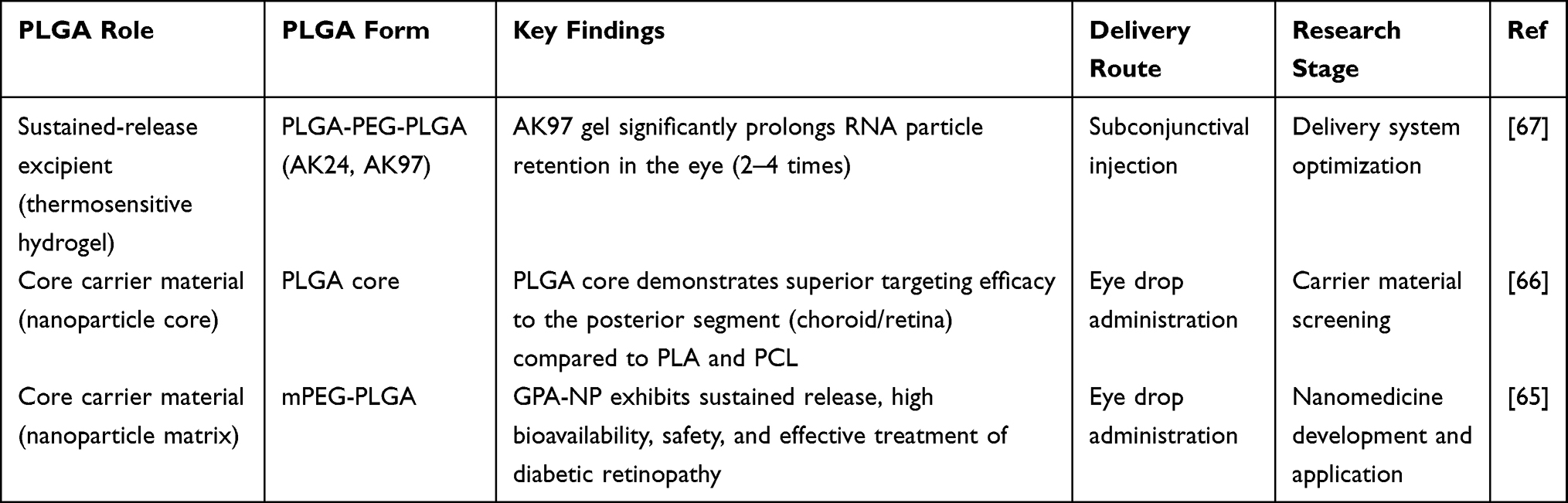 |
Table 1 Comparative Analysis and Evolutionary Trajectory of PLGA and Its Derivatives in Ophthalmic Nanodelivery Applications |
Chitosan
Chitosan (CS) is a naturally occurring hydrophilic cationic polysaccharide.68 Chitosan-based polymer NP exhibits advantages such as ease of preparation, non-toxicity, bioactivity, biocompatibility, biodegradability, and cationic properties, making it an excellent drug carrier.69 Ming et al developed a catalase (CAT) eye drop nanoformulation that self-assembles with cysteine-modified chitosan (CS-Cys) for the treatment of dry eye disease (DED) in mouse and rabbit models. This system achieves prolonged retention of CAT on the ocular surface through disulfide bond-mediated covalent adhesion. It thereby enables the sustained clearance of excessively accumulated reactive oxygen species (ROS), interrupts the oxidative stress-driven vicious cycle in DED, and promotes corneal epithelial repair and restoration of tear secretion. This approach can extend the ocular retention time of protein-based therapies, demonstrating significant potential for DED treatment.70 Mahaling and Katti’s research demonstrated that chitosan (CHI) as a shell material for nanoparticles significantly enhanced their targeted delivery efficiency to posterior segment tissues. Compared to gelatin (GEL) and Pluronic F68 (PF68), the chitosan shell, with its positive charge and strong mucosal adhesion, enables nanoparticles to achieve the highest bioavailability in the conjunctiva, sclera, choroid, and retina,66 which offers a highly promising strategy for treating posterior segment eye diseases through the non-invasive route of eye drops. To enhance the ocular retention and efficacy of glaucoma medications, Maulvi et al developed an enzyme-triggered controlled-release contact lens based on chitosan nanoparticles. The system encapsulates the anti-glaucoma drug timolol maleate within chitosan nanoparticles, which are then loaded onto contact lenses. Research confirmed that this design could effectively prevent drug leakage during sterilization and storage. It degrades and releases the drugs when triggered by lysozyme in the tears only during wearing, achieving sustained intraocular pressure control for up to 120 hours in rabbit models. This strategy offers a promising non-invasive solution for the long-term, effective management of glaucoma.71
Gelatin Nanoparticles
Gelatin is a natural biopolymer prepared and purified by acid or alkaline hydrolysis of collagen (typically sourced from pig skin, bovine bones, or fish scales).72 Zhou et al developed functionalized gelatin nanogels by chemically grafting rosmarinic acid onto the gelatin backbone. After loading with fosfomycin, this platform not only extends corneal dwell time by 2 to 4 times but also endows the system with intrinsic antioxidant activity, enabling synergistic treatment for dry eye syndrome. Its efficacy surpasses commercial drugs while halving the required dosing frequency.73 Ch et al developed mucosal-adherent polyelectrolyte nanoparticles composed of cationized gelatin and sodium alginate for ocular delivery of moxifloxacin. The cationic properties of the gelatin scaffold enable strong electrostatic interactions with the negatively charged ocular surface, resulting in a 2.4-fold increase in corneal retention time compared to conventional eye drops in mice. This enhanced retention effect contributes to its outstanding efficacy in bacterial keratitis models.74
Carbon Nanoparticles
Carbon nanoparticles (CNPs) play a vital role as carriers for various types of drugs, garnering particular attention due to their exceptional properties and multifunctional structure. These properties include physicochemical, thermal, optical, and electrical properties.75 The carbon nanomaterial series encompasses a diverse class of inorganic nanomaterials composed of multiple substances, including carbon quantum dots (CQD), mesoporous carbon nanoparticles, carbon nanotubes (CNT), graphene and graphene oxide (GO) and other 2D materials, as well as 3D structures such as carbon nanohorns (CNH) and nanodiamonds (ND).
Carbon Dots
Carbon dots are a class of quasi-spherical carbon nanoparticles typically less than 10 nanometers in diameter. Characterized by photoluminescence, excellent water solubility, and biocompatibility, they hold significant promise for biomedical applications such as bioimaging, drug delivery, and gene delivery. They represent a novel nanocarrier for delivering genes to retinal cells.76,77 Yu et al designed a nanopore-based nanozyme hydrogel that effectively treated dry eye disease by synergistically combining the inherent antioxidant activity of nanopores with the enhanced retention capacity of the hydrogel. This treatment increased tear secretion by approximately 2.5-fold and significantly reduced corneal fluorescein scores, nearly restoring them to normal levels. It also promoted the repolarization of macrophages toward the anti-inflammatory M2 phenotype.78
Mesoporous Carbon Nanoparticles
Mesoporous carbon nanoparticles, as highly efficient drug carriers, feature high loading capacity, orderly and controllable structure, ease of functionalization, and excellent biocompatibility.79 For refractory intraocular diseases such as choroidal melanoma, mesoporous carbon nanoparticles represent a highly promising platform due to their unique combination of therapeutic and delivery capabilities. Yang et al developed a mesoporous carbon based upconversion nanocomposite platform, designated MCNs/Ln/GD/FR, for the synergistic photothermal chemotherapy of ocular melanoma. Under 980 nm laser irradiation, this platform achieved a photothermal conversion efficiency of 82.86%. By loading gambogic acid and doxorubicin and modifying the surface with folic acid and R8 peptide, the system effectively crossed the blood ocular barrier after intravenous injection and accumulated specifically in the tumor. The therapeutic mechanism involves the activation of tumor suppressor signaling pathways, the upregulation of PHLDA1 to enhance drug sensitivity, and the downregulation of heat shock proteins to reduce tumor thermotolerance. This platform demonstrates both high antitumor efficacy and favorable bio safety, offering a novel strategy for the treatment of ocular tumors.80 Gu et al developed a mesoporous carbon-based nanocarrier co-loaded with natamycin and silver nanoparticles for the treatment of fungal keratitis. This system enables sustained drug release, enhances antifungal activity by inhibiting fungal growth and biofilm formation, and reduces inflammation by adsorbing cytokines. In mouse models, this system achieved superior therapeutic effects at a lower dosing frequency compared to natamycin alone.81
Carbon Nanotubes
Carbon nanotubes (CNTs) are a type of fullerene—hollow spheres, ellipsoids, tubes, and many other shapes, and represent a class of carbon allotropes. Possessing high drug loading capacity, high specific surface area, high mechanical strength, and sufficient chemical stability, carbon nanotubes make them ideal candidates for therapeutic and diagnostic applications, including as excellent nanocarriers for drug delivery.82 Carbon nanotubes have emerged as an ideal material for retinal prostheses due to the high specific surface area afforded by their unique three-dimensional structure. This structural characteristic enables efficient and reversible charge injection via the electrical double-layer capacitance mechanism, avoiding the corrosion risks associated with Faradaic reactions in traditional metal electrodes. This efficient charge injection directly translates into lower neural stimulation thresholds, allowing for further miniaturization of electrodes to enhance prosthetic resolution while ensuring the long-term operational safety and energy efficiency of the implanted device. Studies indicate that CNT electrodes can progressively integrate with the retinal inner limiting membrane, gradually reducing the distance between electrodes and neurons over time without inducing significant glial proliferation. This is crucial for the long-term biocompatibility and functionality of epithelial retinal implants.83 To overcome the challenges of drug resistance and inflammatory damage in existing treatments for fungal keratitis, Zhang et al developed a nanocatalyst-loaded (carbon nanotube) thixotropic hydrogel (NTH@CNT/IZ) loaded with itraconazole. Research indicates that this system not only effectively eliminates fungi but also exerts anti-inflammatory effects by inhibiting the Dectin-1/p38 MAPK pathway. It significantly improves disease prognosis in animal models, making it a highly promising local treatment platform.84
Graphene-Based Nanomaterials
Graphene-based nanomaterials (GFN) constitute a class of materials composed of single-layer or multi-layer graphene sheets. Owing to their exceptional physicochemical properties, they are emerging as a versatile platform for ophthalmic applications.85 In therapeutic applications, GFNs can enhance drug loading capacity and enable sustained-release systems, such as for antifungal contact lenses86 and targeted therapy for choroidal melanoma. Overexpression of matrix metalloproteinase MMP-9 constitutes a molecular mechanism underlying AMD inflammation, particularly in wet AMD characterized by choroidal neovascularization (CNV). Huang et al developed a MMP9-responsive nanoscale drug delivery system (C18PGM) based on graphene oxide quantum dots (GOQDs). Using a laser-induced CNV mouse model, the prepared C18PGM demonstrated significant MMP9 inhibitory activity and anti-inflammatory effects, along with anti-angiogenic properties. This offers a safe and promising therapeutic approach for AMD.87
Nanodiamond
Nano-diamonds (NDs) are minute diamond crystals with diameters of only a few nanometers, composed of regularly arranged carbon atoms. Compared to fullerenes and nanotubes, NDs possess a rough surface that facilitates their adhesion to other molecules. Due to its high surface-to-volume ratio, excellent water solubility and stability, diverse surface electrostatic potential, tunable surface modification, and high biocompatibility, nanodiamond is a promising material for surface modification with various compounds. It holds great potential for use in medicine as a drug delivery vehicle.88,89 Yang et al’s research demonstrated the excellent biocompatibility and delivery efficiency of nanodiamonds for gene editing. They utilized carboxylated nanodiamonds to deliver CRISPR-Cas9 components into the retinal environment of mice, achieving highly efficient gene editing without significant toxicity. This highlights the potential of nanodiamonds as a safe and effective carrier for future retinal gene therapy.90 Kim et al pioneered a smart, enzyme-responsive drug delivery platform by embedding nanodiamond nanogels loaded with maleic acid-thiamolol into contact lenses. This ND nanogel, synthesized by crosslinking PEI-coated NDs with N-acetylated chitosan, functions as a drug reservoir that sequesters glaucoma medication until triggered for release by lysozyme in tears. Upon application, it enables sustained release exceeding 24 hours while maintaining the drug’s therapeutic activity.91
Metal and Metal Compound Nanoparticles
Platinum Nanoparticles
Recent studies indicate that platinum nanoparticles with multiple enzyme-mimetic activities demonstrate significant potential in treating retinal degenerative diseases. As a highly efficient antioxidant nanozyme, it demonstrates significant therapeutic potential in an AMD photodamage model. Cupini et al found that RSA-coated PtNPs effectively penetrate the retina, scavenge ROS, and suppress inflammatory responses. Administration following injury significantly improves electroretinogram responses, protects photoreceptor structures, and downregulates the activation state of Müller cells and microglia, thereby interrupting the vicious cycle of oxidative stress–degeneration–inflammation.This study demonstrates that PtNPs exhibit dual antioxidant and anti-inflammatory effects, offering a promising nanotherapeutic strategy for dry AMD with translational potential92 (As shown in Figure 4). Su et al demonstrated that in a light-induced mouse model of retinal degeneration, PtNPs not only scavenge reactive oxygen species, reduce apoptosis, and improve retinal function, but also enhance endogenous antioxidant defenses by redirecting glucose metabolism toward the pentose phosphate pathway through upregulation of the FoxO signaling pathway.93
 |
Figure 4 Physical–chemical and biological characterization of platinum nanozymes. (A) Representative TEM micrograph of RSA-coated PtNPs (scale bar, 50 nm) and statistical size analysis (at least 200 NPs were counted). (B) Gel-shift assay in 2.5% agarose gel. The gels are shown in bright-field (top) and UV-transillumination (bottom) modes. In the first lane, the electrophoretic run of citrate-stabilized PtNPs is shown. In the second lane, a delayed electrophoretic run of the RSA-coated PtNPs can be observed. In the transillumination mode, the excess of unbound RSA (black band) is visible. (C) Evaluation of CAT-like activity through overtime monitoring of the oxygen gas developed by the reaction (see Methods). In a vial presenting 20% O2 (air), colloidal suspensions of citrate-stabilized PtNPs (cit-PtNPs, 0.2 ppm) and citrate-stabilized CeO2 NPs (cit-CeO2 NPs, 1000 ppm) were exposed to H2O2 (500 mM). The observed O2% induced by the CAT-like activity of PtNPs reached 70%, while that induced by CeO2 NPs (5000-fold more concentrated) was 30% after 100 min. (D and E) Effect of PtNPs on the recovery of chemically induced ROS and apoptosis in primary rat cortex neurons. Treatments with 1 mM H2O2 for 5 min (D) and 5 μM antimycin A for 24 h (E) were used to induce ROS in the culture. ROS values were measured by using appropriate fluorescent probes (H2DCF and DHE, respectively). Apoptosis was measured through caspase 3/7 activity. Results are reported as means ± sem from n = 3 independent experiments and are normalized over untreated controls. **p < 0.01, ***p < 0.001, ****p < 0.0001; one-way ANOVA/Tukey’s tests. In all cases, neurons pretreated with 50 μg/mL RSA-coated PtNPs for 48 h showed a significant recovery of ROS amounts and apoptosis. (F) Representative TEM image of RSA-coated PtNPs internalized in primary rat cortex neurons (scale bar, 1 μm). Two representative intracellular vesicles containing PtNPs are indicated with numbers and magnified. Scale bars of magnified vesicles are 200 nm. N denotes the nucleus. (G) Estimated NP uptake quantification per cell from ICP-MS measurements. The uptake of RSA-coated PtNPs is significantly higher than that of citrate-stabilized PtNPs. Reproduced from Cupini et al, ACS Nano 2023, 17.72 Licensed under CC BY. |
Gold Nanoparticles
Gold nanoparticles have been studied in ophthalmology for enhancing imaging and treating diseases such as corneal and choroidal neovascularization, retinal angiogenesis, and neovascular age-related macular degeneration. Gold nanoparticles exhibit low or no toxicity in most cytotoxicity studies due to their inert nature and the biocompatibility of the selected ligands.94,95 Moreover, they are promising candidate for ophthalmic imaging contrast agents due to its accessibility throughout the entire eye and its ability to be cleared from the eye.96,97 Research by Karthikeyan et al indicated that Au-NPs could effectively inhibit VEGF- and IL-1β-induced proliferation and migration by suppressing the Src kinase pathway in BRPEs. Au-NPs may serve as an effective therapeutic agent for treating ocular diseases such as proliferative vitreoretinopathy.98 Laradji et al synthesized ~20 nm AuNPs and utilized their surface plasmon resonance effect for fluorescence imaging tracking, providing a visual demonstration of AuNP distribution within ocular tissues.99 Further studies demonstrated that surface-engineered AuNPs modified with hyaluronic acid (HA) could significantly enhance their ability to penetrate ocular barriers, enabling targeted delivery to the posterior retina without inducing retinal toxicity. This highlights the broad potential of surface-functionalized AuNPs as an efficient and safe ocular delivery platform.
Silver Nanoparticles
AgNPs possess the advantages of antibacterial and anti-angiogenic effects.100 To address the challenge of antibiotic resistance in bacterial keratitis, Dai et al developed a glycoepitope-mimetic corneal bandage lens (PNH-CBLs). This bandage mirror, loaded with Ag/Cu bimetallic nanoparticles via a polydopamine coating and grafted with heparin, not only continuously releases Ag⁺/Cu2⁺ ions, showing potent antibacterial activity against Staphylococcus aureus and Escherichia coli (inhibition rates >80% and >70%), but also has multifunctional synergistic effects including anti-inflammatory properties, resistance to non-specific protein adsorption, and promotion of beneficial protein adsorption. In rabbit models, it significantly alleviated corneal infection and inflammation.101 The functionality of AgNPs has been innovatively integrated into anti-scarring strategies. For instance, by anchoring them to reduced graphene oxide (rGO), a hydrogel with both long-lasting antibacterial properties and near-infrared light-triggered precise photothermal ablation of fibroblasts was developed, which significantly suppressed scar formation and prolonged surgical efficacy in a glaucoma filtration surgery model.102
Cerium Dioxide Nanoparticles (CeO2)
Cerium oxide (CeO2) nanoparticles have been demonstrated to possess high redox scavenging capacity.103 It exhibits negligible toxicity after delivery to the eye. Wu et al developed hyaluronic acid-modified nano-cerium oxide (HA-CeO2), offering a novel nanocatalyst strategy for DED treatment. This study confirmed that the nano-cerium oxide core effectively scavenges reactive oxygen species (ROS) and downregulates the expression of inflammatory factors MMP9 and IL-1β, thereby breaking the vicious cycle of oxidative stress and inflammation in dry eye disease. In a BAK-induced dry eye mouse model, this composite nanoparticle significantly promoted corneal epithelial repair, increased tear secretion, and restored normal ocular surface structure, demonstrating the immense potential of nano-cerium oxide in synergistic treatment of DED.104 Chen et al developed a multifunctional nanoplatform-miRNA-RSV-CeO2 NPs system, which combined cerium oxide nanoparticles (CeO2 NPs) with self-regenerating antioxidant capabilities with resveratrol (RSV) and exosome-derived microRNA (miRNA), exhibiting exceptional antioxidant and anti-glycation properties in vitro. This platform could effectively scavenge reactive oxygen species, inhibit the formation of advanced glycation end products, and upregulate the expression of lens protective proteins, offering a novel therapeutic approach for diabetic cataracts.105
Mesoporous Silica Nanoparticles
Controlled functionalization of mesoporous silica nanoparticles (MSNs) enables the construction of advanced stimulus-responsive systems. For instance, Elbedwehy et al developed ROS-responsive MSNs for intraocular drug delivery. Their surface charge could reverse from positive to negative in high ROS environments, thereby intelligently switching from vitreous retention to retinal-targeted diffusion, demonstrating the precise controllability of MSNs as smart drug carriers.29 Shao et al developed MSNs loaded with the STING inhibitor C176 for treating retinal neovascularization. This system fully leverages the unique properties of MSNs: Firstly, their high porosity enables efficient loading of hydrophobic drugs, improving their solubility; Secondly, surface modification with phosphatidylserine (PS) enables active targeting of retinal macrophages; Thirdly, the sustained-release property of MSNs facilitates long-lasting therapy with a single injection. This demonstrates the comprehensive advantages of MSNs in enhancing intraocular drug delivery efficiency, targeting precision, and therapeutic persistence106 (As shown in Tables 2 and 3).
 |
Table 2 Characteristics and Potential Applications of Different Types of Nanocarriers in Ophthalmic Drug Delivery |
 |
Table 3 Summary of Nanocarrier Strategies: Administration Routes, Targets, and Mechanisms in Eye Diseases |
Treatment of Anterior Segment Eye Diseases
Cataract
Cataracts are lens opacities that cause visual impairment. They are the leading cause of blindness and the second leading cause of visual impairment worldwide.107 The only available treatment for cataracts is to remove the cloudy lens and replace it with an artificial lens. However, this technique has drawbacks such as implant disintegration, postoperative inflammation, and posterior capsule opacification. Therefore, there is a need to improve minimally invasive surgical techniques and intraocular lens implants, as well as tissue-specific drug delivery methods for postoperative complications.108 Emerging technologies such as nanotechnology and biomaterials based on smart polymers offer promising avenues for developing biocompatible intraocular lenses with enhanced performance.109 Hydrogels are widely used in contact lenses, corneal and scleral repair, and vitreous substitutes due to their biocompatibility with eye components. Some studies indicated that polyacrylamide-sodium acrylate hydrogels (PAHs) are suitable for intraocular lens (IOL) applications due to their excellent biocompatibility, high transparency (94%), refractive index (1.41±0.07) matching that of the human eye’s natural lens (1.42), and good compressive strength (14.00 kPa), extensibility (1400%), and swelling rate (50±2.5%).110 To enhance the bioavailability and therapeutic efficacy of cataract drugs, researchers are developing prodrugs and applying nanotechnology to protect the drugs and optimize their delivery. Curcumin shows potential in treating diabetic cataracts, but its low oral bioavailability limits its clinical application.111 Studies have indicated that encapsulating curcumin within poly(lactic-co-glycolic acid) (PLGA) nanoparticles and administering it orally to rats significantly enhances its oral bioavailability by 9 times compared to free curcumin, thereby enhancing its therapeutic effect. This demonstrates that the application of nanotechnology has a remarkable effectiveness in delaying cataract formation in diabetic rats.112 To overcome the barrier posed by the corneal barrier to drug delivery, Jiang et al developed circular cell-penetrating peptide-modified cerium oxide nanoparticles (cCPP-CeNPs). This study leveraged nanotechnology to achieve synergistic “penetration” and “therapy”: cCPP-CeNPs reversibly opened corneal tight junctions, significantly enhancing their ocular permeability. Upon entering the eye, they targeted mitochondria in lens epithelial cells, not only exerting their inherent regenerative antioxidant enzyme activity but also effectively scavenging reactive oxygen species and mitigating lipid peroxidation by inhibiting ferroptosis (a key pathway). Ultimately, in an ultraviolet-induced cataract model, cCPP-CeNPs administered as non-invasive eye drops significantly delayed cataract formation, fully demonstrating the application potential of functionalized nanoparticles in cataract prevention and treatment.113
Dry Eye Syndrome
Dry Eye Disease (DED), also known as keratoconjunctivitis sicca, is a common ocular surface disorder characterized by tear film instability and/or ocular surface damage, leading to ocular discomfort and visual impairment.114 One of the recently approved nanotechnologies for treating DED is 0.1% cyclosporine (Vevye™ Cyclosporine Eye Drops 0.1%), a preservative-free, non-aqueous formulation of cyclosporine (CsA). A randomized clinical trial investigated the efficacy of 0.1% anhydrous cyclosporine eye drops in treating dry eye syndrome. Research findings indicate that a 0.1% cyclosporine solution effectively treats keratitis associated with dry eye syndrome and is well tolerated. Improvement in total corneal and central corneal staining scores was observed after just two weeks of treatment, with efficacy persisting through day 29115 (As shown in Figure 5). Zhang et al developed a sialic acid-targeted peptide-modified liposome loaded with cyclosporine A (CsA), a typical anti-inflammatory drug, and ferroptosis inhibitor-1 (Fer-1), a selective ferroptosis inhibitor, termed CF@SNPs, for combating DED. Results indicate that CF@SNPs treatment achieves complete remission of DED symptoms, including improvement in corneal defects, increased goblet cell counts, and restoration of tear secretion.116 To overcome barriers to ocular surface drug delivery, Huang et al developed cRGD-conjugated PLGA–PEG nanoparticles (cNPs@BR) for encapsulating bilirubin, which exhibits potent antioxidant and anti-inflammatory activities. Its retention time on the ocular surface extends to 12 hours, effectively overcoming the low bioavailability issue caused by tear clearance in traditional eye drops. It can also restore goblet cells and the corneal epithelial barrier in dry eye models by activating mitochondrial autophagy.117
 |
Figure 5 Total Corneal Fluorescein Staining (tCFS) at Day 29 Responder Analysis and Visual Analog Scale (VAS) Symptoms for tCFS Responders. (A) Proportion of corneal fluorescein staining responders (≥3 score improvement on the National Eye Institute scale) at day 29 using a water-free cyclosporine, 0.1%, solution vs vehicle in the treatment of moderate to severe dry eye disease. (B) Improvement in symptoms in tCFS responders vs nonresponders irrespective of treatment. Reproduced from Akpek et al, JAMA Ophthalmology 2023, 141.96 Licensed under CC BY. |
Inflammatory Diseases
Conjunctivitis
Conjunctivitis (pink eye) is an inflammation or swelling of the conjunctiva. Research indicates that nanotechnology can significantly enhance the efficacy of allergen-specific immunotherapy for allergic conjunctivitis.118 The recombinant Amb a 1-PLGA-PEG nanoparticles developed by Cao et al effectively corrected Th1/Th2 immune imbalance and significantly suppressed the activation of conjunctival mast cells due to their sustained-release properties, demonstrating far superior efficacy compared to allergen proteins alone. This demonstrates the pivotal role of nanocarriers in enhancing drug stability, prolonging duration of action, and boosting immunomodulatory efficacy.119 To overcome the limitations of traditional eye drops in treating bacterial conjunctivitis, Jacinto et al developed a core-shell nanoparticle based on zein and hyaluronic acid (HA) for loading ciprofloxacin. This formulation exhibits optimal particle size (~109 nm) and offers dual advantages: the Zein core ensures high drug encapsulation and sustained release, while the HA shell leverages its mucosal adhesion properties to effectively prolong drug residence time on the ocular surface. In vitro studies have confirmed its excellent biocompatibility and sustained drug release behavior over 24 hours, demonstrating its potential as an upgraded alternative to traditional eye drops120 (As shown in Figure 6).
 |
Figure 6 Release profile of ZeinCPX_HA NPs in STF at 37 °C, pH = 7.4 for 24 h (non-cumulative). Results are presented as mean ± SEM (n = 3). Reproduced from Jacinto et al, Pharmaceutics 2022, 14(8), 1557.101 Licensed under CC BY. |
Keratitis
Keratitis is a corneal infection caused by microorganisms such as bacteria, fungi, and viruses, and it is one of the leading causes of vision impairment and blindness worldwide.7 Functionalized nanoparticles offer diverse therapeutic strategies for keratitis, with targeted bactericidal action and regenerative repair representing two typical approaches. Fan et al developed ε-polylysine-modified polydopamine nanoparticles (EPL@PDA NPs) to achieve targeted photothermal antibacterial effects. This strategy employs cationic peptides to specifically bind bacteria, generating localized high temperatures under near-infrared light to directly eliminate drug-resistant pathogens. Functioning like a “precision-guided missile,” it targets the precise source of bacterial infection.121 In contrast, Sohani et al developed a β-cyclodextrin-encapsulated curcumin nanoparticle-hyaluronic acid hydrogel system, focusing on anti-inflammatory and tissue repair functions. This system effectively suppresses key inflammatory factors such as TNF-α and IL-6 through sustained-release curcumin while promoting corneal epithelial regeneration. Acting like a “repair crew,” it is suitable for controlling inflammation and facilitating tissue defect healing.122 In summary, these two approaches represent distinct strategies: physical sterilization targeting pathogens and chemical repair targeting host tissues, demonstrating the immense potential for on-demand design of nanoparticles in the treatment of keratitis.
Uveitis
Uveitis is a heterogeneous group of intraocular inflammatory diseases affecting the uvea and adjacent structures, ranking among the leading causes of vision loss worldwide.123 Nanotechnology shows tremendous potential in improving the ocular delivery of biomolecular drugs. Vaneev et al developed SOD1 nanoparticles (Nano-SOD1), a nanomedicine formulation that not only significantly prolongs SOD1 retention time on the ocular surface but also enhances its penetration into the anterior chamber of the eye. This approach systematically resolves the fundamental bottlenecks of natural SOD1 in ocular applications—rapid clearance and poor permeability.124 Nanoparticle technology demonstrates significant potential in enhancing the physicochemical properties of traditional drugs. A prime example is curcumin (CUR), which possesses potent antioxidant and anti-inflammatory activity. However, its extremely poor water solubility and low bioavailability severely limit its clinical application. Cao et al developed polyvinylpyrrolidone-curcumin nanoparticles (PVP-CUR NPs). Not only did this formulation enhance curcumin’s water solubility by approximately 400-fold, but it also achieved highly efficient delivery of curcumin’s biological activity in both in vitro and in vivo models. This provides an effective nanoengineering strategy to overcome delivery bottlenecks in the treatment of uveitis using natural products.125
Treatment of Posterior Segment Eye Diseases
Age-Related Macular Degeneration
Age-related macular degeneration (AMD) is a degenerative eye disease affecting the macula. It is one of the most common causes of irreversible vision loss in individuals over 65 years of age. It can be classified into two types—wet AMD and dry AMD. The initial presentation of this disease is dry AMD, which may progress to wet AMD in later stages.126 Its pathophysiological mechanism involves oxidative stress and increased release and expression of vascular endothelial growth factor (VEGF).127 Currently, the treatment for wet AMD primarily relies on intravitreal injections of anti-VEGF drugs such as ranibizumab and conbercept. These medications inhibit VEGF to reduce the growth and leakage of abnormal blood vessels, thereby slowing vision loss. In addition to anti-VEGF therapy, laser treatment and surgical intervention may serve as adjunctive measures, though their efficacy is limited. The primary drawbacks of anti-VEGF therapy stem from various consequences, such as intraocular inflammation, retinal detachment, and occasional ocular hemorrhage due to the short molecular half-life, necessitating repeated intravitreal injections.128 There is growing interest in developing nanoparticles as drug delivery systems for local administration to the posterior chamber of the eye. This approach is non-invasive, eliminates the need for intravitreal injections, and enables drug delivery to the retina without syringes (As shown in Figure 7). It offers additional advantages in delaying drug release and reducing the risk of adverse reactions. In a study, when ranibizumab (Lucentis) was encapsulated in liposomes, DPPC-DPPG liposomes demonstrated significantly higher encapsulation efficiency (approximately 60%) and sustained-release properties compared to other liposomes. They effectively penetrated the sclera, exhibiting promising therapeutic potential. This technology may replace the current invasive intravitreal injection method.129 Alshamrani et al developed a water-based curcumin nanoemulsion formulation (CUR-NMF) for delivery to the posterior segment of the eye. Results demonstrated that CUR-NMF provided enhanced antioxidant stress protection in human retinal pigment epithelial (D407) cells and significantly reduced the release of vascular endothelial growth factor (VEGF).130 A study developed a sustained-release formulation of poly(lactic-co-glycolic acid) (PLGA) nanoparticles loaded with axitinib to reduce the frequency of intravitreal drug administration. The research demonstrated that these nanoparticles exhibit enhanced cellular uptake, significant anti-angiogenic potential, and inhibition of VEGF activity.131
 |
Figure 7 Primary Treatment Methods for AMD (figure was created in https://BioRender.com). |
Diabetic Retinopathy
Diabetic retinopathy (DR) is an eye disease characterized by chronic, progressive vision loss due to long-term hyperglycemia and other diabetes-related abnormalities (such as hypertension and hyperlipidemia), which cause damage to the retinal microvasculature. It is the most common retinal vascular disease and a leading cause of blindness and visual impairment, particularly the primary cause of vision loss in the elderly. Diabetic retinopathy is classified into non-proliferative diabetic retinopathy (NPDR) and proliferative diabetic retinopathy (PDR).132 A research team developed chitosan-hyaluronic acid nanoparticles (CS/HA) as a carrier for EPOβ, delivering it to the retinas of WH rats. Results showed that EPOβ was detected in the retina via immunofluorescence 12 hours after administration and remained detectable at day 21.133 In a 2019 study, Shoval et al developed anti-VEGF aptamer-modified carbon dots (C-dots), which served as effective carriers for anti-VEGF aptamers on the cornea. The results demonstrated that the hybrid C-dots effectively inhibited VEGF-stimulated choroidal vascular angiogenesis.134 Li et al developed a bionic nanoparticle (apoM@mPB@Ra NPs) for immunomodulatory treatment of diabetic retinopathy. This particle utilizes apoptotic cell membranes to target microglia, and through the synergistic action of Prussian blue nanoenzymes clearing reactive oxygen species and loaded rapamycin inhibiting the mTOR pathway, effectively drives microglia polarization toward the anti-inflammatory M2 phenotype. In animal models, it was demonstrated to alleviate retinal hypoxia and suppress pathological angiogenesis, offering a novel strategy for nano-immunotherapy in diabetic retinopathy.135
Retinal Vein Occlusion (RVO)
Retinal vein occlusion (RVO) is the second most common cause of retinal vascular disease after diabetic retinopathy. It is a vascular disorder resulting from obstruction of retinal veins, which impairs retinal blood circulation. This often leads to retinal ischemia, hemorrhage, fluid leakage, and macular edema, causing vision loss that may become permanent. Based on the location of obstruction, retinal vein occlusion (RVO) can be classified into branch retinal vein occlusion (BRVO), hemiretinal vein occlusion (HRVO), and central retinal vein occlusion (CRVO).136 Current treatment options include macular photocoagulation, corticosteroids, and anti-VEGF therapy. Anti-VEGF therapy is the most common treatment approach, encompassing drugs such as Conbercept, Ranibizumab, and Bevacizumab. Potential advantages of nanoengineering for anti-VEGF therapy may include reducing dosing frequency and employing strategies based on sustained-release nanoparticles to maintain therapeutic doses of anti-VEGF drugs at the disease site for extended periods.137 KYandrapu et al evaluated the in vivo delivery efficacy of nanoscale particles NPinPMP within porous microparticles in a rat model using non-invasive fluorometric analysis. Results demonstrated that NPinPMP sustained the release of bevacizumab for up to two months compared to Alexa Fluor 488-conjugated bevacizumab solutions, indicating the NPinPMP system’s significant advantage in prolonging drug action duration.138 Corina-Lenuța Savin et al synthesized a novel polymer, chitosan-g-polyethylene glycol methacrylate (CS-g-PEGMA). Based on magnetic nanoparticles (MNPs), CS-g-PEGMA may serve as a potential carrier for controlled release of bevacizumab (BEV) as an ophthalmic drug delivery system. Results indicated that NPs retained the maximum drug amount (0.327 mg BEV/mg NPs) after 72 h, with an encapsulation efficiency of 39%. This approach enables significantly lower dosing, prolonged release duration, and effective local administration (potentially extending to 14–30 days), demonstrating better therapeutic efficacy in treating inflammation induced by a rabbit model of central retinal vein occlusion.139
Glaucoma
Glaucoma is a disease characterized by optic nerve damage.140 It is the second leading cause of blindness worldwide. It is typically associated with elevated intraocular pressure, and includes types such as primary angle-closure glaucoma, primary open-angle glaucoma, secondary glaucoma, and congenital glaucoma. Early symptoms of glaucoma are often subtle, but as the condition progresses, individuals may experience vision loss, visual field defects, eye pain, and headaches. Treatment options for glaucoma include medication, laser therapy, and surgical intervention. Nanotechnology-based therapies can overcome the limitations of currently available glaucoma treatments by optimizing targeted drug delivery, enhancing bioavailability, and enabling controlled release.141 A study developed an ophthalmic implant loaded with dorzolamide (DRZ) based on carboxymethyl cellulose (CMC) and chitosan (CHI), enabling extended drug delivery. The implant prepared with CMC exhibited significantly slower in vitro release rates and increased drug retention on the ocular surface. This demonstrates that CMC implants loaded with DRZ can provide effective treatment for glaucoma.142 Neha Joshi et al prepared 25 types of acetazolamide nanoemulsions for glaucoma treatment. Results showed that the release rate of acetazolamide from the nanoemulsion increased from 81.59 ± 1.04% to 92.46 ± 0.33% after 24 hours. In vitro drug release studies indicated that the prepared nanoemulsion exhibited excellent bioavailability, sustained release properties, and the ability to target the eye, making it a promising effective ocular delivery system.143 Schnichels et al developed nanoparticles formed by the self-assembly of lipid-DNA amphiphiles. These particles efficiently loaded the glaucoma drug travoprost without chemical modification by hybridizing specific aptamers. Research indicates that these DNA nanoparticles, leveraging their inherent corneal adhesion properties, can significantly prolong drug retention time on the ocular surface. In rat and ex vivo porcine eye models, they achieve at least double the drug delivery efficiency while demonstrating excellent biocompatibility. This technology presents a highly promising platform for sustained-release glaucoma therapy.144
Eye Tumors
Uveal Melanoma
Uveal melanoma is the most common primary intraocular malignancy in adults, characterized by rapid progression and a significant propensity for metastasis, primarily to the liver and lungs.145,146 Traditional treatments such as radiotherapy may cause retinal lesions and neovascular glaucoma; systemic chemotherapy is ineffective and highly toxic; and enucleation surgery results in cosmetic defects and ultimately leads to vision loss.147 To address the highly metastatic nature and treatment challenges of uveal melanoma, Guo et al demonstrated two distinct yet equally effective strategies in their nanotechnology-based research targeting uveal melanoma metastasis. In their early work, they developed an intravenously injectable, reduction-responsive nanoparticle that targets the CD44 receptor via hyaluronic acid (HA) and rapidly releases lapatinib in high-concentration glutathione (GSH) environments, achieving an 82.1% inhibition rate in a lung metastasis model.148 To further address the issues of low drug delivery targeting efficiency and short local drug retention time in nanoparticle systems, the team subsequently designed a locally injectable cascade-release nanocomposite hydrogel. This system integrates pH-responsive HA nanoparticles loaded with doxorubicin and lapatinib into hydrogels, achieving a dual-delivery logic: “hydrogel-mediated sustained-release nanoparticles, nanoparticle-targeted cell killing.” Not only did it reduce postoperative recurrence rates from 100% to 40%, but the released nanoparticles also demonstrated potent anti-metastatic effects, achieving a lung metastasis inhibition rate as high as 85.4%.149 These two studies collectively demonstrate the immense potential of HA-based smart nanoplatforms in UM therapy, offering preferred solutions for systemic and localized treatment, respectively. In the local treatment of primary tumors, several innovative nanoscale strategies have been proposed. Chen et al developed an immunotherapy‑based artificial vitreous hydrogel constructed from tetra-armed polyethylene glycol, which was loaded with melphalan and an anti‑PD‑L1 antibody. When injected after tumor resection in a mouse model of choroidal melanoma, this hydrogel enabled sequential drug release, effectively controlling tumor recurrence while preserving the normal structure and visual function of the eye.150 Furthermore, Barrios-Esteban et al reported the use of peptone-based nanocapsules as a gene delivery vector. Through local administration, these nanocapsules delivered plasmid DNA and miRNA, achieving a transfection efficiency of 36% in uveal melanoma cells. This approach provides a novel delivery platform for gene therapy.151 Bi et al designed biomimetic low-density lipoprotein nanoparticles (LD-DPVP NPs) that co-deliver the photosensitizer verteporfin and the tumor vessel normalization agent dexamethasone. Upon near-infrared laser irradiation, these nanoparticles generate reactive oxygen species to eliminate tumor cells. Concurrently, dexamethasone modulates the tumor microenvironment, thereby enhancing the delivery efficiency of the nanoparticles.152 Particularly noteworthy is the dual efficacy exhibited by some nanosystems, which simultaneously target the primary tumor and inhibit metastasis. Tao et al reported a GSH-responsive nanoparticle, NP@Oxa/Criz, designed for the precise co-delivery of Crizotinib (Criz) and Oxaliplatin (Oxa). This system inhibits the HGF/c-Met axis, thereby preventing the nuclear translocation of β-catenin. This action leads to a reduction in the transcription of metastasis-associated genes and diminishes the stemness and metastatic potential of UM cells. In vivo studies demonstrated that this nanosystem accumulates at the tumor site, effectively eradicating the intraocular primary tumor while concurrently suppressing metastatic tumors in the liver and abdominal cavity. Furthermore, it induces immunogenic cell death, which enhances the antitumor immune response.153
Retinoblastoma
Retinoblastoma is considered the most common primary intraocular malignant tumor in children. Traditional treatments, including enucleation, chemotherapy, and radiotherapy, while effective to varying degrees, are fraught with limitations such as systemic toxicity, recurrence, and long-term side effects.154 In retinoblastoma research, Mudigunda et al designed multifunctional PLGA/PCL polymeric nanoparticles that innovatively co-loaded the chemotherapy drug palbociclib and the near-infrared dye IR820. This nanoparticle not only serves as a drug delivery vehicle but also functions as a photoacoustic imaging probe and photothermal converter. Upon near-infrared light activation, it achieves synergistic effects between chemotherapy and photothermal therapy, demonstrating an 86.5% cell killing rate against retinoblastoma cells. Its mechanism of action has been confirmed to involve inducing DNA damage and apoptosis. This work demonstrates how a simple nanoparticle can be upgraded into a powerful platform integrating diagnosis, treatment, and efficacy monitoring through rational nanoengineering design, providing new tools for precision medicine in retinoblastoma.155 Furthermore, the application of bionic nanoparticles has further advanced targeted therapy for retinoblastoma. Zhang et al achieved specific accumulation and highly efficient killing of drugs (etoposide) in intraocular tumors using tumor cell membrane-coated PLGA nanoparticles, demonstrating the immense potential of homotypic targeting strategies in enhancing localized chemotherapy efficacy.156
Other Applications
Nanostructures for Eye Tissue Regeneration
Tissue regeneration offers a promising approach for repairing damaged tissues or organs.157 Nano scaffolds are self-assembled or electrospun nanofibers composed of synthetic or natural polymers. These scaffolds facilitate adhesion and cell migration during retinal regeneration by mimicking the extracellular matrix (ECM).158 Various types of nanostructures, including electrospun nanofibers, self-assembled peptide nanofibers, and nanotopological structures, have been designed and investigated for the regeneration of retinal, corneal, and lens tissues.159 Warnke et al investigated the culture of human primary retinal pigment epithelial (RPE) cells on substrate materials including PLGA and collagen nanofiber membranes, PLGA films, and coverslips. The study revealed that RPE cells on nanofiber membranes exhibited a monolayer morphology resembling in vivo structures, featuring long, lamellar microvilli—a characteristic similar to natural human RPE cells. In contrast, RPE cells cultured on films and coverslips displayed a flatter morphology, with less than 10% visible microvilli.160 Naghmeh Abbasi et al prepared a poly-L-lactic acid (PLLA) nanofiber membrane via electrospinning, mimicking the structure of the natural Bruch membrane (fiber diameter approximately 50 nm, thickness approximately 5–7 μm). Research indicates that PLLA membranes pretreated with poly-L-ornithine (PLO) and subsequently coated with laminin significantly enhance the adhesion, proliferation, differentiation, and functional expression of human embryonic stem cell-derived retinal pigment epithelial cells (hESC-RPE), forming a monolayer of RPE cells with barrier function and phagocytic activity, demonstrating its potential in retinal tissue engineering and cell transplantation therapy for age-related macular degeneration (AMD). The three-dimensional scaffold supported the growth and proliferation of hESC-RPE cells for up to 8 weeks161 (As shown in Figure 8).
 |
Figure 8 Growth of hESC-RPE cells above scaffolds. (A) Phase contrast microscopy showing hESC-RPE cell morphology during culture on various membranes across weeks 0–8. (B) Live laser scanning images of hESC-RPE cells labeled with both fluorescent phalloidin (green) for Actin staining and 4′,6-diamidino-2-phenylindole (blue) for nuclei staining following growth on various coated and uncoated poly-L-lactic acid (PLLA) scaffolds after 1 day and at 1, 2, 4 and 8 weeks (Scale bar: 100 μm); (C) SEM micrographs showing the morphology and pigmentation of hESC-RPE cells on the uncoated and coated PLLA scaffolds after 1 day and at 1, 2, 4, and 8 weeks of culture (Scale bar: 10 μm). Reproduced from Abbasi et al, Journal of Biomedical Materials Research Part A 2024, 112.135 Licensed under CC BY. |
Retinal Gene Therapy Technology Based on Nanomaterials
One of the goals driving the development of new therapies is to find ways to optimize the local and sustained delivery of drugs or genes to the eye and retina. Gene therapy is a technique that transfers genetic material to remove, replace, repair, or introduce genes for the treatment of diseases. Gene therapy involves modifying damaged DNA within recipient cells to produce the desired therapeutic effect. The eye possesses key characteristics that make it highly suitable for gene therapy: well-defined anatomical structure, relative immune privilege, accessibility, ease of diagnosis, and the ability to use one eye as an experimental target while the other serves as a control within the same subject. Advances in gene therapy hold tremendous promise for the management of ophthalmic diseases.162,163 Mitra et al investigated a non-viral nanoparticle (GCS NPs) composed of glycerol-based chitosan (GCS) and plasmid DNA (pDNA) for retinal gene therapy. The pDNA carrying the CBA-eGFP expression cassette was compacted and injected subretinaly into adult mice. Results showed that significant GFP expression was observed only in the retinal pigment epithelium (RPE) of the GCS NP treatment group, with no significant expression detected in other groups. GCS NPs demonstrated excellent biocompatibility and safety, with no severe retinal toxicity observed. Furthermore, the synthesis and characterization of GCS NPs revealed that it has good solubility, stability, and gene expression capacity, enabling effective promotion of gene expression following subretinal delivery.163 Martens et al employed hyaluronic acid (HA) as an electrostatic coating for non-viral polymeric gene nanomedicine p(CBA-ABOL)/pDNA complexes, providing them with an anionic hydrophilic surface to enhance intravitreal migration. HA-polymer complexes induced GFP expression in this in vitro cell line without significant cytotoxicity, with low molecular weight HA (22 kDa) coating demonstrating the highest expression induction. This demonstrates that hyaluronic acid (HA) as an electrostatic coating for non-viral polymeric gene nanodrugs effectively enhances their migration within the vitreous cavity and cellular uptake efficiency, while exhibiting excellent biocompatibility and safety.164
Conclusions and Outlook
The rise of nanotechnology has revolutionized the treatment of ophthalmic diseases. This review systematically summarizes the application progress of various nanomaterials (including lipid nanoparticles, polymer nanoparticles, carbon nanoparticles, metal and metal compound nanoparticles, etc.) in the treatment of ophthalmic diseases. Overall, nanotechnology demonstrates significant advantages in overcoming ocular physiological barriers (such as the cornea and blood-ocular barrier), enhancing drug bioavailability, and enabling targeted delivery and controlled release through its unique size effects and functional designability.
In the treatment of anterior segment eye diseases (such as cataracts, dry eye syndrome, and inflammatory conditions), nanocarriers have significantly prolonged drug retention time on the ocular surface, reduced dosing frequency, and improved patient compliance. In the treatment of ocular tumors such as retinoblastoma and uveal melanoma, nanotechnology enables targeted delivery and enhanced efficacy of chemotherapeutic drugs while empowering innovative therapeutic strategies like photothermal therapy. This approach significantly reduces systemic toxicity while improving treatment outcomes. In the treatment of posterior segment eye diseases such as age-related macular degeneration, diabetic retinopathy, retinal vein occlusion, and glaucoma, nanotechnology has successfully delivered therapeutic drugs through multiple barriers to target sites like the retina and choroid. This breakthrough offers new solutions for previously difficult-to-treat blinding eye conditions. In terms of integrated diagnosis and treatment, the multifunctional nanoplatform combines imaging and therapeutic capabilities, laying the foundation for real-time disease monitoring and precision therapy. In summary, from the anterior to the posterior segment, and from diffuse lesions to focal tumors, nanotechnology offers a powerful and diverse toolkit for addressing the core challenge of ophthalmic drug delivery, heralding a new era of precision medicine in ophthalmology.
The successful application of nanotechnology in the treatment of eye diseases is mainly attributed to its unique mechanism of overcoming the physiological barriers of the eye in multiple dimensions. First of all, the physicochemical properties of nanoparticles (such as the small size effect and surface modifiability) are the foundation for achieving efficient delivery: by regulating particle size and surface charge, nanocarriers can effectively penetrate the corneal barrier or the blood-retinal barrier, prolong ocular surface retention time, and avoid being rapidly cleared by tears.165–167 Secondly, different nanomaterials exhibit distinct delivery advantages: liposomes achieve active targeting through a biomimetic membrane structure;168 polymer nanoparticles can provide sustained release and be integrated into in situ gel systems to extend corneal contact time;34,166 inorganic nanoparticles achieve excellent scleral permeability through surface modification, offering non-invasive treatment options for posterior segment diseases.169 Third, targeted delivery and controlled release mechanisms together ensure the therapeutic effect: through ligand modification or environment-responsive design, nanocarriers can achieve precise drug release at the lesion site, reducing systemic side effects; at the same time, regulating cellular uptake behavior can further optimize drug efficacy.170 Finally, strategies for penetrating the blood-retinal barrier for late-stage diseases are becoming increasingly mature. Their mechanisms are highly similar to those for crossing the blood-brain barrier, providing important references for drug delivery to the central nervous system.171,172 These mechanisms together form the theoretical foundation for nanotechnology to address issues such as low bioavailability and frequent dosing of traditional ophthalmic formulations, and they have been validated in numerous preclinical models and early clinical translations.
In recent years, the clinical translation of nanotechnology in the field of ophthalmic disease treatment has achieved substantial progress. Regarding commercially available products, the clinical evidence for the dexamethasone intravitreal implant (Ozurdex®) continues to expand. A prospective randomized controlled trial demonstrated that the adjunctive use of this implant during vitrectomy in patients with proliferative diabetic retinopathy complicated by retinal detachment significantly reduced the incidence of postoperative preretinal proliferative membranes (23.5% vs. 88.2%) and epiretinal membranes at the macula (11.8% vs. 41.2%). At the 12-month follow-up, best-corrected visual acuity was improved, highlighting the potential value of nano-sustained release technology in complex ophthalmic surgery.173 At the forefront of advanced technology research and development, various novel nano-delivery systems are accelerating their clinical translation. For instance, a study has developed a thermosensitive hydrogel loaded with triamcinolone-loaded PLGA nanoparticles (approximately 153 nm). This composite system serves both as a vitreous substitute and a platform for sustained drug release, achieving in vitro release for over nine weeks and maintaining intraocular drug concentrations within the therapeutic window for 28 days in a rabbit model, with good biocompatibility. This represents a significant exploration of multifunctional nanocomposite systems toward clinical translation.174 To address the clinical challenge of poor water solubility of the antifungal drug Natamycin, researchers have developed 1% Natamycin nanomicelles. Compared to the commercially available 5% suspension, these nanomicelles exhibit superior corneal permeability, a 24-hour sustained-release profile, and prolonged tear film retention time, thereby offering an innovative solution for ocular surface delivery of poorly soluble drugs.175
Despite promising prospects, the clinical translation of nano-ophthalmic technology still faces challenges. Future research will focus on the following key directions: i. Enhancing Targeting and Intelligence: Develop novel active-targeting ligands and “smart” nanocarriers with heightened responsiveness to environmental stimuli (such as pH and enzymes) to achieve precise, on-demand drug delivery at the target site;176 ii. Ensuring long-term biosafety: Establishing a standardized evaluation system and conducting in-depth research on the long-term retention, degradation, and biocompatibility of nanomaterials within ocular tissues is the primary prerequisite for clinical translation; iii. Advancing interdisciplinary convergence and innovation: Focusing on breakthroughs in integrating nanotechnology with cutting-edge fields such as gene editing and cell therapy to provide curative strategies for hereditary and neurodegenerative eye diseases;177 iv. Breaking Through Industrialization Bottlenecks: Addressing the challenges of process and quality control in the large-scale production of nanomedicines to accelerate the efficient translation of laboratory discoveries into clinical drugs. With the deepening of multidisciplinary collaboration and the breakthrough of related bottlenecks, nanotechnology is expected to usher ophthalmic treatment into a new era characterized by greater precision, efficiency, and safety.
Abbreviations
AMD, Age-related macular degeneration; DR, diabetic retinopathy; NPDR, non-proliferative diabetic retinopathy; PDR, proliferative diabetic retinopathy; RVO, Retinal Vein Occlusion; BRVO, branch retinal vein occlusion; HRVO, hemiretinal vein occlusion; CRVO, central retinal vein occlusion; VEGF, vascular endothelial growth factor; BAB, blood-aqueous barrier; BRB, blood-retinal barrier; LG, Lisosan G; RGCs, Retinal Ganglion Cells; SLNs, Solid lipid nanoparticles; HuR, human antigen R; NLCs, Nano-structured lipid carriers; PNPs, Polymeric nanoparticles; PLA, polylactic acid; PLGA, polylactic-polyglycolic acid copolymer; PGA, polylacticcoglycollic acid; CS, Chitosan; GCS, glycerol-based chitosan; DED, dry eye disease; PF68, Pluronic F68; CNPs, Carbon nanoparticles; CQD, carbon quantum dots; CNT, carbon nanotubes; GO, graphene oxide; CNH, carbon nanohorns; ND, nanodiamonds; GFN, Graphene-based nanomaterials; ROS, Reactive oxygen species; HA, hyaluronic acid; PNH-CBLs, glycoepitope-mimetic corneal bandage lens; rGO, reduced graphene oxide; miRNA, microRNA; MSNs, mesoporous silica nanoparticles; PS, phosphatidylserine; PAHs, polyacrylamide-sodium acrylate hydrogels; cCPP-CeNPs, circular cell-penetrating peptide-modified cerium oxide nanoparticles; EPL@PDA NPs, ε-polylysine-modified polydopamine nanoparticles; CUR, curcumin; PVP-CUR NPs, polyvinylpyrrolidone-curcumin nanoparticles; CS-g-PEGMA, chitosan-g-polyethylene glycol methacrylate; MNPs, magnetic nanoparticles; BEV, bevacizumab; DRZ, dorzolamide; CMC, carboxymethyl cellulose; GSH, glutathione; ECM, extracellular matrix; RPE, primary retinal pigment epithelial; hESC-RPE, human embryonic stem cell-derived retinal pigment epithelial cells; pDNA, plasmid DNA.
Data Sharing Statement
No new data were collected, and no new ethical approval was required.
Consent for Publication
Informed consent for publication was received from all participants.
Acknowledgments
Thanks to all the authors for their help and contributions to this paper.
Author Contributions
All authors made a significant contribution to the work reported, whether that is in the conception, study design, execution, acquisition of data, analysis and interpretation, or in all these areas; took part in drafting, revising or critically reviewing the article; gave final approval of the version to be published; have agreed on the journal to which the article has been submitted; and agree to be accountable for all aspects of the work.
Funding
The National Science Foundation of China (81573621), Key Discipline Project on Chinese Pharmacology of Hunan University of Chinese Medicine (202302), and Key Research and Development Plan of Hunan Province (2017SK2122).
Disclosure
The authors declare that they have no competing interests.
References
1. Burton MJ, Ramke J, Marques AP, et al. The Lancet Global Health Commission on Global Eye Health: vision beyond 2020. Lancet Glob Health. 2021;9(4):e489–31. doi:10.1016/S2214-109X(20)30488-5
2. GBD 2019 Blindness and Vision Impairment Collaborators, Vision Loss Expert Group of the Global Burden of Disease Study. Trends in prevalence of blindness and distance and near vision impairment over 30 years: an analysis for the Global Burden of Disease Study. Lancet Glob Health. 2021;9(2):e130–e143. doi:10.1016/S2214-109X(20)30425-3
3. Zhang S, Ren J, Chai R, Yuan S, Hao Y. Global burden of low vision and blindness due to age-related macular degeneration from 1990 to 2021 and projections for 2050. BMC Public Health. 2024;24(1):3510. doi:10.1186/s12889-024-21047-x
4. Hashemi H, Pakzad R, Yekta A, et al. Global and regional prevalence of age-related cataract: a comprehensive systematic review and meta-analysis. Eye. 2020;34(8):1357–1370. doi:10.1038/s41433-020-0806-3
5. Papas EB. The global prevalence of dry eye disease: a Bayesian view. Ophthalmic Physiol Opt. 2021;41(6):1254–1266. doi:10.1111/opo.12888
6. Azari AA, Arabi A. Conjunctivitis: a systematic review. J Ophthalmic Vis Res. 2020;15(3):372–395. doi:10.18502/jovr.v15i3.7456
7. Qu S, Zheng S, Muhammad S, Huang L, Guo B. An exploration of the ocular mysteries linking nanoparticles to the patho-therapeutic effects against keratitis. J Nanobiotechnol. 2025;23(1):184. doi:10.1186/s12951-025-03230-3
8. Fricke TR, Jong M, Naidoo KS, et al. Global prevalence of visual impairment associated with myopic macular degeneration and temporal trends from 2000 through 2050: systematic review, meta-analysis and modelling. Br J Ophthalmol. 2018;102(7):855–862. doi:10.1136/bjophthalmol-2017-311266
9. Weinreb RN, Aung T, Medeiros FA. The pathophysiology and treatment of glaucoma: a review. JAMA. 2014;311(18):1901–1911. doi:10.1001/jama.2014.3192
10. Wu KY, Wang XC, Anderson M, Tran SD. Advancements in nanosystems for ocular drug delivery: a focus on pediatric retinoblastoma. Molecules. 2024;29(10):2263. doi:10.3390/molecules29102263
11. Chien Y, Hsiao YJ, Chou SJ, et al. Nanoparticles-mediated CRISPR-Cas9 gene therapy in inherited retinal diseases: applications, challenges, and emerging opportunities. J Nanobiotechnol. 2022;20(1):511. doi:10.1186/s12951-022-01717-x
12. Kels BD, Grzybowski A, Grant-Kels JM. Human ocular anatomy. Clin Dermatol. 2015;33(2):140–146. doi:10.1016/j.clindermatol.2014.10.006
13. Ma Y, Bao J, Zhang Y, et al. Mammalian near-infrared image vision through injectable and self-powered retinal nanoantennae. Cell. 2019;177(2):243–255.e15. doi:10.1016/j.cell.2019.01.038
14. Cosert KM, Kim S, Jalilian I, et al. Metallic engineered nanomaterials and ocular toxicity: a current perspective. Pharmaceutics. 2022;14(5):981. doi:10.3390/pharmaceutics14050981
15. Sánchez-López E, Espina M, Doktorovova S, Souto EB, García ML. Lipid nanoparticles (SLN, NLC): overcoming the anatomical and physiological barriers of the eye - Part I - Barriers and determining factors in ocular delivery. Eur J Pharm Biopharm. 2017;110:70–75. doi:10.1016/j.ejpb.2016.10.009
16. Roudnicky F, Zhang JD, Kim BK, et al. Inducers of the endothelial cell barrier identified through chemogenomic screening in genome-edited hPSC-endothelial cells. Proc Natl Acad Sci USA. 2020;117(33):19854–19865. doi:10.1073/pnas.1911532117
17. Vaneev A, Tikhomirova V, Chesnokova N, et al. Nanotechnology for topical drug delivery to the anterior segment of the eye. Int J Mol Sci. 2021;22(22):12368. doi:10.3390/ijms222212368
18. Bracha P, Moore NA, Ciulla TA, WuDunn D, Cantor LB. The acute and chronic effects of intravitreal anti-vascular endothelial growth factor injections on intraocular pressure: a review. Surv Ophthalmol. 2018;63(3):281–295. doi:10.1016/j.survophthal.2017.08.008
19. Spooner KL, Guinan G, Koller S, Hong T, Chang AA. Burden of treatment among patients undergoing intravitreal injections for diabetic macular oedema In Australia. Diabetes Metab Syndr Obes. 2019;12:1913–1921. doi:10.2147/DMSO.S214098
20. Su Y, Fan X, Pang Y. Nano-based ocular drug delivery systems: an insight into the preclinical/clinical studies and their potential in the treatment of posterior ocular diseases. Biomater Sci. 2023;11(13):4490–4507. doi:10.1039/d3bm00505d
21. Elhabal SF, Mahfouz OM, Elrefai MFM, Teaima MH, Abdalla A, El-Nabarawi M. Sustained intraocular pressure reduction using bisoprolol-loaded PLGA nanoparticles: a promising strategy for enhanced ocular delivery with reduced GFAP expression indicative of lower glial activation. Pharmaceutics. 2025;17(11):1418. doi:10.3390/pharmaceutics17111418
22. Lei J, Wang L, Yang C, et al. Dasatinib and erianin co-loaded ion-responsive in-situ hydrogel for effective treatment of corneal neovascularization. J Control Release. 2024;376:94–107. doi:10.1016/j.jconrel.2024.10.001
23. Abbasi M, Aghamollaei H, Vaez A, et al. Bringing ophthalmology into the scientific world: novel nanoparticle-based strategies for ocular drug delivery. Ocul Surf. 2025;37:140–172. doi:10.1016/j.jtos.2025.03.004
24. Sarfraz M, Behl G, Rani S, et al. Development and in vitro and ex vivo characterization of a twin nanoparticulate system to enhance ocular absorption and prolong retention of dexamethasone in the eye: from lab to pilot scale optimization. Nanoscale Adv. 2025;7(10):3125–3142. doi:10.1039/d4na01086h
25. Mohy K, Elhabak M, Elkasabgy NA, Fahmy RH, Younes NF. Bilosomes-mediated trans-ocular delivery of azelastine hydrochloride for the treatment of allergic conjunctivitis: in vitro, ex vivo and in vivo investigations. J Drug Delivery Sci Technol. 2026;115:107673. doi:10.1016/j.jddst.2025.107673
26. Khiev D, Mohamed ZA, Vichare R, et al. Emerging nano-formulations and nanomedicines applications for ocular drug delivery. Nanomaterials. 2021;11(1). doi:10.3390/nano11010173
27. Kaur S, Sohnen P, Swamynathan S, Du Y, Espana EM, Swamynathan SK. Molecular nature of ocular surface barrier function, diseases that affect it, and its relevance for ocular drug delivery. Ocul Surf. 2023;30:3–13. doi:10.1016/j.jtos.2023.08.001
28. Martinez-Carrasco R, Argüeso P, Fini ME. Membrane-associated mucins of the human ocular surface in health and disease. Ocul Surf. 2021;21:313–330. doi:10.1016/j.jtos.2021.03.003
29. Elbedwehy AM, Wu J, Na HK, et al. ROS-responsive charge reversal mesoporous silica nanoparticles as promising drug delivery system for neovascular retinal diseases. J Control Release. 2024;373:224–239. doi:10.1016/j.jconrel.2024.07.022
30. Tsai CH, Hoang LN, Lin CC, et al. Evaluation of topical and subconjunctival injection of hyaluronic acid-coated nanoparticles for drug delivery to posterior eye. Pharmaceutics. 2022;14(6):1253. doi:10.3390/pharmaceutics14061253
31. Guarise C, Acquasaliente L, Pasut G, et al. The role of high molecular weight hyaluronic acid in mucoadhesion on an ocular surface model. J Mech Behav Biomed Mater. 2023;143:105908. doi:10.1016/j.jmbbm.2023.105908
32. Ablamowicz AF, Nichols JJ. Ocular surface membrane-associated mucins. Ocul Surf. 2016;14(3):331–341. doi:10.1016/j.jtos.2016.03.003
33. Ma X, Cui Y, Zhang M, Lyu Q, Zhao J. A multifunctional nanodrug co-delivering VEGF-siRNA and dexamethasone for synergistic therapy in ocular neovascular diseases. Int J Nanomed. 2024;12369–12387.
34. Nafar H, Mahdavi SP, Rezaei N. Advances in nanomedicine-based retinal drug delivery: mechanisms and translational applications. J Nanobiotechnol. 2025.
35. Chavakis E, Riecke B, Lin J, et al. Kinetics of integrin expression in the mouse model of proliferative retinopathy and success of secondary intervention with cyclic RGD peptides. Diabetologia. 2002;45(2):262–267. doi:10.1007/s00125-001-0727-z
36. Fernandes-Cunha GM, Na KS, Putra I, et al. Corneal wound healing effects of mesenchymal stem cell secretome delivered within a viscoelastic gel carrier. Stem Cells Transl Med. 2019;8(5):478–489. doi:10.1002/sctm.18-0178
37. Menko AS, Romisher A, Walker JL. The pro-fibrotic response of mesenchymal leader cells to lens wounding involves hyaluronic acid, its receptor RHAMM, and Vimentin. Front Cell Dev Biol. 2022;10:862423. doi:10.3389/fcell.2022.862423
38. Ganugula R, Arora M, Kumar MNVR. Ex vivo rat eye model for investigating transport of next generation precision-polyester nanosystems. ACS Appl Mater Interfaces. 2017;9(31):25668–25671. doi:10.1021/acsami.7b07896
39. Kompella UB, Sundaram S, Raghava S, Escobar ER. Luteinizing hormone-releasing hormone agonist and transferrin functionalizations enhance nanoparticle delivery in a novel bovine ex vivo eye model. Mol Vis. 2006;12:1185–1198.
40. Schaeffer HE, Breitfeller JM, Krohn DL. Lectin-mediated attachment of liposomes to cornea: influence on transcorneal drug flux. Invest Ophth Vis Sci. 1982;23(4):530–533.
41. Dymek M, Sikora E. Liposomes as biocompatible and smart delivery systems - the current state. Adv Colloid Interface Sci. 2022;309:102757. doi:10.1016/j.cis.2022.102757
42. Chen X, Wu J, Lin X, et al. Tacrolimus loaded cationic liposomes for dry eye treatment. Front Pharmacol. 2022;13:838168. doi:10.3389/fphar.2022.838168
43. Amato R, Rossino MG, Cammalleri M, et al. Lisosan G protects the retina from neurovascular damage in experimental diabetic retinopathy. Nutrients. 2018;10(12):1932. doi:10.3390/nu10121932
44. Amato R, Melecchi A, Pucci L, et al. Liposome-mediated delivery improves the efficacy of Lisosan G against retinopathy in diabetic mice. Cells. 2023;12(20):2448. doi:10.3390/cells12202448
45. Luo B, Cheng T, Xiang Y, et al. Promoting retinal ganglion cell regeneration with targeted liposome-based delivery of MHY1485 for optic nerve repair. J Control Release. 2025;383:113778. doi:10.1016/j.jconrel.2025.113778
46. Mishra V, Bansal KK, Verma A, et al. Solid lipid nanoparticles: emerging colloidal nano drug delivery systems. Pharmaceutics. 2018;10(4):191. doi:10.3390/pharmaceutics10040191
47. Chetoni P, Burgalassi S, Monti D, et al. Solid lipid nanoparticles as promising tool for intraocular tobramycin delivery: pharmacokinetic studies on rabbits. Eur J Pharm Biopharm. 2016;109:214–223. doi:10.1016/j.ejpb.2016.10.006
48. Liu S, Han X, Liu H, et al. Incorporation of ion exchange functionalized-montmorillonite into solid lipid nanoparticles with low irritation enhances drug bioavailability for glaucoma treatment. Drug Deliv. 2020;27(1):652–661. doi:10.1080/10717544.2020.1756984
49. Yadav M, Schiavone N, Guzman-Aranguez A, et al. Correction to: Atorvastatin-loaded solid lipid nanoparticles as eye drops: proposed treatment option for age-related macular degeneration (AMD). Drug Deliv Transl Res. 2020;10(5):1531. doi:10.1007/s13346-020-00777-6
50. Nair AB, Shah J, Al-Dhubiab BE, et al. Clarithromycin solid lipid nanoparticles for topical ocular therapy: optimization, evaluation and in vivo studies. Pharmaceutics. 2021;13(4):523. doi:10.3390/pharmaceutics13040523
51. Amadio M, Pascale A, Cupri S, et al. Nanosystems based on siRNA silencing HuR expression counteract diabetic retinopathy in rat. Pharmacol Res. 2016;111:713–720. doi:10.1016/j.phrs.2016.07.042
52. Ghasemiyeh P, Mohammadi-Samani S. Solid lipid nanoparticles and nanostructured lipid carriers as novel drug delivery systems: applications, advantages and disadvantages. Res Pharm Sci. 2018;13(4):288–303. doi:10.4103/1735-5362.235156
53. Musielak E, Feliczak-Guzik A, Nowak I. Synthesis and potential applications of lipid nanoparticles in medicine. Materials. 2022;15(2):682. doi:10.3390/ma15020682
54. Zhu R, Chen W, Gu D, Wang T, Li J, Pan H. Chondroitin sulfate and L-Cysteine conjugate modified cationic nanostructured lipid carriers: pre-corneal retention, permeability, and related studies for dry eye treatment. Int J Biol Macromol. 2023;228:624–637. doi:10.1016/j.ijbiomac.2022.12.238
55. Mo Z, Ban J, Zhang Y, et al. Nanostructured lipid carriers-based thermosensitive eye drops for enhanced, sustained delivery of dexamethasone. Nanomedicine. 2018;13(11):1239–1253. doi:10.2217/nnm-2017-0318
56. Sabale V, Belokar V, Jiwankar M, Sabale P. Nanostructured lipid carrier-loaded in situ gel for ophthalmic drug delivery: preparation and in vitro characterization studies. Pharm Nanotechnol. 2025;13(1):171–183. doi:10.2174/0122117385266639231029192409
57. Floyd TG, Gurnani P, Rho JY. Characterisation of polymeric nanoparticles for drug delivery. Nanoscale. 2025;17(13):7738–7752. doi:10.1039/d5nr00071h
58. Beach MA, Nayanathara U, Gao Y, et al. Polymeric nanoparticles for drug delivery. Chem Rev. 2024;124(9):5505–5616. doi:10.1021/acs.chemrev.3c00705
59. Kim J, Kudisch M, Mudumba S, et al. Biocompatibility and pharmacokinetic analysis of an intracameral polycaprolactone drug delivery implant for glaucoma. Invest Ophthalmol Vis Sci. 2016;57(10):4341–4346. doi:10.1167/iovs.16-19585
60. Kim J, Kudisch M, Da silva NRK, et al. Long-term intraocular pressure reduction with intracameral polycaprolactone glaucoma devices that deliver a novel anti-glaucoma agent. J Control Release. 2018;269:45–51. doi:10.1016/j.jconrel.2017.11.008
61. Nasr FH, Khoee S, Dehghan MM, Chaleshtori SS, Shafiee A. Preparation and evaluation of contact lenses embedded with polycaprolactone-based nanoparticles for ocular drug delivery. Biomacromolecules. 2016;17(2):485–495. doi:10.1021/acs.biomac.5b01387
62. Mandal A, Bisht R, Rupenthal ID, Mitra AK. Polymeric micelles for ocular drug delivery: from structural frameworks to recent preclinical studies. J Control Release. 2017;248:96–116. doi:10.1016/j.jconrel.2017.01.012
63. Shalem A, Yehezkeli O, Fishman A. Enzymatic degradation of polylactic acid (PLA). Appl Microbiol Biotechnol. 2024;108(1):413. doi:10.1007/s00253-024-13212-4
64. Su Y, Zhang B, Sun R, et al. PLGA-based biodegradable microspheres in drug delivery: recent advances in research and application. Drug Deliv. 2021;28(1):1397–1418. doi:10.1080/10717544.2021.1938756
65. Chen Q, Qiu FS, Xie W, et al. Gypenoside A-loaded mPEG-PLGA nanoparticles ameliorate high-glucose-induced retinal microvasculopathy by inhibiting ferroptosis. Int J Pharm. 2024;666:124758. doi:10.1016/j.ijpharm.2024.124758
66. Mahaling B, Katti DS. Physicochemical properties of core-shell type nanoparticles govern their spatiotemporal biodistribution in the eye. Nanomedicine. 2016;12(7):2149–2160. doi:10.1016/j.nano.2016.05.017
67. Shi Z, Li SK, Charoenputtakun P, Liu CY, Jasinski D, Guo P. RNA nanoparticle distribution and clearance in the eye after subconjunctival injection with and without thermosensitive hydrogels. J Control Release. 2018;270:14–22. doi:10.1016/j.jconrel.2017.11.028
68. Jiménez-Gómez CP, Cecilia JA. Chitosan: a natural biopolymer with a wide and varied range of applications. Molecules. 2020;25(17):3981. doi:10.3390/molecules25173981
69. Jafernik K, Ładniak A, Blicharska E, et al. Chitosan-based nanoparticles as effective drug delivery systems–A review. Molecules. 2023;28(4):1963. doi:10.3390/molecules28041963
70. Shao M, Chai Y, Jiang Y, et al. Eye-drop nano-formulation of catalase self-assembled with thiolated chitosan for effective treatment of dry eye disease. Adv Mater. 2025;37(19):e2415353. doi:10.1002/adma.202415353
71. Maulvi FA, Patel AR, Shetty KH, Desai DT, Shah DO, Willcox MDP. Chitosan nanoparticles laden contact lenses for enzyme-triggered controlled delivery of timolol maleate: a promising strategy for managing glaucoma. Drug Deliv Transl Res. 2024;14(11):3212–3224. doi:10.1007/s13346-024-01543-8
72. Madkhali O, Mekhail G, Wettig SD. Modified gelatin nanoparticles for gene delivery. Int J Pharm. 2019;554:224–234. doi:10.1016/j.ijpharm.2018.11.001
73. Zhou T, Li S, Zhu J, et al. Rosmarinic acid-grafted gelatin nanogels for efficient diquafosol delivery in dry eye disease therapy. J Control Release. 2024;373:306–318. doi:10.1016/j.jconrel.2024.07.026
74. Ch S, Paul M, Padaga SG, Ghosh B, Biswas S. Cationized gelatin-sodium alginate polyelectrolyte nanoparticles encapsulating moxifloxacin as an eye drop to treat bacterial keratitis. Int J Biol Macromol. 2024;264(Pt 1):130457. doi:10.1016/j.ijbiomac.2024.130457
75. Hubbard MA, Luyet C, Kumar P, et al. Chiral chromatography and surface chirality of carbon nanoparticles. Chirality. 2022;34(12):1494–1502. doi:10.1002/chir.23507
76. Kumar VB, Sher I, Rencus-Lazar S, Rotenstreich Y, Gazit E. Functional carbon quantum dots for ocular imaging and therapeutic applications. Small. 2023;19(7):e2205754. doi:10.1002/smll.202205754
77. Biswal MR, Bhatia S. Carbon dot nanoparticles: exploring the potential use for gene delivery in ophthalmic diseases. Nanomaterials. 2021;11(4):935. doi:10.3390/nano11040935
78. Yu H, Yu X, Huang Y, et al. Engineering biocompatible carbon dots nano-enzymes hydrogel for efficient antioxidative and anti-inflammatory treatment of dry eye disease. J Control Release. 2025;381:113490. doi:10.1016/j.jconrel.2025.01.081
79. Gisbert-Garzarán M, Berkmann JC, Giasafaki D, et al. Engineered pH-responsive mesoporous carbon nanoparticles for drug delivery. ACS Appl Mater Interfaces. 2020;12(13):14946–14957. doi:10.1021/acsami.0c01786
80. Yang M, Huang Y, Chen Z, et al. Synthetic carbon-based lanthanide upconversion nanoparticles for enhanced photothermal therapy. Nat Commun. 2025;16(1):6343. doi:10.1038/s41467-025-60454-5
81. Gu L, Li C, Lin J, et al. Drug-loaded mesoporous carbon with sustained drug release capacity and enhanced antifungal activity to treat fungal keratitis. Biomater Adv. 2022;136:212771. doi:10.1016/j.bioadv.2022.212771
82. Krätschmer W, Lamb LD, Fostiropoulos K, Huffman DR. Solid C60: a new form of carbon. Nature. 1990;347(6291):354–358. doi:10.1038/347354a0
83. Eleftheriou CG, Zimmermann JB, Kjeldsen HD, David-Pur M, Hanein Y, Sernagor E. Carbon nanotube electrodes for retinal implants: a study of structural and functional integration over time. Biomaterials. 2017;112:108–121. doi:10.1016/j.biomaterials.2016.10.018
84. Zhang C, Li J, Shen X, Wang J. Fabrication of nanozyme thixotropic anionic hydrogel for treating fungal keratitis by Dectin-1/p38 pathway. Appl Microbiol Biotechnol. 2025;109(1):153. doi:10.1007/s00253-025-13529-8
85. Suvarnaphaet P, Pechprasarn S. Graphene-based materials for biosensors: a review. Sensors. 2017;17(10):2161. doi:10.3390/s17102161
86. Borandeh S, Alimardani V, Abolmaali SS, Seppälä J. Graphene family nanomaterials in ocular applications: physicochemical properties and toxicity. Chem Res Toxicol. 2021;34(6):1386–1402. doi:10.1021/acs.chemrestox.0c00340
87. Huang K, Liu X, Lv Z, et al. MMP9-responsive graphene oxide quantum dot-based nano-in-micro drug delivery system for combinatorial therapy of choroidal neovascularization. Small. 2023;19(39):e2207335. doi:10.1002/smll.202207335
88. Xu J, Chow EKH. Biomedical applications of nanodiamonds: from drug-delivery to diagnostics. SLAS Technol. 2023;28(4):214–222. doi:10.1016/j.slast.2023.03.007
89. Ansari SA, Satar R, Jafri MA, Rasool M, Ahmad W, Kashif Zaidi S. Role of nanodiamonds in drug delivery and stem cell therapy. Iran J Biotechnol. 2016;14(3):130–141. doi:10.15171/ijb.1320
90. Yang TC, Chang CY, Yarmishyn AA, et al. Carboxylated nanodiamond-mediated CRISPR-Cas9 delivery of human retinoschisis mutation into human iPSCs and mouse retina. Acta Biomater. 2020;101:484–494. doi:10.1016/j.actbio.2019.10.037
91. Kim HJ, Zhang K, Moore L, Ho D. Diamond nanogel-embedded contact lenses mediate lysozyme-dependent therapeutic release. ACS Nano. 2014;8(3):2998–3005. doi:10.1021/nn5002968
92. Cupini S, Di Marco S, Boselli L, et al. Platinum nanozymes counteract photoreceptor degeneration and retina inflammation in a light-damage model of age-related macular degeneration. ACS Nano. 2023;17(22):22800–22820. doi:10.1021/acsnano.3c07517
93. Su L, Gong X, Fan R, et al. Mechanism of action of platinum nanoparticles implying from antioxidant to metabolic programming in light-induced retinal degeneration model. Redox Biol. 2023;65:102836. doi:10.1016/j.redox.2023.102836
94. Simpson CA, Salleng KJ, Cliffel DE, Feldheim DL. In vivo toxicity, biodistribution, and clearance of glutathione-coated gold nanoparticles. Nanomedicine. 2013;9(2):257–263. doi:10.1016/j.nano.2012.06.002
95. Xia Q, Li H, Xiao K. Factors affecting the pharmacokinetics, biodistribution and toxicity of gold nanoparticles in drug delivery. Curr Drug Metab. 2016;17(9):849–861. doi:10.2174/1389200217666160629114941
96. Park S, Nguyen VP, Wang X, Paulus YM. Gold nanoparticles for retinal molecular optical imaging. Int J Mol Sci. 2024;25(17):9315. doi:10.3390/ijms25179315
97. Chen F, Si P, de la Zerda A, Jokerst JV, Myung D. Gold nanoparticles to enhance ophthalmic imaging. Biomater Sci. 2021;9(2):367–390. doi:10.1039/d0bm01063d
98. Karthikeyan B, Kalishwaralal K, Sheikpranbabu S, Deepak V, Haribalaganesh R, Gurunathan S. Gold nanoparticles downregulate VEGF-and IL-1β-induced cell proliferation through Src kinase in retinal pigment epithelial cells. Exp Eye Res. 2010;91(5):769–778. doi:10.1016/j.exer.2010.09.003
99. Laradji A, Karakocak BB, Kolesnikov AV, Kefalov VJ, Ravi N. Hyaluronic acid-based gold nanoparticles for the topical delivery of therapeutics to the retina and the retinal pigment epithelium. Polymers. 2021;13(19):3324. doi:10.3390/polym13193324
100. Ye Y, He J, Qiao Y, et al. Mild temperature photothermal assisted anti-bacterial and anti-inflammatory nanosystem for synergistic treatment of post-cataract surgery endophthalmitis. Theranostics. 2020;10(19):8541–8557. doi:10.7150/thno.46895
101. Dai Y, Zhang J, Zhang S, et al. Ag/Cu nanoparticles-loaded glycocalyx biomimetic corneal bandage lenses for combatting bacterial keratitis. J Control Release. 2024;376:382–394. doi:10.1016/j.jconrel.2024.10.015
102. Wang Y, Xu Z, Li W, et al. A graphene-Ag based near-infrared defined accurate anti-scarring strategy for ocular glaucoma surgery. Biomater Sci. 2022;10(5):1281–1291. doi:10.1039/d1bm01614h
103. Zhou Y, Li L, Li S, et al. Autoregenerative redox nanoparticles as an antioxidant and glycation inhibitor for palliation of diabetic cataracts. Nanoscale. 2019;11(27):13126–13138. doi:10.1039/c9nr02350j
104. Wu F, Lv Z, Mao Y, et al. Hyaluronan-modified nanoceria for dry eye disease treatment. J Colloid Interface Sci. 2025;683(Pt 2):215–225. doi:10.1016/j.jcis.2024.12.174
105. Chen X, Xi Q, Sun F, Zou L, Li Y. Facile fabrication of redox nanoparticles loaded with exosomal-miRNAs and resveratrol as glycation inhibitor in alleviating the progression and development of diabetic cataract. Naunyn Schmiedebergs Arch Pharmacol. 2025;398(4):4247–4263. doi:10.1007/s00210-024-03535-4
106. Shao A, Jin L, Ge Y, et al. C176-loaded and phosphatidylserine-modified nanoparticles treat retinal neovascularization by promoting M2 macrophage polarization. Bioact Mater. 2024;39:392–405. doi:10.1016/j.bioactmat.2024.05.038
107. Chen Y, Ye Z, Chen H, Li Z. Breaking barriers: nanomedicine-based drug delivery for cataract treatment. Int J Nanomed. 2024;19:4021–4040. doi:10.2147/IJN.S463679
108. Cetinel S, Montemagno C. Nanotechnology for the prevention and treatment of cataract. Asia Pac J Ophthalmol. 2015;4(6):381–387. doi:10.1097/APO.0000000000000156
109. Wu KY, Khammar R, Sheikh H, Marchand M. Innovative polymeric biomaterials for intraocular lenses in cataract surgery. J Funct Biomater. 2024;15(12):391. doi:10.3390/jfb15120391
110. Cui H, Li P, Su Z, Guan S, Dong H, Dong X. Preparation and stability study of an injectable hydrogel for artificial intraocular lenses. Polymers. 2024;16(18):2562. doi:10.3390/polym16182562
111. Grama CN, Suryanarayana P, Patil MA, et al. Efficacy of biodegradable curcumin nanoparticles in delaying cataract in diabetic rat model. PLoS One. 2013;8(10):e78217. doi:10.1371/journal.pone.0078217
112. Shaikh J, Ankola DD, Beniwal V, Singh D, Kumar MNVR. Nanoparticle encapsulation improves oral bioavailability of curcumin by at least 9-fold when compared to curcumin administered with piperine as absorption enhancer. Eur J Pharm Sci. 2009;37(3–4):223–230. doi:10.1016/j.ejps.2009.02.019
113. Jiang L, Liu J, Chen S, et al. Cyclic cell-penetrating peptide-engineered ceria nanoparticles for non-invasive alleviation of ultraviolet radiation-induced cataract. J Nanobiotechnol. 2025;23(1):337. doi:10.1186/s12951-025-03402-1
114. Aragona P, Giannaccare G, Mencucci R, Rubino P, Cantera E, Rolando M. Modern approach to the treatment of dry eye, a complex multifactorial disease: a P.I.C.A.S.S.O. board review. Br J Ophthalmol. 2021;105(4):446–453. doi:10.1136/bjophthalmol-2019-315747
115. Akpek EK, Wirta DL, Downing JE, et al. Efficacy and safety of a water-free topical cyclosporine, 0.1%, solution for the treatment of moderate to severe dry eye disease: the ESSENCE-2 randomized clinical trial. JAMA Ophthalmol. 2023;141(5):459–466. doi:10.1001/jamaophthalmol.2023.0709
116. Zhang Y, Zhou T, Wang K, et al. Corneal mucin-targeting liposome nanoplatforms enable effective treatment of dry eye diseases by integrated regulation of ferroptosis and inflammation. Adv Sci. 2025;12(3):e2411172. doi:10.1002/advs.202411172
117. Huang Y, Wang L, Jin H. cRGD-conjugated bilirubin nanoparticles alleviate dry eye disease via activating the PINK1-mediated mitophagy. Invest Ophthalmol Vis Sci. 2024;65(13):55. doi:10.1167/iovs.65.13.55
118. Liu YC, Lin MTY, Ahc N, Wong TT, Mehta JS. Nanotechnology for the treatment of allergic conjunctival diseases. Pharmaceuticals. 2020;13(11):351. doi:10.3390/ph13110351
119. Cao H, Liu L, Wang J, et al. Effects of rAmb a 1-Loaded PLGA-PEG nanoparticles in a murine model of allergic conjunctivitis. Molecules. 2022;27(3):598. doi:10.3390/molecules27030598
120. Jacinto TA, Oliveira B, Miguel SP, Ribeiro MP, Coutinho P. Ciprofloxacin-loaded Zein/Hyaluronic acid nanoparticles for ocular mucosa delivery. Pharmaceutics. 2022;14(8):1557. doi:10.3390/pharmaceutics14081557
121. Fan W, Han H, Lu Z, et al. ε-poly-L-lysine-modified polydopamine nanoparticles for targeted photothermal therapy of drug-resistant bacterial keratitis. Bioeng Transl Med. 2023;8(1):e10380. doi:10.1002/btm2.10380
122. Sohani Z, Jamshidi S, Koohi MK, et al. Novel ophthalmic hyaluronic acid-hydrogel with curcumin nanoparticles for enhanced healing of ulcerative keratitis in rabbit model. Sci Rep. 2024;14(1):23046. doi:10.1038/s41598-024-74195-w
123. Hou S, Li N, Liao X, Kijlstra A, Yang P. Uveitis genetics. Exp Eye Res. 2020;190:107853. doi:10.1016/j.exer.2019.107853
124. Vaneev AN, Kost OA, Eremeev NL, et al. Superoxide Dismutase 1 nanoparticles (Nano-SOD1) as a potential drug for the treatment of inflammatory eye diseases. Biomedicines. 2021;9(4):396. doi:10.3390/biomedicines9040396
125. Cao F, Liang K, Tang WW, et al. Polyvinylpyrrolidone-curcumin nanoparticles with immune regulatory and metabolism regulatory effects for the treatment of experimental autoimmune uveitis. J Control Release. 2024;372:551–570. doi:10.1016/j.jconrel.2024.06.047
126. Fernandes AR, Zielińska A, Sanchez-Lopez E, et al. Exudative versus nonexudative age-related macular degeneration: physiopathology and treatment options. Int J Mol Sci. 2022;23(5):2592. doi:10.3390/ijms23052592
127. Mitchell P, Liew G, Gopinath B, Wong TY. Age-related macular degeneration. Lancet. 2018;392(10153):1147–1159. doi:10.1016/S0140-6736(18)31550-2
128. Yang B, Li G, Liu J, et al. Nanotechnology for age-related macular degeneration. Pharmaceutics. 2021;13(12):2035. doi:10.3390/pharmaceutics13122035
129. Joseph RR, Tan DWN, Ramon MRM, et al. Characterization of liposomal carriers for the trans-scleral transport of ranibizumab. Sci Rep. 2017;7(1):16803. doi:10.1038/s41598-017-16791-7
130. Alshamrani M, Sikder S, Coulibaly F, Mandal A, Pal D, Mitra AK. Self-assembling topical nanomicellar formulation to improve curcumin absorption across ocular tissues. AAPS Pharm Sci Tech. 2019;20(7):254. doi:10.1208/s12249-019-1404-1
131. Narvekar P, Bhatt P, Fnu G, Sutariya V. Axitinib-loaded Poly(Lactic-Co-Glycolic Acid) nanoparticles for age-related macular degeneration: formulation development and in vitro characterization. Assay Drug Dev Technol. 2019;17(4):167–177. doi:10.1089/adt.2019.920
132. Liu Y, Wu N. Progress of nanotechnology in diabetic retinopathy treatment. Int J Nanomed. 2021;16:1391–1403. doi:10.2147/IJN.S294807
133. Silva B, Gonçalves LM, São Braz B, Delgado E. Topical ocular delivery of nanoparticles with epoetin beta in Wistar Hannover rats. Sci Rep. 2023;13(1):1559. doi:10.1038/s41598-023-28845-0
134. Shoval A, Markus A, Zhou Z, et al. Anti-VEGF-Aptamer modified C-Dots-A hybrid nanocomposite for topical treatment of ocular vascular disorders. Small. 2019;15(40):e1902776. doi:10.1002/smll.201902776
135. Li Z, Wang W, Zhu L, et al. Reprogramming retinal microglia polarization by efferocytosis-mimicking nanoparticles for ameliorating diabetic retinopathy. ACS Appl Mater Interfaces. 2025;17(20):29210–29223. doi:10.1021/acsami.5c03170
136. Romano F, Lamanna F, Gabrielle PH, et al. Update on retinal vein occlusion. Asia Pac J Ophthalmol. 2023;12(2):196–210. doi:10.1097/APO.0000000000000598
137. Formica ML, Awde Alfonso HG, Palma SD. Biological drug therapy for ocular angiogenesis: anti-VEGF agents and novel strategies based on nanotechnology. Pharmacol Res Perspect. 2021;9(2):e00723. doi:10.1002/prp2.723
138. Yandrapu SK, Upadhyay AK, Petrash JM, Kompella UB. Nanoparticles in porous microparticles prepared by supercritical infusion and pressure quench technology for sustained delivery of bevacizumab. Mol Pharm. 2013;10(12):4676–4686. doi:10.1021/mp400487f
139. Savin CL, Popa M, Delaite C, Costuleanu M, Costin D, Peptu CA. Chitosan grafted-poly(ethylene glycol) methacrylate nanoparticles as carrier for controlled release of bevacizumab. Mater Sci Eng C Mater Biol Appl. 2019;98:843–860. doi:10.1016/j.msec.2019.01.036
140. Chan JW, Chan NCY, Sadun AA. Glaucoma as neurodegeneration in the brain. Eye Brain. 2021;13:21–28. doi:10.2147/EB.S293765
141. Lambuk L, Suhaimi NAA, Sadikan MZ, et al. Nanoparticles for the treatment of glaucoma-associated neuroinflammation. Eye Vis. 2022;9(1):26. doi:10.1186/s40662-022-00298-y
142. Özdemir S, Çakırlı E, Sürücü B, Aygüler Cİ, Üner B, Çelebi ARC. Preparation and characterization studies of dorzolamide-loaded ophthalmic implants for treating glaucoma. Turk J Pharm Sci. 2023;20(3):149–156. doi:10.4274/tjps.galenos.2022.95752
143. Joshi N, Juyal V, Arya RKK, Joshi H. Development and characterization of acetazolamide nanoemulsion for effective ocular delivery. Int J Life Sci Pharma Res. 2022. doi:10.22376/ijpbs/lpr.2021.11.5.l108-121
144. Schnichels S, Hurst J, de Vries JW, et al. Self-assembled DNA nanoparticles loaded with travoprost for glaucoma-treatment. Nanomedicine. 2020;29:102260. doi:10.1016/j.nano.2020.102260
145. Bhawale R, Khan O, Vasave R, Yadav R, Mehra NK. Nanotechnology in uveal melanoma: cutting-edge advances to halt progression and metastasis. AAPS Pharm Sci Tech. 2025;26(6):191. doi:10.1208/s12249-025-03184-7
146. Fu S, Li C. Integrating single-cell omics and materials science for uveal melanoma: from mechanistic insights to precision therapeutics. Front Oncol. 2025;15:1661889. doi:10.3389/fonc.2025.1661889
147. Cimino C, Sánchez López E, Bonaccorso A, et al. In vitro and in vivo studies of ocular topically administered NLC for the treatment of uveal melanoma. Int J Pharm. 2024;660:124300. doi:10.1016/j.ijpharm.2024.124300
148. Guo Z, Wei C, Tan J, Xiu L, Zhu R, Li JL. Lapatinib-loaded reductive-responsive hyaluronic acid-cholesterol nanoparticles for inhibiting metastasis of uveal melanoma. Int J Biol Macromol. 2024;283(Pt 1):137028. doi:10.1016/j.ijbiomac.2024.137028
149. Guo Z, Xiu L, Li Y, et al. Injectable nanocomposite hydrogel with cascade drug release for treatment of uveal melanoma. J Control Release. 2024;376:1086–1099. doi:10.1016/j.jconrel.2024.11.001
150. Chen M, Hu J, Gao H, et al. An immunotherapeutic artificial vitreous body hydrogel to control choroidal melanoma and preserve vision after vitrectomy. Sci Adv. 2023;9:eadh1582. doi:10.1126/sciadv.adh1582
151. Barrios-Esteban S, Alcalde I, Chacón M, Merayo-Lloves J, De la fuente M, Csaba N. Protamine nanocapsules as gene delivery carriers for the treatment of intraocular tumors. Drug Deliv Transl Res. 2025. doi:10.1007/s13346-025-01849-1
152. Bi X, Deng Y, Chu C, et al. Precision-targeted explosion of biomimetic nanoparticles for the effective treatment of uveal melanoma. Int J Pharm. 2025;675:125543. doi:10.1016/j.ijpharm.2025.125543
153. Tao H, Liu H, Zhang H, et al. HGF/c-Met axis-targeted nanotherapy via GSH-responsive polymer platforms suppresses uveal melanoma metastasis. Adv Healthc Mater. 2025;14(15):e2405056. doi:10.1002/adhm.202405056
154. Fabian ID, Onadim Z, Karaa E, et al. The management of retinoblastoma. Oncogene. 2018;37(12):1551–1560. doi:10.1038/s41388-017-0050-x
155. Mudigunda SV, Pemmaraju DB, Paradkar S, et al. Multifunctional polymeric nanoparticles for chemo/phototheranostics of retinoblastoma. ACS Biomater Sci Eng. 2022;8(1):151–160. doi:10.1021/acsbiomaterials.1c01234
156. Zhang Y, Long K, Lee KY, et al. Intravitreal injection of cell membrane-coated nanoparticles for retinoblastoma treatment. J Control Release. 2025;385:113939. doi:10.1016/j.jconrel.2025.113939
157. Yu C, Dong L, Lv Y, et al. Nanotherapy for neural retinal regeneration. Adv Sci. 2025;12(24):e2409854. doi:10.1002/advs.202409854
158. Sharma R, Sharma D, Hazlett LD, Singh NK. Nano-Biomaterials for Retinal Regeneration. Nanomaterials. 2021;11(8):1880. doi:10.3390/nano11081880
159. Sahle FF, Kim S, Niloy KK, et al. Nanotechnology in regenerative ophthalmology. Adv Drug Deliv Rev. 2019;148:290–307. doi:10.1016/j.addr.2019.10.006
160. Warnke PH, Alamein M, Skabo S, et al. Primordium of an artificial Bruch’s membrane made of nanofibers for engineering of retinal pigment epithelium cell monolayers. Acta Biomater. 2013;9(12):9414–9422. doi:10.1016/j.actbio.2013.07.029
161. Abbasi N, O’Neill H. Cytocompatibility of electrospun poly-L-lactic acid membranes for Bruch’s membrane regeneration using human embryonic stem cell-derived retinal pigment epithelial cells. J Biomed Mater Res A. 2024;112(11):1902–1920. doi:10.1002/jbm.a.37736
162. Solinís MÁ, Del Pozo-Rodríguez A, Apaolaza PS, Rodríguez-Gascón A. Treatment of ocular disorders by gene therapy. Eur J Pharm Biopharm. 2015;95(Pt B):331–342. doi:10.1016/j.ejpb.2014.12.022
163. Foster T, Lim P, Wagle SR, et al. Nanoparticle-based gene therapy strategies in retinal delivery. J Drug Target. 2025;33(4):508–527. doi:10.1080/1061186X.2024.2433563
164. Martens TF, Remaut K, Deschout H, et al. Coating nanocarriers with hyaluronic acid facilitates intravitreal drug delivery for retinal gene therapy. J Control Release. 2015;202:83–92. doi:10.1016/j.jconrel.2015.01.030
165. Silva RO, Universidade Federal de Minas Gerais BH. Liquid Crystal Nanoparticle Containing Pirfenidone for the Treatment of Corneal Lesions: Development, Characterization and in Vivo Evaluation. Universidade Federal de Minas Gerais, Belo Horizonte, MG (Brazil). Faculdade de Farmácia. Programa de Pós-Graduação em Ciências Farmacêuticas; 2019. Available from: https://inis.iaea.org/records/3vx2a-7fs15.
166. Jansook P, Soe HMSH, Tun T, et al. Development of piperine/HPβCD-loaded PVA-coated iron oxide nanoparticles in in situ gel for enhanced retinal delivery and anti-VEGF activity. Eur J Pharm Biopharm. 2026;218:114931. doi:10.1016/j.ejpb.2025.114931
167. Kanu LN, Chatterjee A, Mahmoud MY, et al. Uptake inhibition to improve bioavailability of nanoparticle-mediated growth factor delivery to the corneal epithelium. ACS Pharmacol Transl Sci. 2025;8(7):2247–2257. doi:10.1021/acsptsci.5c00287
168. López-Cano JJ, González-Cela-Casamayor MA, Andrés-Guerrero V, Herrero-Vanrell R, Molina-Martínez IT. Liposomes as vehicles for topical ophthalmic drug delivery and ocular surface protection. Expert Opin Drug Deliv. 2021;18(7):819–847. doi:10.1080/17425247.2021.1872542
169. Wang T, Wang Y, Li S, Wang Y, Lan X. Nanomedicine in ophthalmology: conquering anatomical barriers and enhancing therapeutic efficacy. Biomater Sci. 2026;14(3):611–631. doi:10.1039/d5bm00325c
170. Bairagi RD, Reon RR, Hasan MM, et al. Ocular drug delivery systems based on nanotechnology: a comprehensive review for the treatment of eye diseases. Discov Nano. 2025;20(1):75. doi:10.1186/s11671-025-04234-6
171. Parashar R, Vyas A, Sah AK, Hemnani N, Thangaraju P, Suresh PK. Recent updates on nanocarriers for drug delivery in posterior segment diseases with emphasis on diabetic retinopathy. Curr Diabetes Rev. 2024;20(6):e171023222282. doi:10.2174/0115733998240053231009060654
172. Parashar R, Suresh PK. Emerging liposomal therapies for diabetic retinopathy: a review of novel targeting approaches and advances in retinal health outcomes. Drug Deliv. 2025;32(1):2509973. doi:10.1080/10717544.2025.2509973
173. Cao Y, Wang B, Li M, et al. Outcome of silicone oil tamponade combined dexamethasone implantation in patients with proliferative diabetic retinopathy and retinal detachment undergoing vitrectomy: a prospective randomized controlled clinical trial. BMC Ophthalmol. 2025;25(1):523. doi:10.1186/s12886-025-04344-w
174. Naik K, du Toit LC, Ally N, Choonara YE. In vivo evaluation of a Nano-enabled therapeutic vitreous substitute for the precise delivery of triamcinolone to the posterior segment of the eye. Drug Deliv Transl Res. 2024;14(10):2668–2694. doi:10.1007/s13346-024-01566-1
175. Sathe P, Kailasam V, Nagarjuna V, et al. Nanomicelles empower natamycin in treating fungal keratitis: an in vitro, ex vivo and in vivo study. Int J Pharm. 2024;656:124118. doi:10.1016/j.ijpharm.2024.124118
176. Cui W, Chen S, Hu T, et al. Nanoceria-mediated cyclosporin A delivery for dry eye disease management through modulating immune-epithelial crosstalk. ACS Nano. 2024;18(17):11084–11102. doi:10.1021/acsnano.3c11514
177. Hołubowicz R, Du SW, Felgner J, et al. Safer and efficient base editing and prime editing via ribonucleoproteins delivered through optimized lipid-nanoparticle formulations. Nat Biomed Eng. 2025;9(1):57–78. doi:10.1038/s41551-024-01296-2
 © 2026 The Author(s). This work is published and licensed by Dove Medical Press Limited. The
full terms of this license are available at https://www.dovepress.com/terms
and incorporate the Creative Commons Attribution
- Non Commercial (unported, 4.0) License.
By accessing the work you hereby accept the Terms. Non-commercial uses of the work are permitted
without any further permission from Dove Medical Press Limited, provided the work is properly
attributed. For permission for commercial use of this work, please see paragraphs 4.2 and 5 of our Terms.
© 2026 The Author(s). This work is published and licensed by Dove Medical Press Limited. The
full terms of this license are available at https://www.dovepress.com/terms
and incorporate the Creative Commons Attribution
- Non Commercial (unported, 4.0) License.
By accessing the work you hereby accept the Terms. Non-commercial uses of the work are permitted
without any further permission from Dove Medical Press Limited, provided the work is properly
attributed. For permission for commercial use of this work, please see paragraphs 4.2 and 5 of our Terms.