Back to Journals » International Journal of Nanomedicine » Volume 21
Nanoparticle-Enabled Photothermal Therapy Integrated with Gene Delivery, Immunotherapy, and Chemotherapy: A Comprehensive Review
Authors Hwang J, Oh N, Kang MS ![]() , Yoo CY, Park JH, Han DW
, Yoo CY, Park JH, Han DW ![]() , Kwak M
, Kwak M ![]()
Received 9 October 2025
Accepted for publication 6 January 2026
Published 21 January 2026 Volume 2026:21 573093
DOI https://doi.org/10.2147/IJN.S573093
Checked for plagiarism Yes
Review by Single anonymous peer review
Peer reviewer comments 5
Editor who approved publication: Prof. Dr. RDK Misra
Juyoung Hwang,1,2,* Nuri Oh,3,* Moon Sung Kang,4,* Chung-Yul Yoo,5 Ji-Ho Park,6,7 Dong-Wook Han,4,8 Minseok Kwak1,9
1Department of Chemistry and Industry 4.0 Convergence Bionics Engineering, Pukyong National University, Busan, 48513, Republic of Korea; 2Ajou Energy Science Research Center, Ajou University, Yeongtong-gu, Suwon, 16499, Republic of Korea; 3Department of Chemistry and Biology, Korea Science Academy of Korea Advanced Institute of Science & Technology, Busan, 47162, Republic of Korea; 4Institute of Nano-Bio Convergence, Pusan National University, Busan, 46241, Republic of Korea; 5Department of Energy Systems Research and Chemistry, Ajou University, Yeongtong-gu, Suwon, 16499, Republic of Korea; 6Department of Bio and Brain Engineering, Korea Advanced Institute of Science & Technology, Daejeon, 34141, Republic of Korea; 7KAIST Institute for Health Science and Technology, Korea Advanced Institute of Science & Technology, Daejeon, 34141, Republic of Korea; 8Department of Cogno-Mechatronics Engineering, Pusan National University, Busan, 46241, Republic of Korea; 9Smart Gym-Based Translational Research Center for Active Senior’s Healthcare, Pukyong National University, Busan, 48513, Republic of Korea
*These authors contributed equally to this work
Correspondence: Dong-Wook Han, Email [email protected] Minseok Kwak, Email [email protected]
Abstract: Photothermal therapy (PTT) utilizes near-infrared (NIR)–responsive nanoparticles to induce controlled hyperthermia for tumor ablation and modulation of cellular and microenvironmental processes. Advances in nanomaterial engineering have enabled the integration of PTT with gene therapy, immunotherapy, and chemotherapy, wherein photothermal heating enhances membrane perturbation, accelerates endosomal escape, and initiates thermally triggered release of therapeutic payloads. This review summarizes the photothermal mechanisms of metallic, polymeric, hybrid, and semiconducting nanomaterials and examines how these platforms improve nucleic acid transport, immune activation, and chemotherapeutic performance. In addition, key translational challenges, including NIR penetration limits and thermotolerance, are briefly highlighted. By consolidating mechanistic insights and material-specific strategies, this review outlines current progress and future requirements for advancing clinically translatable PTT-based combination therapies.
Keywords: combination therapy, gene therapy, immunotherapy, chemotherapy, nanoparticles, immunogenic cell death
Introduction
Photothermal therapy (PTT) has emerged as a promising minimally invasive cancer treatment modality that utilizes near-infrared (NIR) light–absorbing agents to generate localized hyperthermia.1–3 Upon NIR irradiation, photothermal agents convert optical energy into heat, inducing apoptosis, disrupting tumor vasculature, and modulating the tumor microenvironment.4–12 With advances in nanotechnology, nanoparticles (NPs) have become integral to PTT due to their tunable optical properties, high photothermal conversion efficiency, and capacity to integrate imaging or therapeutic functions.13–16 These features have enabled PTT to be combined with gene therapy, immunotherapy, and chemotherapy in ways that overcome key limitations of each modality.17–19
Gene therapy aims to regulate or replace aberrant genetic pathways but faces challenges such as inefficient intracellular delivery, endosomal degradation, and cytotoxicity associated with transfection reagents.20 Immunotherapies—including cancer vaccines, immune checkpoint inhibitors, and innate immune agonists—offer durable antitumor responses yet remain constrained by poor antigen presentation and immunosuppressive tumor microenvironments.21,22 Chemotherapy remains a clinical mainstay but is hindered by poor tumor selectivity, multidrug resistance, and dose-limiting systemic toxicity.23 Addressing these challenges requires combination strategies capable of improving therapeutic selectivity and enhancing functional efficacy.
PTT offers several mechanisms that complement these therapeutic modalities. Localized hyperthermia increases membrane fluidity to facilitate cytosolic gene entry, accelerates drug release from thermoresponsive carriers, enhances vascular perfusion, and sensitizes tumor cells to chemotherapeutic agents.24–28 In addition, PTT also induces immunogenic cell death (ICD), characterized by the release of damage-associated molecular patterns (DAMPs) such as calreticulin and high mobility group box 1 (HMGB1), which enhance Ag uptake and promote antitumor immunity.29 Unlike photodynamic therapy, which relies on oxygen-dependent reactive oxygen species, PTT generates heat directly and retains efficacy in hypoxic tumor regions.
Despite these advantages, several barriers limit the clinical translation of PTT-based combination therapies. NIR light penetration remains restricted to the millimeter-to-centimeter range depending on wavelength and tissue type, limiting treatment of deep-seated tumors.25,30 Photothermal overexposure can induce heat shock protein (HSP)–mediated thermotolerance, reducing treatment responsiveness.31 Additional concerns include NP biodistribution, long-term biodegradation, and regulatory requirements for complex multifunctional nanoplatforms.32–34 Moreover, differences in tumor anatomy and microenvironmental heterogeneity influence the feasibility of PTT across cancer types.34,35
In this review, we summarize the photothermal mechanisms of various nanomaterials—including metallic, organic/polymeric, hybrid, and emerging Ag2S-based NPs—and discuss how PTT enhances gene delivery, immunotherapy, and chemotherapy (Figure 1A). We also highlight representative nanoplatforms, current limitations, and future opportunities for improving the precision, safety, and translational relevance of NP-enabled combinatorial PTT (Figure 1B).
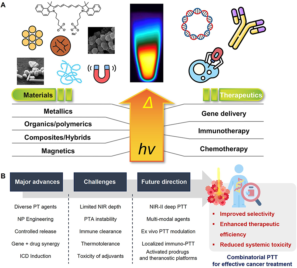 |
Figure 1 Schematic overview of PTT as a nanomaterial-enabled platform for combinatorial cancer treatment. (A) A variety of nanomaterials—including metallic, polymeric, hybrid, and magnetic systems—can be engineered for efficient PT conversion under light irradiation (hν). (B) Schematics illustrating the major advances, challenges, and future directions of combinatorial PTT-based agents for effective cancer treatment. Created by the authors. |
Underlying Mechanism on Photothermal Effects of Different NPs
NPs used in PTT convert absorbed NIR light into heat through distinct physicochemical mechanisms. Depending on their composition, structure, and electronic properties, NPs exhibit different photothermal behaviors that determine their efficiency, stability, and suitability for biomedical applications. This section summarizes the representative classes of photothermal nanomaterials—metallic, organic/polymeric, hybrid/composite, and semiconducting nanoparticles—and highlights their corresponding heat-generation mechanisms and practical considerations (Figure 2).
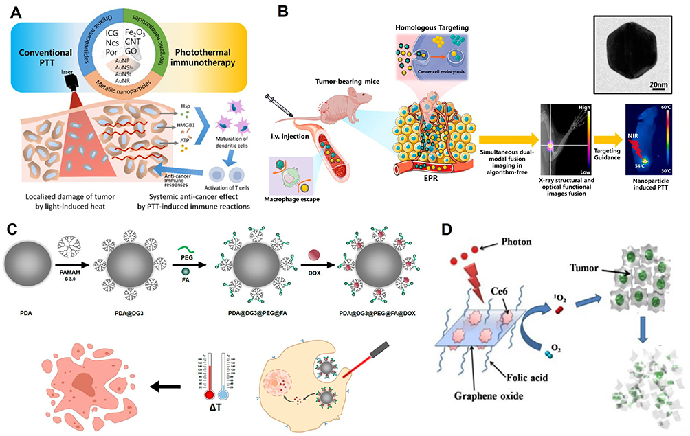 |
Figure 2 NP-based PTT. (A) A schematic diagram of NPs for PTT, where conventional PTT targets cancer cell death, while PTT enhances immune responses for a systemic effect.36 Copyright MDPI 2022. (B) Application of cancer cell membrane-coated AuNPs for PTT application.37 Copyright MDPI 2020. Inset image shows morphology of AuNPs. (C) Dual chemo- and PTT-assisted synergistic cancer therapy using functionalized polydopamine.38 Copyright MDPI 2021. (D) Graphene oxide-based composite PTT probe for tumor ablation.39 Copyright Ivyspring International Publisher 2011. All Figures were reproduced with permission. |
Metallic NPs
Metallic nanoparticles—including gold (Au), silver (Ag), platinum (Pt), and palladium (Pd)—generate heat primarily through localized surface plasmon resonance.40–42 Upon NIR irradiation, collective oscillations of conduction electrons lead to efficient light absorption and rapid thermal relaxation.30 Among these, gold nanorods (AuNRs), in particular, offer tunable longitudinal plasmon peaks and strong NIR absorption, making them widely used PTT agents.43 Their photothermal conversion is efficient, but potential concerns include plasmon damping, reshaping under high-intensity laser exposure, and limitations in deeper tissue penetration depending on their absorption wavelength.44 Recent advances in AuNR research have further optimized their photothermal performance, demonstrating improved plasmonic stability, enhanced heat conversion efficiency, and tunable aspect ratio control for cancer phototherapy applications.45,46
Silver nanoparticles exhibit comparable plasmonic behavior but suffer from oxidative degradation and higher cytotoxicity, whereas iron oxide nanoparticles offer lower photothermal efficiency but improved biocompatibility and magnetic functionality.47–51
Organic and Polymeric NPs
Organic/polymer-based NPs rely on intramolecular vibrations and π–π stacking interactions to dissipate absorbed energy as heat.35 These systems generally offer favorable biocompatibility and biodegradability compared with metallic nanomaterials. Polydopamine (PDA) NPs are a representative example, characterized by strong broadband NIR absorption, high chemical stability, and versatile surface functionality for cargo loading.52,53 Semiconducting polymer nanoparticles and dye-based nanoassemblies offer tunable optical properties but can be limited by photobleaching, aggregation, or reduced irradiation stability over prolonged use.54
Composite and Hybrid NPs
Composite/hybrid NPs integrate metal, polymer, or inorganic components to achieve enhanced photothermal properties and multifunctionality. Black phosphorus (BP) nanosheets (BPNS) can form synergistic hybrid structures with metals, enhancing their properties for PTT. The authors introduced BP-Au-thiosugar NSs (BATNS) composites to enhance stability and anticancer efficiency. Importantly, Au-thiosugar coating mitigated rapid oxidation and degradation of BPNS, which made them stable under laser irradiation. Also, BATNS (BP-Au-thiosugar NSs) offer better photon absorption and heat conversion than BPNS due to the shrinking bandgap (0.97 eV), allowing to convert more photon energy into heat, which was evidenced by the significantly enhanced ablation efficiency in an NIR-irradiated Hep1-6 cells. Additionally, BATNS-mediated PT promoted robust infiltration of tumor-killing NK cells without affecting T- or B-cell populations, thereby enhancing tumor necrosis with hypoxia markers, ultimately improving mouse survival rate.55
Ag2S -Based Semiconducting Nanomaterials
Ag2S NPs represent an emerging class of semiconducting photothermal agents distinguished by their narrow bandgap (~1.0–1.1 eV), which enables strong NIR-I and NIR-II absorption and intrinsic NIR-II fluorescence. This dual optical behavior facilitates both photothermal heating and deep-tissue imaging.56–58 Unlike metallic Ag NPs, Ag2S exhibits higher chemical stability and substantially lower cytotoxicity, improving its suitability for in vivo theranostics. Ag2S can be incorporated into multifunctional structures—including Ag2S@SiO2, Ag2S@PDA, polymeric micelles, and Ag2S–photosensitizer hybrids— allowing simultaneous PTT, chemotherapy, or photodynamic therapy.58,59
Despite these advantages, Ag2S-based platforms remain relatively underexplored compared to traditional PTT agents such as gold nanorods. Key challenges include synthetic complexity, insufficient biodegradation data, and a lack of standardized protocols for photothermal characterization and NIR-II imaging. Nevertheless, their combined photothermal and fluorescent capabilities highlight Ag2S as promising candidate for next-generation combinational PTT systems.
PTT Contribution with Gene Delivery to Cancer Treatment
The properties of the cell membrane (eg, stiffness, melting temperature), where genes or drugs, need to penetrate, can be changed based on wavelength, intensity, or duration of NIR-I (650–950 nm) and NIR-II (1000–1700 nm) laser windows within a specific wavelength depending on the disease site, including tumor depth and area.60 It is commonly exploited using metallic nanostructures (eg, AuNRs, Au nanoshells), organic and polymer-based photothermal dyes (eg, ICG, semiconducting polymers), or hybrid platforms that integrate inorganic cores with organic coatings. The photothermal conversion efficiency, absorption cross-section, biodegradability, and thermal stability of these agent classes vary considerably, influencing both performance and translational potential. At the cellular level, the photothermal heat generated under NIR irradiation, modulates the fluidity of cell membranes composed of phospholipids, proteins, etc, for transient regulation purposes using photothermal heat generated by laser irradiation. Based on these phenomena, the photothermal heat could possibly alter the permeability of the cell membrane or create pores temporally, enabling the direct delivery of genes and drugs of interest to the cytosol rather than the endocytosis pathway. Heat-induced signaling further activates pathways involving HSPs and, under certain regimes, can trigger ICD, providing an additional mechanistic basis for synergy with gene therapy, immunotherapy, and chemotherapeutic agents. Similarly, in terms of the use of the polymer properties, the release rate of the payload can be controlled by changing the conformation of polymer-based NPs, which are light- and thermal-sensitive materials, after laser irradiation.61,62 Thus, in this section, cancer treatment using genes delivery will be discussed through the application of PTT (Figure 3).
 |
Figure 3 Schematic illustration of the intracellular trafficking pathways of plasmid–lipofectamine complexes. The figure depicts two major pathways by which plasmid–lipofectamine complexes enter the cytosol: Direct transportation across the plasma membrane (left) and Endocytosis-mediated internalization (right). Red directional arrows (→) indicate the movement of plasmid–lipofectamine complexes along each intracellular trafficking route. Red upward arrows (↑) denote an increase in specific cellular processes, including membrane fluidity, membrane permeability, and endosomal escape efficiency. Created by the authors. |
Cell Membrane Modulation for Gene Delivery with Photothermal Effects
Positively charged liposomes, such as lipofectamine and Mirus, are typically used as a commercially available method for gene delivery. It has a high transfection efficiency regardless of the size and shape of nucleic acids, such as siRNA, mRNA, and plasmid DNA, in numerous cell types.63,64 However, when nucleic acids are delivered to primary cultured cells or neurons using general transfection agents, transfection efficiency and cell viability are frequently lower than anticipated.65 To use a PTT for nucleic acid delivery, it should be considered in terms of power density per area, duration of laser irradiation, and desirable wavelengths of light. Oh et al and Hosseinpour et al focused on the permeability change of the cell membrane by photothermal heat to deliver genes. The former group used an 808 nm diode laser with different types of cells, such as primary culture fibroblasts (D551), hepatocytes, and Jurkat cells, which are known to be difficult to deliver genes into cells to deliver commercially available lipofectamine and plasmid DNA complexes. To generate a photothermal effect, mPEGylated AuNRs were used, and the laser was irradiated for 30 min, which was sufficient for mPEGylated GNR to stay only outside of the cells. The transfection efficiency of primary cultured fibroblasts, Jurkat cells, and hepatocytes was enhanced 2.72-, 7.19-, and 2.54-fold.66 Another research group of Hosseinpour et al applied various wavelengths such as 445, 685, 810, or 970 nm for 2 h by using a diode laser on preosteoblast MC3T3 cells to improve cationic lipids to transfect ability. When the cells were irradiated with 810 nm at 12 J and 970 nm at 6 J, the proportion of GFP-positive cells significantly increased compared with Lipofectamine alone, reaching approximately 72% and 71%, respectively.67
The other groups focused on making transient pores on the cell membrane to allow impermeable dye to get into the cells. Based on this strategy, the light-triggered self-assembled nanobombs (NBs) gene transfection system was developed (Figure 4A). Iron oxide consists of NB cores, which play a key role as a photothermal component to generate water vapor bubbles when a 561-pulse laser is applied. Compared to electroporation and photoporation by using mRNA and plasmid DNA (Figure 4B), light-triggered NB-mediated photoporation results in a 5.5–7.6-fold increase in transfected cells compared with electroporation, while maintaining higher cell viability. Figure 4C shows parallel results for plasmid DNA, confirming that NB-assisted delivery outperforms both electroporation and repeated irradiation in terms of transfection rate and safety. Based on this result, they applied this strategy to spatial-selective eGFP-mRNA transfection in HeLa cells. As illustrated by the representative fluorescence images (Figure 4D and E), the delivery of genetic material resulted in efficient intracellular expression. Collectively, these data highlight the potential of nanobomb-assisted photoporation as a highly efficient, biosafe, and tunable platform for gene transfection.68 In another strategy, crystalline magnetic carbon NPs (CMCNPs) were introduced to accelerate plasmids into cancer cells under a localized magnetic field and photothermal effect by irradiation of NIR laser simultaneously since CMCNPs showed strong absorption in NIR region and magnetic properties. It was allowed to be an effective localized heating and magnetic field in the target area for delivering impermeable dye as a drug and plasmids.69
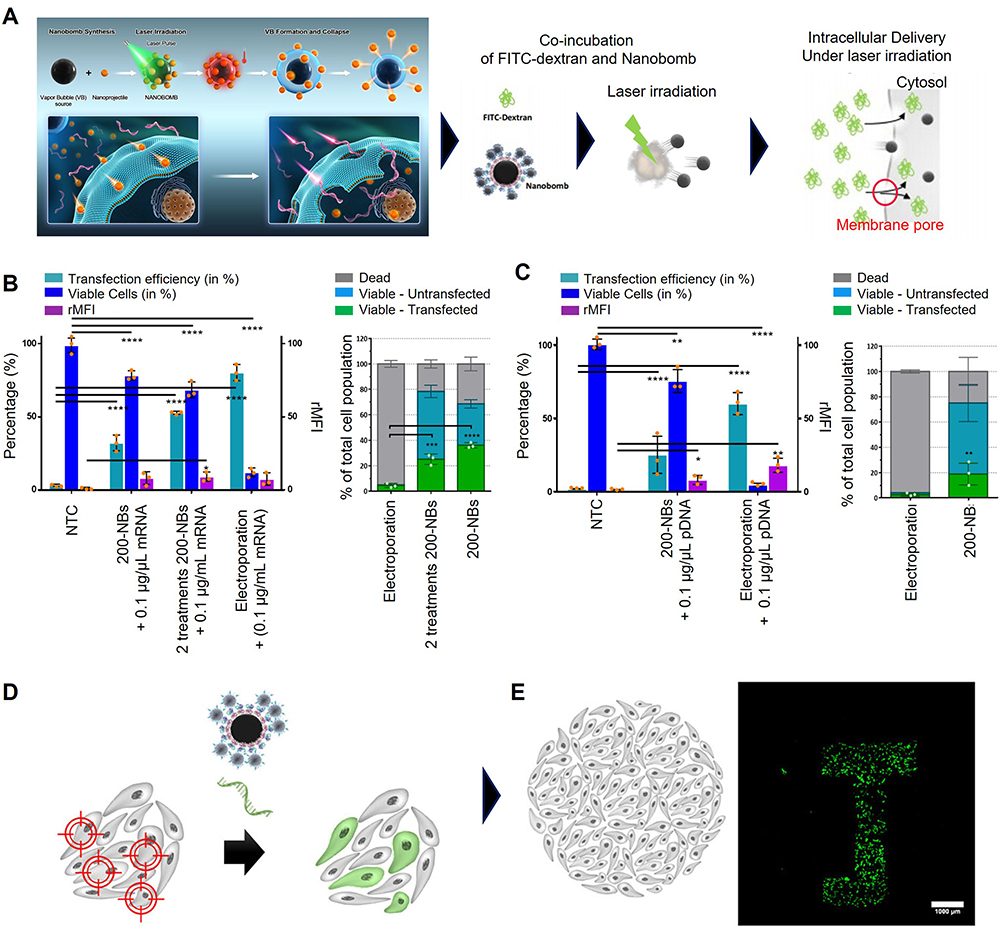 |
Figure 4 Gene delivery strategy with optically triggered nanobombs. (A) the working principle of nanobombs upon irradiation with an intense laser pulse (left) and cytosolic delivery of macromolecules by laser-activated NBs (right). (B) Transfection experiment proceeded with HeLa cells as adherent cells. (C) Transfection experiment proceeded with Jurkat cells as suspension cells (*p < 0.05, **p < 0.01, ***p < 0.001, ****p < 0.0001). (D) The strategy of spatial-selective NB mediated mRNA delivery. (E) Confocal images after transfection in HeLa cells for selective expression of proteins of interest. Scale bar = 1000 µm.68 Copyright Springer Nature Publisher 2022. All Figures were reproduced with permission. |
NP Modulation for Gene Delivery with Photothermal Effects
As shown in Table 1, some research groups used photothermal heat to stimulate a light-sensitive protein such as heat shock protein instead of modulating the properties of the cells. Nakatsuji et al sought to regulate gene expression by 780 nm NIR pulse laser.70 They synthesized DOTAP functionalized AuNRs with HSP promoter-driven vectors and then incubated them in HEK293T cells for 24 h (Figure 5A and B). In order to activate HSP promoters, a 780-nm NIR pulse laser was irradiated for 10s; this transfection condition showed less irradiation time than intracellular carbon nanohorns (0.5 W/cm,2 continuous, 30 min), silica-gold nanoshells and hollow gold NPs (2 W/cm,2 continuous, 15 min) and then transfection efficiency using AuNP functionalized with various cationic coating was quantified (Figure 5C). Flow cytometry analysis demonstrated that DOTAP- and lipofectamine-based formulations produced the highest levels of GFP expression in both HEK293 and HeLa cells. These results correspond to the fluorescence microscopy images (Figure 5D). Because promoter activation is tightly linked to photothermal input, they further optimized irradiation parameters by altering laser power and exposure time (Figure 5E). Given the potential risks associated with intracellular heating, they assessed photothermal cytotoxicity using flow cytometry (Figure 5F). To evaluate functional protein expression driven by photothermal induction, they monitored apoptosis in cells transfected with a pHSP70-TRAIL plasmid. Time-lapse imaging revealed dynamic cell death events among DsRed-positive cells following irradiation (Figure 5G). Some cells were already apoptotic prior to imaging, while others underwent apoptosis during the observation period, indicating active TRAIL expression. Quantitative analysis confirmed a significant increase in the proportion of apoptotic DsRed-positive cells compared with controls (Figure 5H).70 Ali et al have made a new strategy to enhance apoptosis by plasmonic PTT over necrosis because necrosis causes inflammation, known to induce metastasis and cancer growth. This group found that inhibition of HSP 70 was related to an increase in apoptosis.71
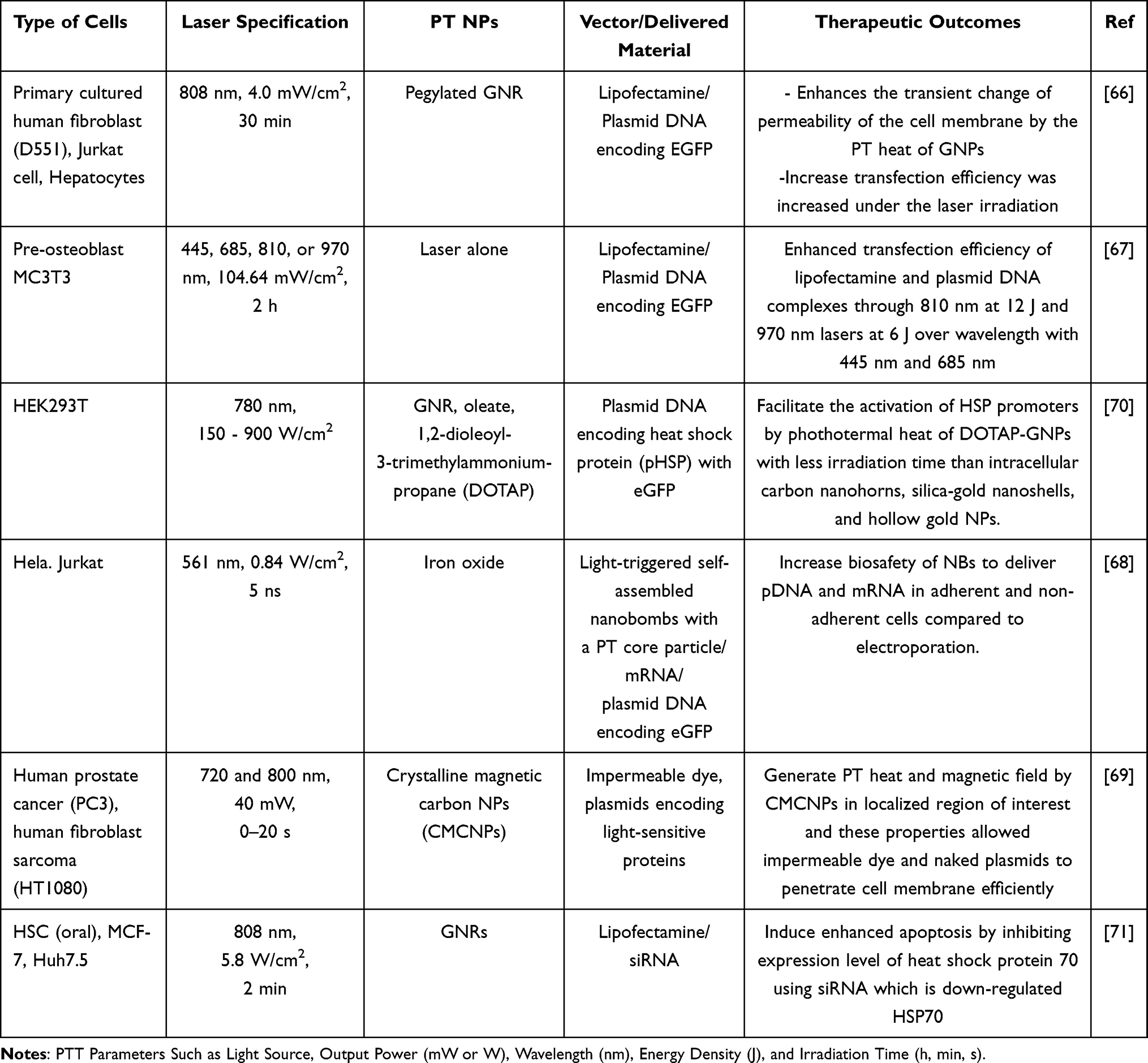 |
Table 1 The Strategy and Effect of Gene Delivery by PT Heat |
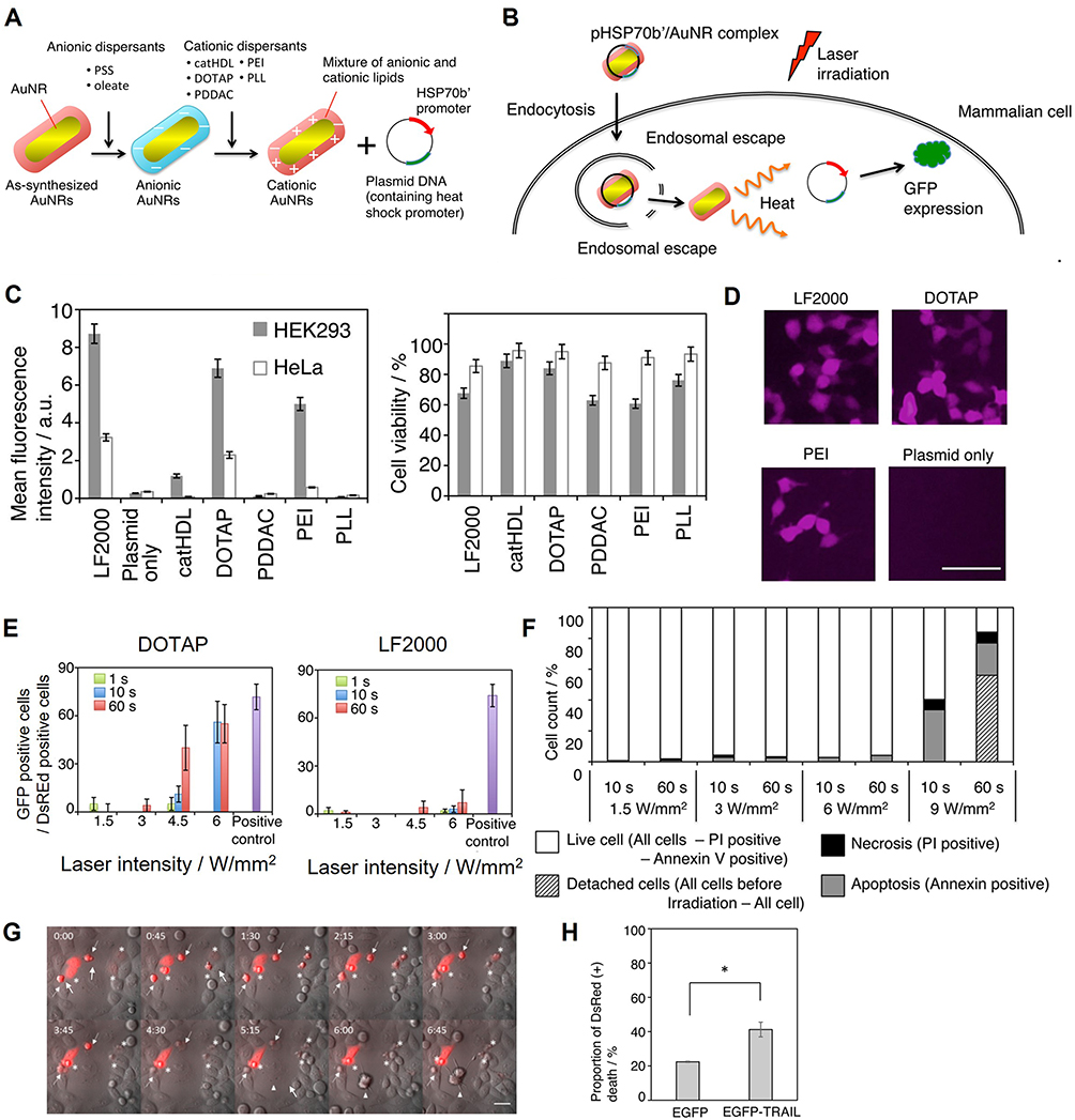 |
Figure 5 Gene delivery strategy with functionalized AuNPs and plasmid DNA. (A) Design of the complex with functionalized AuNPs and plasmid. (B) Schematic of PT induction of protein expression delivered by AuNPs. (C) Transfection efficiency by the formulation of AuNP with PT effect was determined by flow cytometry. (D) Fluorescence images of HEK293T cells treated with cationic AuNPs/plasmid DNA corresponded to flow cytometry data. Scale bar = 100 µm. (E) Transfection efficiency for DOTAP-AuNPs (left) and lipofectamine 2000 (lipo 2k) (right) to investigate optimal condition of laser power and irradiation time. (F) Investigation of phototoxicity by intracellular PT heating. (G) Time-lapse images of 45 minute internal of pHSP70-TRAIL transfected cells. Arrows indicate DsRed positive cells that had died before start of time lapse imaging. Asterisks display DsRed positive cells that die during time lapse imaging. Arrowhead shows DsRed negative cell that dies during time lapse imaging. Scale bar = 40 µm. (H) Quantification data of DsRed positive cells (*p < 0.05).70 Copyright Springer Nature Publisher 2017. All Figures were reproduced with permission. |
Through the use of NPs and the photothermal effect, nucleic acids such as plasmid DNA, siRNA, mRNA, and cationic lipid complexes can be delivered into the cytosol by endocytosis pathways. In the cases, photothermal effect was utilized to promote the escape of NPs form endosome or lysosome.
Unlike this strategy for gene delivery, the method of temporarily forming a pore in the cell membrane and another method of regulating the fluidity of the membrane while generating photothermal heat by irradiating laser onto NPs utilized the melting point of the phospholipids, comprising the cell membrane between −20°C and 60°C.72 This photothermal heat is also known to accelerate the endosomal escape of nucleic acids that have entered the cell via the endocytosis pathway. Both methods have the advantage of being applicable to adherent and nonadherent cells, despite the limited in vivo access due to the limitations of the laser’s tissue penetration depth (< 50 mm for NIR lasers) and are being actively researched for ex vivo cell modulation. These methods have the advantage of being applicable to both cell types to deliver nucleic acid of interest, despite the limited in vivo access due to the limitations of the tissue penetration depth of the laser (<1.0 µm for NIR lasers) and are being actively researched for ex vivo cell modulation.
PTT Contribution with Drug Delivery to Cancer Treatment
Combining PTT with drug delivery systems allows heat-dependent activation of therapeutic agents at tumor sites, improving intratumoral penetration and release kinetics, and tumor-specific accumulation while minimizing off-target exposure.73–75 Controlled hyperthermia alters vascular permeability, softens extracellular structures, and sensitizes tumor cells to pharmacological agents. Through these mechanisms, photothermal heating enhances the performance of both immunotherapy and chemotherapy. This section outlines how PTT interacts with immunogenic adjuvants and chemotherapeutic drugs to produce synergistic anticancer effects.
NP-Mediated Photothermal Immunotherapy
Integrating PTT with immunotherapy leverages localized hyperthermia to enhance antigen release, dendritic cell activation, and overall immune priming within the tumor microenvironment.76 This combined approach builds upon the established modes of immunotherapy—including checkpoint inhibitors, adoptive cell therapies, and vaccines—while addressing key barriers such as insufficient antigen presentation and immune suppression.77–80 PTT-induced ICD releases DAMPs, including HMGB1, ATP, and HSPs, which promote dendritic cell (DC) maturation and antigen presentation.81–86 Photothermal nanomaterials further facilitate targeted delivery of immunostimulatory molecules and enable spatiotemporal control over immune activation. These synergistic interactions have been demonstrated across a range of NPs that improve cytotoxic T-cell responses and inhibit both primary and metastatic tumors. This process is essential for initiating a robust and specific anti-tumor immune response (Figure 6). The localized hyperthermia induced by PTT creates an immunostimulatory microenvironment conducive to immune cell activation.87,88 Following cancer cell death, the released cancer antigens, pro-inflammatory cytokines, and other factors are captured by DCs, which then present the antigens to T cells via major histocompatibility complex (MHC) class I and II molecules.89 This interaction leads to T cell activation, with activated T cells migrating to and infiltrating the tumor site.90 The T cells specifically recognize and bind to cancer cells through the interaction between T cell receptors and antigen-captured MHC class I molecules, leading to the targeted destruction of tumor cells by cytotoxic T lymphocytes.91 However, cancer antigens alone are insufficient to induce a potent immune response. When used in conjunction with immunogenic adjuvants, these antigens can further enhance or prolong antigen-specific immune responses.92,93
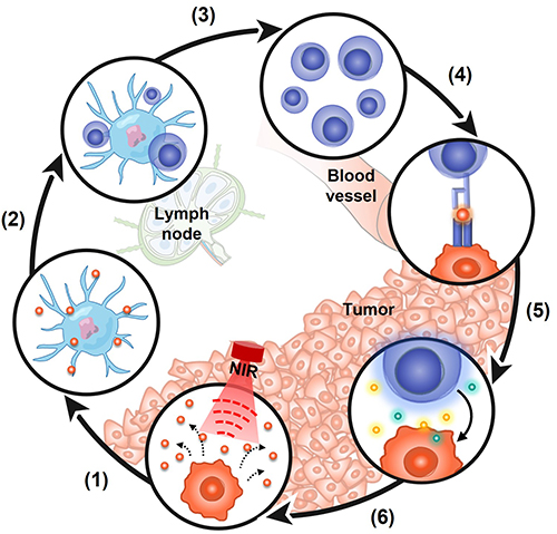 |
Figure 6 Schematic representation of nanomaterial-based photoimmunotherapy. The anti-cancer immune response follows a cyclic and self-propagating process, leading to the accumulation of immune-stimulatory molecules that enhance and amplify T cell responses. This cycle consists of six key stages: (1) Release of apoptotic bodies containing cancer antigens, (2) Presentation of cancer antigens by dendritic cells (DCs), (3) Activation of DCs and T cells, (4) Migration of activated T cells to the tumor site, (5) Recognition of cancer cells by T cells, and (6) Elimination of cancer cells. Created by the authors. |
The most effective immune adjuvants used in PTT-immunotherapy include toll-like receptor (TLR) agonists, which activate DCs and promote tumor-specific T cell responses.94–100 Although systemic administration of these agonists can cause immune-related toxicities, nanoparticle carriers enable localized delivery to the tumor microenvironment, thereby improving safety and immunomodulatory precision. Though these mechanisms, PTT-based combinations can enhance ICD, DC maturation, and antigen presentation, offering a strong rationale for integrating PTT with immunotherapy. Table 2 summarizes the approach and impacts of immunotherapy using hyperthermia. Zhan et al developed an extracellular matrix (ECM)-degrading nanoagonist (dNAc) composed of NIR-II-absorbing ferrous sulfide (FeS2) NPs and the SITNG agonist cyclic guanosine monophosphate–adenosine monophosphate (cyclic GMP-AMP, cGAMP) within thermoresponsive liposomes (Figure 7A).85 To enhance tumor infiltration, the surface of dNAc was functionalized with bromelain, an enzyme that degrades ECM. Upon exposure to an NIR-II laser (1064 nm), dNAc released cGAMP and induced ICD, as shown by calreticulin (CRT) and HMGB1 release (Figure 7B). In vivo studies demonstrated that dNAc treatment significantly robust inhibit and distant tumors, highlighting the robust systemic immune activation (Figure 7C).
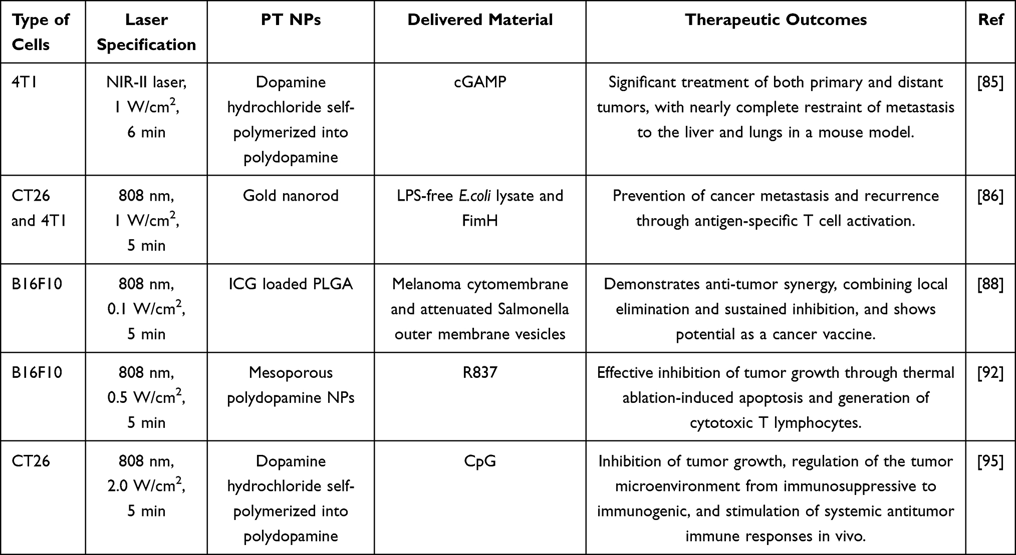 |
Table 2 The Approach and Impacts of Immunotherapy Thorough Using Hyperthermia Involve Adjusting Various Parameters for PTT, Including Laser Power Output, Wavelength, and Duration of Laser Irradiation |
 |
Figure 7 Photothermal-enhanced immunotherapy. (A) Conceptual illustration depicting the preparation of extracellular matrix (ECM)-degrading photothermal nanoagonist (dNAc), generated through a hydration–sonication procedure followed by surface functionalization for combined chemodynamic and immune modulation. (B) Representative immunofluorescence images of 4T1 cancer cells showing calreticulin (CRT) and HMGB1 exposure after treatment with NA0, NAc or dNAc in presence of 100 μM H2O2, followed by NIR-II laser irradiation (1 W/cm2, 6 min). (C) Growth curves of primary and distant tumors after administration of NA0, NAc, or dNAc, with or without NIR-II laser exposure (1 W/cm2, 10 min).85 Copyright 2022, John Wiley and Sons. (D) Schematic overview of E. coli-mimetic gold nanorod (ECA) formation via liposome coating and FimH functionalization. (E) iRFP signal in the lungs of mice on day 14 after the secondary tumor challenge, demonstrating metastatic suppression. (F) Flow cytometry analysis of dendritic cell surface markers in tumor-draining lymph nodes (tdLNs). (G) Quantification of IFN-γ and T-bet mRNA expression levels in tdLNs (**p < 0.01).86 Copyright 2022, American Chemical Society. All Figures were reproduced with permission. |
Hwang et al synthesized an ECA composed of lipopolysaccharides-free clear Escherichia coli (E. coli) lysate and phospholipid-coated AuNRs, conjugated with FimH, a bacterial surface adhesin protein from E. coli (Figure 7D).86 Under NIR irradiation, ECA effectively inhibited tumor growth and significantly prevented secondary pulmonary metastasis (Figure 7E). This approach not only induces direct tumor cell death but also promotes dendritic cell activation in tumor-draining lymph nodes, stimulating antigen-specific T cell immunity and offering protection against secondary tumor challenges (Figure 7F and G).
Notably, PTT-immunotherapy has demonstrated efficacy not only in preventing the progression of primary tumor but also in suppressing metastatic spread and reducing recurrence risk. By inducing ICD and activating adaptive immune responses, combination therapies offer a compelling strategy for treating both primary and metastatic cancers.98,101,102
Collectively, these studies demonstrate that photothermal immunotherapy can potentiate antigen release, dendritic cell maturation, and T-cell–mediated tumor clearance through heat-amplified immunomodulation. Nonetheless, its therapeutic consistency remains influenced by heterogeneous heat distribution, tumor-specific immune suppression, and limited NIR penetration. Further optimization of photothermal dosing, nanomaterial design, and integration with clinically validated immunotherapies—including immune checkpoint blockade—will be required to translate these synergistic effects into reproducible and durable clinical outcomes.
NP-Based Chemo-Photothermal Combination Therapy
When combined with chemotherapeutic agents, PTT provides localized heating that improves drug delivery, accelerates drug release from temperature-responsive carriers, and enhances tumor cell susceptibility to cytotoxic effects.103–105
Photothermal nanomaterials—including gold nanorods, black phosphorus nanosheets, platinum nanoparticles, polydopamine, and carbon-based platforms—act as both drug carriers and heat-generating agents, enabling coordinated delivery and activation (Table 3).106–114
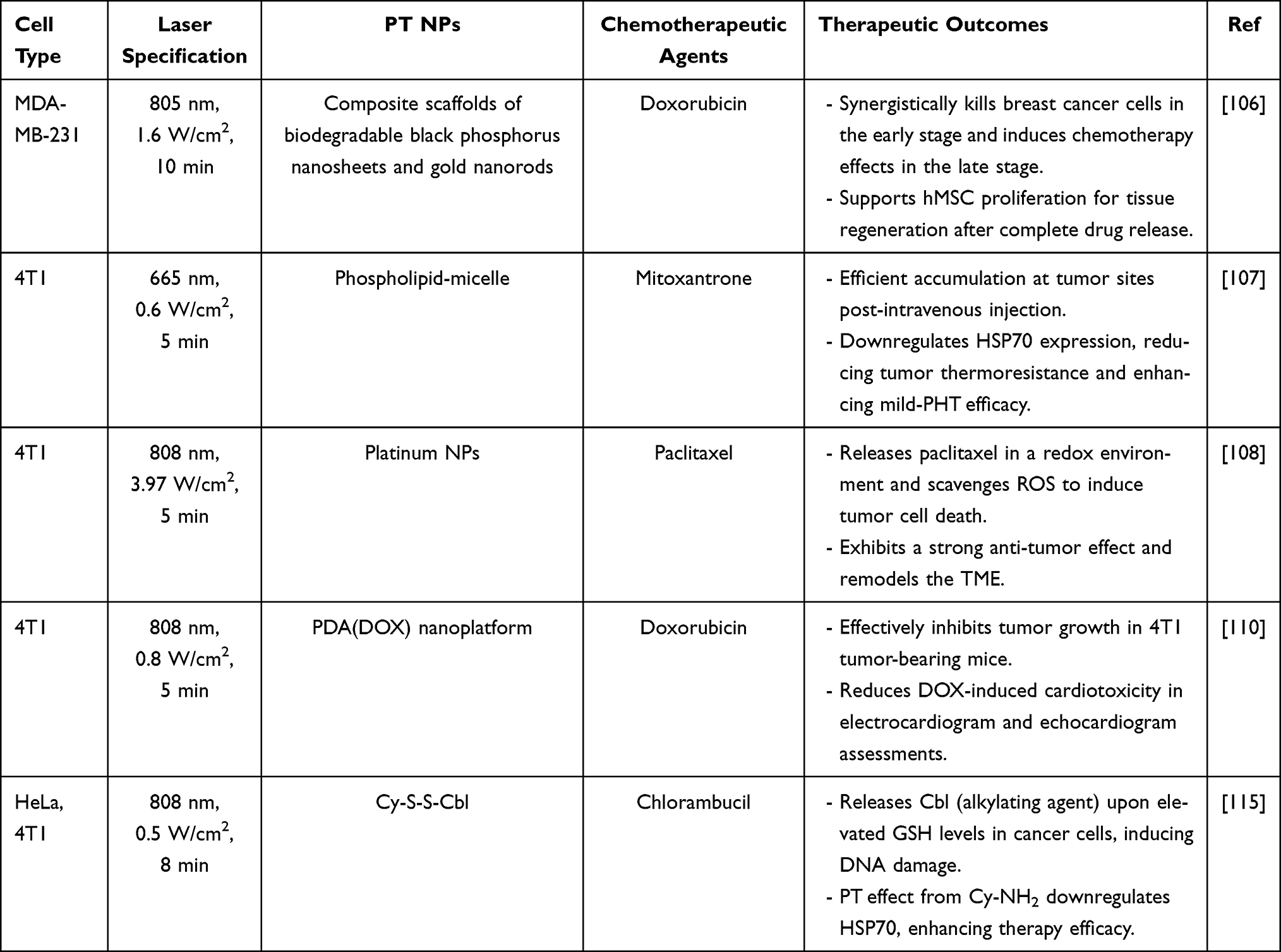 |
Table 3 T Strategies and Outcomes of Chemotherapeutic Drugs Combined with Hyperthermia, Including Various PTT Parameters (Laser Power Density, Wavelength, and Irradiation Duration) |
For example, Chen et al developed mitoxantrone (MTO)-micelles that integrate MTO with mild PTT for improved treatment of triple-negative breast cancer (Figure 8A).107 Following tumor accumulation via enhanced permeability and retention (EPR) effect, NIR irradiation produced mile hyperthermia that triggered MTO release and reduce HSP70 expression (Figure 8B). The combination of MTO-micelles and laser treatment exhibited significant antitumor effects compared to single treatments, highlighting the potential of chemo-PTT as an effective therapy for TNBC (Figure 8C).
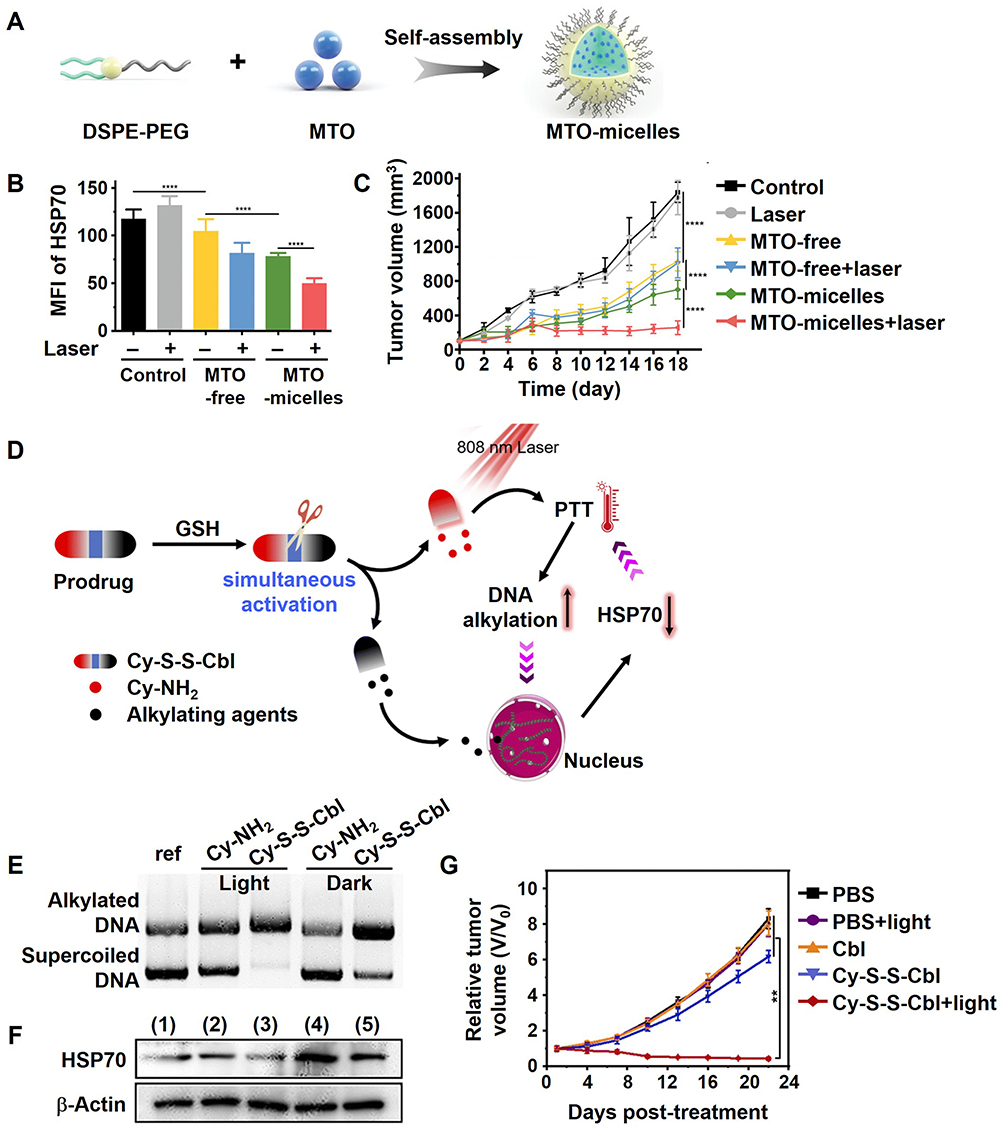 |
Figure 8 Combining Chemotherapy and Immunotherapy for synergistic cancer therapy. (A) Schematic diagram of self-assembled mitoxantrone (MTO)-micelles engineered to deliver MTO in combination with mild-photothermal heating for enhanced therapy in triple-negative breast cancer (TNBC). (B) Quantification of HSP70 expression levels in 4T1 cells following various micelle- or laser-based treatments. (C) Tumor growth curves in mice receiving mild photothermal chemotherapy using either MTO-free or MTO-loaded micelles (****p < 0.0001).107 Copyright 2023, Wiley‐VCH GmbH. (D) Schematic depiction of a glutathione (GSH)-activated Cy-S-S-Cbl prodrug system that able simultaneous activation of photothermal and alkylation functions. (E) Agarose gel electrophoresis showing plasmid DNA (pBR322) after incubation with Cy-S-S-Cbl or Cy-NH2 (50 μM) at 37°C for 4 h in 10 mM GSH, with or without NIR light. (F) Western blot analysis of HSP70 expression in HeLa cells under treatments: (1) PBS, (2) PBS + 808 nm light, (3) Cy-S-S-Cbl, (4) Cy-NH2 + NIR light, and (5) Cy-S-S-Cbl + NIR light. (G) Relative tumor growth in HeLa tumor-bearing mice across different treatment groups (**p < 0.01).115 Copyright 2023, Royal Society of Chemistry. All Figures were reproduced with permission. |
Another innovative approach by Zou et al involved a glutathione (GSH)-activated prodrug, Cy-S-S-Cbl, which coupled a photothermal agent (Cy7) with an alkylating agent (cobalamin, Cbl) (Figure 8D).115
In the high-GSH tumor microenvironment, the disulfide bond is cleaved to yield Cy-NH2 and the active alkylating species. Cy-NH2 demonstrated higher photothermal conversion, while the released alkylating drug induces DNA damage; together, these processes further downregulated HSP70 expression and reduce cancer cell thermotolerance (Figure 8E and F). As a result, combined chemo-PTT produced stronger tumor inhibition than either modality alone (Figure 8G).
These systems illustrate how heat-triggered drug activation and photothermal sensitization create a synergistic therapeutic response capable of overcoming drug resistance and improving intratumoral drug.116–126
However, their translation remains limited by variability in thermal dosing, incomplete data on nanoparticle clearance and long-term toxicity, tumor heterogeneity, and the lack of standardized irradiation parameters across studies. Future progress will require harmonized photothermal protocols, systematic evaluation of chronic safety, and development of deeper-penetrating NIR-responsive nanomaterials to enable reliable efficacy in clinically relevant tumor models.
Conclusion and Perspectives
While PTT shows promising prospects through its integration with gene therapy, immunotherapy, and chemotherapy, this review presents the potential for PTT’s clinical application and critically evaluates its current limitations. Even though the paucity of consistent experimental outcomes based on NP designs and irradiation parameters complicates direct comparison between studies, NP-enabled PTT has progressed from a heat-based tumor ablation method to a multifunctional platform. In this review, we hereby present wavelength-dependent cellular and molecular responses to PTT and correlation with quantifiable optical parameters in comparison with a recent review,127 which mainly addressed the condition and their effect of PTT in cancer treatment. Through localized hyperthermia, PTT increases membrane permeability, promotes endosomal escape, triggers thermally responsive drug release, and induces immunogenic cell death—mechanisms that collectively improve the performance of co-administered therapeutics. Emerging NIR-II–responsive materials, including Ag2S-based semiconductors, further expand the potential of PTT by offering deeper light penetration and integrated imaging capability. Despite these advances, several hurdles limit clinical translation. NIR penetration depth restricts treatment of deep-seated tumors; thermal overexposure may induce HSP–mediated thermotolerance; and long-term biodistribution, biodegradation, and immunotoxicity of nanomaterials remain insufficiently characterized. Moreover, inconsistencies in laser parameters, thermal dosimetry, and treatment protocols hinder reproducibility and complicate cross-study comparison. Additionally, the therapeutic efficacy of PTT-based combinations is highly dependent on tumor anatomy and microenvironmental heterogeneity.
Future progress will benefit from the development of photothermal agents with enhanced optical efficiency and improved tissue penetration—particularly biomimetic and NIR-II–responsive materials—together with standardized laser irradiation parameters and thermal-dosing protocols and more systematic long-term safety evaluations. Mechanistic integration of PTT with clinically validated modalities—such as immune checkpoint inhibitors, nucleic acid therapeutics, or multimodal imaging-guided systems—may yield more reproducible and durable responses. As these translational challenges are addressed, combinatorial PTT has the potential to evolve into a more selective and adaptable treatment framework that complements and extends the capabilities of current cancer therapies.
What’s New in This
A comparison of the present study with existing reviews on photothermal therapy reveals several novel points. The initial point unveils a mechanism-centered, indicator-based framework that establishes a correlation between optical parameters and cellular and molecular responses. The subsequent point concerns the most detailed synthesis to date of PTT-enabled gene delivery mechanisms, encompassing nanobomb-mediated photoporation. The third point pertains to the formulation of a unified mechanistic map that elucidates the manner in which PTT synergizes with gene therapy, immunotherapy, and chemotherapy. The fourth point is an updated and translation-oriented assessment of emerging NIR-II photothermal agents. The fifth point involves a comparative analysis of nanoparticle families aligned with heat-biology interactions rather than material categories alone. The final point reveals that a critical appraisal of current limitations—such as dosimetry inconsistency, thermotolerance, toxicity, and intratumoral heterogeneity—clarifies challenges that must be addressed for clinical translation.
Abbreviations
PTT, Photothermal therapy; NIR, near-infrared; NPs, nanoparticles; ICD, immunogenic cell death; DAMPs, damage-associated molecular patterns; HMGB1, High mobility group box 1; HSP, heat shock protein; AuNRs, gold nanorods; PDA, Polydopamine; BP, Black phosphorus; BPNS, BP nanosheets; BATNS, BP-Au-thiosugar NSs; NBs, nanobombs; DC, dendritic cell; MCH, major histocompatibility complex; TLR, toll-like receptor; ECM, extracellular matrix; cGAMP, cyclic guanosine monophosphate–adenosine monophosphate; CTR, calreticulin; E. coli, Escherichia coli; MTO, mitoxantrone; GSH, glutathione.
Acknowledgments
This work was supported by the National Research Foundation of Korea (NRF) grants funded by the Ministry of Science and ICT (NRF-2022R1A5A8023404 and RS-2023-00281553), by the Ministry of Education (RS-2023-00241311), and by the Research Program at the Korea Science Academy of KAIST with funding from the Korean Government (the Ministry of Science and ICT). This work was also supported by the Technology Development Program (No. RS-2025-25460411) funded by the Ministry of SMEs and Startups (MSS, Korea).
Disclosure
The authors report no conflicts of interest in this work.
References
1. Li J, Zhang W, Ji W, et al. Near infrared photothermal conversion materials: mechanism, preparation, and photothermal cancer therapy applications. J Mater Chem B. 2021;9(38):7909–19. doi:10.1039/d1tb01310f
2. Zhang L, Oudeng G, Wen F, Liao G. Recent advances in near-infrared-II hollow nanoplatforms for photothermal-based cancer treatment. Biomater Res. 2022;26(1):61. doi:10.1186/s40824-022-00308-z
3. Zeng W, Li Z, Chen H, Zeng X, Mei L. An optimal portfolio of photothermal combined immunotherapy. Cell Rep Phy Sci. 2022;3:1.
4. Cui X, Ruan Q, Zhuo X, et al. Photothermal nanomaterials: a powerful light-to-heat converter. Chem Rev. 2023;123(11):6891–6952. doi:10.1021/acs.chemrev.3c00159
5. Shannon SR, Ben-Akiva E, Green JJ. Approaches towards biomaterial-mediated gene editing for cancer immunotherapy. Biomater Sci. 2022;10(23):6675–6687. doi:10.1039/d2bm00806h
6. Chen Y, Yu H, Wang Y, Sun P, Fan Q, Ji M. Thiadiazoloquinoxaline derivative-based NIR-II organic molecules for NIR-II fluorescence imaging and photothermal therapy. Biomater Sci. 2022;10(11):2772–2788. doi:10.1039/d2bm00283c
7. Hwang J, Mhamdi R, Soeriawidjaja BF, et al. Assessment of near-infrared penetration depth and photothermal efficiency of organic and inorganic materials in tissue-mimicking phantoms. Available at SSRN 4439126. 2023;47(5):678–685.
8. Zhang Y, Hao S, Zuo J, et al. NIR-activated thermosensitive liposome-gold nanorod hybrids for enhanced drug delivery and stimulus sensitivity. ACS Biomater Sci Eng. 2023;9(1):340–351. doi:10.1021/acsbiomaterials.2c01142
9. Lu F, Li Z, Kang Y, Su Z, Yu R, Zhang S. Black phosphorus quantum dots encapsulated in anionic waterborne polyurethane nanoparticles for enhancing stability and reactive oxygen species generation for cancer PDT/PTT therapy. J Mater Chem B. 2020;8(46):10650–10661. doi:10.1039/d0tb02101f
10. Revuri V, Mondal J, Lee YK. Graphene as photothermal therapeutic agents. Adv Exp Med Biol. 2022;1351:177–200. doi:10.1007/978-981-16-4923-3_9
11. Xiong Y, Rao Y, Hu J, Luo Z, Chen C. Nanoparticle-based photothermal therapy for breast cancer noninvasive treatment. Adv Mater. 2023;37:e2305140. doi:10.1002/adma.202305140
12. Hwang J, Jin JO. Attachable hydrogel containing indocyanine green for selective photothermal therapy against melanoma. Biomolecules. 2020;10(8):1124. doi:10.3390/biom10081124
13. Kashyap BK, Singh VV, Solanki MK, Kumar A, Ruokolainen J, Kesari KK. Smart nanomaterials in cancer theranostics: challenges and opportunities. ACS Omega. 2023;8(16):14290–14320. doi:10.1021/acsomega.2c07840
14. Overchuk M, Weersink RA, Wilson BC, Zheng G. Photodynamic and photothermal therapies: synergy opportunities for nanomedicine. ACS Nano. 2023;17(9):7979–8003. doi:10.1021/acsnano.3c00891
15. Farzam OR, Mehran N, Bilan F, et al. Nanoparticles for imaging-guided photothermal therapy of colorectal cancer. Heliyon. 2023;9(11):e21334. doi:10.1016/j.heliyon.2023.e21334
16. Khafaji M, Zamani M, Golizadeh M, Bavi O. Inorganic nanomaterials for chemo/photothermal therapy: a promising horizon on effective cancer treatment. Biophys Rev. 2019;11(3):335–352. doi:10.1007/s12551-019-00532-3
17. Xu H, Li S, Liu YS. Nanoparticles in the diagnosis and treatment of vascular aging and related diseases. Signal Transduct Target Ther. 2022;7(1):231. doi:10.1038/s41392-022-01082-z
18. Shao C, Li Z, Zhang C, et al. Optical diagnostic imaging and therapy for thyroid cancer. Mater Today Bio. 2022;17:100441. doi:10.1016/j.mtbio.2022.100441
19. Gowsalya K, Yasothamani V, Vivek R. Emerging indocyanine green-integrated nanocarriers for multimodal cancer therapy: a review. Nanoscale Adv. 2021;3(12):3332–3352. doi:10.1039/d1na00059d
20. Chehelgerdi M, Chehelgerdi M, Allela OQB, et al. Progressing nanotechnology to improve targeted cancer treatment: overcoming hurdles in its clinical implementation. Mol Cancer. 2023;22(1):169. doi:10.1186/s12943-023-01865-0
21. Li Y, Zhang K, Wu Y, et al. Antigen capture and immune modulation by bacterial outer membrane vesicles as in situ vaccine for cancer immunotherapy post-photothermal therapy. Small. 2022;18(14):e2107461. doi:10.1002/smll.202107461
22. Sushnitha M, Evangelopoulos M, Tasciotti E, Taraballi F. Cell membrane-based biomimetic nanoparticles and the immune system: immunomodulatory interactions to therapeutic applications. Front Bioeng Biotechnol. 2020;8:627. doi:10.3389/fbioe.2020.00627
23. Hong L, Li W, Li Y, Yin S. Nanoparticle-based drug delivery systems targeting cancer cell surfaces. RSC Adv. 2023;13(31):21365–21382. doi:10.1039/d3ra02969g
24. Lv W, Xu C, Wu H, et al. Ultrasound-visualized nanocarriers with siRNA for targeted inhibition of M2-like TAM polarization to enhance photothermal therapy in NSCLC. Nano Res. 2023;16(1):882–893. doi:10.1007/s12274-022-4767-7
25. Nag S, Mitra O, Tripathi G, et al. Nanomaterials-assisted photothermal therapy for breast cancer: state-of-the-art advances and future perspectives. Photodiagn Photodyn Ther. 2024;45:103959. doi:10.1016/j.pdpdt.2023.103959
26. Nam J, Son S, Ochyl LJ, Kuai R, Schwendeman A, Moon JJ. Chemo-photothermal therapy combination elicits anti-tumor immunity against advanced metastatic cancer. Nat Commun. 2018;9(1):1074. doi:10.1038/s41467-018-03473-9
27. Dube T, Kompella UB, Panda JJ. Near infrared triggered chemo-PTT-PDT effect mediated by glioma directed twin functional-chimeric peptide-decorated gold nanoroses. J Photochem Photobiol B. 2022;228:112407. doi:10.1016/j.jphotobiol.2022.112407
28. Waheed S, Li Z, Zhang F, Chiarini A, Armato U, Wu J. Engineering nano-drug biointerface to overcome biological barriers toward precision drug delivery. J Nanobiotechnol. 2022;20(1):395. doi:10.1186/s12951-022-01605-4
29. Kim SJ, Park HB, An EK, et al. Artificial immunogenic cell death lipid nanoparticle functions as a therapeutic vaccine for cancer. Adv Funct Mater. 2023;33:2302825. doi:10.1002/adfm.202302825
30. Lv Z, He S, Wang Y, Zhu X. Noble metal nanomaterials for NIR‐triggered photothermal therapy in cancer. Adv Healthcare Mater. 2021;10(6):2001806. doi:10.1002/adhm.202001806
31. Chen C, Ma Y, Du S, et al. Controlled CRISPR-Cas9 ribonucleoprotein delivery for sensitized photothermal therapy. Small. 2021;17(33):e2101155. doi:10.1002/smll.202101155
32. Papini E, Tavano R, Mancin F. Opsonins and dysopsonins of nanoparticles: facts, concepts, and methodological guidelines. Front Immunol. 2020;11:567365. doi:10.3389/fimmu.2020.567365
33. Han X, Gong C, Yang Q, Zheng K, Wang Z, Zhang W. Biomimetic nano-drug delivery system: an emerging platform for promoting tumor treatment. Int J Nanomed. 2024;19:571–608. doi:10.2147/IJN.S442877
34. Ren Y, Yang H, Xu D, Zhang Z, Gao S, Yu R. Application of multifunctional metal nanoparticles in the treatment of glioma. Int J Nanomed. 2025;625–638. doi:10.2147/IJN.S493565
35. Pham -T-TD, Phan LMT, Cho S, Park J. Enhancement approaches for photothermal conversion of donor–acceptor conjugated polymer for photothermal therapy: a review. Sci Technol Adv Mater. 2022;23(1):707–734. doi:10.1080/14686996.2022.2134976
36. Yun WS, Park J-H, Lim D-K, Ahn C-H, Sun I-C, Kim K. How did conventional nanoparticle-mediated photothermal therapy become “hot” in combination with cancer immunotherapy? Cancers. 2022;14(8):2044. doi:10.3390/cancers14082044
37. Wang R, Yang H, Fu R, et al. Biomimetic upconversion nanoparticles and gold nanoparticles for novel simultaneous dual-modal imaging-guided photothermal therapy of cancer. Cancers. 2020;12(11):3136. doi:10.3390/cancers12113136
38. Grześkowiak BF, Maziukiewicz D, Kozłowska A, Kertmen A, Coy E, Mrówczyński R. Polyamidoamine dendrimers decorated multifunctional polydopamine nanoparticles for targeted chemo-and photothermal therapy of liver cancer model. Int J Mol Sci. 2021;22(2):738. doi:10.3390/ijms22020738
39. Huang P, Xu C, Lin J, et al. Folic acid-conjugated graphene oxide loaded with photosensitizers for targeting photodynamic therapy. Theranostics. 2011;1:240. doi:10.7150/thno/v01p0240
40. Feng Z, Jia Y, Cui H. Engineering the surface roughness of the gold nanoparticles for the modulation of LSPR and SERS. J Colloid Interface Sci. 2024;672:1–11. doi:10.1016/j.jcis.2024.05.217
41. Unser S, Bruzas I, He J, Sagle L. Localized surface plasmon resonance biosensing: current challenges and approaches. Sensors. 2015;15(7):15684–15716. doi:10.3390/s150715684
42. Liu S, Chen G, Prasad PN, Swihart MT. Synthesis of monodisperse Au, Ag, and Au–Ag alloy nanoparticles with tunable size and surface plasmon resonance frequency. Chem Mater. 2011;23(18):4098–4101. doi:10.1021/cm201343k
43. Abed A, Mirzaei SA, Hosseini SA, Ghelich E, Rahimian N, Mirzaei H. Metal nanoparticles and sensitivity/resistance to therapy in cancer: two sides of the coin? J Nanopart Res. 2025;27(2):44. doi:10.1007/s11051-025-06228-y
44. Loiseau A, Asila V, Boitel-Aullen G, Lam M, Salmain M, Boujday S. Silver-based plasmonic nanoparticles for and their use in biosensing. Biosensors. 2019;9(2):78. doi:10.3390/bios9020078
45. Ma Z, Han Z, Wan M, et al. Development of a pH/NIR/temperature-responsive drug delivery system using AuNRs@ ZnO@ mPDA nanoparticles for synergistic cancer therapy. Surf Interfaces. 2025;61:106153. doi:10.1016/j.surfin.2025.106153
46. Han Z, Gao M, Wang Z, Peng L, Zhao Y, Sun L. pH/NIR-responsive nanocarriers based on mesoporous polydopamine encapsulated gold nanorods for drug delivery and thermo-chemotherapy. J Drug Delivery Sci Technol. 2022;75:103610. doi:10.1016/j.jddst.2022.103610
47. Fernández-Afonso Y, Asín L, Pardo J, et al. Key factors influencing magnetic nanoparticle-based photothermal therapy: physicochemical properties, irradiation power, and particle concentration in vitro. Nanoscale Adv. 2025;7(1):336–345. doi:10.1039/D4NA00384E
48. Sezer N, Arı İ, Biçer Y, Koç M. Superparamagnetic nanoarchitectures: multimodal functionalities and applications. J Magn Magn Mater. 2021;538:168300.
49. Grancharova T, Zagorchev P, Pilicheva B. Iron oxide nanoparticles: parameters for optimized photoconversion efficiency in synergistic cancer treatment. J Funct Biomat. 2024;15(8):207. doi:10.3390/jfb15080207
50. Mittal A, Roy I, Gandhi S. Magnetic nanoparticles: an overview for biomedical applications. Magnetochemistry. 2022;8(9):107. doi:10.3390/magnetochemistry8090107
51. Albukhaty S, Sulaiman GM, Al-Karagoly H, et al. Iron oxide nanoparticles: the versatility of the magnetic and functionalized nanomaterials in targeting drugs, and gene deliveries with effectual magnetofection. J Drug Delivery Sci Technol. 2024;99:105838.
52. Xing Y, Zhang J, Chen F, Liu J, Cai K. Mesoporous polydopamine nanoparticles with co-delivery function for overcoming multidrug resistance via synergistic chemo-photothermal therapy. Nanoscale. 2017;9(25):8781–8790. doi:10.1039/C7NR01857F
53. Wu Y, Huang Y, Tu C, et al. A mesoporous polydopamine nanoparticle enables highly efficient manganese encapsulation for enhanced MRI-guided photothermal therapy. Nanoscale. 2021;13(13):6439–6446. doi:10.1039/D1NR00957E
54. Chao B, Jiao J, Yang L, et al. Application of advanced biomaterials in photothermal therapy for malignant bone tumors. Biomater Res. 2023;27(1):116. doi:10.1186/s40824-023-00453-z
55. Jia C, Zhang F, Lin J, et al. Black phosphorus-Au-thiosugar nanosheets mediated photothermal induced anti-tumor effect enhancement by promoting infiltration of NK cells in hepatocellular carcinoma. J Nanobiotechnol. 2022;20(1):90. doi:10.1186/s12951-022-01286-z
56. Zhao H, He Y, Wang Z, Zhao Y, Sun L. Mussel-inspired fabrication of PDA@ PAN electrospun nanofibrous membrane for oil-in-water emulsion separation. Nanomaterials. 2021;11(12):3434. doi:10.3390/nano11123434
57. Gao M, Du J, Han Z, et al. Precise preparation of various morphological silver sulfide nanostructures, investigation of formation mechanism, and comparative study of Photothermal performance for cancer treatment. Part Part Syst Charact. 2022;39(2):2100240. doi:10.1002/ppsc.202100240
58. Gao M, Han Z, Wang Z, et al. Fabrication of a smart drug delivery system based on hollow Ag2S@ mSiO2 nanoparticles for fluorescence-guided synergistic photothermal chemotherapy. Mikrochim Acta. 2022;189(10):376. doi:10.1007/s00604-022-05468-2
59. Gao M, Han Z, Zhang X, et al. Construction of double-shelled hollow Ag2S@ polydopamine nanocomposites for fluorescence-guided, dual stimuli-responsive drug delivery and photothermal therapy. Nanomaterials. 2022;12(12):2068. doi:10.3390/nano12122068
60. Tao Y, Chan HF, Shi B, Li M, Leong KW. Light: a magical tool for controlled drug delivery. Adv Funct Mater. 2020;30(49). doi:10.1002/adfm.202005029
61. Qiu M, Wang D, Liang W, et al. Novel concept of the smart NIR-light-controlled drug release of black phosphorus nanostructure for cancer therapy. Proc Natl Acad Sci U S A. 2018;115(3):501–506. doi:10.1073/pnas.1714421115
62. Nakayama A, Sato M, Shinohara M, et al. Efficient transfection of primarily cultured porcine embryonic fibroblasts using the Amaxa Nucleofection system. Cloning Stem Cells. 2007;9(4):523–534. doi:10.1089/clo.2007.0021
63. Wang T, Larcher LM, Ma L, Veedu RN. Systematic screening of commonly used commercial transfection reagents towards efficient transfection of single-stranded oligonucleotides. Molecules. 2018;23(10). doi:10.3390/molecules23102564
64. Cardarelli F, Digiacomo L, Marchini C, et al. The intracellular trafficking mechanism of Lipofectamine-based transfection reagents and its implication for gene delivery. Sci Rep. 2016;6:25879. doi:10.1038/srep25879
65. Fountain JW, Lockwood WK, Collins FS. Transfection of primary human skin fibroblasts by electroporation. Gene. 1988;68(1):167–172. doi:10.1016/0378-1119(88)90610-5
66. Oh N, Park S, Kim JW, Park JH. Photothermal transfection for effective nonviral genome editing. ACS Appl Bio Mater. 2021;4(7):5678–5685. doi:10.1021/acsabm.1c00465
67. Hosseinpour S, Xu C, Walsh LJ. Impact of photobiomodulation using four diode laser wavelengths of on cationic liposome gene transfection into pre-osteoblast cells. J Photochem Photobiol B. 2021;215:112108. doi:10.1016/j.jphotobiol.2020.112108
68. Fraire JC, Shaabani E, Sharifiaghdam M, et al. Light triggered nanoscale biolistics for efficient intracellular delivery of functional macromolecules in mammalian cells. Nat Commun. 2022;13(1):1996. doi:10.1038/s41467-022-29713-7
69. Gu L, Koymen AR, Mohanty SK. Crystalline magnetic carbon nanoparticle assisted photothermal delivery into cells using CW near-infrared laser beam. Sci Rep. 2014;4:5106. doi:10.1038/srep05106
70. Nakatsuji H, Kawabata Galbraith K, Kurisu J, Imahori H, Murakami T, Kengaku M. Surface chemistry for cytosolic gene delivery and photothermal transgene expression by gold nanorods. Sci Rep. 2017;7(1):4694. doi:10.1038/s41598-017-04912-1
71. Ali MR, Ali HR, Rankin CR, El-Sayed MA. Targeting heat shock protein 70 using gold nanorods enhances cancer cell apoptosis in low dose plasmonic photothermal therapy. Biomaterials. 2016;102:1–8. doi:10.1016/j.biomaterials.2016.06.017
72. Muzic T, Tounsi F, Madsen SB, Pollakowski D, Konrad M, Heimburg T. Melting transitions in biomembranes. Biochim Biophys Acta Biomembr. 2019;1861(11):183026. doi:10.1016/j.bbamem.2019.07.014
73. Pan Y, Xue X, Liang XJ. Nanotechnology‐empowered combination cancer immunotherapies: mechanisms, synergies, and perspectives. Adv NanoBiomed Res. 2024;4:2300129. doi:10.1002/anbr.202300129
74. Xu T, Liu Z, Huang L, Jing J, Liu X. Modulating the tumor immune microenvironment with nanoparticles: a sword for improving the efficiency of ovarian cancer immunotherapy. Front Immunol. 2022;13:1057850. doi:10.3389/fimmu.2022.1057850
75. Huang X, Lu Y, Guo M, Du S, Han N. Recent strategies for nano-based PTT combined with immunotherapy: from a biomaterial point of view. Theranostics. 2021;11(15):7546–7569. doi:10.7150/thno.56482
76. Zhang Y, Zhang Z. The history and advances in cancer immunotherapy: understanding the characteristics of tumor-infiltrating immune cells and their therapeutic implications. Cell Mol Immunol. 2020;17(8):807–821. doi:10.1038/s41423-020-0488-6
77. Kiaie SH, Salehi-Shadkami H, Sanaei MJ, et al. Nano-immunotherapy: overcoming delivery challenge of immune checkpoint therapy. J Nanobiotechnol. 2023;21(1):339. doi:10.1186/s12951-023-02083-y
78. Nadukkandy AS, Ganjoo E, Singh A, Dinesh Kumar L. Tracing new landscapes in the arena of nanoparticle-based cancer immunotherapy. Front Nanotechnol. 2022;4:911063. doi:10.3389/fnano.2022.911063
79. Thakur N, Chatterjee S, Thakur S, Das J, Sil PC. Nanoparticles as smart carriers for enhanced cancer immunotherapy. Front Chem. 2020;8:597806. doi:10.3389/fchem.2020.597806
80. Gupta S, Shukla S. Limitations of immunotherapy in cancer. Cureus. 2022;14(10):1.
81. Li Z, Lai X, Fu S, et al. Immunogenic cell death activates the tumor immune microenvironment to boost the immunotherapy efficiency. Adv Sci. 2022;9(22):e2201734. doi:10.1002/advs.202201734
82. Xu P, Liang F. Nanomaterial-Based Tumor Photothermal Immunotherapy. Int J Nanomed. 2020;15:9159–9180. doi:10.2147/IJN.S249252
83. Liu Y, Hardie J, Zhang X, Rotello VM. Effects of engineered nanoparticles on the innate immune system. Semin Immunol. 2017;34:25–32. doi:10.1016/j.smim.2017.09.011
84. Li Q, Liu Y, Huang Z, Guo Y, Li Q. Triggering immune system with nanomaterials for cancer immunotherapy. Front Bioeng Biotechnol. 2022;10:878524. doi:10.3389/fbioe.2022.878524
85. Zhan M, Yu X, Zhao W, et al. Extracellular matrix-degrading STING nanoagonists for mild NIR-II photothermal-augmented chemodynamic-immunotherapy. J Nanobiotechnol. 2022;20(1):23. doi:10.1186/s12951-021-01226-3
86. Hwang J, An EK, Kim SJ, Zhang W, Jin JO. Escherichia coli mimetic gold nanorod-mediated photo- and immunotherapy for treating cancer and its metastasis. ACS Nano. 2022;16(5):8472–8483. doi:10.1021/acsnano.2c03379
87. Zhang Y, Li Z, Huang Y, Zou B, Xu Y. Amplifying cancer treatment: advances in tumor immunotherapy and nanoparticle-based hyperthermia. Front Immunol. 2023;14:1258786. doi:10.3389/fimmu.2023.1258786
88. Chen Q, Huang G, Wu W, et al. A hybrid eukaryotic-prokaryotic nanoplatform with photothermal modality for enhanced antitumor vaccination. Adv Mater. 2020;32(16):e1908185. doi:10.1002/adma.201908185
89. Peng F, Liao M, Qin R, et al. Regulated cell death (RCD) in cancer: key pathways and targeted therapies. Signal Transduct Target Ther. 2022;7(1):286. doi:10.1038/s41392-022-01110-y
90. Wang Y, Xiang Y, Xin VW, et al. Dendritic cell biology and its role in tumor immunotherapy. J Hematol Oncol. 2020;13(1):107. doi:10.1186/s13045-020-00939-6
91. MacNabb BW, Tumuluru S, Chen X, et al. Dendritic cells can prime anti-tumor CD8(+) T cell responses through major histocompatibility complex cross-dressing. Immunity. 2022;55(6):982–97e8. doi:10.1016/j.immuni.2022.04.016
92. Wang L, He Y, He T, et al. Lymph node-targeted immune-activation mediated by imiquimod-loaded mesoporous polydopamine based-nanocarriers. Biomaterials. 2020;255:120208. doi:10.1016/j.biomaterials.2020.120208
93. Zhang W, Xu L, Park HB, et al. Escherichia coli adhesion portion FimH functions as an adjuvant for cancer immunotherapy. Nat Commun. 2020;11(1):1187. doi:10.1038/s41467-020-15030-4
94. Lee D, Huntoon K, Wang Y, Jiang W, Kim BYS. Harnessing innate immunity using biomaterials for cancer immunotherapy. Adv Mater. 2021;33(27):e2007576. doi:10.1002/adma.202007576
95. Duan Y, Yang H, Gao J, et al. Immune modulator and low-temperature PTT-induced synergistic immunotherapy for cancer treatment. ACS Appl Bio Mater. 2021;4(2):1524–1535. doi:10.1021/acsabm.0c01397
96. Jin SM, Yoo YJ, Shin HS, et al. A nanoadjuvant that dynamically coordinates innate immune stimuli activation enhances cancer immunotherapy and reduces immune cell exhaustion. Nat Nanotechnol. 2023;18(4):390–402. doi:10.1038/s41565-022-01296-w
97. Zhang W, Lim SM, Hwang J, Ramalingam S, Kim M, Jin JO. Monophosphoryl lipid A-induced activation of plasmacytoid dendritic cells enhances the anti-cancer effects of anti-PD-L1 antibodies. Cancer Immunol Immunother. 2021;70(3):689–700. doi:10.1007/s00262-020-02715-4
98. Hwang J, Zhang W, Park HB, Yadav D, Jeon YH, Jin JO. Escherichia coli adhesin protein-conjugated thermal responsive hybrid nanoparticles for photothermal and immunotherapy against cancer and its metastasis. J Immunother Cancer. 2021;9(7):e002666. doi:10.1136/jitc-2021-002666
99. Song C, Phuengkham H, Kim YS, et al. Syringeable immunotherapeutic nanogel reshapes tumor microenvironment and prevents tumor metastasis and recurrence. Nat Commun. 2019;10(1):3745. doi:10.1038/s41467-019-11730-8
100. Jin JO, Kim H, Huh YH, Herrmann A, Kwak M. Soft matter DNA nanoparticles hybridized with CpG motifs and peptide nucleic acids enable immunological treatment of cancer. J Control Release. 2019;315:76–84. doi:10.1016/j.jconrel.2019.09.013
101. Jia YP, Shi K, Yang F, et al. Multifunctional nanoparticle loaded injectable thermoresponsive hydrogel as NIR controlled release platform for local photothermal immunotherapy to prevent breast cancer postoperative recurrence and metastases. Adv Funct Mater. 2020;30(25):2001059. doi:10.1002/adfm.202001059
102. Xu L, Zhang W, Park HB, et al. Indocyanine green and poly I:C containing thermo-responsive liposomes used in immune-photothermal therapy prevent cancer growth and metastasis. J Immunother Cancer. 2019;7(1):220. doi:10.1186/s40425-019-0702-1
103. Sordo-Bahamonde C, Lorenzo-Herrero S, Gonzalez-Rodriguez AP, et al. Chemo-immunotherapy: a new trend in cancer treatment. Cancers. 2023;15(11):2912. doi:10.3390/cancers15112912
104. Anand U, Dey A, Chandel AKS, et al. Cancer chemotherapy and beyond: current status, drug candidates, associated risks and progress in targeted therapeutics. Genes Dis. 2023;10(4):1367–1401. doi:10.1016/j.gendis.2022.02.007
105. Talib WH, Alsayed AR, Barakat M, Abu-Taha MI, Mahmod AI. Targeting drug chemo-resistance in cancer using natural products. Biomedicines. 2021;9(10). doi:10.3390/biomedicines9101353
106. Chen H, Sun R, Zeng T, et al. Stepwise photothermal therapy and chemotherapy by composite scaffolds of gold nanoparticles, BP nanosheets and gelatin immobilized with doxorubicin-loaded thermosensitive liposomes. Biomater Sci. 2022;10(24):7042–7054. doi:10.1039/d2bm01155g
107. Chen Z, Li S, Li F, et al. DNA damage inducer mitoxantrone amplifies synergistic mild-photothermal chemotherapy for TNBC via decreasing heat shock protein 70 expression. Adv Sci. 2023;10(16):e2206707. doi:10.1002/advs.202206707
108. Fang T, Zhang J, Zuo T, et al. Chemo-photothermal combination cancer therapy with ROS scavenging, extracellular matrix depletion, and tumor immune activation by telmisartan and diselenide-paclitaxel prodrug loaded nanoparticles. ACS Appl Mater Interfaces. 2020;12(28):31292–31308. doi:10.1021/acsami.0c10416
109. Dai J, Luo Y, Nie D, et al. pH/photothermal dual-responsive drug delivery and synergistic chemo-photothermal therapy by novel porous carbon nanofibers. Chem Eng J. 2020;397:125402.
110. Geng S, Feng Q, Wang C, et al. A versatile PDA(DOX) nanoplatform for chemo-photothermal synergistic therapy against breast cancer and attenuated doxorubicin-induced cardiotoxicity. J Nanobiotechnol. 2023;21(1):338. doi:10.1186/s12951-023-02072-1
111. Seynhaeve ALB, Amin M, Haemmerich D, van Rhoon GC, Ten Hagen TLM. Hyperthermia and smart drug delivery systems for solid tumor therapy. Adv Drug Deliv Rev. 2020;163-164:125–144. doi:10.1016/j.addr.2020.02.004
112. Baranwal J, Barse B, Di Petrillo A, Gatto G, Pilia L, Kumar A. Nanoparticles in cancer diagnosis and treatment. Materials. 2023;16(15):5354. doi:10.3390/ma16155354
113. Al Bostami RD, Abuwatfa WH, Husseini GA. Recent advances in nanoparticle-based co-delivery systems for cancer therapy. Nanomaterials. 2022;12(15). doi:10.3390/nano12152672
114. Villaverde G, Gómez‐Graña S, Guisasola E, et al. Targeted chemo‐photothermal therapy: a nanomedicine approximation to selective melanoma treatment. Part Part Syst Charact. 2018;35(7):1800148. doi:10.1002/ppsc.201800148
115. Zou Y, Huang D, He S, et al. Cooperatively enhanced photothermal-chemotherapy via simultaneously downregulating HSPs and promoting DNA alkylation in cancer cells. Chem Sci. 2023;14(4):1010–1017. doi:10.1039/d2sc06143k
116. Zhou T, Wu L, Ma N, et al. Photothermally responsive theranostic nanocomposites for near-infrared light triggered drug release and enhanced synergism of photothermo-chemotherapy for gastric cancer. Bioeng Transl Med. 2023;8(1):e10368. doi:10.1002/btm2.10368
117. Tian H, Zhang T, Qin S, et al. Enhancing the therapeutic efficacy of nanoparticles for cancer treatment using versatile targeted strategies. J Hematol Oncol. 2022;15(1):132. doi:10.1186/s13045-022-01320-5
118. Wang X, Xuan Z, Zhu X, Sun H, Li J, Xie Z. Near-infrared photoresponsive drug delivery nanosystems for cancer photo-chemotherapy. J Nanobiotechnol. 2020;18(1):108. doi:10.1186/s12951-020-00668-5
119. Zeng Y, Zhan Y, Liu X, et al. Highly efficient Chemo/Photothermal therapy alleviating tumor hypoxia against cancer and attenuate liver metastasis in vivo. Chem Eng J. 2022;448:137724. doi:10.1016/j.cej.2022.137724
120. Kong X, Cheng R, Wang J, Fang Y, Hwang KC. Nanomedicines inhibiting tumor metastasis and recurrence and their clinical applications. Nano Today. 2021;36:101004.
121. Sun P, Qu F, Zhang C, et al. NIR-II excitation phototheranostic platform for synergistic photothermal therapy/chemotherapy/chemodynamic therapy of breast cancer bone metastases. Adv Sci. 2022;9(33):e2204718. doi:10.1002/advs.202204718
122. Bhatt HN, Diwan R, Borrego EA, et al. A photothermal driven chemotherapy for the treatment of metastatic melanoma. J Control Release. 2023;361:314–333. doi:10.1016/j.jconrel.2023.08.005
123. Altorki NK, Markowitz GJ, Gao D, et al. The lung microenvironment: an important regulator of tumour growth and metastasis. Nat Rev Cancer. 2019;19(1):9–31. doi:10.1038/s41568-018-0081-9
124. Chen J, Zeng Z, Huang L, et al. Photothermal therapy technology of metastatic colorectal cancer. Am J Transl Res. 2020;12(7):3089–3115.
125. Chen Q, Liang C, Wang C, Liu Z. An imagable and photothermal “Abraxane-like” nanodrug for combination cancer therapy to treat subcutaneous and metastatic breast tumors. Adv Mater. 2015;27(5):903–910. doi:10.1002/adma.201404308
126. Qiu Y, Wu Z, Chen Y, et al. Nano ultrasound contrast agent for synergistic chemo-photothermal therapy and enhanced immunotherapy against liver cancer and metastasis. Adv Sci. 2023;10(21):e2300878. doi:10.1002/advs.202300878
127. Cai Y, Chai T, Nguyen W, et al. Phototherapy in cancer treatment: strategies and challenges. Signal Transduct Target Ther. 2025;10(1):115. doi:10.1038/s41392-025-02140-y
 © 2026 The Author(s). This work is published and licensed by Dove Medical Press Limited. The
full terms of this license are available at https://www.dovepress.com/terms
and incorporate the Creative Commons Attribution
- Non Commercial (unported, 4.0) License.
By accessing the work you hereby accept the Terms. Non-commercial uses of the work are permitted
without any further permission from Dove Medical Press Limited, provided the work is properly
attributed. For permission for commercial use of this work, please see paragraphs 4.2 and 5 of our Terms.
© 2026 The Author(s). This work is published and licensed by Dove Medical Press Limited. The
full terms of this license are available at https://www.dovepress.com/terms
and incorporate the Creative Commons Attribution
- Non Commercial (unported, 4.0) License.
By accessing the work you hereby accept the Terms. Non-commercial uses of the work are permitted
without any further permission from Dove Medical Press Limited, provided the work is properly
attributed. For permission for commercial use of this work, please see paragraphs 4.2 and 5 of our Terms.
Recommended articles
Nanoparticles for Chemoimmunotherapy Against Triple-Negative Breast Cancer
Liu S, Li J, Gu L, Wu K, Xing H
International Journal of Nanomedicine 2022, 17:5209-5227
Published Date: 7 November 2022
Nanoparticle-Based Combination Therapy for Ovarian Cancer
Wu Y, Yang Y, Lv X, Gao M, Gong X, Yao Q, Liu Y
International Journal of Nanomedicine 2023, 18:1965-1987
Published Date: 12 April 2023
Breast Cancer: An Overview of Current Therapeutic Strategies, Challenge, and Perspectives
Wang J, Wu SG
Breast Cancer: Targets and Therapy 2023, 15:721-730
Published Date: 20 October 2023
Diagnosis, Prognosis, and Treatment of Triple-Negative Breast Cancer: A Review
Jie H, Ma W, Huang C
Breast Cancer: Targets and Therapy 2025, 17:265-274
Published Date: 17 March 2025
Advances of Drug-Loaded Microsphere Technology for Targeted Immunotherapy Against Prostate Cancer
Feng W
International Journal of Nanomedicine 2025, 20:11479-11489
Published Date: 20 September 2025