Back to Journals » International Journal of Nanomedicine » Volume 21
Nanomaterial-Enhanced Conductive Hydrogels for Peripheral Nerve Repair: Biomimetic Design, Mechanisms, and Translational Challenges
Authors Shi S ![]() , Yu X, Ou X, Wang Q, Huang Y, Hu D
, Yu X, Ou X, Wang Q, Huang Y, Hu D
Received 27 October 2025
Accepted for publication 6 February 2026
Published 13 February 2026 Volume 2026:21 577139
DOI https://doi.org/10.2147/IJN.S577139
Checked for plagiarism Yes
Review by Single anonymous peer review
Peer reviewer comments 4
Editor who approved publication: Professor Eng San Thian
Shaoyan Shi,1 Xingxing Yu,2 Xuehai Ou,1 Qian Wang,1 Yansheng Huang,3 Dong Hu4
1Department of Hand Surgery, Honghui Hospital, Xi’an Jiaotong University, Xi’an, Shaanxi, 710000, People’s Republic of China; 2Department of Laboratory Medicine, Xi’an Medical College, Xi’an, Shaanxi, 710000, People’s Republic of China; 3Department of Spine Surgery, Honghui Hospital, Xi’an Jiaotong University, Xi’an, Shaanxi, 710000, People’s Republic of China; 4Department of Foot and Ankle Surgery, Honghui Hospital, Xi’an Jiaotong University, Xi’an, Shaanxi, 710000, People’s Republic of China
Correspondence: Dong Hu, Department of Foot and Ankle Surgery, Honghui Hospital, Xi’an Jiaotong University, Xi’an, Shaanxi, 710000, People’s Republic of China, Email [email protected]
Abstract: Peripheral nerve injuries often lead to permanent functional deficits, and current surgical or grafting techniques offer only partial recovery. Conductive hydrogels have recently emerged as a versatile platform that integrates tissue-like softness with electrical conductivity to actively promote nerve regeneration. By providing both mechanical support and electroactive cues, these materials enhance axonal extension, Schwann cell function, and neuroimmune modulation. Advances in hydrogel design—such as self-healing networks, injectability, and controlled release—further expand their therapeutic potential. Incorporating conductive polymers, nanomaterials, or ion-based systems enables precise tuning of conductivity and biological interactions. Preclinical studies demonstrate accelerated nerve repair and functional restoration, highlighting conductive hydrogels as a promising interface between biology and bioelectronics. Nonetheless, critical challenges remain, including long-term biocompatibility, controlled degradation, and scalable manufacturing for clinical translation. This review summarizes current design strategies, mechanisms, critically identifies evidence-based design principles most relevant for near-term clinical translation, while distinguishing speculative bioelectronic concepts from validated strategies.
Keywords: nanomaterials, hydrogels, nerve regeneration, conductive, repair
Introduction
Peripheral nerve injuries (PNIs) represent a prevalent and often devastating clinical problem, arising from trauma, surgical complications, or metabolic disorders. Epidemiological studies estimate an annual incidence of 13–23 cases per 100,000 population, with trauma-related PNIs accounting for up to 5% of all extremity injuries worldwide. Despite advances in microsurgical repair, functional recovery remains incomplete, especially for injuries involving long nerve gaps. Standard synthetic nerve conduits approved by the FDA, such as collagen-based or polyglycolic acid conduits, are effective for short gaps (<3 cm) but exhibit failure rates of approximately 30–40% in longer defects, and nerve regeneration typically proceeds at only 1–3 mm per day.1,2 For repairing large peripheral nerve defects, nerve grafting remains the gold standard. Autografts, taken from the patient’s own nerves, provide ideal regeneration with native Schwann cells and extracellular matrix but are limited by donor site morbidity and scarce availability. Allografts, from decellularized cadaveric donors, avoid donor issues yet lose beneficial cells and face immune rejection risks. Xenografts, sourced from animals, offer abundant supply but struggle with immunogenicity and regulatory barriers. Therefore, these limitations drive the demand for synthetic or hybrid biomaterials that can replicate autograft advantages without their inherent drawbacks.3,4 Now a days synthetic nerve guidance conduits provide physical scaffolding yet frequently fail to recapitulate the intricate biochemical and electrical milieu essential for effective axonal regeneration.5 Consequently, engineering next-generation biomaterials that emulate or surpass the native nerve microenvironment has emerged as a central pursuit in peripheral nerve tissue engineering.
Accumulating evidence emphasizes the significant role of bioelectric signaling in neural regeneration.6,7 Beyond biochemical cues, the peripheral nervous system relies heavily on endogenous electrical activity to orchestrate axonal elongation, Schwann cell function, and synaptic remodeling.8 To support this, emerging conductive hydrogels have been developed to provide both mechanical support and electroactive cues, promoting axonal outgrowth and remyelination.9 Incorporation of nanostructured conductive fillers, such as graphene, MXene nanosheets, and poly(3,4-ethylenedioxythiophene) (PEDOT) nanoparticles, enhances hydrogel conductivity (typically 0.1–1 S/m, compatible with neural tissue), improves mechanical stability, and enables precise tuning of cell–material interactions.10 The emerging hydrogel formulations further integrate bioadhesion, self-healing, and injectability, enabling minimally invasive administration and intimate integration with host tissues.11
At the biological level, the mechanisms by which conductive hydrogels facilitate nerve repair are increasingly being elucidated. These materials promote Schwann cell proliferation and myelination, guide axonal alignment via anisotropic architectures, and modulate immune polarization to favor a pro-regenerative macrophage phenotype.12 Several formulations also stimulate angiogenesis, recognizing that revascularization is indispensable for sustained neural recovery.13 Despite these advances, considerable challenges persist. Achieving an optimal balance between electrical conductivity, biodegradability, and mechanical robustness remains a core materials design problem. Furthermore, large-scale manufacturability, long-term biocompatibility, and regulatory translation continue to constrain clinical adoption.14
Although several advanced therapeutic strategies have been explored for peripheral nerve repair over the past decade, including electrospun nanofibers, nerve guidance conduits, decellularized matrices, and bioelectronic interfaces. For instance, electrospun nanofibers offer excellent topographical guidance due to their aligned architecture and high surface area, which support Schwann cell elongation and directional axonal growth. However, nanofiber scaffolds often suffer from limited cell infiltration, poor injectability, and inadequate integration with irregular nerve defects.15 In contrast, hydrogels possess inherent advantages for nerve tissue engineering, including high water content, tissue-matched softness, injectability, and the ability to encapsulate cells, drugs, and bioactive molecules. Conductive hydrogels uniquely combine these features with electroactivity, enabling simultaneous mechanical support, biochemical delivery, and electrical stimulation. This multifunctionality renders hydrogels particularly suitable for complex or minimally invasive nerve repair scenarios, where conformability and bioelectrical integration are essential. In this review, we synthesize recent progress in conductive hydrogel design and mechanistic understanding, critically evaluates translational opportunities and constraints, and outlines prospective directions toward intelligent, patient-tailored biomaterials for clinical nerve regeneration.
Hydrogels and Conductive Hydrogels for Peripheral Nerve Repair
Peripheral nerve regeneration is a highly coordinated, multistep biological process involving Wallerian degeneration, Schwann cell activation, axonal sprouting, remyelination, and functional reinnervation. Following injury, distal axons undergo degeneration while Schwann cells dedifferentiate, proliferate, and align longitudinally to form Bands of Büngner that guide regenerating axons. Concurrently, macrophages clear myelin debris and secrete cytokines that regulate inflammation and repair. Bioelectric signaling plays a critical but often underappreciated role in this process. Endogenous electric fields influence neurite outgrowth, Schwann cell migration, and cytoskeletal organization by modulating calcium influx, membrane polarization, and intracellular signaling pathways such as PI3K/Akt and MEK/ERK.16,17 Conductive hydrogels amplify or restore these bioelectrical cues at the injury site, enabling continuous electrical communication between proximal and distal nerve stumps. By providing a permissive electroactive microenvironment, conductive hydrogels enhance axonal elongation, promote Schwann cell myelination, and accelerate functional recovery compared with electrically inert scaffolds.
Design Strategies for Conductive Hydrogels
The rational design of conductive hydrogels for peripheral nerve repair requires the integration of mechanical compliance, electrical conductivity, and biological compatibility within a single platform. An ideal system should reproduce the hydrated, viscoelastic nature of neural tissue while enabling efficient signal transduction across damaged regions. Achieving this delicate balance demands precise control over material composition, network architecture, and electroactive functionality. Several complementary strategies have therefore emerged, including the optimization of polymer backbones, the selection of conduction mechanisms, and the incorporation of multifunctional nanomaterials and bioadhesive chemistries.18–20
Natural versus Synthetic Polymer Backbones
The hydrogel backbone defines the scaffold’s structural framework, dictating its mechanical resilience, degradation behavior, and interaction with host tissue. Natural polymers such as gelatin, hyaluronic acid, alginate, and chitosan have long been favored for their intrinsic biocompatibility and resemblance to extracellular matrix (ECM) constituents. For example, alginate-based hydrogels have been shown to facilitate Schwann cell migration while mitigating fibrotic encapsulation, thereby enhancing functional nerve recovery.21 Likewise, chitosan exhibits excellent biodegradability and hemostatic properties, and chitosan-derived hydrogels loaded with PEDOT nanoparticles markedly improve axonal repair and remyelination.10
In contrast, synthetic polymers—such as polyacrylamide, poly(vinyl alcohol) (PVA), and polyethylene glycol (PEG)—offer superior control over network uniformity, mechanical stiffness, and degradation kinetics. Their modular chemistry enables the introduction of adhesive moieties, photopolymerizable groups, or drug-loading functionalities. Hybrid hydrogels that combine natural and synthetic components are emerging as particularly promising, as they couple the bioactivity of natural matrices with the tunability and reproducibility of synthetic polymers.22 Such composite architectures exemplify the current shift toward biohybrid design paradigms that aim to mimic the complexity of native neural tissue.
Conductivity Mechanisms: Electronic versus Ionic Conduction
A defining feature of conductive hydrogels lies in their mechanism of charge transport. Traditional designs employ electronic conductors, including PEDOT, graphene, and MXene. These materials transmit electrons through percolating conductive networks, enabling rapid signal propagation reminiscent of neuronal conduction. Incorporation of PPy or PEDOT into hydrogel matrices has been shown to stimulate neurite extension, enhance Schwann cell activity, and accelerate myelin formation.23
However, biological tissues predominantly utilize ionic conduction, transmitting signals via fluxes of Na+, K+, and Ca2+ ions. To better emulate this native modality, researchers have developed ionically conductive hydrogels incorporating zwitterionic or polyelectrolyte components. These systems facilitate continuous ion transport, maintain electrochemical stability in physiological environments, and demonstrate regenerative outcomes comparable to autografts in preclinical studies.9 Recent ionogel systems, such as cellulose-based rubber-like stretchable conductors, further illustrate the potential of soft ionically conductive networks in bioelectronic interfaces.24
The optimal conduction mode is context-dependent: electronic systems excel in amplifying external electrical cues, while ionic systems achieve higher bioaffinity and long-term compatibility. Hybrid hydrogels that couple both electron and ion transport are now emerging, offering synergistic conductivity and improved integration with the neural microenvironment—a direction likely to dominate the next generation of electroactive biomaterials.
Integration of Conductive Polymers, Carbon-Based Nanomaterials, and MXenes
Conductive polymers remain the cornerstone of hydrogel electroactivity. PPy and PEDOT have been extensively employed owing to their tunable conductivity, stability, and established biocompatibility. When integrated into biopolymer frameworks such as chitin or hyaluronic acid, they endow the scaffold with self-healing, injectability, and electroresponsiveness—features essential for minimally invasive neural repair.11
In parallel, carbon-based nanomaterials—including graphene and carbon nanotubes—have expanded the functional landscape of conductive hydrogels. Their high aspect ratios create efficient electron pathways while reinforcing mechanical strength. Moreover, their intrinsic photothermal and antioxidant properties confer additional therapeutic benefits, such as bacterial inhibition and oxidative stress mitigation (Figure 1).13
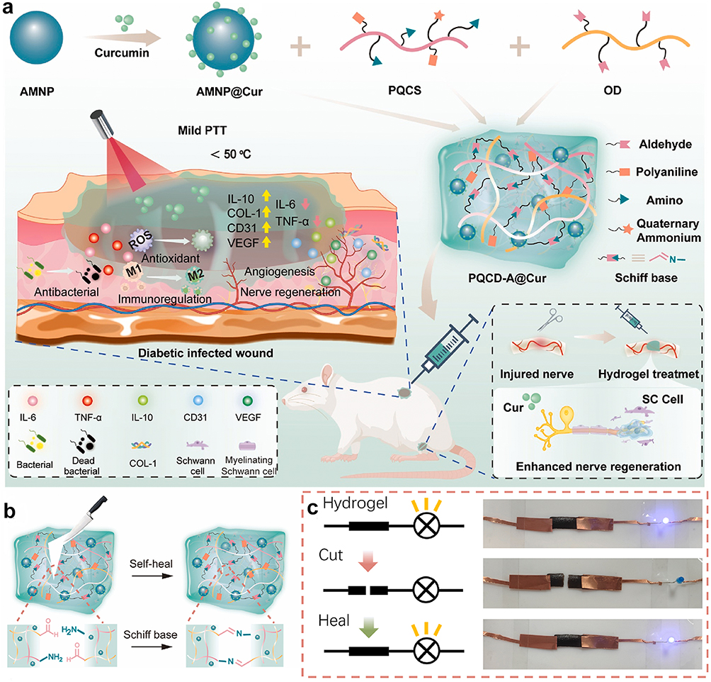 |
Figure 1 An example of carbon-based nanomaterials for peripheral nerve repair. (a) Diagram showing the fabrication of the hydrogel and its functional outcomes in repairing peripheral nerve damage and accelerating diabetic wound closure. Yellow upward represents upregulation and pink downward arrow represents downregulation. (b) Schematic illustration of the proposed self-healing mechanism of the PQCD-A@Cur hydrogel. (c) Electrical conductivity characterization of PQCD-A@Cur hydrogels, demonstrating their stable and efficient charge-transfer capability. Reproduced with permission.13 Copyright 2025, Elsevier. |
More recently, MXene nanosheets have emerged as next-generation conductive nanofillers. With their exceptional electrical conductivity, hydrophilicity, and surface modifiability, MXenes can be homogeneously dispersed within hydrogel matrices to yield stable and tunable electroactive networks. MXene-modulated GelMA hydrogels, for instance, exhibit optimized conductivity, controlled degradation, and enhanced axonal regeneration in sciatic nerve injury models.25 Collectively, these approaches illustrate how the integration of diverse electroactive fillers can simultaneously advance mechanical, biological, and electrical performance, guiding the evolution of multifunctional nerve repair platforms.
Balancing Conductivity with Softness, Degradability, and Bioadhesion
A persistent challenge in conductive hydrogel design is achieving a delicate equilibrium between electroactivity and mechanical/biological compatibility. Excessive loading of conductive fillers often leads to brittleness or cytotoxicity, whereas insufficient loading compromises signal transmission. The most successful systems maintain tissue-like softness and elasticity while degrading in synchrony with nerve regeneration and adhering firmly to the nerve stump to prevent micro-motion or fluid infiltration.
Recent innovations highlight several effective strategies. Phenylborate-modified hydrogels, for example, demonstrate improved stretchability and nerve adhesion without sacrificing conductivity.22 Likewise, catechol-functionalized hydrogels inspired by mussel adhesion achieve robust bioadhesion and suture-free implantation, while preserving high electrical performance and long-term stability.14 These studies exemplify the importance of a holistic, functionally integrated design philosophy—one that treats conductivity not as an isolated parameter but as an element inherently coupled to mechanics, degradation, and biological interaction. Such multidimensional optimization is crucial for advancing conductive hydrogels from preclinical feasibility toward clinical translation.
Advanced Functionalities and Smart Systems
While early generations of conductive hydrogels primarily focused on combining electrical conductivity with mechanical compliance, recent advances have shifted toward multifunctional and adaptive systems designed to meet the clinical realities of complex PNIs.26 These emerging hydrogel architectures go beyond passive scaffolding to actively interact with biological tissues, respond dynamically to environmental cues, and integrate with bioelectronic systems. The overarching goal is to develop clinically translatable, intelligent materials capable of minimizing surgical trauma, modulating the neuro-immune milieu, and supporting personalized regenerative therapies.
Self-Healing, Injectable, and Sprayable Hydrogels for Minimally Invasive Use
Conventional nerve repair procedures typically involve open surgery and mechanical suturing, which increase the risk of infection, scarring, and delayed recovery. In contrast, self-healing and injectable conductive hydrogels are being designed to enable minimally invasive or image-guided delivery directly to the injury site, reducing operative complexity and improving patient outcomes.27,28
Self-healing systems rely on reversible dynamic bonds—such as boronate ester, Schiff base, or hydrogen bonding—to restore structural integrity after deformation or mechanical disruption.29 A notable example is self-healing electroconductive hydrogels (ECHs), which offer a flexible, adaptive, and clinically promising alternative to conventional rigid conductive scaffolds. These materials possess excellent self-healing and tissue-adhesive properties, allowing them to adhere tightly to injured nerve fibers and spontaneously form tubular structures without sutures or invasive procedures. Their stable conductive network establishes an intimate electrical bridge with neural tissues, facilitating effective signal transmission and cellular communication.30,31 Studies have shown that ECHs significantly enhance Schwann cell migration and adhesion, activate the MEK/ERK signaling pathway, and thereby accelerate axonal regeneration and remyelination, while preventing muscle denervation atrophy and improving functional recovery (Figure 2).27
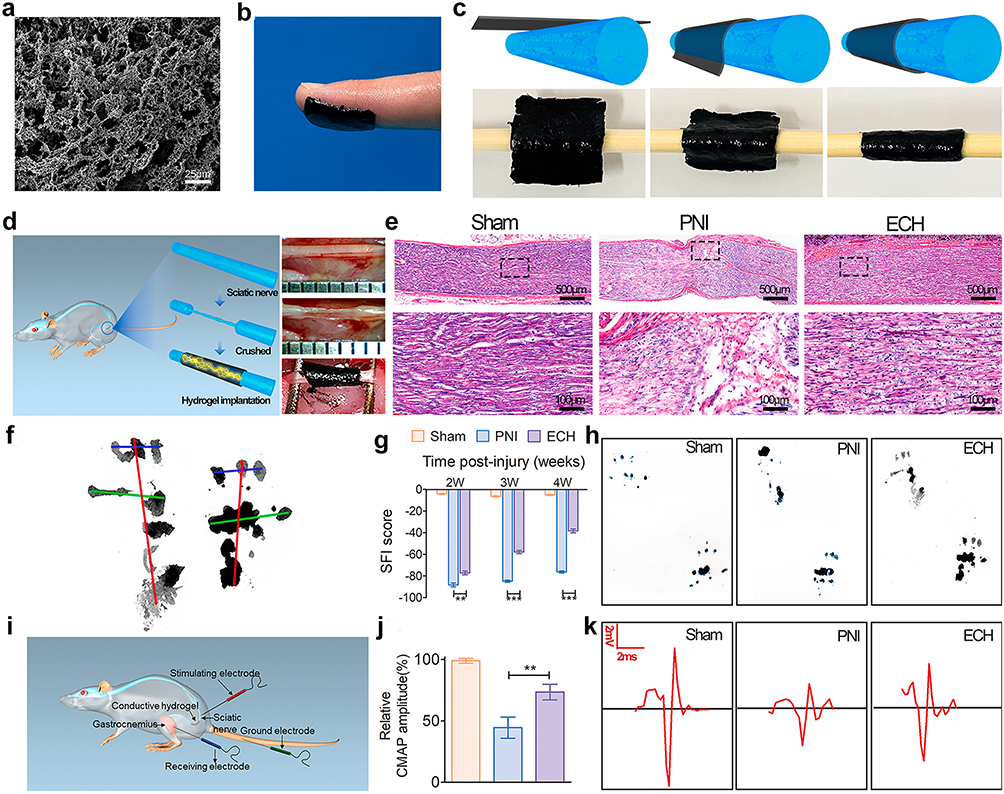 |
Figure 2 An example of Self-healing systems for peripheral nerve repair. (a) SEM micrograph of the electroconductive hydrogel. (b) Photograph showing the ECH film attached to a finger, demonstrating its strong adhesive capability. (c) Schematic and corresponding images depicting the self-rolling behavior of the ECH, forming a tubular configuration matching the nerve size. (d) Implantation of the hydrogel into a diabetic rat model with a sciatic nerve defect. (e) H&E-stained sections showing structural regeneration of the sciatic nerve 28 days post-injury. Black dotted lines indicate the locally magnified regions. (f) Formula and parameters used for calculating the sciatic functional index based on footprint measurements. (g) Comparison of SFI scores among groups at 14, 21, and 28 days after surgery (n = 5). (h) Representative footprint analyses illustrating motor function recovery. (i) Schematic diagram of the compound muscle action potential testing procedure. (j). Quantitative analysis of CMAP amplitudes recorded in each group (n = 3). (k) Representative CMAP waveforms from the injured sciatic nerve, with significance levels indicated (**p < 0.01, ***p < 0.001). Reproduced under the terms of a Creative Commons Attribution 4.0 International License.27 Copyright 2021, the authors. |
In parallel, sprayable hydrogel formulations are emerging as an innovative approach for addressing irregular, small-diameter, or difficult-to-access nerve defects. A recent study developed an in situ-sprayed adhesive conductive hydrogel incorporating carbon nanotubes, which could conformally coat the nerve surface and re-establish electrical continuity across transected cavernous nerves.32 This spray-deposited hydrogel achieved functional restoration in rodent models and demonstrated compatibility with robotic or laparoscopic surgical platforms, illustrating the transformative potential of spray-assisted, patient-specific hydrogel delivery in next-generation regenerative neurosurgery.
Adhesive and Suture-Free Bandages for on-Nerve Application
Traditional microsurgical techniques for peripheral nerve repair often depend on delicate suturing to secure grafts or conduits—a process that can exacerbate mechanical trauma and prolong operative time. Adhesive hydrogels present a suture-free alternative that simplifies the procedure while maintaining intimate electrical and mechanical contact with neural tissue.33
For instance, a fibrous hydrogel bandage composed of electrospun hyaluronic acid fibers coated with polypyrrole provided excellent flexibility, conformability, and electrical conductivity.22 When applied in vivo in a rat model of sciatic nerve crush, this adhesive hydrogel formed a stable wrap around the injury without sutures, enhancing axonal regeneration and preventing muscle atrophy.
Expanding upon this principle, conductive hydrogel cuff systems have been engineered to act as both adhesive dressings and bioelectronic interfaces. A highly conductive, adhesive, and biocompatible hydrogel was shown to form seamless contact with nerve stumps while serving as an electroactive conduit for closed-loop neuromodulation.14 Such devices not only provide mechanical fixation but also enable real-time electrical feedback and targeted stimulation, bridging the gap between regenerative biomaterials and implantable neural interface technologies. Collectively, these innovations signify a paradigm shift toward integrated, suture-free nerve repair systems capable of combining adhesion, conductivity, and therapeutic modulation in a single platform.
Multifunctional Scaffolds Integrating Drug Delivery, Antibacterial Activity, and Photothermal Therapy
The latest advance of conductive hydrogel design emphasizes multifunctionality—the capacity to simultaneously support electrical signaling, control the biochemical environment, and counteract pathological challenges such as infection, inflammation, and ischemia. These hybrid scaffolds combine electroconductivity with controlled drug delivery, antioxidant activity, and immunomodulation, achieving synergistic therapeutic effects.28,34
A representative example is a versatile conductive hydrogel co-loaded with curcumin and melanin-inspired nanoparticles. This multifunctional system not only enhanced schwann cell proliferation and axonal outgrowth but also conferred photothermal antibacterial capability and reactive oxygen species scavenging, resulting in accelerated recovery of infected diabetic wounds.13 Stimuli-responsive systems, such as glucose-responsive nanozyme hydrogels, illustrate how biomaterials can dynamically modulate inflammatory and metabolic microenvironments, which is especially relevant for diabetic nerve repair.35 Such designs exemplify how coupling electroactive and biochemical stimuli can orchestrate neuro-immune homeostasis during regeneration.
Another promising direction integrates anti-inflammatory and neurotrophic strategies. Modulation of neuroinflammation has become increasingly recognized as a key therapeutic axis in neural repair, as demonstrated by Lan et al, where curcumin-primed MSCs attenuated neuronal PANoptosis through microglial polarization.36 A conductive hydrogel embedding PEDOT within a chitosan–cellulose network exhibited stable conductivity while releasing simvastatin to suppress excessive inflammation and promote M2-type macrophage polarization.37 This dual-function architecture established a more permissive microenvironment for nerve repair, emphasizing that electroconductivity and immune regulation can act in concert to optimize regeneration outcomes.
Furthermore, recent studies have introduced angiogenic or photothermal functionalities into conductive hydrogel matrices. By incorporating vascular endothelial growth factors or photothermal nanocomponents, these scaffolds simultaneously stimulate revascularization and neuronal growth—two interdependent processes crucial for functional recovery.12 Such systems exemplify a new generation of bioelectronic-multifunctional hybrids that treat nerve injury not as an isolated defect but as part of a broader systemic and microenvironmental process.
Bioinspired and Biomimetic Approaches
An emerging paradigm in the design of conductive hydrogels for peripheral nerve repair is the application of bioinspired and biomimetic principles.38,39 The peripheral nervous system is characterized by a precisely orchestrated combination of biochemical, topographical, and electrophysiological cues that together govern axonal regeneration and remyelination. Reproducing these native features in engineered materials has therefore become a cornerstone for developing next-generation scaffolds that move beyond passive support toward dynamic biological emulation. Such designs aim not only to replace damaged tissues but also to actively instruct and synchronize cellular behavior, thereby achieving more complete and functional recovery.40
ECM-Mimicking Designs and Decellularized Matrix Composites
The ECM of peripheral nerves provides an intricate 3D microenvironment that regulates Schwann cell adhesion, proliferation, and axonal pathfinding through biochemical and mechanical cues.41 Mimicking this complexity remains a key challenge in biomaterial design. Hydrogels have similarly been applied to dental pulp regeneration, underscoring the cross-tissue relevance of ECM-mimetic hydrogel scaffolds for neurovascular repair.42 Conductive hydrogels composed of natural polymers—such as gelatin, collagen, and hyaluronic acid—closely resemble ECM composition and architecture while allowing further functionalization with conductive fillers.43 These hybrid systems restore both bioelectric communication and structural integrity, facilitating synchronized nerve regeneration.
For a representative example, Fan et al present the development of a multifunctional ECM-based conductive nerve guidance conduit (HE-NGC) for PNI repair.44 By integrating E-jet 3D printing and electrospinning, the researchers fabricated an aligned topographical scaffold to guide nerve regeneration, while filling the lumen with umbilical cord-derived decellularized ECM (dECM) hydrogels and extracellular vesicles (EVs) to create a biomimetic microenvironment. The sustained release of EVs from the dECM hydrogel enhanced Schwann cell (SC) and PC12 cell proliferation and migration in vitro. In vivo, the HE-NGC successfully bridged a 12-mm rat sciatic nerve defect, restoring motor function and promoting remyelination (Figure 3).
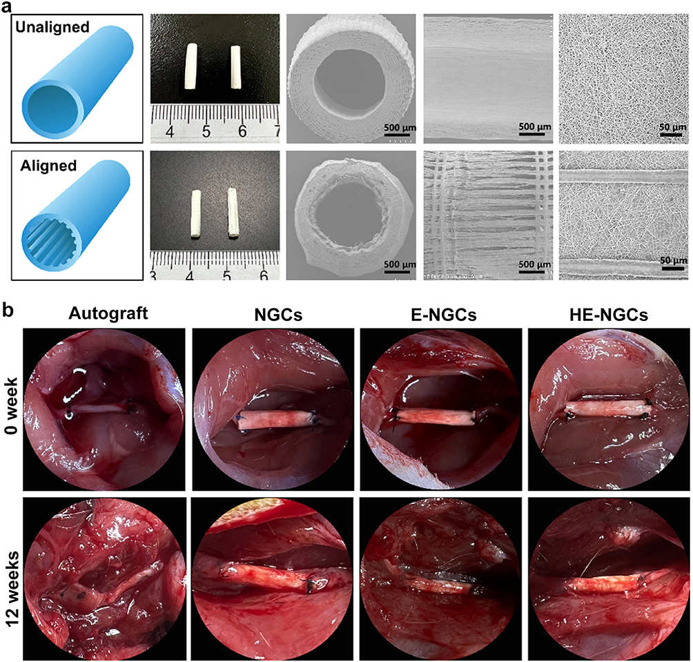 |
Figure 3 An example of multifunctional ECM-based conductive nerve guidance conduit for peripheral nerve repair. (a) Comparison between unaligned (top) and aligned (bottom) NGC scaffolds. From left to right are shown the schematic illustration, actual photograph, cross-sectional image, longitudinal section image, and magnified structural view. (b) Images showing NGC implantation in the autograft, NGC, E-NGC, and HE-NGC groups. The top row displays the grafts immediately after implantation, while the bottom row presents the grafts prior to removal after 12 weeks of implantation. Reproduced with permission.44 Copyright 2025, Elsevier. |
Furthermore, the combination of decellularized components with synthetic polymer backbones offers improved mechanical stability and degradation control without compromising biocompatibility. This approach allows fine-tuning of viscoelastic properties to match those of peripheral nerves, promoting optimal cell–matrix crosstalk and directional axonal growth.45 Collectively, ECM-mimicking conductive hydrogels exemplify how natural design principles can be harnessed to recreate the structural and functional hallmarks of native neural tissue.
Aligned Fibers, Multichannel Conduits, and Topographical Cues
Topographical guidance plays an equally vital role in peripheral nerve regeneration.46,47 In native nerves, aligned basal lamina tubes provide continuous physical tracks that direct axonal elongation and Schwann cell migration. To emulate this, conductive hydrogels are now being engineered with anisotropic architectures, including aligned fibers, grooves, and multichannel conduits.48,49
In a prior study, Fang et al developed a multiscale conductive nerve guidance conduit (MF-NGC) composed of electrospun PCL/collagen nanofibers as the outer sheath, rGO/PCL conductive microfibers as the backbone, and PCL microfibers as the internal support.50 The 3D-printed MF-NGC exhibited excellent permeability, mechanical stability, and electrical conductivity, which significantly promoted Schwann cell elongation and PC12 neurite outgrowth in vitro. In a rat sciatic nerve defect model, the MF-NGC enhanced angiogenesis and induced M2 macrophage polarization, leading to improved axon myelination, muscle mass recovery, and functional restoration (Figure 4).
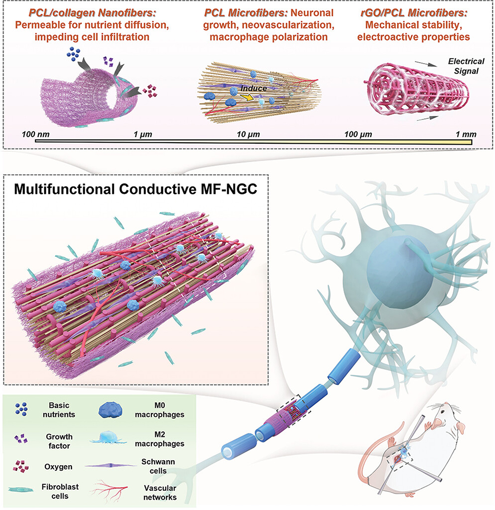 |
Figure 4 Schematic illustration of the multifunctional conductive MF-NGC designed for peripheral nerve injury repair. The conduit features a hierarchical fibrous architecture with diameters spanning 500 nm to 120 µm, fabricated through the combined use of melt electrowriting and electrospinning techniques. Reproduced under the terms of a Creative Commons Attribution 4.0 International License.50 Copyright 2023, the authors. |
Beyond fibrous matrices, multichannel hydrogel conduits have been introduced to mimic the fascicular organization of nerves.51,52 By dividing the lumen into multiple parallel microchannels, these scaffolds effectively prevent axonal misrouting and create a topography conducive to synchronized regeneration. A recent design integrating multichannel geometry, conductivity, and biochemical gradients—a so-called triple-cue system—achieved superior nerve conduction velocity and morphological regeneration.53
At the nanoscale, introducing surface ridges, grooves, or conductive nanopatterns has been shown to modulate Schwann cell orientation and cytoskeletal organization, further enhancing directional axonal growth. These findings collectively underscore that structural anisotropy and topographical mimicry are indispensable elements for translating conductive hydrogels into clinically effective nerve repair strategies.
Multi-Gradient Strategies Combining Conductivity, Biochemical Signals, and Mechanical Guidance
In vivo, nerve regeneration proceeds along multiple intertwined gradients—electrical, biochemical, and mechanical—that dynamically evolve during healing.54,55 Therefore, reproducing these multi-gradient environments within hydrogel scaffolds represents a sophisticated biomimetic strategy to synchronize repair processes. Previous studies have shown that activating neurotrophic pathways such as BDNF–TrkB can counteract neuroinflammation, as evidenced by Apelin-13–mediated rescue in AD models.56
Furthermore, Cai et al developed a multichannel conductive gradient hydrogel conduit (NGF-AGHC) that integrates topographical alignment, a conductivity gradient, and a nerve growth factor (NGF) gradient to promote peripheral nerve regeneration.53 Fabricated through directional lyophilization of a GO/PVA solution and dopamine-assisted reduction, the conduit achieves spatially controlled conductivity and NGF distribution. In vitro, NGF-AGHC effectively directs neurite outgrowth and neuronal differentiation of PC12 cells, while in vivo it enhances axonal alignment, remyelination, and functional recovery, performing comparably to autografts. These findings demonstrate that conductive gradient hydrogels offer a simple yet powerful biomimetic platform for coordinated electrical, structural, and biochemical guidance in peripheral nerve repair (Figure 5).
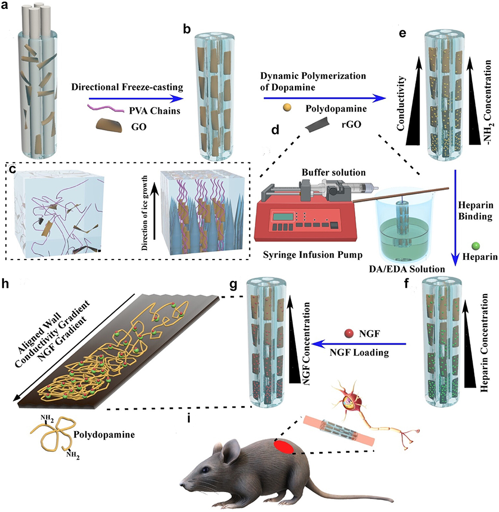 |
Figure 5 Schematic illustration of the fabrication process and functional design of the NGF-gradient/aligned multichannel hydrogel conduit. (a) Preparation of PVA/GO solution within a custom-designed mold that generates four parallel microchannels. (b) Directional freeze-drying of the PVA/GO solution produces anisotropic conduits with aligned porous structures. (c) Mechanism of anisotropic structure formation: ice crystals nucleate on the copper substrate and grow vertically, forming single-domain honeycomb-like architectures. (d) During the DA/EDA reaction, amine groups form a spatial gradient while GO is progressively reduced, establishing a conductivity gradient. (e) Formation of the anisotropic gradient PVA/rGO/PDA hydrogel conduit (AGHC). (f) Heparin is covalently linked to the amine-rich surface of the conduit. (g) NGF is further immobilized to generate a biochemical gradient. (h) Illustration of the integrated guidance strategy combining topographical, chemical, and electrical cues for nerve regeneration. (i) Diagram of the rat sciatic nerve defect model used for in vivo implantation. Reproduced with permission.53 Copyright 2025, America Chemical Society. |
Simultaneously, biochemical gradients—such as controlled release of neurotrophic factors or chemokines—guide Schwann cell migration and direct axonal extension. When integrated with mechanical gradients, where stiffness gradually transitions from compliant nerve tissue to more rigid anchoring regions, these systems can dynamically modulate cell behavior and mechanical load distribution.57,58 A representative example involves a multifunctional biodegradable conductive hydrogel designed to regulate the microenvironment for stem cell therapy. This system combined conductivity, growth factor gradients, and mechanical adaptability, leading to enhanced vascularization and nerve tissue regeneration.12 Besides, innovations such as Janus hydrogels with asymmetric protective properties exemplify how gradient and directionally designed architectures can regulate tissue-specific microenvironments.59
Such multi-gradient scaffolds embody the convergence of materials science and developmental neurobiology, where the goal is not merely to replace tissue but to recapitulate the self-organizing dynamics of natural regeneration. As fabrication technologies advance, including 3D bioprinting and microfluidic patterning, the precise spatial control of multiple gradients is expected to open a new era of adaptive, intelligent hydrogel systems for nerve repair.60
Translational Barriers and Clinical Outlook
Despite the significant advances in conductive hydrogels for peripheral nerve repair, the process from bench to bedside remains complex and uncertain. While preclinical models have consistently demonstrated functional recovery and biocompatibility, the translation of these materials into reliable, regulatory-approved clinical products faces numerous scientific, technical, and ethical hurdles. Addressing these translational barriers is critical not only for ensuring patient safety but also for realizing the full therapeutic promise of electroactive biomaterials in regenerative medicine.
Long-Term Safety and Immune Compatibility
For any implantable biomaterial, long-term safety and immunological integration are of paramount importance. Conductive hydrogels should maintain their bioelectrical functionality while minimizing chronic inflammation, fibrotic encapsulation, and cytotoxic degradation by-products. The conductive fillers that enable these materials—such as polypyrrole, PEDOT, graphene, and MXene nanosheets—can provoke immune activation or oxidative stress if not properly stabilized or functionalized.10,29,61,62
Recent studies have shown encouraging short-term results, demonstrating both electrical stability and favorable host responses.6,63,64 For instance, a zwitterionic conductive hydrogel-based nerve guidance conduit effectively promoted peripheral nerve regeneration while significantly suppressing fibrotic encapsulation and macrophage-driven inflammation in rat models.9 However, such outcomes have yet to be validated in chronic or large-animal models, where degradation kinetics, filler clearance, and systemic immune responses may diverge significantly from rodent studies.
Future generations of conductive hydrogels are likely to incorporate immunomodulatory design strategies—including surface-grafted zwitterionic moieties, bioactive peptides, or controlled-release anti-inflammatory agents—to achieve long-term biocompatibility. Additionally, degradable conductive networks that break down into non-toxic, renally excretable fragments will be critical for ensuring clinical safety.65,66 The challenge lies in striking a precise balance between durable electroactivity and immune homeostasis, a goal that demands close integration of materials chemistry, immunology, and translational pathology.
Scalable and Reproducible Manufacturing Processes
A persistent bottleneck in the clinical translation of conductive hydrogels is the scalability and reproducibility of their synthesis. Laboratory formulations often rely on multi-step polymerization, intricate crosslinking mechanisms, or expensive conductive nanomaterials that are difficult to reproduce under industrial conditions. Batch-to-batch variability in composition or crosslinking density can alter electrical conductivity, mechanical behavior, and degradation rate—parameters that are critical for regulatory approval and consistent therapeutic performance. Crosslinking crucially determines hydrogel properties for nerve repair. Physical methods (hydrogen bonding, ionic interactions) provide injectability, self-healing, and stress relaxation, beneficial for minimally invasive procedures, but often lack long-term stability. Chemical crosslinking (eg, Schiff base reactions, photopolymerization) offers superior mechanical strength and durability. In conductive hydrogels, crosslinking density critically affects conductivity; excessive crosslinking hinders ion transport, while insufficient crosslinking weakens the structure. Therefore, an optimal nerve repair scaffold requires a balanced crosslinking strategy that maintains electrical conductivity while matching the viscoelasticity of native nerve tissue.
For example, MXene-modulated hydrogels demonstrate remarkable electroactivity and regenerative potential, yet they require specialized photopolymerization conditions and controlled inert environments that complicate good manufacturing practice (GMP) implementation.25 Similarly, high-purity graphene and PEDOT derivatives often face supply chain limitations and scalability issues due to batch-specific variation in particle size and surface chemistry.
To overcome these obstacles, translational research should emphasize standardized fabrication protocols and process validation across laboratories. Techniques such as 3D printing, microfluidic templating, and continuous-flow polymerization are emerging as scalable routes for precise structural and compositional control. Moreover, integrating machine learning-based process optimization could help establish predictive models for material consistency and performance, paving the way for industrial reproducibility and regulatory compliance.67,68
Integration with Wearable Devices and Personalized Medicine
The clinical future of conductive hydrogels will likely unfold within integrated therapeutic ecosystems rather than as standalone implants. These materials can serve as bioelectronic interfaces linking regenerating nerves with wearable or implantable devices capable of real-time electrophysiological monitoring and adaptive stimulation control.69,70
A representative example is the highly conductive, adhesive, and biocompatible hydrogel developed for closed-loop neuromodulation, which dynamically interfaces with damaged nerves to deliver feedback-controlled stimulation and accelerate regeneration.71 Such systems foreshadow a new era of personalized neurorehabilitation, where stimulation patterns and repair strategies are continuously optimized based on patient-specific physiological data.38,72
In parallel, 3D printing and biodesign technologies are enabling the fabrication of patient-tailored conduits and scaffolds based on individualized imaging and injury geometry.73,74 Integration with wearable sensors could further allow longitudinal monitoring of recovery dynamics, electromyography activity, and biochemical markers of inflammation. The importance of personalized neuromuscular control systems is further highlighted by advanced exoskeleton frameworks, underscoring opportunities for integrating conductive hydrogel interfaces with robotic rehabilitation.75 These data-rich approaches not only enhance clinical precision but also provide feedback loops for adaptive therapy—blurring the boundary between material-based repair and digital therapeutics.
Future Perspectives
The rapid progress in conductive hydrogel research has laid a solid foundation for their translation toward peripheral nerve repair. Yet, this field is still in its early stages of clinical realization, and several transformative directions are emerging at the interface of materials science, bioengineering, and neurobiology. The next generation of conductive hydrogels will likely evolve into intelligent, patient-specific, and multifunctional systems that not only passively support tissue regeneration but also actively sense, respond, and adapt to the biological milieu.
Intelligent Hydrogels with Adaptive, Stimuli-Responsive Properties
Traditional hydrogels offer static physicochemical environments, yet nerve regeneration is an inherently dynamic and multistage process, requiring materials that evolve in sync with the biological repair timeline.76,77 Intelligent conductive hydrogels endowed with adaptive and stimuli-responsive characteristics—reacting to changes in pH, temperature, reactive oxygen species, enzymatic activity, or applied electrical fields—represent the next generation of biofunctional scaffolds.
For instance, self-healing hydrogels capable of autonomously restoring their structural integrity under mechanical deformation can maintain conductive pathways and ensure stable electrical coupling throughout regeneration. Such systems, as exemplified by a prior study, demonstrate how dynamic crosslinking and reversible bonding chemistries can generate “living” scaffolds that release bioactive cues upon inflammation or adjust stiffness and conductivity across healing stages.11 Recent work also shows that zwitterionic hydrogels can undergo structural remodeling under mechanical training, highlighting their potential as adaptable scaffolds for dynamic neural environments.78 By closely mimicking the temporally orchestrated repair microenvironment, these materials could provide unprecedented precision in regulating Schwann cell behavior, axonal guidance, and immune modulation.
3D Printing and Patient-Tailored Conduit Fabrication
The convergence of additive manufacturing with conductive hydrogel technology enables a new paradigm of personalized nerve regeneration.79,80 Advances in 3D bioprinting make it feasible to fabricate anatomically accurate nerve conduits directly derived from patient imaging data, offering unparalleled control over conduit geometry, porosity, and alignment of microchannels.
Recent studies highlight how printable conductive nanomaterials such as MXene or graphene can be embedded to establish continuous electroactive networks within hydrogel matrices.81 These architectures can spatially guide regenerating axons and promote bidirectional electrical communication between proximal and distal nerve stumps.82,83 In the long term, such patient-specific, data-driven fabrication strategies could reduce immune complications, improve graft integration, and facilitate scalable manufacturing of bespoke nerve repair devices tailored to individual defect morphologies and electrophysiological requirements.
Combination Therapies with Bioactive Agents
While conductive hydrogels alone provide structural and electrical cues, combining them with biochemical or cellular therapeutics could yield synergistic outcomes. Recent studies have explored the integration of stem cells—such as MSCs and neural stem/progenitor cells—into conductive hydrogel matrices to synergistically enhance nerve regeneration.84 Conductive hydrogels provide a supportive niche that improves stem cell survival, promotes neural differentiation, and facilitates paracrine signaling through the release of neurotrophic and angiogenic factors. Electrical conductivity further augments stem cell function by modulating membrane potential and intracellular signaling pathways associated with neurogenesis. Despite these advantages, stem cell–loaded conductive hydrogels face notable challenges, including potential tumorigenicity, immune rejection, limited cell retention, and regulatory complexity. Additionally, electrical overstimulation may adversely affect stem cell viability if not precisely controlled. Encapsulation of stem cell-derived exosomes offers a promising approach to deliver a spectrum of paracrine signals that enhance angiogenesis, neurogenesis, and immune modulation.85,86 Likewise, sustained release of growth factors such as nerve growth factor or brain-derived neurotrophic factor from the hydrogel matrix can establish spatially controlled gradients that orchestrate cellular infiltration and axonal elongation.87,88
In particular, multifunctional scaffolds exemplify the potential of integrating conductive polymers, degradable backbones, and biological agents to modulate the neuroimmune microenvironment.49,89 Moreover, gene-delivery functionalities embedded within these hydrogels could enable localized and transient modulation of transcriptional programs in Schwann cells or macrophages, pushing the frontier toward smart, immuno-modulatory biomaterials capable of self-regulated therapeutic actions.90
Convergence with Bioelectronics and AI-Driven Systems
Perhaps the most visionary direction is the seamless integration of conductive hydrogels with bioelectronic and AI-enabled platforms. Conductive hydrogels serve as ideal soft interfaces between neural tissue and electronic devices, combining mechanical compliance with high signal fidelity. Recent developments demonstrate adhesive, biocompatible hydrogels that allow real-time recording and feedback stimulation without the drawbacks of rigid electrodes.91 In future clinical applications, these hybrid systems could operate as closed-loop neuromodulation platforms that dynamically sense nerve activity, decode electrophysiological signals, and deliver tailored electrical or biochemical stimulation in response. Coupled with AI algorithms, such systems might autonomously adjust stimulation parameters based on patient-specific recovery profiles, paving the way for adaptive, precision-guided neural rehabilitation. The ultimate vision is a bioelectronic–biomaterial symbiosis, where hydrogel-based scaffolds function as both regenerative matrices and intelligent sensing–actuating systems, heralding a new era of personalized nerve repair therapeutics.
While conductive hydrogels have demonstrated significant promise in promoting peripheral nerve regeneration, several unresolved challenges and limitations should be considered. For instance, excessive incorporation of conductive fillers such as MXene or PEDOT can improve electroactivity but may lead to brittleness or cytotoxicity, and long-term in vivo safety data remain limited. Studies on purely ionic conductive hydrogels and degradable conductive polymers are comparatively fewer, and reported regeneration outcomes are sometimes inconsistent across different models or fabrication methods. Furthermore, variability in hydrogel degradation rates and immune responses can affect reproducibility and functional recovery. Comparisons with alternative approaches, including FDA-approved conduits and hybrid scaffolds, indicate that no single material system currently addresses all mechanical, electrical, and biological requirements.
Conclusion
Conductive hydrogels provide versatile scaffolds supporting axonal regeneration, Schwann cell function, and immune modulation, with advances in PEDOT, carbon-based nanomaterials, MXenes, and bioinspired designs expanding their capabilities (Table 1). Despite this progress, significant translational gaps persist, including long-term biocompatibility, degradation control, reproducible manufacturing, and limited validation beyond small-animal models. Near-term clinical translation is most likely for injectable or adhesive conductive hydrogels with simplified compositions, well-defined degradation profiles, and compatibility with existing surgical workflows. In contrast, concepts such as AI-integrated scaffolds and fully autonomous closed-loop bioelectronic systems remain speculative and require substantial technological and regulatory maturation. Future efforts should prioritize standardized preclinical models, quantitative benchmarking against FDA-approved conduits, and scalable fabrication strategies. By aligning material design with biological mechanism and clinical feasibility, conductive hydrogels are poised to transition from experimental constructs to practical bioelectronic therapies for peripheral nerve regeneration.
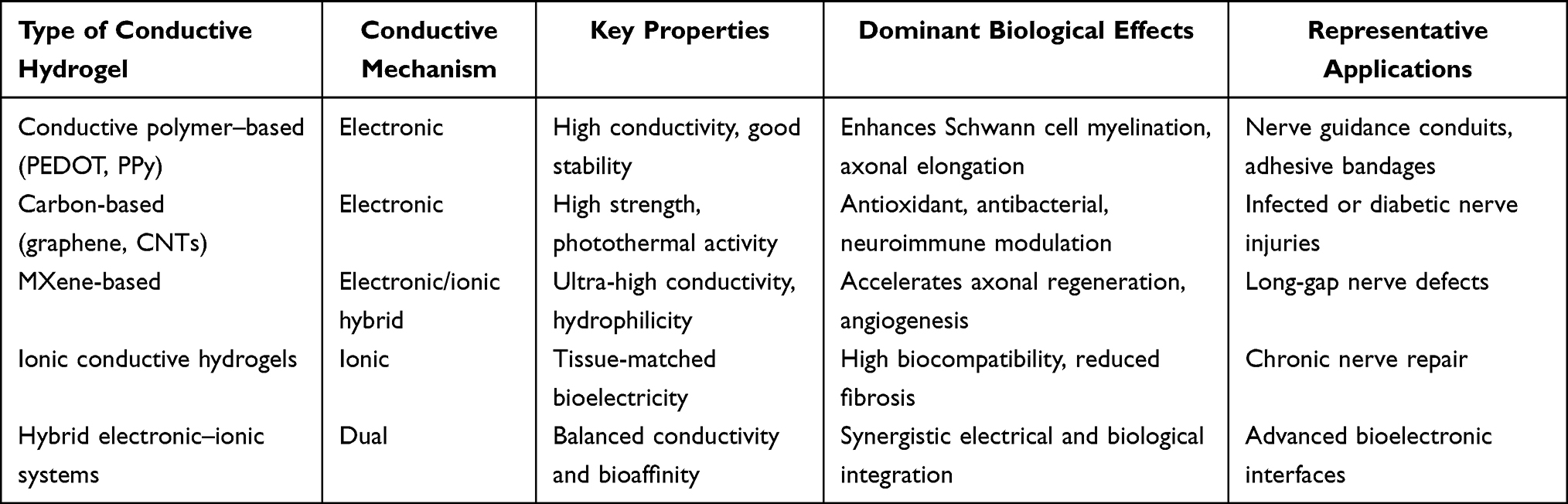 |
Table 1 Comparison of Different Types of Conductive Hydrogels for Peripheral Nerve Repair |
Acknowledgments
Artificial intelligence tools were used only for the correction of spelling and grammatical errors in the manuscript. No AI participated in the creation of any original content presented in this work.
Funding
This study was supported by the Natural Science Foundation of Shaanxi Province, Grant/Award Number: 2025JC-YBQN-1269.
Disclosure
The author(s) report no conflicts of interest in this work.
References
1. Lavorato A, Aruta G, De Marco R, et al. Traumatic peripheral nerve injuries: a classification proposal. J Orthop Traumatol. 2023;24(1):20. doi:10.1186/s10195-023-00695-6
2. Osborne NR, Anastakis DJ, Davis KD. Peripheral nerve injuries, pain, and neuroplasticity. J Hand Ther. 2018;31(2):184–18. doi:10.1016/j.jht.2018.01.011
3. Kedar DJ, Shani N, Fliss E, et al. Long-term hyperbaric oxygen treatment enhances nerve regeneration and remyelination in a rat sciatic nerve graft model. Plast Reconstr Surg Glob Open. 2025;13(8):e7039. doi:10.1097/GOX.0000000000007039
4. Yang X, Huo N, Zhou H, Li S, Fang M, Zhou N. Application strategies of autologous and decellularized nerve grafts: structural and functional recovery. Neural Regen Res. 2025. doi:10.4103/NRR.NRR-D-25-00607
5. Ma X, Wang M, Ran Y, et al. Design and fabrication of polymeric hydrogel carrier for nerve repair. Polymers (Basel). 2022;14(8):1549. doi:10.3390/polym14081549
6. Feng Y, Shan L, Wang Y, Chen X, Wang C, Liu J. Conductive hydrogels with topographical geometry and mechanical robustness for enhanced peripheral nerve regeneration. ACS Nano. 2025;19(17):16675–16684. doi:10.1021/acsnano.5c00845
7. Wei G, Chen C, Li X, et al. In situ piezoelectricity induces M2 polarization of macrophages to regulate schwann cells for alleviating neuropathic pain of CCI rats. Biomater Adv. 2025;174:214319. doi:10.1016/j.bioadv.2025.214319
8. Liu J, Lin Z, Wu H, et al. Dual-regulation biomimetic composite nerve scaffold with oriented structure and conductive function for skin peripheral nerve injury repair. Colloids Surf B Biointerfaces. 2025;253:114768. doi:10.1016/j.colsurfb.2025.114768
9. Xu Y, Liu J, Zhang P, et al. Zwitterionic Conductive hydrogel-based nerve guidance conduit promotes peripheral nerve regeneration in rats. ACS Biomater Sci Eng. 2023;9(12):6821–6834. doi:10.1021/acsbiomaterials.3c00761
10. Huang L, Yang X, Deng L, et al. Biocompatible chitin hydrogel incorporated with PEDOT nanoparticles for peripheral nerve repair. ACS Appl Mater Interfaces. 2021;13(14):16106–16117. doi:10.1021/acsami.1c01904
11. Xuan H, Wu S, Jin Y, et al. A bioinspired self-healing conductive hydrogel promoting peripheral nerve regeneration. Adv Sci (Weinh). 2023;10(28):e2302519. doi:10.1002/advs.202302519
12. Xu C, Wu P, Yang K, et al. Multifunctional biodegradable conductive hydrogel regulating microenvironment for stem cell therapy enhances the nerve tissue repair. Small. 2024;20(23):e2309793. doi:10.1002/smll.202309793
13. Bi S, He C, Zhou Y, et al. Versatile conductive hydrogel orchestrating neuro-immune microenvironment for rapid diabetic wound healing through peripheral nerve regeneration. Biomaterials. 2025;314:122841. doi:10.1016/j.biomaterials.2024.122841
14. Chu T, Xiao Y, Lai H, et al. Highly conductive, adhesive and biocompatible hydrogel for closed-loop neuromodulation in nerve regeneration. ACS Nano. 2025;19(19):18729–18746. doi:10.1021/acsnano.5c03336
15. Wu S, Qi Y, Shi W, Kuss M, Chen S, Duan B. Electrospun conductive nanofiber yarns for accelerating mesenchymal stem cells differentiation and maturation into schwann cell-like cells under a combination of electrical stimulation and chemical induction. Acta Biomater. 2022;139:91–104. doi:10.1016/j.actbio.2020.11.042
16. Pham VM. Targeting PI3K/AKT and MEK/ERK pathways for synergic effects on improving features of peripheral diabetic neuropathy. J Diabetes Investig. 2024;15(11):1537–1544. doi:10.1111/jdi.14289
17. Ye K, Li Z, Yin Y, et al. LIPUS-SCs-Exo promotes peripheral nerve regeneration in cavernous nerve crush injury-induced ED rats via PI3K/Akt/FoxO signaling pathway. CNS Neurosci Ther. 2023;29(11):3239–3258. doi:10.1111/cns.14256
18. Xia B, Gao X, Qian J, et al. A novel superparamagnetic multifunctional nerve scaffold: a remote actuation strategy to boost in situ extracellular vesicles production for enhanced peripheral nerve repair. Adv Mater. 2024;36(3):e2305374. doi:10.1002/adma.202305374
19. Yuan T, Li W, Zhou M, Wang X, Wang B, Zhao Y. Biomimetic multichannel silk nerve conduits with multicellular spatiotemporal distributions for spinal cord injury repair. Adv Mater. 2024;36(44):e2411628. doi:10.1002/adma.202411628
20. Zhou Y, Xu T, Zhou Y, et al. A myelin debris cleaner for spinal cord injury recovery: polycaprolactone/cell membrane assembled scaffolds. Adv Sci. 2025;12(36):e03269. doi:10.1002/advs.202503269
21. Eisenberg MT, Hustedt JW. Alginate use in orthopedics and peripheral nerve repair: a systematic review. Cureus. 2024;16(10):e72480. doi:10.7759/cureus.72480
22. Jin S, Jung H, Song J, et al. Adhesive and conductive fibrous hydrogel bandages for effective peripheral nerve regeneration. Adv Healthc Mater. 2025;14(7):e2403722. doi:10.1002/adhm.202403722
23. Yin S, Zhou J, Wang J, Xia B, Chen G. Preparation and performance of electrically conductive decellularized nerve matrix hydrogel conduits. J Biomater Appl. 2023;38(4):471–483. doi:10.1177/08853282231200963
24. Long Q, Jiang G, Zhou J, Zhao D, Yu H. A cellulose ionogel with rubber-like stretchability for low-grade heat harvesting. Research. 2024;7:0533. doi:10.34133/research.0533
25. Lotfi R, Dolatyar B, Zandi N, Tamjid E, Pourjavadi A, Simchi A. Electrically conductive and photocurable MXene-modulated hydrogel conduits for peripheral nerve regeneration: in vitro and in vivo studies. Biomater Adv. 2025;170:214197. doi:10.1016/j.bioadv.2025.214197
26. Zhang M, An H, Gu Z, et al. Multifunctional wet-adhesive chitosan/acrylic conduit for sutureless repair of peripheral nerve injuries. Int J Biol Macromol. 2023;253(Pt 6):126793. doi:10.1016/j.ijbiomac.2023.126793
27. Liu C, Fan L, Tian Z, et al. Self-curling electroconductive nerve dressing for enhancing peripheral nerve regeneration in diabetic rats. Bioact Mater. 2021;6(11):3892–3903. doi:10.1016/j.bioactmat.2021.03.034
28. Qian Y, Cheng Y, Song J, et al. Mechano-informed biomimetic polymer scaffolds by incorporating self-powered zinc oxide nanogenerators enhance motor recovery and neural function. Small. 2020;16(32):e2000796. doi:10.1002/smll.202000796
29. Wang Z, Zheng Y, Qiao L, et al. 4D-Printed MXene-Based artificial nerve guidance conduit for enhanced regeneration of peripheral nerve injuries. Adv Healthc Mater. 2024;13(23):e2401093. doi:10.1002/adhm.202401093
30. Lv Q, Zhou D, He Y, Xu T, Qiu X, Zeng J. Engineering functional electroconductive hydrogels for targeted therapy in myocardial infarction repair. Bioact Mater. 2025;49:172–192. doi:10.1016/j.bioactmat.2025.01.013
31. Yang Q, Su S, Liu S, et al. Exosomes-loaded electroconductive nerve dressing for nerve regeneration and pain relief against diabetic peripheral nerve injury. Bioact Mater. 2023;26:194–215. doi:10.1016/j.bioactmat.2023.02.024
32. Wang S, Wang Z, Yang W, et al. In situ-sprayed bioinspired adhesive conductive hydrogels for cavernous nerve repair. Adv Mater. 2024;36(19):e2311264. doi:10.1002/adma.202311264
33. Zhang M, An H, Gu Z, et al. Mimosa-inspired stimuli-responsive curling bioadhesive tape promotes peripheral nerve regeneration. Adv Mater. 2023;35(32):e2212015. doi:10.1002/adma.202212015
34. Tang X, Gu X, Huang T, et al. Anisotropic silk-inspired nerve conduit with peptides improved the microenvironment for long-distance peripheral nerve regeneration. ACS Macro Lett. 2021;10(12):1501–1509. doi:10.1021/acsmacrolett.1c00533
35. Tai QD, Tang Y, Xie ST, et al. Glucose-responsive nanozyme hydrogel for glycemic control and catalytic anti-infective therapy in diabetic wound healing. Mater Today Bio. 2025;35:102405. doi:10.1016/j.mtbio.2025.102405
36. Lan Z, Tan F, He J, et al. Curcumin-primed olfactory mucosa-derived mesenchymal stem cells mitigate cerebral ischemia/reperfusion injury-induced neuronal PANoptosis by modulating microglial polarization. Phytomedicine. 2024;129:155635. doi:10.1016/j.phymed.2024.155635
37. Song J, Liao C, Yuan Z, et al. Electrically conductive and anti-inflammatory nerve conduits based on chitosan/hydroxyethyl cellulose hydrogel for enhanced peripheral nerve regeneration. Carbohydr Polym. 2025;368(Pt 2):124178. doi:10.1016/j.carbpol.2025.124178
38. Cai Y, Huang Q, Wang P, et al. Conductive hydrogel conduits with growth factor gradients for peripheral nerve repair in diabetics with non-suture tape. Adv Healthc Mater. 2022;11(16):e2200755. doi:10.1002/adhm.202200755
39. Hu Y, Chen Z, Wang H, et al. Conductive nerve guidance conduits based on morpho butterfly wings for peripheral nerve repair. ACS Nano. 2022;16(2):1868–1879. doi:10.1021/acsnano.1c11627
40. Mankavi F, Ibrahim R, Wang H. Advances in biomimetic nerve guidance conduits for peripheral nerve regeneration. Nanomaterials (Basel). 2023;13(18):2528. doi:10.3390/nano13182528
41. Tansley S, Gu N, Guzman AU, et al. Microglia-mediated degradation of perineuronal nets promotes pain. Science. 2022;377(6601):80–86. doi:10.1126/science.abl6773
42. Guo X, Li J, Wu Y, Xu L. Recent advancements in hydrogels as novel tissue engineering scaffolds for dental pulp regeneration. Int J Biol Macromol. 2024;264(Pt 2):130708. doi:10.1016/j.ijbiomac.2024.130708
43. Xu Y, Liu X, Ahmad MA, et al. Engineering cell-derived extracellular matrix for peripheral nerve regeneration. Mater Today Bio. 2024;27:101125. doi:10.1016/j.mtbio.2024.101125
44. Fan N, Song D, Ding H, et al. E-jet 3D printed aligned nerve guidance conduits incorporated with decellularized extracellular matrix hydrogel encapsulating extracellular vesicles for peripheral nerve repair. Acta Biomater. 2025;194:122–139. doi:10.1016/j.actbio.2025.01.025
45. Shi S, Yu X, Ou X, Zheng C, Xie F, Huang Y. Advanced nanoparticle-engineered platforms for peripheral nerve repair: multimodal therapeutic strategies and clinical translation. Int J Nanomedicine. 2025;20:12041–12056. doi:10.2147/IJN.S547018
46. Jenkins PM, Laughter MR, Lee DJ, Lee YM, Freed CR, Park D. A nerve guidance conduit with topographical and biochemical cues: potential application using human neural stem cells. Nanoscale Res Lett. 2015;10(1):972. doi:10.1186/s11671-015-0972-6
47. Yu B, Bai J, Guan Y, et al. Fully biodegradable and self-powered nerve guidance conduit based on zinc-molybdenum batteries for peripheral nerve repair. Biosens Bioelectron. 2024;263:116578. doi:10.1016/j.bios.2024.116578
48. Jin B, Yu Y, Lou C, et al. Combining a density gradient of biomacromolecular nanoparticles with biological effectors in an electrospun fiber-based nerve guidance conduit to promote peripheral nerve repair. Adv Sci (Weinh). 2023;10(4):e2203296. doi:10.1002/advs.202203296
49. Zhang Z, Jorgensen ML, Wang Z, et al. 3D anisotropic photocatalytic architectures as bioactive nerve guidance conduits for peripheral neural regeneration. Biomaterials. 2020;253:120108. doi:10.1016/j.biomaterials.2020.120108
50. Fang Y, Wang C, Liu Z, et al. 3D printed conductive multiscale nerve guidance conduit with hierarchical fibers for peripheral nerve regeneration. Adv Sci. 2023;10(12):e2205744. doi:10.1002/advs.202205744
51. Joshi A, Choudhury S, Majhi A, et al. 4D-printed multifunctional hydrogels as flexible strain sensors and nerve conduits. Biomater Sci. 2025;13(17):4706–4716. doi:10.1039/D5BM00166H
52. Maeng WY, Lee Y, Chen SH, et al. 3D printed biodegradable hydrogel-based multichannel nerve conduits mimicking peripheral nerve fascicules. Mater Today Bio. 2025;31:101514. doi:10.1016/j.mtbio.2025.101514
53. Cai Y, Wang P, Li Y, et al. Triple-cue-guided multichannel hydrogel conduit to synergistically enhance peripheral nerve repair. ACS Nano. 2025;19(24):22163–22178. doi:10.1021/acsnano.5c03215
54. Carvalho CR, Silva-Correia J, Oliveira JM, Reis RL. Nanotechnology in peripheral nerve repair and reconstruction. Adv Drug Deliv Rev. 2019;148:308–343. doi:10.1016/j.addr.2019.01.006
55. Vijayavenkataraman S. Nerve guide conduits for peripheral nerve injury repair: a review on design, materials and fabrication methods. Acta Biomater. 2020;106:54–69. doi:10.1016/j.actbio.2020.02.003
56. Luo H, Xiang Y, Qu X, et al. Apelin-13 suppresses neuroinflammation against cognitive deficit in a streptozotocin-induced rat model of alzheimer’s disease through activation of BDNF-TrkB signaling pathway. Front Pharmacol. 2019;10:395. doi:10.3389/fphar.2019.00395
57. Huang L, Gao J, Wang H, et al. Fabrication of 3D scaffolds displaying biochemical gradients along longitudinally oriented microchannels for neural tissue engineering. ACS Appl Mater Interfaces. 2020;12(43):48380–48394. doi:10.1021/acsami.0c15185
58. Huang T, Mu J, Wu J, et al. A functionalized scaffold facilitates neurites extension for spinal cord injury therapy. Small. 2024;20(45):e2401020. doi:10.1002/smll.202401020
59. Xu L, Zhang J, Luo J, et al. Double-sided protector” Janus hydrogels for skin and mucosal wound repair: applications, mechanisms, and prospects. J Nanobiotechnology. 2025;23(1):387. doi:10.1186/s12951-025-03438-3
60. Huang WC, Lin CC, Chiu TW, Chen SY. 3D gradient and linearly aligned magnetic microcapsules in nerve guidance conduits with remotely spatiotemporally controlled release to enhance peripheral nerve repair. ACS Appl Mater Interfaces. 2022;14(41):46188–46200. doi:10.1021/acsami.2c11362
61. Trueman RP, Guillemot-Legris O, Lancashire HT, et al. Aligned bioelectronic polypyrrole/collagen constructs for peripheral nerve interfacing. Adv Eng Mater. 2024;26(6). doi:10.1002/adem.202301488
62. Wang Y, Yang B, Huang Z, et al. Progress and mechanism of graphene oxide-composited materials in application of peripheral nerve repair. Colloids Surf B Biointerfaces. 2024;234:113672. doi:10.1016/j.colsurfb.2023.113672
63. Guan W, Gao H, Sun S, et al. Multi-scale, multi-level anisotropic silk fibroin/metformin scaffolds for repair of peripheral nerve injury. Int J Biol Macromol. 2023;246:125518. doi:10.1016/j.ijbiomac.2023.125518
64. Hu C, Liu B, Huang X, et al. Sea cucumber-inspired microneedle nerve guidance conduit for synergistically inhibiting muscle atrophy and promoting nerve regeneration. ACS Nano. 2024;18(22):14427–14440. doi:10.1021/acsnano.4c00794
65. Magaz A, Faroni A, Gough JE, Reid AJ, Li X, Blaker JJ. Bioactive silk-based nerve guidance conduits for augmenting peripheral nerve repair. Adv Healthc Mater. 2018;7(23):e1800308. doi:10.1002/adhm.201800308
66. Serger E, Luengo-Gutierrez L, Chadwick JS, et al. The gut metabolite indole-3 propionate promotes nerve regeneration and repair. Nature. 2022;607(7919):585–592. doi:10.1038/s41586-022-04884-x
67. Guo Y, Sun L, Zhong W, Zhang N, Zhao Z, Tian W. Artificial intelligence-assisted repair of peripheral nerve injury: a new research hotspot and associated challenges. Neural Regen Res. 2024;19(3):663–670. doi:10.4103/1673-5374.380909
68. Stewart CE, Kan CFK, Stewart BR, et al. Machine intelligence for nerve conduit design and production. J Biol Eng. 2020;14(1):25. doi:10.1186/s13036-020-00245-2
69. Choudhury S, Wang ZL, Kim SW. Hydrogel-based piezoelectric materials and devices for implantable bioelectronics. Biomaterials. 2025;327:123768. doi:10.1016/j.biomaterials.2025.123768
70. Muhlestein WE, Chang KWC, Smith BW, Yang LJS, Brown SH. Quantifying long-term upper-limb activity using wearable motion sensors after nerve reconstruction for neonatal brachial plexus palsy. J Neurosurg Pediatr. 2022;29(6):727–732. doi:10.3171/2022.2.PEDS21478
71. Zhang H, Lu L, Wang Y, Zhao Y. Developing conductive materials for peripheral nerve interfaces. Small Methods. 2025;e01219.
72. Srinivasan SS, Gfrerer L, Karandikar P, et al. Adaptive conductive electrotherapeutic scaffolds for enhanced peripheral nerve regeneration and stimulation. Med. 2023;4(8):541–553e5. doi:10.1016/j.medj.2023.05.007
73. Liu K, Yan L, Li R, et al. 3D printed personalized nerve guide conduits for precision repair of peripheral nerve defects. Adv Sci. 2022;9(12):e2103875. doi:10.1002/advs.202103875
74. Vijayavenkataraman S, Thaharah S, Zhang S, Lu WF, Fuh JYH. 3D-printed PCL/rGO conductive scaffolds for peripheral nerve injury repair. Artif Organs. 2019;43(5):515–523. doi:10.1111/aor.13360
75. Chen Y, Yu W, Benali A, Lu D, Kok SY, Wang R. Towards human-like walking with biomechanical and neuromuscular control features: personalized attachment point optimization method of cable-driven exoskeleton. Front Aging Neurosci. 2024;16:1327397. doi:10.3389/fnagi.2024.1327397
76. Han S, Gao L, Dou X, et al. Chiral hydrogel nerve conduit boosts peripheral nerve regeneration via regulation of schwann cell reprogramming. ACS Nano. 2024;18(41):28358–28370. doi:10.1021/acsnano.4c10653
77. Xu D, Fu S, Zhang H, et al. Ultrasound-responsive aligned piezoelectric nanofibers derived hydrogel conduits for peripheral nerve regeneration. Adv Mater. 2024;36(28):e2307896. doi:10.1002/adma.202307896
78. Liu J, Chen J, Liu S, et al. Mechanical training drives structural remodeling of zwitterionic hydrogels. Mater Horiz. 2025;12(18):7473–7485. doi:10.1039/D5MH00465A
79. Kong L, Gao X, Yao X, et al. Multilevel neurium-mimetic individualized graft via additive manufacturing for efficient tissue repair. Nat Commun. 2024;15(1):6428. doi:10.1038/s41467-024-49980-w
80. Selim OA, Lakhani S, Midha S, Mosahebi A, Kalaskar DM. Three-dimensional engineered peripheral nerve: toward a new era of patient-specific nerve repair solutions. Tissue Eng Part B Rev. 2022;28(2):295–335. doi:10.1089/ten.teb.2020.0355
81. Maeng WY, Tseng WL, Li S, Koo J, Hsueh YY. Electroceuticals for peripheral nerve regeneration. Biofabrication. 2022;14(4):042002. doi:10.1088/1758-5090/ac8baa
82. Dixon AR, Jariwala SH, Bilis Z, Loverde JR, Pasquina PF, Alvarez LM. Bridging the gap in peripheral nerve repair with 3D printed and bioprinted conduits. Biomaterials. 2018;186:44–63. doi:10.1016/j.biomaterials.2018.09.010
83. Vijayavenkataraman S, Vialli N, Fuh JYH, Lu WF. Conductive collagen/polypyrrole-b-polycaprolactone hydrogel for bioprinting of neural tissue constructs. Int J Bioprint. 2019;5(2.1):229. doi:10.18063/ijb.v5i2.1.229
84. Salehi M, Bagher Z, Kamrava SK, et al. Alginate/chitosan hydrogel containing olfactory ectomesenchymal stem cells for sciatic nerve tissue engineering. J Cell Physiol. 2019;234(9):15357–15368. doi:10.1002/jcp.28183
85. Tang H, Li J, Wang H, et al. Human umbilical cord mesenchymal stem cell-derived exosomes loaded into a composite conduit promote functional recovery after peripheral nerve injury in rats. Neural Regen Res. 2024;19(4):900–907. doi:10.4103/1673-5374.380911
86. Wang H, Zhao H, Chen Z, et al. Hypoxic bone mesenchymal stem cell-derived exosomes direct schwann cells proliferation, migration, and paracrine to accelerate facial nerve regeneration via circRNA_Nkd2/miR-214-3p/MED19 Axis. Int J Nanomedicine. 2024;19:1409–1429. doi:10.2147/IJN.S443036
87. Sun Y, Sun X, Wang R, et al. Oxidized sodium alginate hydrogel-mouse nerve growth factor sustained release system promotes repair of peripheral nerve injury. J Biomater Sci Polym Ed. 2024;35(10):1550–1570. doi:10.1080/09205063.2024.2339636
88. Xu W, Zhang Z, Lu H, et al. Biocompatible polyurethane conduit grafted with vascular endothelial growth factor-loaded hydrogel repairs the peripheral nerve defect in rats. Macromol Biosci. 2022;22(3):e2100397. doi:10.1002/mabi.202100397
89. Zhang J, Ge H, Li J, et al. Effective regeneration of rat sciatic nerve using nanofibrous scaffolds containing rat ADSCs with controlled release of rhNGF and melatonin molecules for the treatment of peripheral injury model. Regen Ther. 2023;24:180–189. doi:10.1016/j.reth.2023.06.009
90. Moore JT, Wier CG, Lemmerman LR, et al. Nanochannel-based poration drives benign and effective nonviral gene delivery to peripheral nerve tissue. Adv Biosyst. 2020;4(11):e2000157. doi:10.1002/adbi.202000157
91. Moghaddasi M, Oktay B, Bingol AB, et al. Conductive nanocomposite hydrogels for neural tissue engineering: a systematic scoping review of recent trends. Adv Sci. 2025;12(38):e16085. doi:10.1002/advs.202416085
 © 2026 The Author(s). This work is published and licensed by Dove Medical Press Limited. The
full terms of this license are available at https://www.dovepress.com/terms
and incorporate the Creative Commons Attribution
- Non Commercial (unported, 4.0) License.
By accessing the work you hereby accept the Terms. Non-commercial uses of the work are permitted
without any further permission from Dove Medical Press Limited, provided the work is properly
attributed. For permission for commercial use of this work, please see paragraphs 4.2 and 5 of our Terms.
© 2026 The Author(s). This work is published and licensed by Dove Medical Press Limited. The
full terms of this license are available at https://www.dovepress.com/terms
and incorporate the Creative Commons Attribution
- Non Commercial (unported, 4.0) License.
By accessing the work you hereby accept the Terms. Non-commercial uses of the work are permitted
without any further permission from Dove Medical Press Limited, provided the work is properly
attributed. For permission for commercial use of this work, please see paragraphs 4.2 and 5 of our Terms.
Recommended articles
Nanomaterial-Based Electrically Conductive Hydrogels for Cardiac Tissue Repair

Lee M, Kim MC, Lee JY
International Journal of Nanomedicine 2022, 17:6181-6200
Published Date: 9 December 2022