Back to Journals » Clinical, Cosmetic and Investigational Dermatology » Volume 16
Multiple Dense Papules on the Entire Glans: Profound Pearly Penile Papules
Authors Li H, Tian Q, Shan X, Wang Z ![]() , Fang X
, Fang X ![]()
Received 6 June 2023
Accepted for publication 3 August 2023
Published 7 August 2023 Volume 2023:16 Pages 2089—2092
DOI https://doi.org/10.2147/CCID.S421272
Checked for plagiarism Yes
Review by Single anonymous peer review
Peer reviewer comments 2
Editor who approved publication: Dr Jeffrey Weinberg
Huixian Li,1 Qinghua Tian,2 Xiujuan Shan,2 Zhenhua Wang,2 Xiangang Fang2
1Department of Sterilization Supply, Weifang People’s Hospital, Weifang, Shandong, People’s Republic of China; 2Department of Dermatology, Weifang People’s Hospital, Weifang, Shandong, People’s Republic of China
Correspondence: Xiangang Fang, Department of Dermatology, Weifang People’s Hospital, 151 Guangwen Street, Weifang, Shandong, 261041, People’s Republic of China, Tel +86-0536-8192561, Email [email protected]
Abstract: A 23-year-old man presented for evaluation of multiple dense asymptomatic papules on the entire glans. Histologically, the lesions resembled acral angiofibroma. A diagnosis of profound pearly penile papules was made. This is the third reported case and more serious and typical than described in previous reports.
Keywords: pearly, penile papules, profound
A 23-year-old man presented with asymptomatic papules on the glans was first noted at the age of 15 that were previously diagnosed as condylomata acuminata (CA) and subsequently treated with topical imiquimod cream for several months but without improvement. He was especially concerned about transmitting the lesions to his sexual partner. Physical examination revealed rows of monomorphic, soft flesh-colored filiform papules confluent over the entire glans with a “cobblestone” appearance accompanied by smaller papules in a linear arrangement on the corona (Figure 1). There was no enhancement of the papules with 5% acetic acid. Testing for human papillomavirus (HPV) infection was negative.
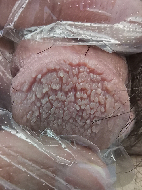 |
Figure 1 Physical examination revealed dense monomorphic soft flesh-colored filiform papules over the entire glans with a “cobblestone” appearance. |
Histopathologic examinations of shave biopsy specimens of the papules on the ventral glans demonstrated a polypoidal architecture with hyperkeratosis, acanthosis, and focally elongated rete ridges (Figure 2a). The upper dermis featured abundant small vessels with perivascular fibroplasia (Figure 2b). Histologically, the lesions resembled acral angiofibroma. The final diagnosis was profound pearly penile papules (PPP), and the patient was assured of the non-contagious nature of the condition.
 |
Figure 2 Histopathologic manifestations of the lesions. (a) Histopathologic examination demonstrated polypoidal architecture with acanthosis and hyperkeratosis. Focally elongated rete ridges were noted. (HE, 40×). (b) The upper dermis featured an increase in small vessels with perivascular fibroplasia and increased dermal stellate-shaped fibroblasts with spindle- to triangular-shaped nuclei (HE 200×). |
Discussion
PPP are painless and benign lesions that occur in rows around the corona and/or the sulcus of the glans.1 Although usually asymptomatic, PPP are sometimes misdiagnosed as a sexually transmitted disease, such as CA, and cause anxiety to the patient.
Although it was first described by Littré in 1700, Johnson coined the term “pearly penile papule” in 1964. More recently, PPP were classified as a type of acral angiofibroma.2 PPP are reported to affect 14%–48% of men.3,4 PPP usually develop in late adolescence or early adulthood, as manifestation in prepubertal children is rare. Also, the prevalence of PPP decreases with age.
PPP typically manifest as 1–3 rows of small (diameter, 1–2 mm; height, 1–4 mm) pink or white, dome-shaped or filiform papules around the corona. Although most prominent on the dorsal side, PPP may completely surround the corona and have been reported ectopically on the penis shaft.
The main differential diagnosis of PPP includes CA, ectopic sebaceous glands, and molluscum contagiosum. CA are often not infectious or uniform in size and shape. PPP can be histologically differentiated from CA by the lack of parakeratosis and koilocytes. Furthermore, PPP do not possess HPV sequences. Ectopic sebaceous glands are usually distributed along the penile shaft and characterized by secretion of a “cheesy substance” during extrusion. Molluscum contagiosum is clinically distinguished from PPP by the larger size of umbilicated papules and easily histologically differentiated.
Although PPP are benign, treatment is usually recommended for patients with severe psychological stress. Several physical therapies can achieve low rates of recurrence, including cryotherapy, electrodessication and curettage, pulsed dye laser application, ablative CO2, and erbium-doped yttrium aluminium garnet laser treatment.5
To date, only two case reports of PPP involving the entire glans have been published, with the first by Vesper in 1995.6,7 This variant was described as profound PPP, thereby expanding the disease spectrum. As compared to previous reports, the presentation of our case is more obvious and typical.
In conclusion, profound PPP are rare and especially atypical. For atypical lesions, biopsy is helpful to ensure a correct diagnosis and typical lesions elsewhere on the corona present a clue to diagnosis. Timely and accurate diagnosis and management are crucial to reduce patient anxiety.
Data Sharing Statement
The data that support the findings of this study are openly available.
Consent Statement
Signed informed consent was obtained from the patient for the publication of the case details including publication of the images.
Funding
There is no funding to report.
Disclosure
All authors report no conflicts of interest in this work.
References
1. Ramirez-Lluch M, Hernandez-Martin A. Pearly penile papules. N Engl J Med. 2021;385(15):1420. doi:10.1056/NEJMicm2106984
2. Ackerman AB, Kornberg R. Pearly penile papules: acral angiofibromas. Arch Dermatol. 1973;108:673–675.
3. Glicksman JM, Freeman RG. Pearly penile papules. A statistical study of incidence. Arch Dermatol. 1966;93(1):56–59. doi:10.1001/archderm.1966.01600190062012
4. Yildiz H, Demirer Z, Ozmen I. The prevalence of penile pearly papules among young men. Acta Dermatovenerol Croat. 2017;25(1):46–49.
5. Honigman AD, Dubin DP, Chu J, et al. Management of pearly penile papules: a review of the literature. J Cutan Med Surg. 2019;24(1):79–85. doi:10.1177/1203475419887730
6. Vesper JL, Messina J, Glass LF, et al. Profound proliferating pearly penile papules. Int J Dermatol. 1995;34(6):425–426. doi:10.1111/j.1365-4362.1995.tb04445.x
7. Yang A, Blaya Alvarez B, Makhija M, Sebaratnam DF. Profound pearly penile papules. Australas J Dermatol. 2020;61(3):280–282.
 © 2023 The Author(s). This work is published and licensed by Dove Medical Press Limited. The
full terms of this license are available at https://www.dovepress.com/terms
and incorporate the Creative Commons Attribution
- Non Commercial (unported, 3.0) License.
By accessing the work you hereby accept the Terms. Non-commercial uses of the work are permitted
without any further permission from Dove Medical Press Limited, provided the work is properly
attributed. For permission for commercial use of this work, please see paragraphs 4.2 and 5 of our Terms.
© 2023 The Author(s). This work is published and licensed by Dove Medical Press Limited. The
full terms of this license are available at https://www.dovepress.com/terms
and incorporate the Creative Commons Attribution
- Non Commercial (unported, 3.0) License.
By accessing the work you hereby accept the Terms. Non-commercial uses of the work are permitted
without any further permission from Dove Medical Press Limited, provided the work is properly
attributed. For permission for commercial use of this work, please see paragraphs 4.2 and 5 of our Terms.