Back to Journals » International Journal of Nanomedicine » Volume 13 » T-NANO 2014 Abstracts
Modification of porous polyethylene scaffolds for cell attachment and proliferation
Authors Sengupta P, Surwase SS, Prasad BLV
Received 18 October 2016
Accepted for publication 31 October 2016
Published 15 March 2018 Volume 2018:13(T-NANO 2014 Abstracts) Pages 87—90
DOI https://doi.org/10.2147/IJN.S125000
Checked for plagiarism Yes
Review by Single anonymous peer review
Peer reviewer comments 2
Editor who approved publication: Professor Lei Yang
Poulomi Sengupta, Sachin S Surwase, Bhagavatula LV Prasad
Physical Chemistry Division, CSIR–National Chemical Laboratory, Pune, India
Abstract: Synthetic polymers are widely researched for their use in tissue engineering. Control in size, surface area, pore size, and elasticity are the biggest advantages of using a man-made polymer. However, often the polymers are hydrophobic (do not encourage cell attachment); hence, it is hugely challenging to integrate them with the normal tissues. Herein, we have tried to overcome this disadvantage of polymers by coating them with citrate-stabilized gold nanoparticles and arginine. High-density polyethylene, upon multiple treatments, shows low water contact angle, which encourages cell attachment and proliferation in comparison to the untreated polymers.
Keywords: tissue engineering, gold nanoparticle, HDPE hydrophilicity
Introduction
High-density polyethylene (HDPE) is a great choice for the preparation of variety of materials due to its facile malleability and strength. It is hard and also non-biodegradable, which enable it to maintain its location after suturing. Furthermore, generation of interconnected microsized pores in HDPE is also relatively easy. For these reasons, HDPE has widely been harnessed in making artificial limbs,1 organs for tissue engineering purpose. Unfortunately, HDPE’s hydrophobic nature (coming from the inherent –CH2– backbones) discourages cell adhesion, resulting in poor integration with the healthy tissues. Keeping aside the immunological challenges, the initial hurdle of mismatch between the foreign body and live tissues is crucial and needs attention for further research. Herein, we propose to “hydrophilize” the HDPE surface by sequential plasma treatment, citrate-stabilized gold nanoparticle (AuNP) attachment,2–4 followed by treating them with the amino acid “arginine”. Layer-by-layer assembly (Figure 1) of AuNPs and arginine is expected to transform the porous HDPE surface hydrophilic from a hydrophobic one, without compromising with the inherent useful properties of HDPE.
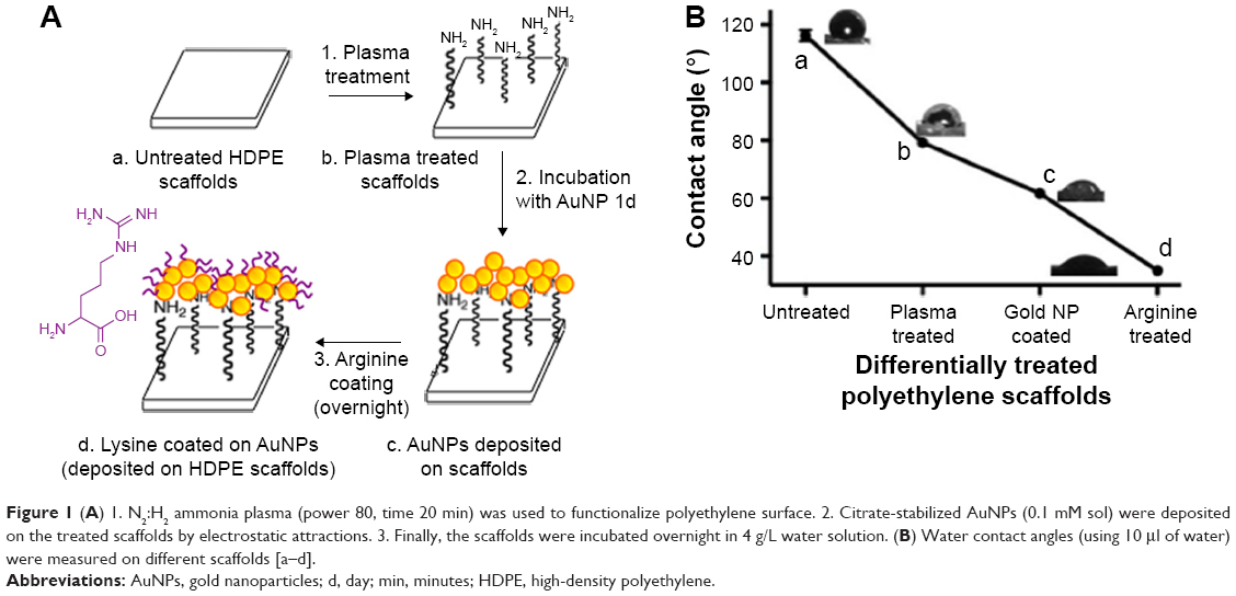 | Figure 1 (A) 1. N2:H2 ammonia plasma (power 80, time 20 min) was used to functionalize polyethylene surface. 2. Citrate-stabilized AuNPs (0.1 mM sol) were deposited on the treated scaffolds by electrostatic attractions. 3. Finally, the scaffolds were incubated overnight in 4 g/L water solution. (B) Water contact angles (using 10 μl of water) were measured on different scaffolds [a–d]. |
Materials and methods
Reagents and chemicals
HDPE was a generous donation from a Mumbai-based company “Biopore Surgicals”. Arginine and fetal bovine serum were obtained from Sigma-Aldrich. Citric acid was obtained from Merck and used as such. Chloroauric acid was purchased from Sisco Research Laboratories. 3-(4,5-Dimethylthiazoyl-2-yl)-2,5-diphenyltetrazolium bromide (MTT) dye was procured from Invitrogen. Dulbecco’s Modified Eagle’s Medium was supplied by Hi-Media. PSN 100× (penicillin, streptomycin, neomycin antibiotic cocktail) and trypsin ethylenediaminetetraacetic acid mixture were purchased from Gibco Life Technologies. Water used in the benchtop chemical reactions was of milli-q grade. HeLa cells (passage no 15/16) were purchased from the animal cell repository of NCCS (National Centre For Cell Sciences), Pune, India.
Synthesis of AuNPs
In a typical synthesis, AuNPs were synthesized by the reduction of chloroauric acid (0.1 mM solution) by citric acid. Fifty milliliters of HAuCl4 (0.1 mM) was refluxed, 500 mg of citric acid was added to it, and the solution was continued to reflux for 10 min. AuNPs formed with the development of a ruby red color with a peak in the optical absorbance spectra (Jasco V-570 ultraviolet-visible/near infrared spectrophotometer) at 520 nm. Transmission electron microscopy (FEI, TECNAI G2 TF 20) analysis indicated the particle size to be around 15–20 nm.
Plasma treatment of HDPE scaffolds
The scaffolds were treated with ammonia plasma (H2:N2 ratio 3:2) under 80 W power setting for 20 min in Emitech Plasma Asher (K1050X).3
Layer-by-layer assembly of AuNP and arginine
After plasma treatment, the scaffolds were immediately dipped in 0.1 mM gold sol and stirred for 1 day, which resulted in AuNP attachment.3 The time was optimized by solid-state ultraviolet spectrophotometry (Jasco V-570 ultraviolet-visible/near infrared/NIR Spectrophotometer). Finally, the scaffolds were incubated overnight in 4 g/L arginine in milli-q water solution. The extent of hydrophilicity was determined by contact angle measurement (GBX model DIGIDROP contact angle instrument, Windrop software).
Cell proliferation (MTT) assay
Polymer scaffolds under different treatments were cut into pieces of 1 cm2 and placed aseptically into a 24-well cell culture plate. They were sterilized with ultraviolet exposure for 40 min under cell culture hood. HeLa cells (10,000 cells/500 μL of complete medium) were seeded on each scaffold and allowed to proliferate for 48 h at 37°C in 5% CO2 atmosphere. Ten microliters of MTT (5 mg/mL) reagent in phosphate-buffered saline (PBS) was added to each well and reincubated for another 3 h at 37°C. Formazan crystals were made soluble by adding 200 μL of dimethyl sulfoxide to each well. After 10 min, the absorbance of the purple color was read at 570 nm. The % mitochondrial activity was calculated as (A570 of treated samples/A570 of untreated samples) ×100, where A is the absorbance. Data shown are mean ± standard error of n=3. Cell counting experiment was performed according to a similar protocol.
Scanning electron microscopic (SEM) images of cells grown on different scaffolds
HeLa cells were incubated with differently treated scaffolds as described in the previous section. Ultraviolet-visible/near infrared scaffolds were removed from culture condition, washed 2x with PBS, fixed with 4% PFA (paraformaldehyde). They were exposed to gold sputtering (for SEM) and directly taken into SEM instrument.
Results and discussion
The general strategy adopted to convert hydrophobic HDPE surface to a hydrophilic one is depicted in Figure 1A. After each treatment (plasma, AuNP, and arginine), the scaffolds showed gradually lowered contact angle (Figure 1B), where a huge shift from 110° to 33° at an ambient condition suggests hydrophilic properties induced by the differential layer by layer procedure followed here. Apart from that, infrared and solid-state nuclear magnetic resonance spectra (data not shown here) confirm the attachment of arginine molecules on the citrate-capped AuNP. According to the in vitro experiments (MTT and cell counting, Figure 2A and B), arginine-coated scaffolds show significantly increased live cell population. We hypothesize, this is due to the fact that adhering cells found a strong support to hold on to and “spread” thereafter. Arginine, due to its guanidinium group’s lone electrons (Figure 1A), imparts delocalized positive charge, which makes the scaffolds hydrophilic. On the other hand, the cellular membranes are the self-assembly of amphiphilic lipidic units where the negatively charged phosphate groups are exposed to the outside. We summarize that the resulting electrostatic attraction between the cellular membrane and the treated HDPE surface helps in proper anchorage. The polyethylene scaffolds having the right porosity and interconnection allow the flow of nutrients (media) in an optimal way to help in proliferation of cells – almost mimicking the in vivo situation. SEM images (Figure 3) show the cells grown on the treated scaffold to be healthier and they were clearly seen to form pili in order to attach to the solid surface. Surprisingly, in vitro results also show that only plasma-treated scaffolds have high degree of cellular attachment. But with plasma treatment, the scaffold lost its hydrophilicity with time (around 8 days), while the fully treated scaffolds (plasma, AuNP, arginine) retained their hydrophilicity for a very long period. The choice of nanoparticle was also unique. AuNP is nontoxic and possesses easier surface chemistry in comparison to other nanomaterials. Moreover, attachment of AuNPs (15–20 nm diameter) increases the surface area by a few folds, which ultimately increases the chance of cell adherence.
 | Figure 2 Effect of different scaffolds on cellular proliferation: (A) MTT assay and (B) cell counting experiment. |
 | Figure 3 SEM images of cells grown on different scaffolds. |
Conclusion
The arginine-treated scaffolds proved to be a permanent solution to the problem of hydrophobicity of the polymers. For a long period (2 months), the scaffolds did not lose their hydrophilicity (proven by contact angle, data not shown). Moreover, being a nondegradable and hard solid, it has the potential to be stable as an implant. Apart from HDPE, different polymers (eg, poly-L-lactic acid, poly-lactic-co-glycolic acid) have been widely researched as a material to be used for tissue engineering purpose. This platform technique (plasma treatment, AuNP, and arginine layer-by-layer assembly) is equally applicable on any polymeric material. On the other hand, arginine (especially R9) is a well-established transfection agent which encourages cell entry when tagged to a moiety of interest (like siRNA). These properties have been well harnessed in this work, and it opens up many new routes to challenge similar problems.
Acknowledgments
The authors sincerely acknowledge Dr. Vinay Agrawal (Biopore Surgicals) and Dr. V Premnath for polyethylene scaffolds. Poulomi Sengupta thanks CSIR-SRF for fellowship and DST WOS-A grant number SR/WOS-A/CS-94/2012 for fellowship and consumables. Sachin Surwase thanks “CSIR-Molecules to Materials to Devices” project for project assistantship. The authors thank CSIR-National Chemical Laboratory for providing lab facilities.
Disclosure
The authors report no conflicts of interest in this work.
References
Leong KF, Cheah CM, Chua CK. Solid freeform fabrication of three-dimensional scaffolds for engineering replacement tissues and organs. Biomaterials. 2003;24(13):2363–2378. | ||
D’Britto V, Tiwari S, Purohit V, et al. Composites of plasma treated poly (etherimide) films with gold nanoparticles and lysine through layer by layer assembly: a ‘‘friendly-rough’’ surface for cell adhesion and proliferation for tissue engineering applications. J Mater Chem. 2009;19(4):544–550. | ||
D’Britto, V. Synthesis of metal nanoparticles and polymer/metal nanoparticle composites: investigation towards biological applications. PhD Thesis. CSIR-National Chemical Laboratory, Pune, India, 2010. | ||
D’Britto V, Kapse H, Babrekar H, et al. Silver nanoparticle studded porous polyethylene scaffolds: bacteria struggle to grow on them while mammalian cells thrive. Nanoscale. 2011;3(7):2957–2963. |
 © 2018 The Author(s). This work is published and licensed by Dove Medical Press Limited. The
full terms of this license are available at https://www.dovepress.com/terms
and incorporate the Creative Commons Attribution
- Non Commercial (unported, 3.0) License.
By accessing the work you hereby accept the Terms. Non-commercial uses of the work are permitted
without any further permission from Dove Medical Press Limited, provided the work is properly
attributed. For permission for commercial use of this work, please see paragraphs 4.2 and 5 of our Terms.
© 2018 The Author(s). This work is published and licensed by Dove Medical Press Limited. The
full terms of this license are available at https://www.dovepress.com/terms
and incorporate the Creative Commons Attribution
- Non Commercial (unported, 3.0) License.
By accessing the work you hereby accept the Terms. Non-commercial uses of the work are permitted
without any further permission from Dove Medical Press Limited, provided the work is properly
attributed. For permission for commercial use of this work, please see paragraphs 4.2 and 5 of our Terms.