Back to Journals » International Journal of Women's Health » Volume 17
miR-6844 Regulates Cell Functions and Acts as a Potential Biomarker to Predict Prognosis in Breast Cancer
Authors Peng Y, Zhang X, Wu J, Wang H, Huang X ![]()
Received 29 May 2025
Accepted for publication 31 October 2025
Published 26 December 2025 Volume 2025:17 Pages 5657—5667
DOI https://doi.org/10.2147/IJWH.S543601
Checked for plagiarism Yes
Review by Single anonymous peer review
Peer reviewer comments 3
Editor who approved publication: Dr Everett Magann
Yi Peng, Xin Zhang, Jianbin Wu, Hongmei Wang, Xiaoxi Huang
Department of Breast Surgery, Fujian Provincial Maternity and Children’s Hospital, Fuzhou, 350001, People’s Republic of China
Correspondence: Xiaoxi Huang, Department of Breast Surgery, Fujian Provincial Maternity and Children’s Hospital, No. 18, Daoshan Road, Gulou District, Fuzhou, 350001, People’s Republic of China, Tel +86-0591-87626063, Email [email protected]
Purpose: MicroRNAs can epigenetically regulate numerous cancer-related genes and are recognized as key players in cancer biology. To explore the intrinsic mechanisms by which miR-6844 regulates the functions of BC cells and assess its potential as a prognostic biomarker for BC clinical outcomes.
Methods: A total of 130 BC patients were enrolled as the research subjects. Real-time fluorescence quantitative PCR was used to detect miR-6844 levels in cancer tissues and adjacent non-cancerous tissues. Kaplan-Meier survival curve was employed to analyze the 5-year survival status of BC patients. Multivariate Cox regression analysis was conducted to identify the influencing factors for mortality in BC patients. CCK-8 and Transwell assays were utilized to measure the proliferation, migration, and invasion of MCF-7 and MDA-MB-231 cells.
Results: miR-6844 is markedly upregulated in BC tissues and cell lines. The expression of miR-6844 is closely correlated with the TNM stage and lymph node metastasis in BC patients. Elevated levels of miR-6844 are correlated with diminished overall survival rates. Functional investigations reveal that miR-6844 enhances BC cell proliferation, migration, and invasion while exerting a negative regulatory effect on the expression of the Methylthioadenosine phosphorylase (MTAP). Conversely, silencing miR-6844 markedly inhibits the progression of BC cells, an effect that can be counteracted by concurrent inhibition of MTAP expression.
Conclusion: miR-6844 exhibits elevated expression levels in BC and is correlated with adverse prognostic outcomes. This microRNA promotes BC progression by targeting and negatively regulating MTAP.
Keywords: miR-6844, BC, prognosis, MTAP
Introduction
Breast cancer (BC) is a highly prevalent malignant tumor in women globally, characterized by sustained high incidence and mortality.1 The inherent heterogeneity of BC, coupled with its intricate molecular regulatory networks, results in substantial variations in patient prognosis.2 Consequently, the precise prediction of BC outcomes is of paramount importance in crafting personalized treatment strategies and enhancing the quality of life for patients. While existing indicators for forecasting BC prognosis are utilized in clinical practice, they exhibit certain limitations.3 Thus, the pursuit of novel and more effective prognostic biomarkers has emerged as a pivotal focus in BC research.
In recent years, microRNAs (miRNAs) have emerged as a focal point of research in oncology, owing to their pivotal roles in tumorigenesis, development, and metastasis.4 An extensive array of studies has elucidated the significant impact of miRNAs on the initiation and progression of various tumors.5,6 Evidence suggests that miR-21 may facilitate the promotion and invasion of BC.7 Furthermore, research has indicated that miR-34a holds promise as a non-invasive biomarker for the diagnosis of BC.8 A potential role for miR-130a-5p as both a diagnostic and therapeutic target in BC is suggested.9 Additionally, findings reveal that elevated levels of miR-100-5p mitigate the enhancing effects of CDC25A on the proliferation of BC cells.10
As a member of the miRNA family, miR-6844 has emerged as a pivotal player in the regulation of cellular functions in recent years.11 Existing research has illuminated its significant regulatory roles across various physiological and pathological processes.12,13 Notably, studies indicate that miR-6844 is markedly overexpressed in all clinical subtypes of invasive BC.14 However, investigations into its specific implications within the realm of BC remain relatively limited. Understanding these mechanisms is critical to BC research.
This paper seeks to elucidate the potential mechanisms through which miR-6844 influences BC cell functions and to assess its viability as a biomarker for predicting BC prognosis. This investigation aims to offer novel strategies and methodologies for the early diagnosis, prognostic evaluation, and personalized treatment of BC.
Materials and Methods
Sample Collection
This study enrolled a total of 130 BC patients who underwent radical mastectomy or breast-conserving surgery at Fujian Provincial Maternity and Children’s Hospital between January 2017 and January 2020. The research protocol was approved by the Institutional Ethics Committee of our hospital (No: 2016-129). Inclusion criteria:1) Confirmed BC diagnosis; 2) Complete clinical data and follow-up information; 3) Written informed consent obtained from each participant. Exclusion criteria: 1) Patients who received chemotherapy, radiotherapy, or other anticancer treatments prior to surgery; 2) History of other malignant tumors; 3) Family history of BC.
Surgical specimens including BC tissues and matched adjacent normal tissues (≥2 cm from the tumor margin) were collected intraoperatively. After collection, the specimens were immediately processed and stored at low temperature for subsequent analysis.
Sample size was calculated using G*Power 3.1 software. Based on an effect size of 0.5, a power of 0.95, and a significance level of α=0.05, the minimum required sample size per group was determined to be 88. This study ultimately enrolled 130 BC patients, exceeding the calculated threshold.
Clinical Data Collection and Follow-up
Clinical parameters, including patient age, tumor size and TNM stage (according to the AJCC 8th edition) were meticulously recorded. Each patient participated in a comprehensive 5-year postoperative follow-up, with overall survival designated as the primary endpoint of this study.
Cell Culture
Human BC cell lines MCF-7, MDA-MB-231, MDA-MB-453, MDA-MB-468 and non-tumorigenic breast epithelial cell line MCF-10A were obtained from the Cell Bank of the Chinese Academy of Sciences (Shanghai). Cell lines authentication by STR profiling and routine mycoplasma testing were cultured in DMEM (GIBCO, USA) supplemented with 10% heat-inactivated FBS (GIBCO, USA) and maintained under standard culture conditions at 37°C in a 5% CO2 atmosphere. Cells were passaged when the confluence reached 80%.
Cell Transfection
Transfection was carried out in accordance with the manufacturer’s guidelines for Lipofectamine 2000 (Invitrogen, USA). Following transfection, the expression of miR-6844 was evaluated through RT-qPCR to assess the efficiency of the transfection process. The experimental groups consisted of cells treated with miR-6844 mimic or inhibitor, while the control groups included cells subjected to mimic negative control (NC) or inhibitor NC treatments. Specifically, cells were transfected with 50 nM of miR-6844 mimic/inhibitor or mimic/inhibitor NC for 48 hours prior to functional assays. Additionally, a blank control group was established, wherein cells were cultured in standard medium without any transfection.
Real-Time Quantitative PCR
Total RNA was meticulously extracted from tissues or cells utilizing the Trizol reagent (Invitrogen, USA), strictly adhering to the manufacturer’s protocol. The concentration of RNA was accurately measured using NanoDrop spectrophotometer (Thermo Fisher Scientific, USA). RNA with an A260/A280 ratio between 1.8 and 2.0 was considered suitable for subsequent experiments, followed by the synthesis of cDNA through reverse transcription employing a specialized reverse transcription kit (Vazyme, China). RT-qPCR was performed on a fluorescence detection instrument, with reaction systems carefully prepared following the instructions of the RT-qPCR kit (Toyobo, Tokyo, Japan). U6 served as the internal reference for miR-6844, and the relative expression levels were calculated employing the 2−ΔΔCt method. The corresponding primer sequences are provided in Supplementary Table 1.
CCK-8 Assay
At 24 hours post-transfection, the cells were detached through trypsinization, subsequently collected, and subjected to counting. Thereafter, the cells were seeded into 96-well plates at a density of 5000 cells per well. Following this, at 24, 48, and 72 hours post-seeding, the culture medium was replaced with 100 μL of 10% DMEM supplemented with 10% CCK-8 reagent (Dojindo, Japan). After a 2-hour incubation in a cell culture incubator, the absorbance of each well was measured using a microplate reader at a wavelength of 450 nm.
Transwell Assay
Migration assay: Transfected cells were trypsinized, resuspended in serum-free medium, and adjusted to a density of 5 × 10⁵ cells/mL. A total of 100 µL, equivalent to 50,000 cells, was carefully seeded into the upper chamber of an 8-µm pore Transwell insert (Corning, USA), while the lower chamber was supplemented with 500 µL of complete medium. Following a 24-hour incubation period, non-migrated cells were meticulously removed, and those that had successfully traversed to the lower surface of the membrane were fixed, stained, and quantified under a microscope at 200× magnification, with counts taken from five randomly selected fields. Invasion assay: The upper surface of the Transwell membrane was coated with Matrigel (1:8 dilution; BD Biosciences, USA) and incubated at 37°C for 30 minutes to allow gelation. Cells were then seeded into the upper chamber as described above, and the lower chamber was filled with complete medium. Post-incubation, cells were processed identically to the migration assay for fixation, staining, and microscopic quantification.
Dual-Luciferase Reporter Assay
The TargetScan database was utilized to predict the binding sites between miR-6844 and MTAP, and corresponding reporter gene vectors were constructed by pmirGLO vector (Promega, USA). miR-6844 mimics or inhibitors were co-transfected into cells with Wild-type (WT)-MTAP or mutant (MUT)-MTAP reporter gene vectors. After incubation in an appropriate cell culture environment for 24 hours, luciferase activity was detected to validate the targeting relationship between miR-6844 and MTAP.
Statistical Methods
The SPSS 26.0 software was used for statistical analysis. Categorical variables were expressed as count (n), and intergroup comparisons were conducted using the chi-square test. Continuous variables are presented as mean ± SD. Comparisons between two groups were performed using t-tests, while comparisons among three or more groups were conducted using analysis of variance (ANOVA) combined with Tukey’s post hoc test. The Kaplan-Meier (K-M) survival curve was used to analyze the 5-year survival status of BC patients, and the Cox proportional hazards model was employed to explore the effect of miR-6844 on the postoperative survival time of BC patients. The regulatory relationship between miR-6844 and MTAP was evaluated by Pearson correlation analysis. P < 0.05 was considered statistically significant.
Results
Expression of miR-6844 in BC
RT-qPCR analysis revealed the relative expression of miR-6844 was substantially higher in BC tissues than in adjacent non-tumor tissues (P < 0.001) (Figure 1A). Further comparison of miR-6844 expression between different BC cell lines and normal human breast cell lines revealed its high expression in multiple BC cell lines. Among them, the expression differences in MCF-7 and MDA-MB-231 were the most significant (P < 0.001) (Figure 1B).
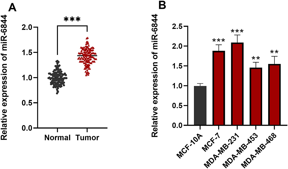 |
Figure 1 Expression of miR-6844 in BC. (A) miR-6844 showed significantly higher relative expression in BC tissues compared to adjacent normal tissues (P < 0.001). (B) Human BC cell lines exhibited notably higher miR-6844 expression than normal mammary epithelial cell lines. Among them, MCF-7 and MDA-MB-231 cells showed the most significant differences (P < 0.001). **P<0.01, ***P<0.001. |
Baseline Characteristics of Patients
A total of 130 BC patients were successfully enrolled in the study, comprising 72 individuals aged under 50 years, 83 patients with tumor sizes less than 5 cm, and 93 patients classified at TNM stages I–II. According to the median expression level of miR-6844, the patients were divided into a high-expression group (n=68) and a low-expression group (n=62). There were no statistically significant differences between the two groups in terms of age, menopausal status, tumor size, or degree of differentiation (P > 0.05), while significant statistical differences were observed in TNM stage and lymph node metastasis (Table 1).
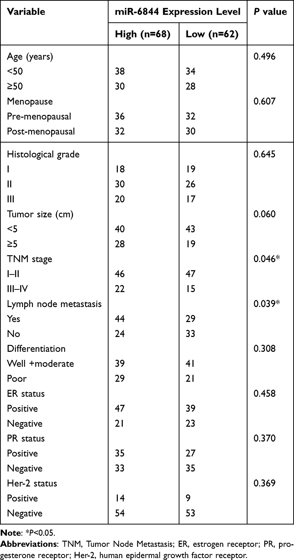 |
Table 1 The Relationship Between miR-6844 Expression and Clinical Pathological Parameters of BC |
Prognostic Impact of MiR-6844 on Survival in BC
After a 5-year follow-up of all patients, it was found that the overall survival rate of patients with high miR-6844 expression was significantly lower than that of patients with low miR-6844 expression (Log-rank P = 0.013) (Figure 2). Cox regression analysis further revealed that high miR-6844 expression was a significant factor affecting patient survival outcomes (P < 0.05) (Table 2).
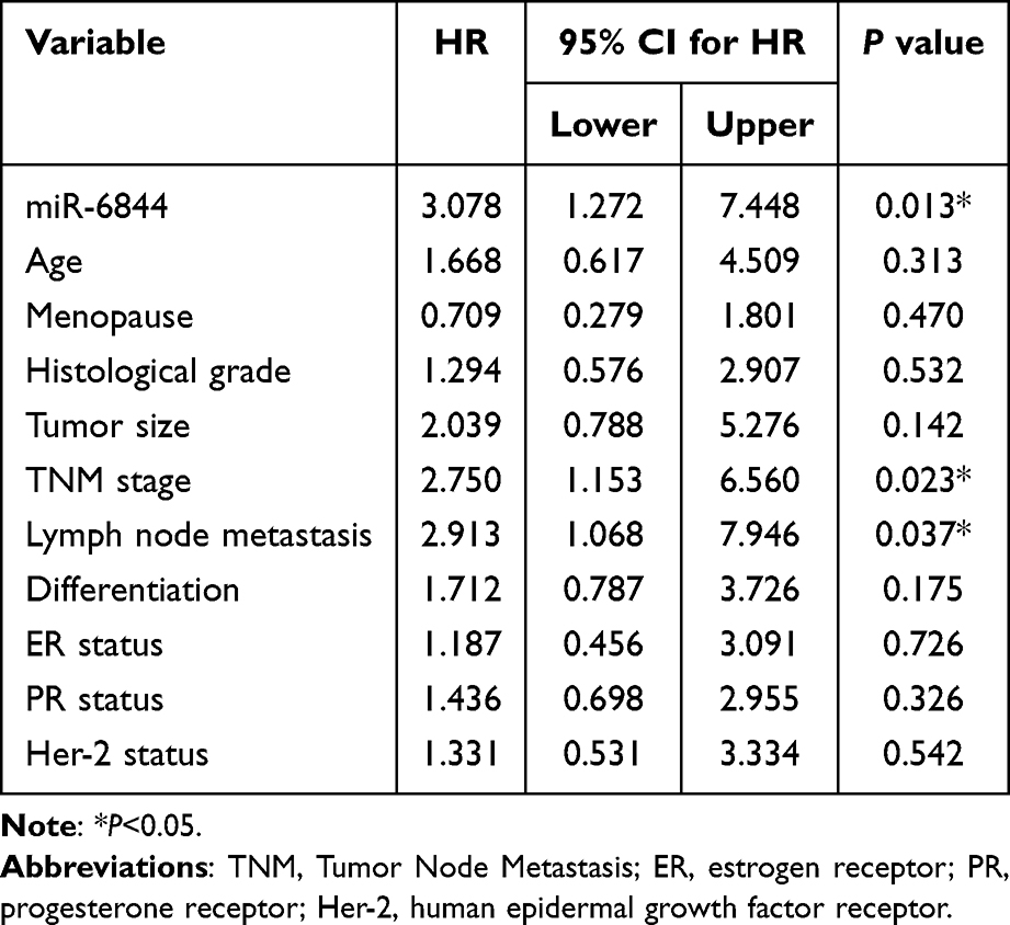 |
Table 2 Multivariate Cox Analysis of Survival Factors for BC Patients |
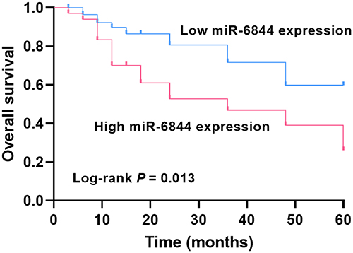 |
Figure 2 K-M Survival Curve of BC Patients with High vs Low miR-6844 Expression. |
Effect of miR-6844 Expression on BC Cells
RT-qPCR was conducted to assess the transfection efficiency in MCF-7 and MDA-MB-231 cells. The findings revealed a significant upregulation by 2-fold of miR-6844 expression in the miR-6844 mimics group compared to the mimics-NC group (P < 0.001). Conversely, miR-6844 expression was markedly diminished in the miR-6844 inhibitors group when compared to the inhibitor-NC group (P < 0.01) (Figure 3A and B). At the cellular functional level, treatment with miR-6844 mimic substantially enhanced the proliferation, migration, and invasion of BC cells by approximately 2-fold. Conversely, the application of miR-6844 inhibitor effectively impeded cell proliferation and significantly curtailed both migration and invasion, thereby exhibiting a pronounced inhibitory effect on the malignant behaviors of BC cells (P < 0.01) (Figure 3C–H). These results indicate that the upregulation of miR-6844 appears to facilitate the malignant progression of BC cells.
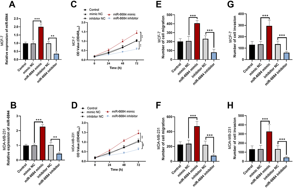 |
Figure 3 Effect of miR-6844 Expression on BC Cells. (A and B) The validation results of miR-6844 expression levels in MCF-7 and MDA-MB-231 cells after transfection with miR-6844 mimic, mimic negative control, inhibitor, and inhibitor negative control, respectively. (C–H) Effects of miR-6844 mimic and miR-6844 inhibitor transfection on cell proliferation, migration and invasion. **P<0.01, *** P<0.001. |
Validation of the Target Relationship Between miR-6844 and MTAP
Bioinformatics prediction showed that miR-6844 has potential binding sites with the 3′-untranslated region (3′-UTR) of MTAP (Figure 4A). Dual-luciferase reporter gene assays confirmed that in MCF7 and MDA-MB-231 cell lines, co-transfection with miR-6844 mimic and MTAP-WT notably reduced luciferase activity (P < 0.01). In contrast, co-transfection with miR-6844 inhibitor and MTAP-WT notably increased luciferase activity (P < 0.01), but no significant difference was observed in co-transfection with MTAP-MUT (P > 0.05) (Figure 4B and C). Furthermore, the relative expression levels of MTAP mRNA were notably lower in the miR-6844 mimic group than in the control group, whereas a significant increase by approximately 1.4-fold was observed in the miR-6844 inhibitor group (P < 0.01) (Figure 4D and E). In BC tissues, the expression level of MTAP mRNA was notably lower compared to adjacent normal tissues (P < 0.001) (Figure 4F). Additionally, the expression of miR-6844 was found to be markedly negatively correlated with the expression of MTAP mRNA (r= −0782, P<0.001) through Pearson correlation analysis (Figure 4G).
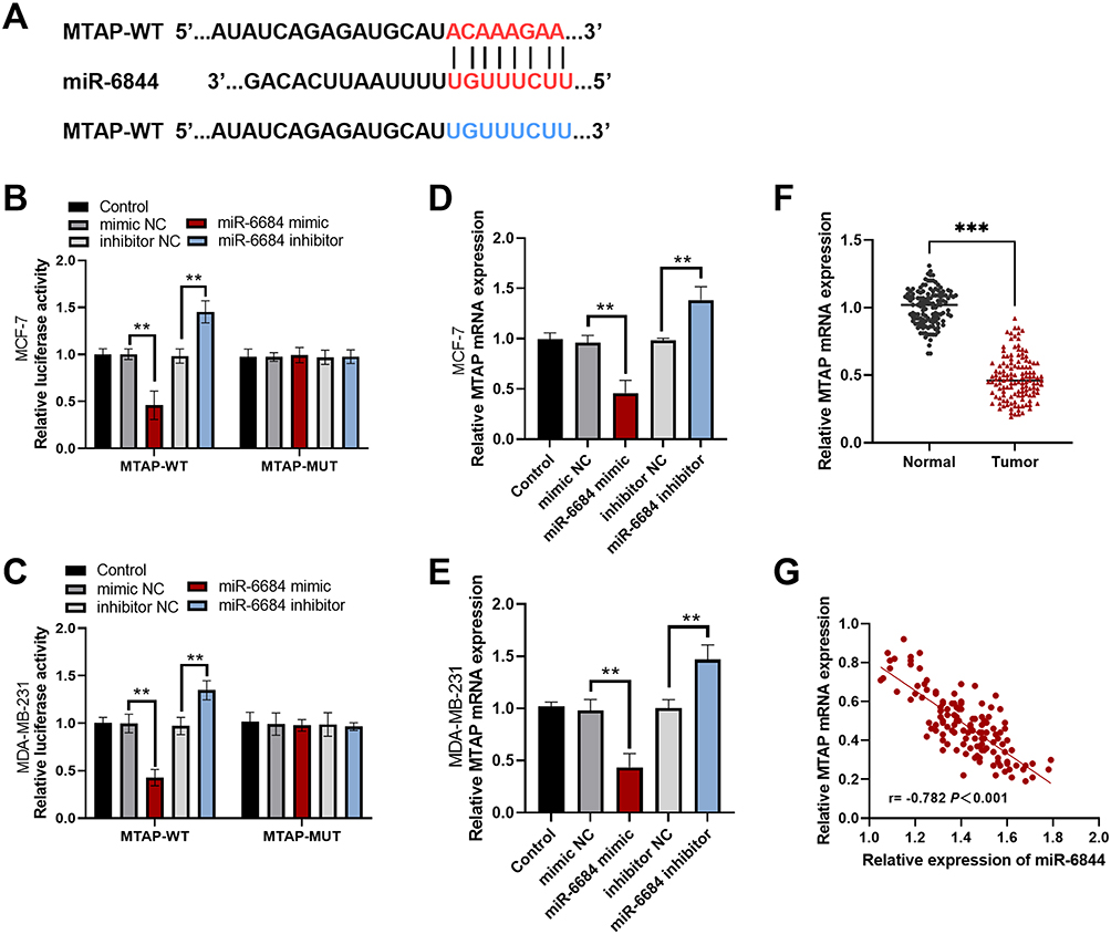 |
Figure 4 Validation of the Target Relationship between miR-6844 and MTAP. (A) Potential binding sites between miR-6844 and the 3′-UTR of MTAP. (B and C) Luciferase activity assays in MCF-7 and MDA-MB-231 cells co-transfected with miR-6844 mimic or inhibitor and wild-type (WT) or mutant (MUT) MTAP luciferase reporter vectors. (D and E) Changes in relative MTAP mRNA expression levels after transfection with miR-6844 mimic or inhibitor. (F) Comparison of MTAP mRNA expression between BC tissues and adjacent normal tissues. (G) Correlation analysis between miR-6844 and MTAP mRNA expression in BC tissues. **P<0.01, ***P<0.001. |
Effect of miR-6844 Targeting MTAP on BC Cells
In the MCF-7 and MDA-MB-231 BC cell lines, transfection with a miR-6844 inhibitor markedly upregulated MTAP mRNA expression when compared to the control (P < 0.01), thereby indicating that miR-6844 negatively regulates MTAP expression. Further experiments showed that co-transfection with the miR-6844 inhibitor and si-MTAP significantly reduced MTAP mRNA levels compared to the single miR-6844 inhibitor transfection group (P < 0.01) (Figure 5A and B). Functional assays revealed that downregulating miR-6844 significantly inhibited BC cell proliferation (P < 0.01) and reduced cell migration and invasion capabilities (P < 0.001), confirming that miR-6844 inhibition exerts a suppressive effect on BC progression. However, when MTAP expression was simultaneously inhibited, the tumor-suppressive effects mediated by miR-6844 downregulation were significantly reversed, with cell proliferation, migration, and invasion abilities returning to levels close to those of the control (P < 0.05) (Figure 5C–H). These findings suggest that miR-6844 could affect the malignant biological behaviors of BC cells through regulating MTAP expression.
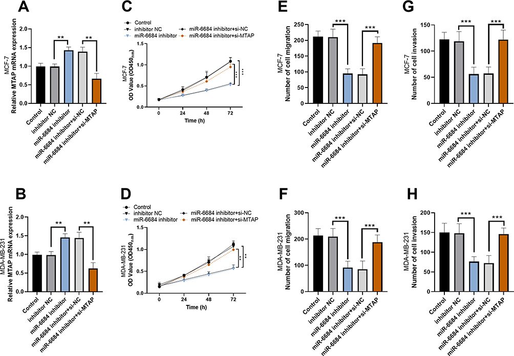 |
Figure 5 Effect of miR-6844 Targeting MTAP on BC Cells. (A and B) Changes in MTAP mRNA expression levels in MCF-7 and MDA-MB-231 cells after transfection with miR-6844 inhibitors, si-MTAP, and their co-transfection. (C–H) Functional assays detecting changes in cell proliferation, migration, and invasion capabilities after co-transfection of miR-6844 inhibitor and si-MTAP. **P<0.01, ***P<0.001. |
Discussion
miRNAs regulate gene expression at the post-transcriptional level. They influence biological processes such as cell proliferation, differentiation, apoptosis, migration, and invasion by complementary base-pairing with messenger RNAs (mRNAs) of target genes.15–17 Research has indicated that elevated expression levels of miR-103a-3p are associated with poorer survival outcomes in BC patients.18 Similarly, findings suggest that the high expression of miR-6844 in liver cancer correlates with detrimental prognostic implications.19 This study confirmed that miR-6844 is also highly expressed in BC and is closely associated with poor prognosis in patients. Compared with the miR-6844 low expression group, the high expression group had a later TNM stage and a higher lymph node metastasis rate. This suggests that upregulation of miR-6844 may promote the invasion and metastatic ability of tumor cells, leading to more advanced tumor staging and lymph node metastasis, providing new insights and important clues for the field of BC research. In the present study, we observed no statistically significant difference in miR-6844 expression between ER-positive and ER-negative patients, implying that miR-6844 may act independently of ER status. However, ER-negative tumors, including aggressive subtypes such as triple-negative breast cancer, often present distinct miRNA regulatory mechanisms.20 For example, miR-145 expression is downregulated in ER-positive BC patients and is lower than in ER-negative breast cancer.21 Although miR-6844 appears to promote BC progression independent of ER status, its downstream targets or pathway activation may differ in ER-negative backgrounds.
Additionally, in BC cells, multiple studies have shown that various miRNAs exert their effects by targeting different genes. For example, miR-518a-5p inhibits BC progression by targeting ZEB2,22 while miR-4513 promotes BC development through interaction with TRIM3.23 This study revealed that miR-6844 promotes BC progression by negatively regulating MTAP through targeting. This not only clarifies the role of miR-6844 in BC development but also elucidates the regulatory relationship between miR-6844 and MTAP, providing potential new targets and intervention directions for BC treatment.
MTAP, a key enzyme involved in purine metabolism, is closely associated with various diseases, particularly malignant tumors, due to its abnormal function.24 In malignant tumors such as BC, the expression and role of MTAP have garnered significant attention.25 Previous studies have demonstrated that downregulation of MTAP enhances specific capabilities of certain cells,26 and MTAP is considered to possess tumor-suppressive functions.25,27 MTAP deficiency has been linked to high invasiveness and poor prognosis in BC.28 This study further confirms that MTAP serves as a critical downstream target for miR-6844’s tumor-promoting effects. miR-6844 influences the malignant biological behaviors of BC cells by negatively regulating MTAP expression. At the molecular level, the regulation of MTAP by miR-6844 is profound: when miR-6844 expression is upregulated, it inhibits the translation of MTAP mRNA, reducing its tumor-suppressive capacity and thereby promoting the proliferation, migration, and invasion of BC cells. Conversely, when miR-6844 expression is suppressed, MTAP levels are restored, reactivating its tumor-suppressive function and effectively inhibiting the malignant progression of BC cells. Elucidation of this regulatory mechanism opens a new perspective for a comprehensive and in-depth exploration of the mechanisms underlying BC development. Moreover, Zhang et al proposed that downregulation of MTAP exerts an inhibitory effect by activating ornithine decarboxylase (ODC) activity.26 This provides an effective reference for our subsequent studies on the downstream pathways of MTAP.
Nevertheless, this study has certain limitations. On one hand, the relatively small sample size may lead to selection bias in the research results. In follow-up studies, multi-center, large-sample studies, and clinical validation in extensive cohorts could be conducted to ensure the universality and reliability of the findings. On the other hand, although the regulatory effect of miR-6844 on MTAP has been clarified, whether miR-6844 also influences the biological behaviors of BC cells through other signaling pathways, as well as the specific molecular mechanisms downstream of MTAP, still require further in-depth investigation through proteomics validation and in vivo biological model construction. In addition, it is necessary to explore whether miR-6844 interacts with ER signaling and evaluate its function in ER status models. Future research could focus on these directions to comprehensively reveal the mechanism of action of miR-6844 in BC and provide more robust support for precision therapy of BC.
Conclusion
In summary, this study confirmed that miR-6844 is highly expressed in BC, clarified its relationship with BC prognosis, and revealed that miR-6844 promotes the proliferation, migration, and invasion of BC cells by negatively regulating MTAP through targeting. These findings provide new biomarkers for the molecular typing of BC and lay an important theoretical foundation for the development of miRNA-based personalized treatment strategies for BC.
Ethics Approval and Consent to Participate
The study was performed in line with the principles of the Declaration of Helsinki. Approval was granted by the Ethics Committee of Fujian Provincial Maternity and Children’s Hospital before the study began (No. K2016-141-01). The participants’ right to be informed about the study was ensured and agreed to participate in the study.
Funding
This research received no specific grant from any funding agency in the public, commercial or not-for-profit sectors.
Disclosure
The authors have no conflicts of interest to declare.
References
1. Arnold M, Morgan E, Rumgay H, et al. Current and future burden of breast cancer: global statistics for 2020 and 2040. Breast. 2022;66:15–23. doi:10.1016/j.breast.2022.08.010
2. Liang Y, Zhang H, Song X, Yang Q. Metastatic heterogeneity of breast cancer: molecular mechanism and potential therapeutic targets. Semi Cancer Biol. 2020;60:14–27. doi:10.1016/j.semcancer.2019.08.012
3. Wang Z, Zhang X, Zhang S, Dai X. An integrative view on breast cancer signature panels. Expert Rev Mol Diagn. 2019;19(8):715–724. doi:10.1080/14737159.2019.1642751
4. Wang X, Hua J, Li J, et al. Mechanisms of non-coding RNA-modulated alternative splicing in cancer. RNA Biol. 2022;19(1):541–547. doi:10.1080/15476286.2022.2062846
5. Gomarasca M, Maroni P, Banfi G, Lombardi G. microRNAs in the antitumor immune response and in bone metastasis of breast cancer: from biological mechanisms to therapeutics. Int J Mol Sci. 2020;21(8):2805. doi:10.3390/ijms21082805
6. Puppo M, Valluru MK, Clézardin P. MicroRNAs and their roles in breast cancer bone metastasis. Curr Osteoporosis Rep. 2021;19(3):256–263. doi:10.1007/s11914-021-00677-9
7. Petrović N. miR-21 might be involved in breast cancer promotion and invasion rather than in initial events of breast cancer development. Mol Diagn Ther. 2016;20(2):97–110. doi:10.1007/s40291-016-0186-3
8. Imani S, Zhang X, Hosseinifard H, Fu S, Fu J. The diagnostic role of microRNA-34a in breast cancer: a systematic review and meta-analysis. Oncotarget. 2017;8(14):23177–23187. doi:10.18632/oncotarget.15520
9. Ilkhani K, Delgir S, Safi A, et al. Clinical and in silico outcomes of the expression of miR-130a-5p and miR-615-3p in tumor compared with non-tumor adjacent tissues of patients with BC. Anti-Cancer Agents Med Chem. 2021;21(7):927–935. doi:10.2174/1871520620666200924105352
10. Li X, Ren Y, Liu D, Yu X, Chen K. Role of miR-100-5p and CDC25A in breast carcinoma cells. PeerJ. 2022;9:e12263. doi:10.7717/peerj.12263
11. Wang F, Kang X, Li Y, Lu J, Liu X, Yan H. Elucidating hepatocellular carcinoma progression: a novel prognostic miRNA-mRNA network and signature analysis. Sci Rep. 2024;14(1):5042. doi:10.1038/s41598-024-55806-y
12. Rahmadi A, Fasyah I, Sudigyo D, et al. Comparative study of predicted miRNA between Indonesia and China (Wuhan) SARS-CoV-2: a bioinformatics analysis. Genet Genom. 2021;43(9):1079–1086. doi:10.1007/s13258-021-01119-7
13. Tamkini M, Nourbakhsh M, Movahedi M, Golestani A. Exploring genetic signatures of obesity: hub genes and miRNAs unveiled through comprehensive bioinformatic analysis. J Diabetes Metab Disord. 2024;23(2):2225–2232. doi:10.1007/s40200-024-01490-8
14. Prajapati KS, Shuaib M, Kushwaha PP, Singh AK, Kumar S. Identification of cancer stemness related miRNA(s) using integrated bioinformatics analysis and in vitro validation. 3 Biotech. 2021;11(10):446. doi:10.1007/s13205-021-02994-3
15. Ding L, Gu H, Xiong X, et al. MicroRNAs involved in carcinogenesis, prognosis, therapeutic resistance and applications in human triple-negative breast cancer. Cells. 2019;8(12):1492. doi:10.3390/cells8121492
16. Ors-Kumoglu G, Gulce-Iz S, Biray-Avci C. Therapeutic microRNAs in human cancer. Cytotechnology. 2019;71(1):411–425. doi:10.1007/s10616-018-0291-8
17. Osaki M, Takeshita F, Ochiya T. MicroRNAs as biomarkers and therapeutic drugs in human cancer. Biomarkers. 2008;13(7):658–670. doi:10.1080/13547500802646572
18. Liu H, Bian QZ, Zhang W, Cui HB. Circulating microRNA-103a-3p could be a diagnostic and prognostic biomarker for breast cancer. Oncol Lett. 2022;23(1):38. doi:10.3892/ol.2021.13156
19. Liao L, Yi Q, Zhao Z, et al. miR-6844/HSD17B13 axis contributes the malignant phenotype of hepatocellular carcinoma cells. Cell Biol Int. 2025. doi:10.1002/cbin.70025
20. Ebata A, Suzuki T, Shoji-Harada N, et al. Immunolocalization of cytoplasmic ER in ER-negative breast carcinoma as a potent favorable prognostic predictor. Acta Histochem Cytochem. 2023;56(4):59–66. doi:10.1267/ahc.23-00016
21. Lv P, Zhang Z, Hou L, et al. Meta-analysis of the clinicopathological significance of miRNA-145 in breast cancer. Biosci Rep. 2020;40(9):BSR20193974. doi:10.1042/bsr20193974
22. Wang L, Guo Z, Zhang S, et al. MIR-518a-5p targets ZEB2 to suppress the migration and invasion of breast-cancer cells. Altern Ther Health Med. 2023;29(1):137–143.
23. Li Y, Zhu H, Wang J, Qian X, Li N. miR-4513 promotes breast cancer progression through targeting TRIM3. Am J Transl Res. 2019;11(4):2431–2438.
24. Valera PS, Plou J, García I, et al. SERS analysis of cancer cell-secreted purines reveals a unique paracrine crosstalk in MTAP-deficient tumors. Proc Natl Acad Sci USA. 2023;120(52):e2311674120. doi:10.1073/pnas.2311674120
25. Fan N, Zhang Y, Zou S. Methylthioadenosine phosphorylase deficiency in tumors: a compelling therapeutic target. Front Cell Dev Biol. 2023;11:1173356. doi:10.3389/fcell.2023.1173356
26. Zhang Y, Zhang TT, Gao L, et al. Downregulation of MTAP promotes tumor growth and metastasis by regulating ODC activity in breast cancer. Int J Bio Sci. 2022;18(7):3034–3047. doi:10.7150/ijbs.67149
27. Bertino JR, Waud WR, Parker WB, Lubin M. Targeting tumors that lack methylthioadenosine phosphorylase (MTAP) activity: current strategies. Cancer Biol Ther. 2011;11(7):627–632. doi:10.4161/cbt.11.7.14948
28. Bou Zerdan M, Ashok Kumar P, Haroun E, Srivastava N, Ross J, Sivapiragasam A. Genomic landscape of metastatic breast cancer (MBC) patients with methylthioadenosine phosphorylase (MTAP) loss. Oncotarget. 2023;14:178–187. doi:10.18632/oncotarget.28376
 © 2025 The Author(s). This work is published and licensed by Dove Medical Press Limited. The
full terms of this license are available at https://www.dovepress.com/terms
and incorporate the Creative Commons Attribution
- Non Commercial (unported, 4.0) License.
By accessing the work you hereby accept the Terms. Non-commercial uses of the work are permitted
without any further permission from Dove Medical Press Limited, provided the work is properly
attributed. For permission for commercial use of this work, please see paragraphs 4.2 and 5 of our Terms.
© 2025 The Author(s). This work is published and licensed by Dove Medical Press Limited. The
full terms of this license are available at https://www.dovepress.com/terms
and incorporate the Creative Commons Attribution
- Non Commercial (unported, 4.0) License.
By accessing the work you hereby accept the Terms. Non-commercial uses of the work are permitted
without any further permission from Dove Medical Press Limited, provided the work is properly
attributed. For permission for commercial use of this work, please see paragraphs 4.2 and 5 of our Terms.