Back to Journals » OncoTargets and Therapy » Volume 12
MiR-19a Promotes Migration And Invasion By Targeting RHOB In Osteosarcoma
Authors Liu R, Shen L, Qu N, Zhao X, Wang J, Geng J
Received 2 June 2019
Accepted for publication 5 September 2019
Published 23 September 2019 Volume 2019:12 Pages 7801—7808
DOI https://doi.org/10.2147/OTT.S218047
Checked for plagiarism Yes
Review by Single anonymous peer review
Peer reviewer comments 2
Editor who approved publication: Dr XuYu Yang
Ruidong Liu,1 Liefeng Shen,2 Niyan Qu,3 Xia Zhao,4 Jialiang Wang,5 Jun Geng6
1Department of Clinical Laboratory, Jinan City People’s Hospital, Jinan 271100, People’s Republic of China; 2Department of Spinal Surgery, The People’s Hospital of Zhangqiu Area, Jinan 250200, People’s Republic of China; 3Department of PICU, Qingdao Women and Children’s Hospital, Qingdao 266012, People’s Republic of China; 4Medical Insurance Department, Yantai Affiliated Hospital of Binzhou Medical University, Yantai 264100, People’s Republic of China; 5Department of Radiology, The People’s Hospital of Zhangqiu Area, Jinan 250200, People’s Republic of China; 6Medical Laboratory Diagnosis Center, Jinan Central Hospital, Jinan 250013, People’s Republic of China
Correspondence: Jun Geng
Medical Laboratory Diagnosis Center, Jinan Central Hospital, 105 Jiefang Road, Lixia District, Jinan 250013, Jinan, People’s Republic of China
Tel +86 531 8569 5114
Email [email protected]
Introduction: Osteosarcoma is the most common bone tumor with high metastasis and recurrence rate. MicroRNA-19a (miR-19a) has been reported to act as tumor oncogene in multiple cancers. The objective of the study was to explore the molecular mechanisms of miR-19a in osteosarcoma cell migration and invasion.
Materials and methods: Real-time quantitative polymerase chain reaction (RT-qPCR) and Western blotting were employed to measure the levels of miR-19a and RhoB in osteosarcoma tissues and cell lines. Transwell assay was employed to analyze the tissues and cell lines’ migratory and invasive abilities. Dual luciferase reporter assay was utilized to analyze the association between miR-19a and RhoB.
Results: MiR-19a was overexpressed in osteosarcoma tissues and cell lines. MiR-19a promoted osteosarcoma cell migration and invasion in vitro. RhoB was thus confirmed as a direct and functional target of miR-19a, and it could partially reverse the function of miR-19a. Knockdown miR-19a inhibited osteosarcoma cell epithelial-mesenchymal transition (EMT) and suppressed osteosarcoma xenograft growth.
Conclusion: MiR-19a enhanced cell migration, invasion and EMT through RhoB in osteosarcoma. The newly identified miR-19a/RhoB axis provides novel insight into the progression of osteosarcoma and offers a promising target for osteosarcoma therapy.
Keywords: miR-19a, RhoB, EMT, osteosarcoma, migration, invasion
Introduction
Osteosarcoma (OS), the most primary bone tumor with annual 400,000 incidences, has a highly malignant tendency of damaging the surrounding tissues and occurring metastasis.1,2 The metastasis of osteosarcoma was so high and over 80% patients may metastasize therefore the survival rate is only 10–20% after surgery.3–5 Thus, it is urgently necessary to discover novel biomarkers for osteosarcoma early diagnosis and treatment.
MicroRNAs (miRNAs), a category of non-coding short RNA, could promote or inhibit cell progress through binding to target mRNAs at the 3ʹ-untranslated region (3ʹ-UTR).6,7 Accumulating evidence demonstrated that altered expression of miRNAs has been reported to have a relationship with the proliferation, metastasis, and prognosis of osteosarcoma, including miR-223, miR-675, miR-127 and miR-22.8–11 MiR-19a, a member of miR-17-92 cluster that is situated at chromosome 13q31.3, was upregulated and acted as an oncogene in several tumors, including bladder cancer, clear cell renal cell carcinoma, prostate cancer and colorectal cancer.12–15 Liu et al16 illuminated that miR-19a promoted colorectal cancer cell proliferation, colony formation, migration and xenograft growth. Similar findings were demonstrated by Lu et al17 that miR-19a promoted cell proliferation, migration, invasion and epithelial-mesenchymal transition (EMT) through phosphatidilinositol 3-kinase/protein kinase B (PI3K/AKT) pathway in gastric cancer. Additionally, miR-19a functioned as a potential biomarker for osteosarcoma18 and esophageal squamous cell carcinoma and prognosis.20 Therefore, we strongly believe that miR-19a may enhance osteosarcoma cell migration and invasion.
RhoB, small GTPase of the Rho family, was a kind of GTP-binding protein.20,22 RhoB, usually acted as a tumor suppressor, which was associated with multiple cellular progresses, including cell growth, adhesion and transformation.23–25 In lung cancer, RhoB regulated the activity of PP2A to modulate mesenchymal phenotype and invasion.26,27 Diao et al28 discovered that RhoB mediated cell proliferation, adhesion and migration in osteoblastic cells. Similarly, Tan et al29 indicated RhoB suppressed cell growth, migration and induced cell apoptosis in pancreatic cancer. Therefore, we strongly believe that RhoB could inhibit osteosarcoma cell migration, invasion and associated with osteosarcoma patients’ survival.
Patients And Methods
Patients And Clinical Specimens
The osteosarcoma and corresponding adjacent tissue specimens were collected from 51 patients who underwent operation from January 2015 to December 2017 in Jinan City People’s Hospital. The specimens were instantly frozen in liquid nitrogen and then stored at −80°C freezer. None of the patients had received radiotherapy or chemotherapy before surgery. The study was approved by the Ethical Committee of Jinan City People’s Hospital. All patients provided written informed consent. This study was conducted in accordance with the Declaration of Helsinki.
Cell Lines And Culture Condition
The normal osteoblast cell line NHOst and two osteosarcoma cell lines SaOS2 and MG63 were obtained from American Type Culture Collection (ATCC; Manassas, VA, USA). Roswell Park Memorial Institute 1640 (RPMI 1640) (Gibco, Rockville, MD, USA) supplemented with 10% fetal bovine serum (FBS) (Gibco, Rockville, MD, USA) was utilized to culture the cells at 37°C in a humidified atmosphere of 5% CO2.
Vectors And Transfection
The miR-19a mimic, miR-19a inhibitor and negative control (NC) oligos were purchased from Ribobio (Guangzhou, China). MG63 cells were seeded into 6-well plate and cultured at 37°C overnight. The cells transient transfected with miR-19a mimic and miR-19a inhibitor oligos were harvest after 48 h. And the miR-19a inhibitor oligo was inserted into pmirGlo vector (Promega, Madison, WI, USA) and then screened by Geneticin (G418; Thermo Scientific, Waltham, MA, USA). The transfections were carried out using Lipofectamine 3000 reagent (Invitrogen, Carlsbad, CA, USA). The sequences of the corresponding small non-coding RNAs are as follows: miR-19a mimics: 5ʹ-AGUUUUGCAUAGUUGCACUACA-3ʹ; miR-19a inhibitor: 5ʹ-UGUAGUGCAACUAUGCAAAACU-3ʹ, NC: 5ʹ-UUCUCCGAACGUGUCACGUTT-3ʹ.
RNA Isolation And Real-Time Quantitative Polymerase Chain Reaction (RT-qPCR)
We verified the total mRNAs or miRNAs expression using qRT-PCR assays via the TRIzol reagent (Invitrogen, Carlsbad, CA, USA) or the MIR Cut and Separation of miRNAs Kit (Tiangen, Beijing, China) from osteosarcoma tissues and cells. The first-strand cDNA was synthesized by Reverse Transcription System (Thermo Fisher Scientific, Waltham, MA, USA). MiRNAs Rapid Extraction Kit (BioTeke, Beijing, China) was employed to extract total miRNAs. RT-qPCR was performed using SYBR® Premix Ex Taq™ (TaKaRa, Dalian, China) in ABI7500 sequence detector (Applied Biosystems, Foster City, CA, USA). The relative expression of miR-19a and RhoB were calculated by 2−ΔΔCt method with U6 small nuclear RNA (U6) and glyceraldheyde 3-phosphate dehydrogenase (GAPDH) as the internal reference relatively. The cycling conditions for qPCR were as follows: 95°C for 10 min, followed by 40 cycles of 95°C for 15 sec and 60°C for 1 min. Primers were as follows: miR-19a forward, 5ʹ-ACACTCCAGCTGGGTGTGCAAATCCATGCAA-3ʹ, reverse, 5ʹ-CTCACAGTACGTTGGTATCCTTGTGATGTTTCGATGCCATATTGTACTGTGAGTCAGTTTT-3ʹ; U6 forward, 5ʹ-GCTTCGGCAGCACATATACTAAAAT-3ʹ, reverse, 5ʹ-CGCTTCACGAATTTGCGTGTCAT-3ʹ; RhoB forward, 5ʹ-GCCTGTCCTAGAAGTGAA-3ʹ, reverse, 5ʹ-GAATGCTACTGTCGTATGC-3ʹ, GAPDH forward, 5ʹ-CTGGGCTACACTGAGCACC-3ʹ, reverse, 5ʹ-AAGTGGTCGTTGAGGGCAATG-3ʹ.
Protein Extraction And Western Blotting
The osteosarcoma cells were lysed in radioimmunoprecipitation assay (RIPA) Lysis Buffer (Beyotime, Shanghai, China) containing Phenylmethanesulfonyl fluoride (PMSF; Sigma, St. Louis, MO, USA). Then the sample was washed with phosphate buffered saline (PBS). 10% sodium dodecyl sulfate-polyacrylamide gel electrophoresis (SDS-PAGE) was employed to separate equal amounts of proteins and followed the proteins transferred a polyvinylidene difluoride (PVDF) membranes (Millipore, Billerica, MA, USA). Subsequently, primary antibodies were used to incubate the membranes at 4°C overnight. And the antibodies were against rabbit polyclonal anti-RHOB antibody (ab155149, 1:1000; Abcam, Cambridge, CA, USA), rabbit polyclonal to N-cadherin (ab18203, 1:1000; Abcam, Cambridge, CA, USA), rabbit monoclonal to E-cadherin (ab133597, 1:1000; Abcam, Cambridge, CA, USA) and rabbit monoclonal to Vimentin (ab92547, 1:1000; Abcam, Cambridge, CA, USA) and GAPDH (GW22763, 1:3000; Sigma-Aldrich Chemicals, St. Louis, MO, USA). After washed with TBST three times, the membranes were incubated by anti-rabbit HRP-conjugated antibody for 2h at room temperature. The Western blot was then measured by Enhanced Chemiluminescence (ECL, Pharmacia Biotech, Arlington, USA) performed on the Bio-Rad Gel Doc XR instrument (Bio-Rad, Hercules, CA, USA).
Transwell Assay
Transwell assay was conducted to measure the capacities of migration and invasion with or without Matrigel (BD Biosciences, San Jose, CA, USA). The upper chamber was filled with a total of 5×10⁵ MG63 cells that re-suspended in serum free RPMI 1640 medium. Whereas, the medium with 15% FBS was added into the lower chamber acted as chemoattractant. After incubation at 37°C for 48 h, the non-penetrating cells on the upper side of the membrane were wiped off by a cotton swab, while the migrated or invaded cells were fixated in 4% paraformaldehyde for 20 min and followed stained with 0.1% crystal violet for 10 min. The membranes were separated and photographed under a microscope (Olympus Corporation, Tokyo, Japan).
Plasmid Construction And Luciferase Reporter Assay
The TargetScan predicted miR-19a targeted RhoB and the binding sequences were from 857 to 864 located at 3ʹ-UTR. The RhoB 3ʹ-UTR fragment containing the binding sequences was cloned by PCR and inserted into the pmirGlo luciferase activity vector. Followed, the binding sequences of miR-19a on RhoB 3ʹ-UTR were mutated from UUUGCAC to AAACGUG by Quick Change Site-Directed Mutagenesis Kit (Agilent, Roseville City, CA, USA). MG63 cells were co-transfected miR-19a mimic or NC and wild type (WT) or the mutant (MUT) using Lipofectamine 3000 (Invitrogen, Carlsbad, CA, USA) and then compared the differences of luciferase activities. The luciferase activity was measured 48 h after transfection using a Dual-Luciferase Reporter Assay System (Promega, Madison, WI, USA) according to the manufacturer’s protocols.
Xenograft Tumor Formation Assay
The nude mice of 4 weeks old were obtained from Charles River Laboratories (Beijing, China). MG63 cells stably transfected with miR-19a inhibitor plasmid or negative control were injected subcutaneous into the axillae. After completed the transplant tumor model, the tumor volume and the body weight were evaluated and recorded every 3 days. After cultured 26 days, the mice were dissected and Xenografts were utilized for the further study. All animal experiments were approved by the Animal Care and Utilization Committee of Jinan City People’s Hospital and the protocols complied with the guidelines for the welfare and use of animals in cancer research.
Statistical Analysis
Statistical analysis was performed by the Graph-Pad Prism version 5.0 software (La Jolla, CA, USA), and all quantitative data are presented as the mean ± standard deviation (SD). Differences between two groups were analyzed by using the Student’s t-test. Comparison between multiple groups was done using One-way ANOVA test followed by Post Hoc Test (Least Significant Difference). Statistical significance was denoted as P-values of less than 0.05. Each experiment was repeated at least three times.
Results
MiR-19a Was Upregulated In Osteosarcoma Tissues And Upregulation Of miR-19a Predicted Poor Prognosis
To investigate the important role of miR-19a in osteosarcoma, 51 paired osteosarcoma and corresponding paracancerous tissue specimens were collected. The expression of miR-19a was calculated in 51 pairs of osteosarcoma tissues and adjacent normal tissues by RT-qPCR. As expected, the mRNA level of miR-19a was higher in all osteosarcoma tissues compared with the corresponding adjacent normal tissues (P<0.0001) (Figure 1A). What’s more, the overall survival of 51 osteosarcoma patients was evaluated by Kaplan-Meier method that the 5-year survival rate was higher in miR-19a(-) group than that in miR-19a(+) group (P=0.0313) (Figure 1B). Thus, the results demonstrated that miR-19a might be a biomarker for predicting prognosis in osteosarcoma.
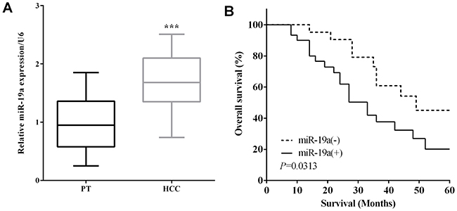 |
Figure 1 MiR-19a was upregulated in osteosarcoma tissues and upregulation of miR-19a predicted poor prognosis. (A) MiR-19a mRNA level was higher in osteosarcoma tissues than corresponding adjacent normal tissues (***P<0.001). (B) The 5-year survival rate of miR-19a (−) group was higher than that in miR-19a(+) group. |
MiR-19a Promoted The Migration And Invasion In Osteosarcoma
To further evaluate the biological function of miR-19a in osteosarcoma, the expression of miR-19a in osteosarcoma cell lines were calculated by RT-qPCR. Same with the results in tissues, the expression of miR-19a in osteosarcoma cell MG63 (P=0.0001) and SaOS2 (P=0.0010) were higher than normal osteoblast cell NHOst (Figure 2A). MiR-19a mimic and miR-19a inhibitor were transfected to up- (P<0.0001) or down-regulate (P=0.0028) miR-19a expression into MG63 cells (Figure 2B). Transwell assay was conducted to evaluate the MG63 cells’ migratory and invasion abilities. As expected, the migratory and invasive abilities were increased when miR-19a overexpression in MG63 cells (P=0.0015 and 0.0020). On the contrary, transfection of miR-19a inhibitor could suppress cell migration and invasion in MG63 cells (P=0.0103 and 0.0066) (Figure 2C). Thus, the above results illuminated that miR-19a enhanced cell migration and invasion in osteosarcoma.
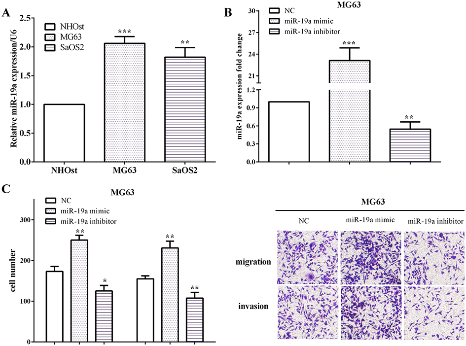 |
Figure 2 MiR-19a enhanced cell migration and invasion in osteosarcoma. (A) The expression OF miR-19a in osteosarcoma cell MG63 and SaOS2 were higher than normal osteoblast cell NHOst (compared with NHOst, **P<0.01; ***P<0.001). (B) MiR-19a mimic and miR-19a inhibitor were conducted to up- or down-regulate miR-19a in MG63 cells. (C) MiR-19a promoted cell migration and invasion in osteosarcoma (compared with NC, *P<0.05; **P<0.01; ***P<0.001). |
miR-19a Targeted To RhoB In Osteosarcoma Cells
To investigate the molecular mechanism of miR-19a promoted osteosarcoma cell migration and invasion, TargetScan was conducted to predict the larvaceous target gene of miR-19a, and we found that RhoB was potential target gene of miR-19a (Figure 3A). To verify whether miR-19a regulated cell progress by targeting RhoB, two types of vectors which containing the wild-type (pmiR-RhoB-WT, WT) or the mutant (pmiR-RhoB-MUT, MUT) 3ʹ-UTR fragment of RhoB were used. MG63 cells were co-transfected miR-19a mimic and WT or MUT RhoB 3ʹ-UTR and then we calculated the luciferase ability. As expected, compared with negative control, the relative luciferase activity was reduced (P=0.0023) in MG63 cells which co-transfected with miR-19a mimic and WT 3ʹ-UTR, but not in cells co-transfected with miR-19a and MUT 3ʹ-UTR (P=0.9009) (Figure 3B). Additionally, RhoB mRNA levels was reduced by transfecting miR-19a mimic, but was increased by transfecting miR-19a inhibitor in MG63 cells (P=0.0029 and 0.0004) (Figure 3C), which demonstrated that RhoB was a target gene of miR-19a in osteosarcoma cell.
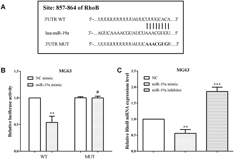 |
Figure 3 RhoB was a target gene of miR-19a in osteosarcoma cells. (A) Predicted binding sites for miR-19a in the 3ʹUTR of RhoB and the mutations in the binding sites. (B) The relative luciferase activity was reduced in wide type RhoB 3ʹ-UTR MG63 cells, but not in mutant cells. (C) RhoB mRNA levels was mediated by miR-19a in MG63 cells (compared with NC, #P>0.05; **P<0.01; ***P<0.001). |
MiR-19a Promoted Osteosarcoma Cell Epithelial-Mesenchymal Transition Through Targeting RhoB
RhoB expression was calculated in 51 paired osteosarcoma and corresponding paracancerous tissue specimens, and the results indicated that RhoB was downregulated in osteosarcoma tissue versus paracancerous tissue specimens (P<0.0001) (Figure 4A). What’s more, the 5-year overall survival was measured according to RhoB expression, and discovered that RhoB low expression predicted poor prognosis in osteosarcoma (P=0.0449) (Figure 4B). In addition, RhoB mRNA levels were lower in osteosarcoma cell lines MG63 and SaOS2 compared with normal osteoblast cell line NHOst (P=0.0007 and 0.0035) (Figure 4C). To explore the mechanism of miR-19a in regulation of osteosarcoma cell migration and invasion, EMT markers were evaluated after exogenous altered miR-19a. The results of Western blotting indicated that knockdown of miR-19a promoted RhoB expression and epithelial marker E-cadherin expression, while N-cadherin and Vimentin were reduced in MG63 cells (Figure 4D). Thus, these results illuminated that knockdown of miR-19a suppressed osteosarcoma cell EMT through targeting RhoB.
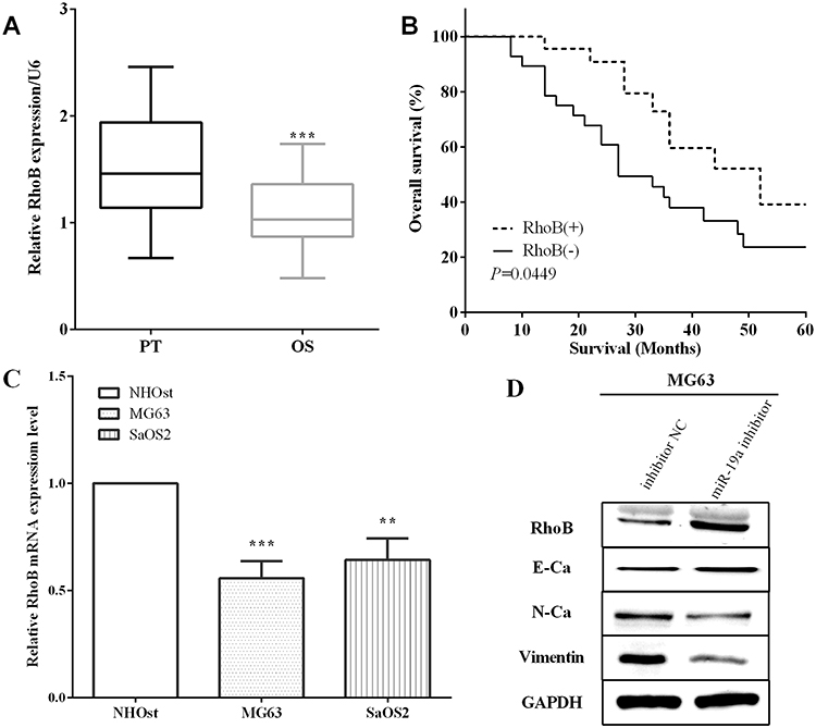 |
Figure 4 MiR-19a promoted osteosarcoma cell epithelial-mesenchymal transition through targeting RhoB. (A) RhoB was downregulated in osteosarcoma versus tissue specimens (compared with PT, ***P<0.001). (B) RhoB low expression predicted poor prognosis in osteosarcoma. (C) RhoB mRNA levels were lower in osteosarcoma cells MG63 and SaOS2 versus normal osteoblast cell NHOst (compared with NHOst, **P<0.01; ***P<0.001). (D) Knockdown miR-19a suppressed osteosarcoma cell EMT through targeting RhoB. |
MiR-19a Promoted The Xenograft Growth In Vivo
MG63 cells stably transfected with miR-19a inhibitor or negative control plasmids were injected subcutaneously under the axillae of nude mice and established the xenograft had formed. Ended the experiment 26 days after injected the cells and the xenograft size of miR-19a inhibitor injected nude mice were 321.95 and 276.06 mm3, whereas the size of control group were 469.14 and 543.72 mm3. The xenograft volume were measured every two days and the results showed that the growth rate was slower in miR-19a inhibitor group than that in control group (Figure 5A), which demonstrated that miR-19a promoted osteosarcoma xenograft growth in vivo. Knockdown of miR-19a had a remarkable smaller tumor volume versus the control group (P=0.0418) (Figure 5B).
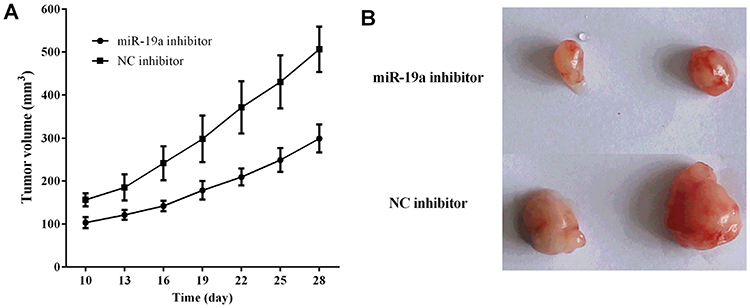 |
Figure 5 MiR-19a promoted the xenograft growth in vivo. (A) Knockdown of miR-19a had a remarkable smaller tumor volume versus the control group. (B) The xenograft growth rate was slower in miR-19a inhibitor group than that in control group. |
Discussion
Osteosarcoma was the primary bone tumor with a highly malignant tendency to metastasis and had a lower 5-year survival rate.1,2 Thus, it is necessary to find novel biomarkers for osteosarcoma early diagnosis and treatment. miRNAs could regulate tumor progress through binding to target gene mRNAs at the 3ʹ-untranslated region (3ʹ-UTR).6,7 MiR-19a is a novel microRNA, which located at chromosome 13q31.3, also act as an oncogene in several tumors.30 In colorectal cancer, miR-19a promoted cell proliferation, colony formation, migration and promoted xenograft growth.16 Similarly, miR-19a has been reported to contribute to cell proliferation, migration, invasion and EMT in gastric cancer.17 Even in osteosarcoma, previous studies reported that silencing of miR-19a-3p enhanced chemosensitivity of OS cells to Cisplatin, through suppressing cell proliferation and promoting cell apoptosis during treatment with Cisplatin.18 Our results were consistent with with previous findings that miR-19a was overexpressed in osteosarcoma tissues and cell lines. What’s more, we also found that miR-19a promoted osteosarcoma cells migration and invasion. Consistent with the findings in esophageal squamous cell carcinoma,20 we explored that upregulation of miR-19a predicted shorter overall survival in osteosarcoma. In addition, knockdown of miR-19a inhibited osteosarcoma cell EMT and suppressed osteosarcoma xenograft growth.
RhoB, a kind of GTP-binding protein, is an early response gene, which regulates by various stimuli and usually acts as a tumor suppressor.22,24,31 In pancreatic cancer and clear cell renal cell carcinoma, RhoB was a target gene of miR-19a and miR-19a could promote cell growth, migration and induced cell apoptosis through RhoB.13,29 Our results were consistent with the previous findings,31 RhoB is a target gene of miR-19a and its expression is correlated with miR-19a. We also showed that RhoB was downregulated in osteosarcoma tissues and low expression of RhoB predicted poor prognosis of osteosarcoma patients. Furthermore, in lung cancer and osteoblastic cell RhoB mediated cell proliferation, adhesion, migration and invasion in lung cancer and osteoblastic cell.27,28 In our study, knockdown of miR-19a inhibited osteosarcoma cell EMT through RhoB. However, due to the limited number of cases, the correlation between miR-19a and RhoB expression is low, which is the limit of this experiment. We will collect more cases next time to improve the correlation of expression.
Conclusions
In brief, miR-19a was overexpressed in osteosarcoma tissues and cells. Furthermore, we showed that miR-19a was a prognostic factor of osteosarcoma. MiR-19a enhanced osteosarcoma cell migration and invasion. RhoB was verified as a target of miR-19a and the RhoB expression was regulated by exogenous altered miR-19a. Knockdown of miR-19a inhibited osteosarcoma cell EMT and suppressed osteosarcoma xenograft growth. This newly identified miR-19a may provide further insight into the progression of osteosarcoma and offers a promising therapeutic target for the treatment of osteosarcoma. Additional studies to investigate the function of the miR-19a/RhoB axis in the tumorigenesis and progression of osteosarcoma are needed.
Disclosure
The authors report no conflicts of interest in this work.
References
1. Mirabello L, Troisi RJ, Savage SA. Osteosarcoma incidence and survival rates from 1973 to 2004: data from the surveillance, epidemiology, and end results program. Cancer-Am Cancer Soc. 2009;115(7):1531–1543. doi:10.1002/cncr.24121
2. Link MP, Goorin AM, Miser AW, et al. The effect of adjuvant chemotherapy on relapse-free survival in patients with osteosarcoma of the extremity. N Engl J Med. 1986;314(25):1600–1606. doi:10.1056/NEJM198606193142502
3. Luetke A, Meyers PA, Lewis I, Juergens H. Osteosarcoma treatment - where do we stand? A state of the art review. Cancer Treat Rev. 2014;40(4):523–532. doi:10.1016/j.ctrv.2013.11.006
4. Marina N, Gebhardt M, Teot L, Gorlick R. Biology and therapeutic advances for pediatric osteosarcoma. Oncologist. 2004;9(4):422–441. doi:10.1634/theoncologist.9-4-422
5. Moore DD, Luu HH. Osteosarcoma. Cancer Treat Res. 2014;162:65–92. doi:10.1007/978-3-319-07323-1_4
6. Gargalionis AN, Basdra EK. Insights in microRNAs biology. Curr Top Med Chem. 2013;13(13):1493–1502. doi:10.2174/15680266113139990098
7. Lu J, Getz G, Miska EA, et al. MicroRNA expression profiles classify human cancers. Nature. 2005;435(7043):834–838. doi:10.1038/nature03702
8. Ji Q, Xu X, Song Q, et al. miR-223-3p inhibits human osteosarcoma metastasis and progression by directly targeting CDH6. Mol Ther. 2018;26(5):1299–1312. doi:10.1016/j.ymthe.2018.03.009
9. Gong L, Bao Q, Hu C, et al. Exosomal miR-675 from metastatic osteosarcoma promotes cell migration and invasion by targeting CALN1. Biochem Biophys Res Commun. 2018;500(2):170–176. doi:10.1016/j.bbrc.2018.04.016
10. Wang D, Tang L, Wu H, Wang K, Gu D. MiR-127-3p inhibits cell growth and invasiveness by targeting ITGA6 in human osteosarcoma. IUBMB Life. 2018;70(5):411–419. doi:10.1002/iub.1710
11. Zhou X, Natino D, Zhai X, Gao Z, He X. MicroRNA22 inhibits the proliferation and migration, and increases the cisplatin sensitivity, of osteosarcoma cells. Mol Med Rep. 2018;17(5):7209–7217. doi:10.3892/mmr.2018.8790
12. Feng Y, Liu J, Kang Y, et al. miR-19a acts as an oncogenic microRNA and is up-regulated in bladder cancer. J Exp Clin Cancer Res. 2014;33:67. doi:10.1186/PREACCEPT-9242556491295527
13. Niu S, Ma X, Zhang Y, et al. MicroRNA-19a and microRNA-19b promote the malignancy of clear cell renal cell carcinoma through targeting the tumor suppressor RhoB. PLoS One. 2018;13(2):e192790. doi:10.1371/journal.pone.0192790
14. Fu F, Wan X, Wang D, et al. MicroRNA-19a acts as a prognostic marker and promotes prostate cancer progression via inhibiting VPS37A expression. Oncotarget. 2018;9(2):1931–1943. doi:10.18632/oncotarget.23026
15. Chen M, Lin M, Wang X. Overexpression of miR-19a inhibits colorectal cancer angiogenesis by suppressing KRAS expression. Oncol Rep. 2018;39(2):619–626. doi:10.3892/or.2017.6110
16. Liu Y, Liu R, Yang F, et al. miR-19a promotes colorectal cancer proliferation and migration by targeting TIA1. Mol Cancer. 2017;16(1):53. doi:10.1186/s12943-017-0625-8
17. Lu WD, Zuo Y, Xu Z, Zhang M. MiR-19a promotes epithelial-mesenchymal transition through PI3K/AKT pathway in gastric cancer. World J Gastroenterol. 2015;21(15):4564–4573. doi:10.3748/wjg.v21.i15.4564
18. Zou Q, Xiao X, Liang Y, et al miR-19a-mediated downregulation of RhoB inhibits the dephosphorylation of AKT1 and induces osteosarcoma cell metastasis. Cancer Lett. 2018;428:147–159. doi:10.1016/j.canlet.2018.04.027
19. Bai Y, Lin H, Fang Z, et al. Plasma microRNA-19a as a potential biomarker for esophageal squamous cell carcinoma diagnosis and prognosis. Biomark Med. 2017;11(5):431–441. doi:10.2217/bmm-2016-0286
20. Karlsson R, Pedersen ED, Wang Z, Brakebusch C. Rho GTPase function in tumorigenesis. Biochim Biophys Acta. 2009;1796(2):91–98. doi:10.1016/j.bbcan.2009.03.003
21. Vega FM, Ridley AJ. Rho GTPases in cancer cell biology. FEBS Lett. 2008;582(14):2093–2101. doi:10.1016/j.febslet.2008.04.039
22. Chen Z, Sun J, Pradines A, Favre G, Adnane J, Sebti SM. Both farnesylated and geranylgeranylated RhoB inhibit malignant transformation and suppress human tumor growth in nude mice. J Biol Chem. 2000;275(24):17974–17978. doi:10.1074/jbc.C000145200
23. Liu AX, Rane N, Liu JP, Prendergast GC. RhoB is dispensable for mouse development, but it modifies susceptibility to tumor formation as well as cell adhesion and growth factor signaling in transformed cells. Mol Cell Biol. 2001;21(20):6906–6912. doi:10.1128/MCB.21.20.6906-6912.2001
24. Mazieres J, Tillement V, Allal C, et al. Geranylgeranylated, but not farnesylated, RhoB suppresses Ras transformation of NIH-3T3 cells. Exp Cell Res. 2005;304(2):354–364. doi:10.1016/j.yexcr.2004.10.019
25. Calvayrac O, Pradines A, Favre G. RHOB expression controls the activity of serine/threonine protein phosphatase PP2A to modulate mesenchymal phenotype and invasion in non-small cell lung cancers. Small GTPases. 2018;9(4):339–344. doi:10.1080/21541248.2016.1234429
26. Bousquet E, Calvayrac O, Mazieres J, et al. RhoB loss induces Rac1-dependent mesenchymal cell invasion in lung cells through PP2A inhibition. Oncogene. 2016;35(14):1760–1769. doi:10.1038/onc.2015.240
27. Diao F, Chen K, Wang Y, et al. Involvement of small G protein RhoB in the regulation of proliferation, adhesion and migration by dexamethasone in osteoblastic cells. PLoS One. 2017;12(3):e174273. doi:10.1371/journal.pone.0174273
28. Tan Y, Yin H, Zhang H, et al. Sp1-driven up-regulation of miR-19a decreases RHOB and promotes pancreatic cancer. Oncotarget. 2015;6(19):17391–17403. doi:10.18632/oncotarget.3975
29. Olive V, Jiang I, He L. mir-17-92, a cluster of miRNAs in the midst of the cancer network. Int J Biochem Cell Biol. 2010;42(8):1348–1354. doi:10.1016/j.biocel.2010.03.004
30. Jahner D, Hunter T. The ras-related gene rhoB is an immediate-early gene inducible by v-Fps, epidermal growth factor, and platelet-derived growth factor in rat fibroblasts. Mol Cell Biol. 1991;11(7):3682–3690. doi:10.1128/mcb.11.7.3682
31. Chen Q, Guo W, Zhang Y, Wu Y, Xiang J. MiR-19a promotes cell proliferation and invasion by targeting RhoB in human glioma cells. Neurosci Lett. 2016;628:161–166. doi:10.1016/j.neulet.2016.06.031
 © 2019 The Author(s). This work is published and licensed by Dove Medical Press Limited. The
full terms of this license are available at https://www.dovepress.com/terms
and incorporate the Creative Commons Attribution
- Non Commercial (unported, 3.0) License.
By accessing the work you hereby accept the Terms. Non-commercial uses of the work are permitted
without any further permission from Dove Medical Press Limited, provided the work is properly
attributed. For permission for commercial use of this work, please see paragraphs 4.2 and 5 of our Terms.
© 2019 The Author(s). This work is published and licensed by Dove Medical Press Limited. The
full terms of this license are available at https://www.dovepress.com/terms
and incorporate the Creative Commons Attribution
- Non Commercial (unported, 3.0) License.
By accessing the work you hereby accept the Terms. Non-commercial uses of the work are permitted
without any further permission from Dove Medical Press Limited, provided the work is properly
attributed. For permission for commercial use of this work, please see paragraphs 4.2 and 5 of our Terms.