
Back to Journals » Infection and Drug Resistance » Volume 19
MiR-181a, IL-6, and TNF-α: Biomarkers for Diagnosis and Long-Term Neurodevelopmental Prognosis in Neonatal Sepsis
Authors Shen J, Yu M, Zhu S, Xue M 
Received 13 November 2025
Accepted for publication 15 March 2026
Published 27 March 2026 Volume 2026:19 574673
DOI https://doi.org/10.2147/IDR.S574673
Checked for plagiarism Yes
Review by Single anonymous peer review
Peer reviewer comments 2
Editor who approved publication: Dr Hemant Joshi
Jialin Shen, Min Yu, Shirui Zhu, Mei Xue
Department of Neonatology, The Affiliated Taizhou People’s Hospital of Nanjing Medical University, Taizhou, Jiangsu, People’s Republic of China
Correspondence: Mei Xue, The Affiliated Taizhou People’s Hospital of Nanjing Medical University, No. 366, Taihu Road, Hailing District, Taizhou City, Jiangsu Province, People’s Republic of China, Email [email protected]
Objective: To investigate the value of serum microRNA-181a (miR-181a), interleukin-6 (IL-6), and tumor necrosis factor-alpha (TNF-α) in early prediction of neonatal sepsis and their correlation with long-term neurodevelopmental outcomes.
Methods: A retrospective analysis of 131 septic and 129 non-infected neonates from January 2023 to August 2024 assessed serum miR-181a, TNF-α, and IL-6 levels, with neurodevelopmental outcomes evaluated at 12 months. Multivariate logistic regression was employed to identify risk factors for sepsis and adverse neurodevelopmental outcomes. Receiver operating characteristic (ROC) curve analysis was used to evaluate the predictive value of each indicator for sepsis and poor neurodevelopmental outcomes.
Results: The septic group had lower miR-181a and higher IL-6 and TNF-α levels (P < 0.0001), with an AUC of 0.860 for sepsis diagnosis. Among septic neonates, 31.30% had neurodevelopmental abnormalities, linked to lower miR-181a and higher IL-6 and TNF-α levels (P < 0.01), with an AUC of 0.850 for predicting these abnormalities. Multivariable logistic regression identified miR-181a as a protective factor for neonatal sepsis, while IL-6, TNF-α, and NLR were independent risk factors. For long-term adverse neurodevelopmental outcomes, miR-181a and gestational age served as protective factors, whereas IL-6, TNF-α, and prenatal inflammation were independent risk factors.
Conclusion: Serum miR-181a, IL-6, and TNF-α are sensitive indicators for the early prediction of neonatal sepsis. Low expression of miR-181a and high expression of IL-6 and TNF-α are closely associated with adverse neurodevelopmental outcomes. Combined detection of these three markers holds significant clinical value for prognostic assessment, facilitating early identification of high-risk infants and enabling timely intervention.
Keywords: neonatal sepsis, miR-181a, interleukin-6, tumor necrosis factor-alpha, neurodevelopmental outcomes
Introduction
Neonatal sepsis (NS) remains one of the leading causes of mortality and long-term neurological sequelae during the neonatal period. Its global incidence remains persistently high, and surviving infants often face severe neurodevelopmental impairments such as cognitive deficits and motor delays, imposing a substantial burden on families and society.1,2 Blood culture remains the gold standard for diagnosis. Clinicians also widely use markers like C-reactive protein (CRP) and procalcitonin (PCT). However, these indicators have significant limitations in early diagnostic sensitivity, specificity, and the ability to predict neurological outcomes.3,4
Recent studies have shown that the systemic inflammatory response triggered by sepsis is a core mechanism of brain injury. Cytokines like interleukin-6 (IL-6) and tumor necrosis factor-alpha (TNF-α) can damage the blood-brain barrier. They also trigger neuronal cell death. Consequently, these factors are linked to poor neurodevelopmental results.5 Nevertheless, individual inflammatory responses vary significantly, and the predictive power of a single indicator is limited, making precise risk stratification challenging.
With advances in molecular mechanism research, microRNAs (miRNAs), as key regulators of gene expression, are increasingly recognized for their role in the pathogenesis of sepsis.6 Among them, microRNA-181a (miR-181a) has been shown to participate in the regulation of innate immunity and inflammatory responses.7 It may influence the release of inflammatory factors such as IL-6 and TNF-α by modulating signaling pathways like NF-κB, theoretically forming an inflammatory cascade from upstream regulation to downstream effects.8,9 However, its overall value in neonatal sepsis remains unclear.
Against this background, this study was initiated in January 2023. It aims to evaluate the diagnostic and prognostic value of serum miR-181a, IL-6, and TNF-α—both individually and in combination—for early disease detection and long-term neurodevelopmental outcomes in neonates with sepsis. Furthermore, it seeks to explore their potential as multidimensional biomarkers for clinical application, providing a new theoretical basis and strategic support for early identification of high-risk infants and targeted neuroprotective interventions.
Materials and Methods
Study Subjects
A total of 131 neonates with sepsis (gestational age ≥32 weeks) and 129 non-infected neonates admitted to the Department of Neonatology, The Affiliated Taizhou People’s Hospital of Nanjing Medical University between January 2023 and August 2024 were enrolled. The case group comprised 82 males and 49 females, while the control group included 76 males and 53 females. Inclusion criteria were as follows: (1) Cases were diagnosed according to the Expert consensus on the diagnosis and management of neonatal sepsis (version 2019);10 (2) Controls were non-infected neonates admitted during the same period, with no clinical signs of infection and confirmed non-infectious conditions such as mild hyperbilirubinemia, hypoglycemia, or non-infectious diarrhea via blood culture. The selection of symptomatic non-infected neonates, rather than strictly healthy asymptomatic infants, was necessitated by ethical restrictions regarding venipuncture in healthy newborns. While we acknowledge that conditions like hypoglycemia may induce physiological stress, this control group represents the real-world clinical scenario where clinicians must distinguish sepsis from other symptomatic non-infectious conditions; (3) Complete clinical data of both neonates and their mothers were available. Exclusion criteria included neonates with severe congenital malformations, inherited metabolic diseases, grade III or higher intracranial hemorrhage, severe perinatal asphyxia (1-minute Apgar score ≤3), congenital infections, or those who received anti-infective therapy prior to enrollment. Approved by the Hospital Ethics Committee (Approval No. KY-2023-169-01), this study was conducted in accordance with the Declaration of Helsinki, with written informed consent obtained from all guardians. The study flowchart is presented in Figure 1.
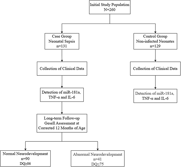 |
Figure 1 Study Enrollment and Follow-up Flowchart for Neonates with Sepsis. |
Clinical Data
Clinical data were collected from electronic medical records, including neonatal parameters such as sex, gestational age, delivery mode, birth weight, Apgar scores at 1 and 5 minutes, inflammatory markers like CRP and PCT, laboratory results including WBC, PLT, NLR, LMR, and PLR, cerebrospinal fluid analysis, blood culture findings, and occurrences of NRDS or intrauterine distress. Maternal data covered history of miscarriage, twin pregnancy, conception method such as assisted reproductive technology, prenatal inflammation, placental and amniotic fluid status, threatened preterm labor, and pregnancy complications including diabetes, hypertension, hypothyroidism, cervical insufficiency, and anemia.
Research Methods
Serum Sample Collection and Testing
Peripheral venous blood (2.5 mL) was collected within 24 hours of admission into enzyme-free EP tubes, centrifuged at 3000 rpm for 15 minutes at 4°C, and the supernatant was stored at −80°C for RNA extraction and detection of miR-181a and TNF-α. Serum miR-181a levels were quantified using quantitative real-time PCR, with total RNA extracted using TRIzol reagent, purity and concentration assessed via NanoDrop 2000, reverse transcription performed with a MicroRNA Reverse Transcription Kit, and amplification carried out using the miRCURY LNA SYBR Green PCR Kit; U6 served as the internal reference, and relative expression was calculated by the 2−ΔΔCt method, normalized to the mean Ct value of the control group, using primer sequences for miR-181a forward 5’-ACACTCCAGCTGGGAACATTCAACGCTGTCG-3’ and reverse 5’-CTCAACTGGTGTCGTGGAGTCGGCAATTCAGTTGAGACTCACCG-3’, and for U6 forward 5′-CTCGCTTCGGCAGCACA-3′ and reverse 5′-AACGCTTCACGAATTTGCGT-3′. TNF-α levels were measured via enzyme-linked immunosorbent assay with a kit from Solarbio (Catalog Number: SEKH-0047), and IL-6 concentrations were determined using an automated electrochemiluminescence analyzer.
Neurodevelopmental Assessment
Follow-ups were conducted for the case group after discharge, with neurodevelopment assessed at a corrected age of 12 months using the Gesell Developmental Diagnosis Scale,11 administered independently by two trained developmental pediatricians. The assessment covered five domains: adaptability, gross motor, fine motor, language, and personal-social skills. Based on the results, neonates were categorized into normal neurodevelopment with a Developmental Quotient (DQ) of 86 or higher and abnormal neurodevelopment with a DQ of 75 or lower.
Statistical Analysis
Data were analyzed using SPSS 27.0 software and R language (version 4.5.2). Continuous variables were assessed for normality using the Shapiro–Wilk test. As most variables were non-normally distributed, data are presented as median (interquartile range, IQR) and compared using the Mann–Whitney U-test. Categorical data were described as counts with percentages and compared via χ2 or Fisher’s exact test. Spearman correlation analysis was used for variable associations. Firstly, based on univariate logistic regression analysis, potential predictors of the outcome event were screened out (P < 0.05). Further, for the selected variables, the Least Absolute Shrinkage and Selection Operator (LASSO) regression model was used to select significant features (non-zero coefficients), and 10-fold cross-validation was used to determine the optimal parameter configuration. The lambda value corresponding to the minimum mean squared error (min) was used to determine the coefficients, and variables with non-zero coefficients were selected. For the selected variables, multivariate logistic regression identified risk factors for sepsis and neurodevelopmental abnormalities, and receiver operating characteristic (ROC) curves evaluated predictive performance of indicators with area under the curve calculated. The combined biomarker panel (miR-181a + IL-6 + TNF-α) was constructed using predicted probabilities from a multivariate logistic regression model. The ROC curve for the combined panel was generated based on these predicted probabilities, and the optimal cut-off value was determined using the Youden index. A two-sided P-value less than 0.05 was considered statistically significant.
Results
Comparison of Clinical Data Between Case and Control Groups
This retrospective study enrolled a total of 260 infants, who were divided into two groups based on sepsis diagnostic criteria: a case group (n=131) and a control group (n=129). As summarized in Table 1, no statistically significant differences were observed between the case and control groups in terms of sex, gestational age, birth weight, or delivery mode (P > 0.05). However, significant differences were found in Apgar scores, CRP, PCT, WBC, NLR, LMR, and PLR (P < 0.05). Among the case group, 7 patients were diagnosed with purulent meningitis, and 15 had positive blood culture results.
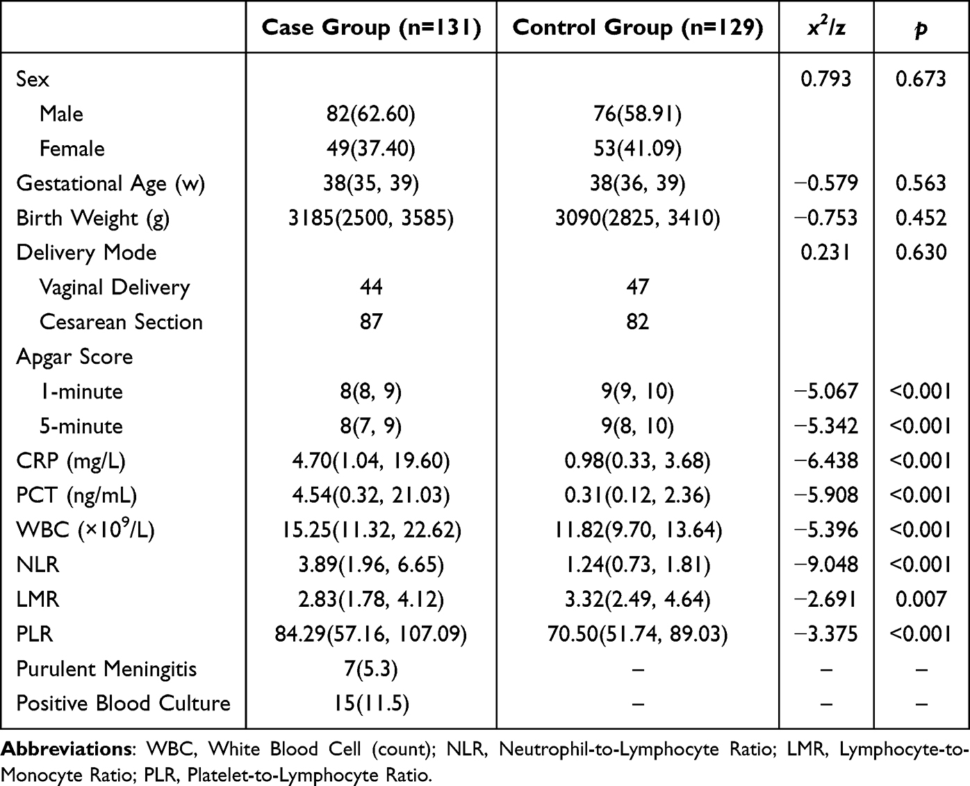 |
Table 1 Comparison of Clinical Characteristics Between Case and Control Groups (M (P25, P75), n[%]) |
Comparison of Serum miR-181a, IL-6, and TNF-α Levels Between Case and Control Groups
To investigate their potential roles in the pathogenesis of neonatal sepsis, we first compared the serum levels of miR-181a and two key inflammatory cytokines between the two groups. The relative expression of serum miR-181a was significantly lower in the case group (0.71 ± 0.31) than in the control group (1.19 ± 0.50; P < 0.0001; Figure 2). Conversely, the case group exhibited markedly elevated levels of both IL-6 (98.14 ± 52.54 pg/mL vs. 52.81 ± 35.73 pg/mL) and TNF-α (108.58 ± 48.80 pg/mL vs. 67.82 ± 45.86 pg/mL) compared to the control group (both P < 0.0001; Figure 2). This distinct expression pattern—characterized by suppressed miR-181a alongside amplified pro-inflammatory mediators—suggests a potential disruption of immunoregulatory mechanisms and a pronounced systemic inflammatory response in neonates with sepsis.12
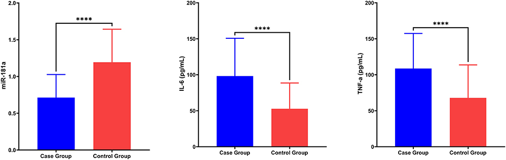 |
Figure 2 Comparison of biomarkers levels in serum between two groups ( Note: ****Indicates P < 0.0001 between groups. |

Feature Selection via LASSO Regression
To address potential overfitting and multicollinearity given the sample size, we further employed the Least Absolute Shrinkage and Selection Operator (LASSO) regression model using the “glmnet” package in R software. Ten predictors retained non-zero coefficients: miR-181a, IL-6, TNF-α, NLR, Apgar 1min, Apgar 5min, CRP, PCT, WBC, LMR (Supplementary Figure 1A and B).
Risk Factor Analysis for NS
Subsequently, the variables selected by the lasso regression model were input into the multivariate logistic regression model to adjust for potential confounding factors and identify the independent predictors. The results of this analysis confirmed that miR-181a was a protective factor (OR: 0.351; 95% CI: 0.193–0.640; P < 0.001), whereas elevated IL-6 (OR: 2.586; 95% CI: 1.642–4.071; P < 0.001), elevated TNF-α (OR: 1.506; 95% CI: 1.149–1.975; P = 0.003), and a high NLR (OR: 2.068; 95% CI: 1.569–2.727; P < 0.001) were independent risk factors for neonatal sepsis after multivariable adjustment. The complete results of the logistic regression analysis, including all covariates and their corresponding statistical metrics, are detailed in Table 2.
 |
Table 2 Logistic Regression Analysis of Risk Factors for NS |
Diagnostic Performance of Biomarkers for NS
The primary objective of this study was to assess the diagnostic value of serum miR-181a, IL-6, and TNF-α, both individually and in combination, for the early identification of neonatal sepsis. ROC curve analysis was employed to evaluate their performance (Table 3 and Figure 3). The areas under the curve (AUC) for the individual biomarkers were 0.804 for miR-181a, 0.763 for IL-6, and 0.745 for TNF-α, indicating good diagnostic ability for each.
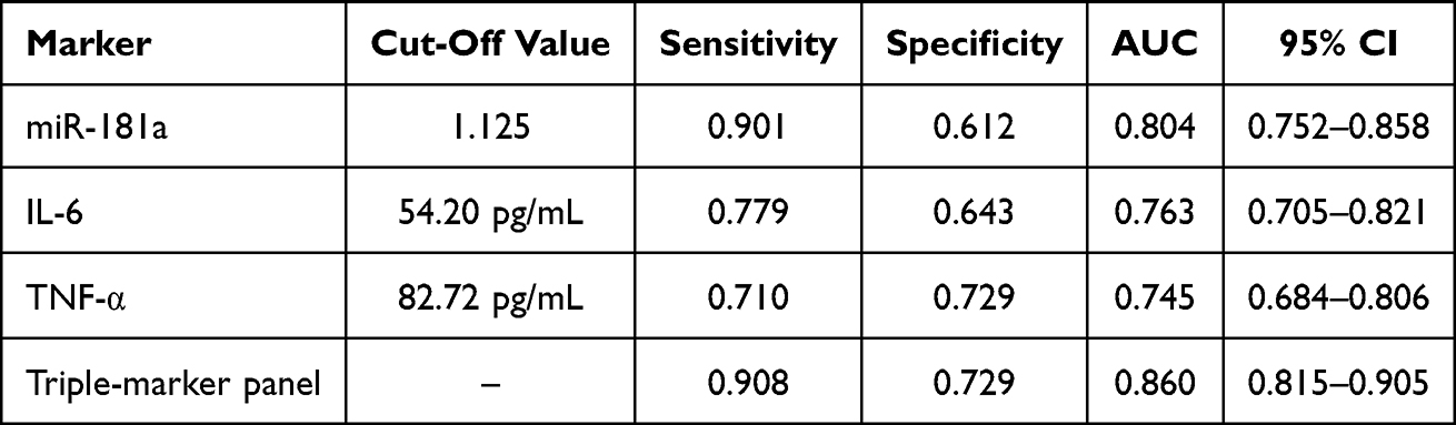 |
Table 3 Diagnostic Value of Serum miR-181a, IL-6, and TNF-α for NS |
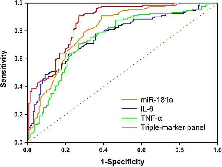 |
Figure 3 ROC curves for the diagnostic value of each marker for NS. |
Notably, the combination of all three biomarkers achieved the highest diagnostic efficacy, with an AUC of 0.860, sensitivity of 90.8%, and specificity of 72.9%. This combined model demonstrated significantly improved discriminatory power compared to any single biomarker alone, suggesting that miR-181a, IL-6, and TNF-α provide complementary information that enhances the detection of neonatal sepsis when used together.
Neurodevelopmental Outcomes at 12 Months in Neonates with Sepsis: Association with Perinatal and Inflammatory Factors
All 131 neonates diagnosed with sepsis were successfully followed up to a corrected age of 12 months. Based on the Gesell Developmental Scores, 41 infants (31.3%) were classified into the abnormal neurodevelopment group (total DQ ≤ 75), while 90 infants (68.7%) comprised the normal neurodevelopment group (DQ ≥ 86).
Comparative analysis of baseline and clinical characteristics revealed no statistically significant differences between the two groups in terms of sex distribution, mode of delivery, or admission inflammatory markers, including CRP, PCT, WBC, NLR, LMR, and PLR (all P > 0.05).
In contrast, several key perinatal factors demonstrated significant associations with neurodevelopmental outcomes. The abnormal neurodevelopment group had a significantly lower mean gestational age and birth weight compared to the normal development group. Furthermore, they exhibited lower Apgar scores at both 1 and 5 minutes, and a higher incidence of NRDS and intrauterine distress (all P < 0.05). The detailed results of these comparative analyses are presented in Tables 4 and 5.
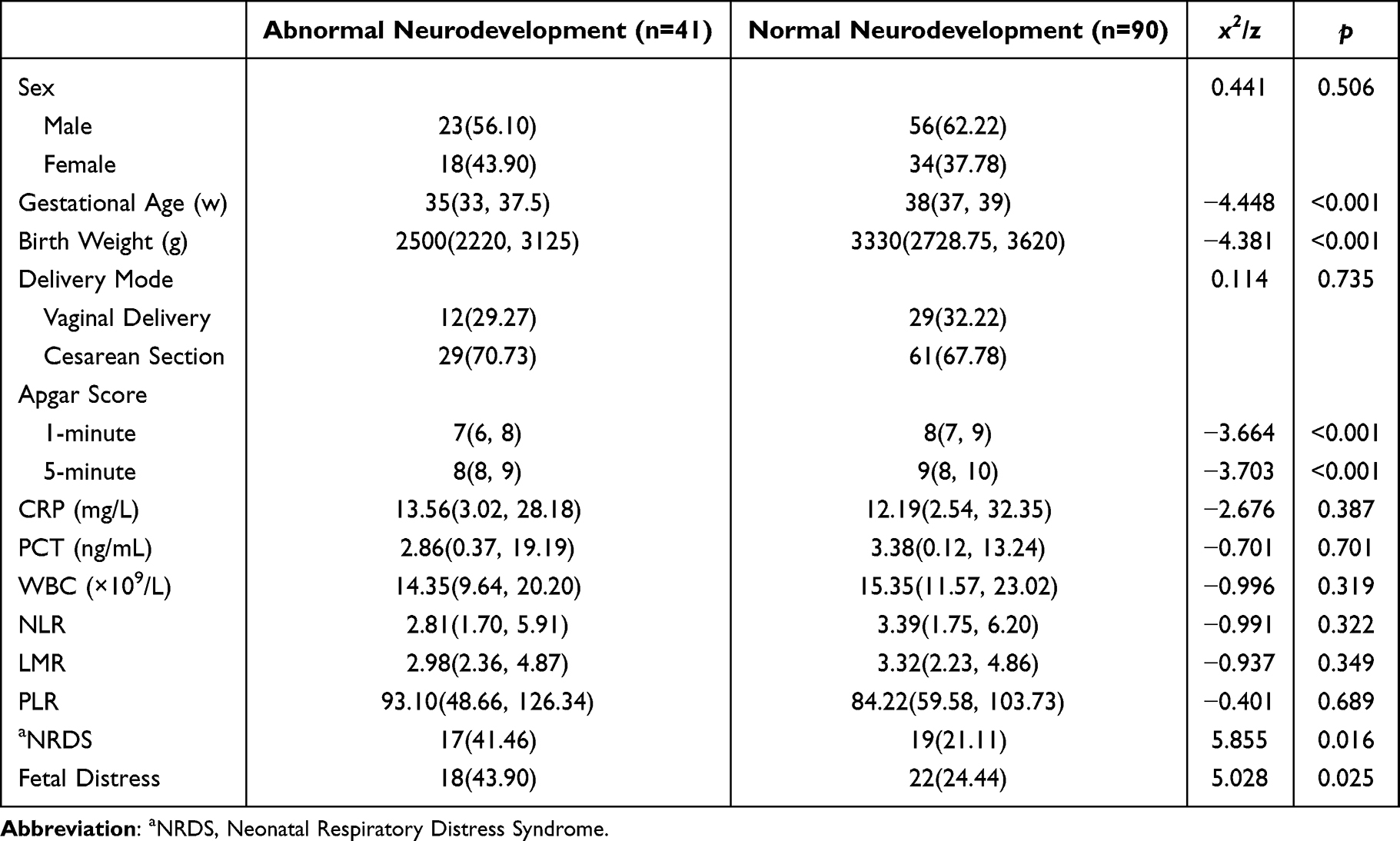 |
Table 4 Comparison of Clinical Characteristics in Septic Children with Different Neurodevelopmental Outcomes (M (P25, P75), n[%]) |
 |
Table 5 Comparison of Clinical Characteristics of Mothers of Septic Children with Different Neurodevelopmental Outcomes (n[%]) |
Distinct Serum Biomarker Profiles Associated with Neurodevelopmental Outcomes in Neonates with Sepsis
To investigate the potential link between dysregulated inflammation and impaired neurodevelopment following sepsis, we compared the expression levels of serum miR-181a, IL-6, and TNF-α between the normal and abnormal neurodevelopment (ND) groups. The relative expression level of miR-181a was significantly lower in the abnormal ND group (0.54 ± 0.45) compared to the normal group (0.79 ± 0.31) (P < 0.001; Figure 4). Consistently, the abnormal ND group exhibited markedly elevated concentrations of both IL-6 (120.48 ± 54.85 vs. 87.96 ± 48.42 in the normal group) and TNF-α (133.36 ± 46.11 vs. 97.29 ± 45.94 in the normal group) (all P < 0.01; Figure 4), reflecting a state of heightened inflammatory response.
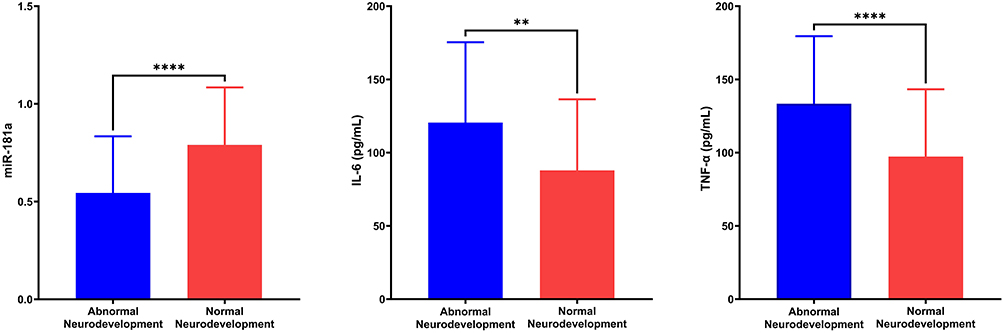 |
Figure 4 Comparative analysis of biomarkers between two groups ( Notes: ****Indicates P < 0.0001 between groups. **Indicates P < 0.01 between groups. |

Correlation Analysis Between Serum miR-181a, IL-6, TNF-α, and Gesell Scores
Pearson correlation analysis was performed to evaluate the relationships between serum biomarker levels and developmental outcomes. A significant positive correlation was observed between the relative expression of miR-181a and Gesell scores (r = 0.461, P < 0.001). In contrast, IL-6 and TNF-α levels demonstrated significant negative correlations with developmental outcomes (r = −0.389, P < 0.001 and r = −0.452, P < 0.001). Complete correlation statistics are detailed in Table 6 and Figure 5.
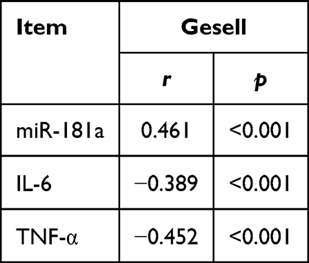 |
Table 6 Correlation Analysis Between Serum miR-181a, IL-6, TNF-α and Gesell Scores |
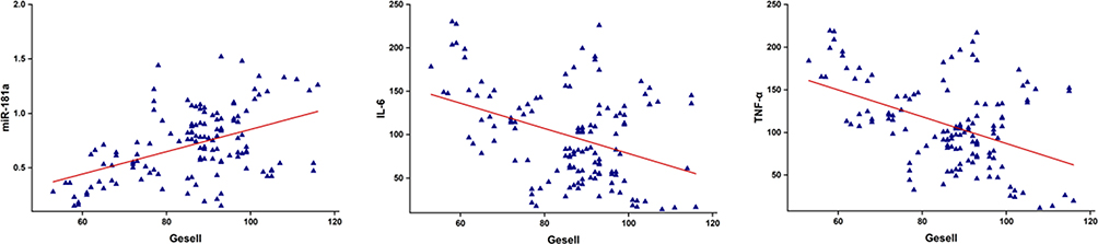 |
Figure 5 Scatter plots showing correlation between Gesell scores and levels of miR-181a, IL-6, and TNF-α. |
Feature Selection via LASSO Regression
To address multicollinearity, variables with P < 0.05 from univariate analysis underwent LASSO regression. Nine predictors retained non-zero coefficients: miR-181a, IL-6, TNF-α, Gestational Age, Prenatal Inflammation, Birth Weight, Apgar 5min, NRDS, Fetal Distress (Supplementary Figure 1C and D).
Multivariate Logistic Regression Analysis of Factors Influencing Neurodevelopmental Outcomes in Sepsis Patients
Multivariable logistic regression analysis, incorporating the robust variables identified by the LASSO model and adjusting for other clinical confounders, identified miR-181a (OR: 0.524; 95% CI: 0.415–0.662) and gestational age (OR: 0.655; 95% CI: 0.548–0.781) as protective factors against abnormal neurodevelopment in neonatal sepsis patients. Conversely, elevated IL-6 (OR: 1.079; 95% CI: 1.004–3.393), elevated TNF-α (OR: 1.130; 95% CI: 1.021–2.090), and prenatal inflammation (OR: 4.052; 95% CI: 1.609–10.205) were independent risk factors. Complete regression results are presented in Table 7.
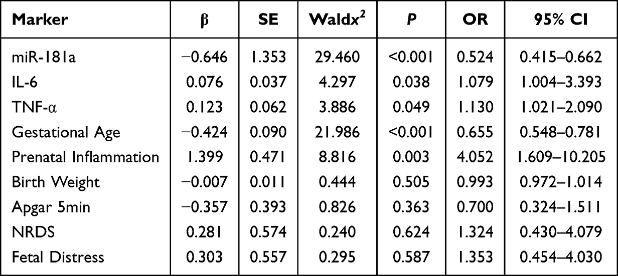 |
Table 7 Multivariate Logistic Regression Analysis of Influencing Factors for Adverse Neurodevelopment in NS |
Predictive Performance of Biomarkers for Abnormal Neurodevelopment
The diagnostic utility of miR-181a, IL-6, and TNF-α—both individually and in combination—was assessed using ROC curve analysis. The combined model integrating all three biomarkers demonstrated superior predictive efficacy, achieving an area under the curve (AUC) of 0.850, with 80.5% sensitivity and 85.6% specificity, significantly outperforming any single biomarker alone (all P < 0.05). These results underscore the complementary value of combining inflammatory and immunoregulatory markers for early risk stratification. Corresponding ROC curves and detailed performance metrics are included in Table 8 and Figure 6.
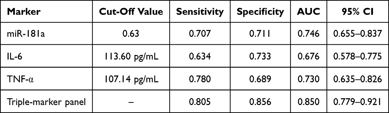 |
Table 8 Diagnostic Performance of Biomarkers for Predicting Abnormal Neurodevelopment |
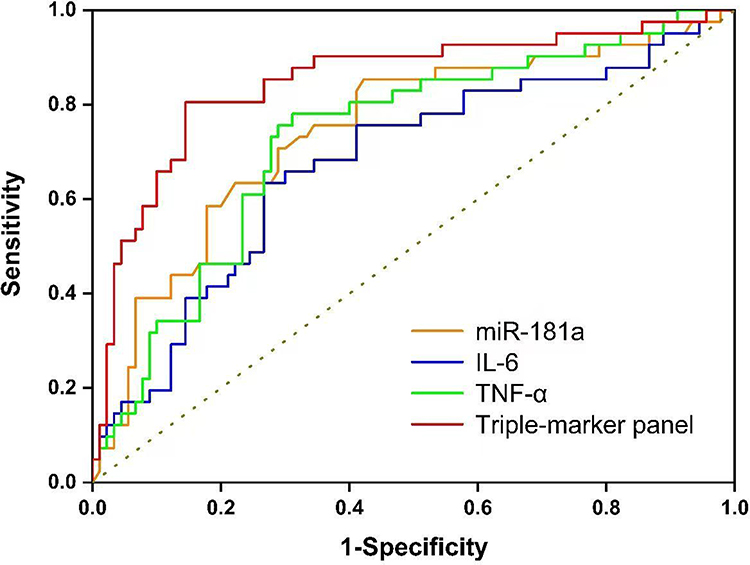 |
Figure 6 ROC curves of the biomarkers for predicting adverse neurodevelopment. |
Discussion
Neonatal sepsis remains a devastating perinatal infection. It causes high mortality and lasting neurological damage. Consequently, this disease presents a major challenge in intensive care units.13 Treatments have certainly improved. Yet, survivors still frequently suffer from cognitive, motor, and behavioral deficits. These persistent risks highlight an urgent need. We must uncover the mechanisms behind brain injury. Simultaneously, we need to develop early biomarkers and effective neuroprotective strategies. The immature immune system and weakened immune function of neonates make them highly susceptible to systemic inflammatory responses.14,15 Simultaneously, their underdeveloped blood-brain barrier allows inflammatory mediators to easily affect the central nervous system. The mechanisms of sepsis-associated brain injury are complex, involving multiple pathological processes such as ischemia-hypoxia, excitotoxicity, oxidative stress, and neuroinflammation. Systemic infection can lead to hypotension and inadequate cerebral perfusion, triggering neuronal death.16 Activated microglia release pro-inflammatory factors and reactive oxygen species under inflammatory stimulation, exacerbating neuroinflammation and ultimately resulting in white matter damage and neurodevelopmental disorders.17
Our findings link serum miR-181a, IL-6, and TNF-α to the pathology of neonatal sepsis. These biomarkers also forecast neurodevelopmental results. MiR-181a functions as a key immune regulator. It governs cell development and activation. Crucially, this microRNA provides negative feedback to dampen inflammatory responses.18 This study found that serum miR-181a was significantly downregulated in septic neonates, and its low expression was an independent risk factor for both disease occurrence and poor neurological prognosis. By targeting multiple key signaling molecules, miR-181a may participate in the negative regulation of NF-κB and inflammasome activation. Under physiological conditions, its expression helps maintain immune homeostasis and prevent excessive inflammatory responses. In neonatal sepsis, its downregulation leads to overactivation of downstream pro-inflammatory signaling pathways, resulting in the massive release of inflammatory factors such as IL-6 and TNF-α and the formation of a “exaggerated inflammatory response”.19 Additionally, miR-181a is highly expressed in the central nervous system, where it regulates synaptic plasticity, neuronal survival, and microglial polarization. Its deficiency may weaken neuronal protective mechanisms, thereby exacerbating neuroinflammation and injury.20
IL-6 and TNF-α are core effector molecules in the pathophysiology of sepsis. This study confirmed that their serum levels were significantly elevated in septic neonates and served as risk markers predicting poor outcomes. TNF-α acts as an early trigger for inflammation. It activates endothelial cells. Simultaneously, it boosts adhesion molecule expression. This process recruits and activates leukocytes. Crucially, TNF-α directly breaks down vascular tight junctions. This damage increases blood-brain barrier permeability. Consequently, peripheral inflammatory mediators invade the central nervous system.21 Within the brain, TNF-α can activate death receptors on neuronal surfaces, initiating apoptotic cascades that lead to neuronal and oligodendrocyte death.22
IL-6 is a pleiotropic cytokine with concentration- and context-dependent functions. In the early stages of acute infection, IL-6 promotes the hepatic synthesis of acute-phase proteins and is an essential component of host defense. However, persistently high levels of IL-6 are closely associated with disease severity and poor prognosis. It promotes terminal differentiation of B and T cells, amplifying adaptive immune responses. Furthermore, elevated IL-6 levels activate microglia and astrocytes, leading to the release of pro-inflammatory cytokines and neurotoxic molecules, thereby exacerbating neuroinflammation.23,24
Our multivariate analysis highlighted independent predictors for sepsis and neurodevelopment. These include NLR, prenatal inflammation, and gestational age. NLR acts as a composite marker for systemic inflammation. It captures two critical immune shifts. First, high neutrophil counts indicate an acute bacterial response. Second, low lymphocyte levels reflect stress-induced immunosuppression. This lymphocyte depletion stems primarily from cortisol surges during infection. Thus, as a composite indicator, NLR captures both the “pro-inflammatory” and “immune paralysis” aspects of sepsis pathophysiology, offering superior diagnostic value compared to simple white blood cell or neutrophil counts, consistent with recent findings highlighting the prognostic utility of inflammatory ratios.25–27 Additionally, prenatal inflammation adversely affects fetal brain development. Maternal inflammatory mediators cross the placenta into the fetal circulation, activating immune cells in the brain, inducing systemic inflammation, disrupting the blood-brain barrier, prompting premature microglial activation, and interfering with critical processes like neurogenesis.5 A synergistic effect occurs if neonatal sepsis develops postnatally. This study also confirmed that higher gestational age is a significant protective factor against both neonatal sepsis and poor neurological outcomes, as more mature infants have better developed cardiovascular, pulmonary, and immune systems. Furthermore, gestational age directly influences brain maturity, promoting the formation of neuronal networks and endogenous neuroprotective mechanisms.28
Understanding how peripheral inflammation leads to central nervous system damage is key to unraveling sepsis-associated encephalopathy. The factors identified in this study bridge systemic infection and brain injury. High levels of TNF-α and IL-6 are primary effectors disrupting blood-brain barrier integrity. TNF-α increases barrier permeability by inducing endothelial cell contraction and downregulating tight junction protein expression. IL-6 exacerbates vascular leakage by activating intracellular signaling pathways that promote the production of factors like vascular endothelial growth factor in endothelial cells. This breach allows serum proteins, inflammatory cells, and cytokines to enter the brain parenchyma, directly exposing neurons and glial cells.29 Brain function relies on the intricate collaboration within the “neurovascular unit,” comprising neurons, blood-brain barrier endothelial cells, pericytes, astrocytic end-feet, and microglia.30 Systemic inflammation and oxidative stress in sepsis can cause pericyte contraction, aberrant capillary perfusion, and local microcirculatory dysfunction. Disrupted astrocyte function impairs their ability to maintain ionic homeostasis and uptake synaptic glutamate, potentially leading to excitotoxicity.31
In clinical practice, combined early detection of these biomarkers upon admission could enable rapid disease identification and risk stratification. For high-risk infants, aggressive anti-infective therapy is essential, and they should be primary targets for neuroprotective strategies. This may involve early comprehensive neurological monitoring and exploring the feasibility of adjunctive anti-inflammatory treatments and neurotrophic support.32 Post-discharge, these infants should be integrated into high-risk follow-up programs to ensure timely rehabilitative assessment and intervention. Future research should include multicenter prospective cohort studies to validate the predictive efficacy of these biomarkers, employ serial sampling to map molecular dynamics, utilize cell and animal models to verify their causal roles in inflammation regulation and neuronal injury, and integrate multi-omics analysis with advanced neuroimaging to build a comprehensive understanding from molecule to phenotype.33
In summary, this study systematically elucidates the roles of miR-181a, IL-6, and TNF-α in the pathogenesis of neonatal sepsis and subsequent brain injury. Low miR-181a expression coupled with high IL-6 and TNF-α levels not only reflects disease severity but is also closely associated with long-term neurodevelopmental outcomes. The combined detection of these biomarkers provides a novel tool for the early identification of high-risk neonates and the implementation of personalized interventions, holding significant clinical importance for improving the long-term prognosis of neonatal sepsis.
Limitations and Future Perspectives
Several limitations warrant acknowledgment. First, excluding infants <32 weeks gestation limits generalizability to the highest-risk very preterm population. Second, sepsis diagnosis relied primarily on clinical criteria, with only 11.5% culture-confirmed—reflecting real-world practice but potentially compromising diagnostic specificity. Third, unmeasured confounders (eg, maternal fever, delivery-related inflammatory) may independently influence cytokine profiles. Fourth, limited sample size precluded stratified analyses of early- versus late-onset sepsis, which have distinct pathophysiology. Fifth, the absence of neuroimaging data (cranial ultrasound/MRI) limits our ability to draw firm conclusions about brain injury. Sixth, the Gesell Developmental Scale, while informative, has less established psychometric properties than the Bayley Scales, potentially underestimating subtle deficits. Finally, technical barriers currently limit routine miRNA implementation, though advancing PCR technologies are progressively mitigating these challenges.
Future research requires prospective multicenter cohorts including infants <32 weeks to ensure generalizability across gestational ages. Stratified enrollment by gestational age, onset type (early-onset vs. late-onset), and culture status (culture-positive vs. culture-negative) will enable granular subgroup analyses and tailored diagnostic algorithms. External validation in independent populations is underway through multicenter collaborations. Longitudinal follow-up with Bayley-IV assessments at 18 and 24 months corrected age will capture developmental trajectories and the full continuum of neurodevelopmental impairment. Multimodal neuroimaging (cranial ultrasound/MRI) will provide essential structural correlates. Mechanistic studies (in vitro/in vivo) are needed to delineate the functions of these biomarkers and identify therapeutic targets, which we are currently pursuing. As PCR-based miRNA detection becomes rapid and cost-effective, point-of-care applications warrant investigation. These integrated efforts will address current limitations and advance precision-based risk stratification and targeted intervention in neonatal sepsis.
Conclusion
This study provides compelling evidence that dysregulation of key immunomodulatory and inflammatory mediators—specifically decreased miR-181a expression and elevated IL-6 and TNF-α levels—plays a central role in the pathophysiology of neonatal sepsis and its associated neurodevelopmental sequelae. The significant correlations observed between these biomarkers and Gesell developmental scores, together with their identification as independent risk factors in multivariate analysis, underscore their dual utility as indicators of both disease severity and neurological vulnerability.
The integration of these biomarkers into a combined predictive model demonstrated superior diagnostic and prognostic performance compared to any single marker alone, offering a clinically feasible approach for early identification of high-risk neonates. This multi-parameter strategy captures the complex interplay between systemic inflammation and neurological injury, reflecting both the pro-inflammatory drive and impaired immunoregulation characteristic of sepsis-related brain injury.
These findings have important clinical implications. The assessment of miR-181a, IL-6, and TNF-α upon admission could facilitate timely risk stratification, enabling more targeted neuroprotective interventions and personalized follow-up strategies for vulnerable infants. Future research should focus on validating these biomarkers in larger multicenter cohorts, elucidating their precise mechanistic roles in blood-brain barrier disruption and neuronal injury, and exploring their potential as therapeutic targets to improve long-term neurodevelopmental outcomes in neonatal sepsis survivors.
Funding
Project Title: Jiangsu Provincial Health Commission Maternal and Child Health Research Project (No. F202331); Jiangsu Provincial Maternal and Child Health Association Research Project (No. FYX202334); Jiangsu Provincial Medical Association Pediatric Medicine Research Project (No. SYH-32034-0076(20230023)).
Disclosure
The authors report no conflicts of interest in this work.
References
1. De Souza DC, Paul R, Mozun R, et al. Quality improvement programmes in paediatric sepsis from a global perspective. Lancet Child Adolesc Health. 2024;8(9):695–14. doi:10.1016/S2352-4642(24)00142-1
2. Shane AL, Sánchez PJ, Stoll BJ. Neonatal sepsis. Lancet. 2017;390(10104):1770–1780. doi:10.1016/S0140-6736(17)31002-4
3. Barboza AZ, Flannery DD, Shu D, et al. Trends in C-reactive protein use in early-onset sepsis evaluations and associated antibiotic use. J Pediatr. 2024;273:114153. doi:10.1016/j.jpeds.2024.114153
4. Huang C, Chen J, Zhan X, et al. Clinical value of laboratory biomarkers for the diagnosis and early identification of culture-positive sepsis in neonates. JIR. 2023;16:5111–5124. doi:10.2147/JIR.S419221
5. Serdar M, Walther KA, Gallert M, et al. Prenatal inflammation exacerbates hyperoxia-induced neonatal brain injury. J Neuroinflammation. 2025;22(1):57. doi:10.1186/s12974-025-03389-4
6. Fatmi A, Chabni N, Cernada M, et al. Clinical and immunological aspects of microRNAs in neonatal sepsis. Biomed Pharmacother. 2022;145:112444. doi:10.1016/j.biopha.2021.112444
7. Kim C, Ye Z, Weyand CM, Goronzy JJ. miR-181a-regulated pathways in T-cell differentiation and aging. Immun Ageing. 2021;18(1):28. doi:10.1186/s12979-021-00240-1
8. Zhang H, Wang J, Qu Y, et al. Brain injury biomarkers and applications in neurological diseases. Chinese Med J. 2025;138(1):5–14. doi:10.1097/CM9.0000000000003061
9. Guo Q, Jin Y, Chen X, et al. NF-κB in biology and targeted therapy: new insights and translational implications. Sig Transduct Target Ther. 2024;9(1):53. doi:10.1038/s41392-024-01757-9
10. Subspecialty Group of Neonatology, the Society of Pediatric, Chinese Medical Association, et al. Expert consensus on the diagnosis and management of neonatal sepsis (version 2019). Zhonghua Er Ke Za Zhi. 2019;57(4):252–257. doi:10.3760/cma.j.issn.0578-1310.2019.04.005
11. Liu D, Duan M, Chen C, et al. Efficacy analysis of comprehensive rehabilitation therapy based on the Gesell developmental schedules among infants with global developmental delay of different ages: a retrospective study. Front Neurol. 2025;16:1568643. doi:10.3389/fneur.2025.1568643
12. Zhang C, Li X, Liu N, et al. MicroRNA-96 is downregulated in sepsis neonates and attenuates LPSInduced inflammatory response by inhibiting IL-16 in monocytes. CCHTS. 2021;25(1):90–96. doi:10.2174/1386207323666201211091312
13. Kim F, Polin RA, Hooven TA. Neonatal sepsis. BMJ. 2020;m3672. doi:10.1136/bmj.m3672
14. Dong Y, Basmaci R, Titomanlio L, et al. Neonatal sepsis: within and beyond China. Chinese Med J. 2020;133(18):2219–2228. doi:10.1097/CM9.0000000000000935
15. Liang X, Luo H, Chen X, et al. Prognostic value of blood inflammatory parameters for predicting mortality in neonates with sepsis. Int Immunopharmacol. 2023;123:110780. doi:10.1016/j.intimp.2023.110780
16. Zhang Y, Shan Z, Zhao Y, et al. Sevoflurane prevents miR-181a-induced cerebral ischemia/reperfusion injury. Chem Biol Interact. 2019;308:332–338. doi:10.1016/j.cbi.2019.06.008
17. Mohamed Ahmed Hamid SM, Ibrahim Shaban KS, Abdulhamid Abdulaziz SB, et al. The impact of neonatal sepsis on long-term neurodevelopment: a systematic review of cognitive and sensory outcomes. Cureus. 2025. doi:10.7759/cureus.90176
18. Indrieri A, Carrella S, Carotenuto P, et al. The pervasive role of the miR-181 family in development, neurodegeneration, and cancer. IJMS. 2020;21(6):2092. doi:10.3390/ijms21062092
19. Hutchison ER, Kawamoto EM, Taub DD, et al. Evidence for miR‐181 involvement in neuroinflammatory responses of astrocytes. Glia. 2013;61(7):1018–1028. doi:10.1002/glia.22483
20. Agostini S, Bolognesi E, Mancuso R, et al. miR-23a-3p and miR-181a-5p modulate SNAP-25 expression. PLoS One. 2023;18(1):e0279961. doi:10.1371/journal.pone.0279961
21. Lawrence T. The nuclear factor NF- B pathway in inflammation. Cold Spring Harbor Perspect. Biol. 2009;1(6):a001651. doi:10.1101/cshperspect.a001651
22. Brehmer F, Bendix I, Prager S, et al. Interaction of inflammation and hyperoxia in a rat model of neonatal white matter damage. PLoS One. 2012;7(11):e49023. doi:10.1371/journal.pone.0049023
23. Varga NI, Bagiu IC, Vulcanescu DD, et al. IL-6 baseline values and dynamic changes in predicting sepsis mortality: a systematic review and meta-analysis. Biomolecules. 2025;15(3):407. doi:10.3390/biom15030407
24. Kerkis I, Silva ÁPD, Araldi RP. The impact of interleukin-6 (IL-6) and mesenchymal stem cell-derived IL-6 on neurological conditions. Front Immunol. 2024;15:1400533. doi:10.3389/fimmu.2024.1400533
25. Xin Y, Shao Y, Mu W, et al. Accuracy of the neutrophil-to-lymphocyte ratio for the diagnosis of neonatal sepsis: a systematic review and meta-analysis. BMJ Open. 2022;12(12):e060391. doi:10.1136/bmjopen-2021-060391
26. Keskin A, Aci R. Procalcitonin to albumin ratio as a biomarker for predicting mortality in sepsis. J Coll Physicians Surg Pak. 2024;34(3):360–363. doi:10.29271/jcpsp.2024.03.360
27. Arı HF, Keskin A, Arı M, et al. Importance of lactate/albumin ratio in pediatric nosocomial infection and mortality at different times. Future Microbiol. 2024;19(1):51–59. doi:10.2217/fmb-2023-0125
28. Afonso EDP, Blot S. Effect of gestational age on the epidemiology of late-onset sepsis in neonatal intensive care units - a review. Exp Rev Anti-Infect Ther. 2017;15(10):917–924. doi:10.1080/14787210.2017.1379394
29. Grebenciucova E, VanHaerents S. Interleukin 6: at the interface of human health and disease. Front Immunol. 2023;14:1255533. doi:10.3389/fimmu.2023.1255533
30. Muoio V, Persson PB, Sendeski MM. The neurovascular unit - concept review. Acta Physiol. 2014;210(4):790–798. doi:10.1111/apha.12250
31. Jin C, Londono I, Mallard C, et al. New means to assess neonatal inflammatory brain injury. J Neuroinflammation. 2015;12(1):180. doi:10.1186/s12974-015-0397-2
32. Kelly SB, Gunn AJ, Hunt RW, et al. What is the pathophysiology of inflammation-induced cortical injury in the perinatal brain? Neural Regenerat Res. 2026;21(2):502–505. doi:10.4103/NRR.NRR-D-24-01091
33. Celik IH, Hanna M, Canpolat FE, et al. Diagnosis of neonatal sepsis: the past, present and future. Pediatr Res. 2022;91(2):337–350. doi:10.1038/s41390-021-01696-z
 © 2026 The Author(s). This work is published and licensed by Dove Medical Press Limited. The
full terms of this license are available at https://www.dovepress.com/terms
and incorporate the Creative Commons Attribution
- Non Commercial (unported, 4.0) License.
By accessing the work you hereby accept the Terms. Non-commercial uses of the work are permitted
without any further permission from Dove Medical Press Limited, provided the work is properly
attributed. For permission for commercial use of this work, please see paragraphs 4.2 and 5 of our Terms.
© 2026 The Author(s). This work is published and licensed by Dove Medical Press Limited. The
full terms of this license are available at https://www.dovepress.com/terms
and incorporate the Creative Commons Attribution
- Non Commercial (unported, 4.0) License.
By accessing the work you hereby accept the Terms. Non-commercial uses of the work are permitted
without any further permission from Dove Medical Press Limited, provided the work is properly
attributed. For permission for commercial use of this work, please see paragraphs 4.2 and 5 of our Terms.