Back to Journals » Diabetes, Metabolic Syndrome and Obesity » Volume 18
Mesoporous Silica Nanoparticle as a Potential Nanocarrier to Improve the Effectiveness of Antiobesity Drugs
Authors Aulifa DL ![]() , Nurulaini SN, Ayodduki ZNSH, Anggi JF, Chandra JH
, Nurulaini SN, Ayodduki ZNSH, Anggi JF, Chandra JH ![]() , Budiman A
, Budiman A
Received 6 May 2025
Accepted for publication 5 September 2025
Published 4 October 2025 Volume 2025:18 Pages 3751—3770
DOI https://doi.org/10.2147/DMSO.S538644
Checked for plagiarism Yes
Review by Single anonymous peer review
Peer reviewer comments 2
Editor who approved publication: Dr Donald McClain
Diah Lia Aulifa,1 Siti Nunung Nurulaini,1 Zahra Noor Silmi Hermawan Ayodduki,1 Joseph Fide Anggi,1 Joshua Harry Chandra,2 Arif Budiman2
1Department of Pharmaceutical Analysis and Medicinal Chemistry, Faculty of Pharmacy, Universitas Padjadjaran, Sumedang, 45363, Indonesia; 2Department of Pharmaceutics and Pharmaceutical Technology, Faculty of Pharmacy, Universitas Padjadjaran, Sumedang, 45363, Indonesia
Correspondence: Diah Lia Aulifa, Department of Pharmaceutical Analysis and Medicinal Chemistry, Faculty of Pharmacy, Universitas Padjadjaran, Sumedang, 45363, Indonesia, Email [email protected]
Abstract: Obesity is a global health issue characterized by an abnormal or excessive accumulation of body fat, defined as a body mass index (BMI) that exceeds 30 kg/m². Many antiobesity drugs, such as orlistat and quercetin, are often limited by poor solubility and bioavailability. These physicochemical limitations can lead to poor gastrointestinal absorption, erratic plasma drug concentrations, and ultimately, suboptimal therapeutic efficacy. This review employed a narrative approach with a structured literature search to summarize current advances in the use of mesoporous silica nanoparticles (MSNs) as drug delivery systems to improve the dissolution and pharmacological profiles of antiobesity compounds. The literature search was conducted using PubMed and Scopus databases with the keywords “obesity OR antiobesity AND mesoporous silica”. The search included free full-text articles from the past ten years. A total of 106 articles were screened, and 11 were selected based on relevance to MSN-based delivery in in vitro or in vivo obesity studies. MSNs enhanced the dissolution profile of various antiobesity compounds, including orlistat, quercetin, epigallocatechin gallate, and liraglutide. In vitro assays showed improved lipase inhibition, adipogenesis suppression, and DPP-4 enzyme inhibition. In vivo studies demonstrated significant improvements in triglyceride levels, blood glucose levels, body weight reduction, and food intake suppression. Functionalization of MSNs further enabled targeted and pH-responsive delivery, enhancing bioavailability and reducing systemic side effects. MSNs offer a promising nanocarrier platform to overcome limitations of conventional antiobesity therapies by improving drug solubility, stability, and targeted pharmacological activity. These findings support the potential of MSN-based formulations as innovative strategies in obesity treatment and need for further studies.
Keywords: antiobesity, mesoporous silica, nanocarrier, dissolution, bioavailability
Introduction
Obesity is a major global health issue, with the World Obesity Foundation (2024) reporting that one billion individuals are obese and 2.5 billion have elevated body mass index (BMI) levels. It is characterized by an abnormal or excessive accumulation of body fat, typically diagnosed when BMI exceeds 30 kg/m².1,2 Treatment includes using medication in addition to lifestyle changes.3 Antiobesity medications (AOMs), including intra gastrointestinal medications (orlistat), centrally acting medications (phentermine, phentermine-topiramate, naltrexone-bupropion), and nutrient-stimulated hormone-based medications (liraglutide, semaglutide, tirzepatide), have been developed to support weight loss by addressing appetite dysregulation.4–6 However, their clinical use is often limited by issues such as modest long-term efficacy, gastrointestinal side effects, lean mass loss, high costs, and societal misconceptions about obesity treatment.4,7 Furthermore, certain AOMs, such as orlistat, exhibit low aqueous solubility, categorized under BCS Class II, which further restricts their therapeutic potential.8,9
Mesoporous silica nanoparticles (MSNs) have emerged as a promising nanocarrier to enhance the effectiveness of antiobesity medications by addressing solubility and bioavailability limitations. MSNs can enhance drug solubility, prevent medications from early degradation, and facilitate targeted and controlled delivery of drugs because to their high surface area, large pore volume, and adjustable surface characteristics.10–12 Compared to other nanocarriers commonly explored in obesity research, such as liposomes, polymeric nanoparticles, and lipid-based systems, which often faces challenges like structural instability, rapid systemic clearance, low drug-loading capacity, or complex formulation requirement, MSNs offer superior thermal and chemical stability, tunable pore size for precise drug loading, and enhanced control over drug release kinetics. These features make MSNs a more versatile and robust platform for overcoming the solubility and bioavailability challenges associated with antiobesity medications.13–15 Through targeted delivery, MSNs have the potential to increase therapeutic efficacy of AOMs while minimizing systemic side effects, offering a strategic solution to the limitations faced by conventional antiobesity therapies.16 Therefore, this review aims to present current insights, summarize, and elucidate the mechanism of antiobesity drugs within MSNs, focusing on the improvements in dissolution profiles, in vitro, and in vivo activity to highlight their potential as innovative strategies for advancing obesity treatment.
Methodology
Briefly, a literature search using the (1) PubMed database using the keywords obesity OR antiobesity AND mesoporous silica within Abstract, free full text, in the last 10 years, and resulted in n = 14; (2) in the Scopus database using the same keywords within All fields, resulted in n = 92, with a total of all publications found through these searches were reviewed. The authors retrieved and evaluated all documents that met the inclusion criteria, such as using MSN for in vitro or in vivo study in treating obesity. Finally, 11 articles were selected for review, as depicted in Figure 1.
 |
Figure 1 Flow chart of the methodology. |
Pathophysiological Pathway of Obesity
The pathophysiological pathway of obesity is presented in Figure 2. At its core, one of the factors that causes overweight, and obesity is chronic imbalance between energy intake and expenditure.17–19 A diet high in fats and calories, when combined with insufficient physical activity, disrupts the body’s energy balance, ultimately leading to excessive fat accumulation in adipose tissue, including in the heart, pancreas, and kidneys.20–22
 |
Figure 2 Pathophysiology pathways of obesity and its associated comorbidities.19,23,24 |
As adipose tissue expands, it initiates a series of physiological disturbances, notably affecting the hormonal regulation of appetite.25 Appetite-regulating hormones are critical for maintaining energy homeostasis through the modulation of hunger and satiety signals directed to the central nervous system. In the context of obesity, this regulatory system becomes markedly impaired, primarily due to the development of leptin resistance and elevated ghrelin secretion.26 Leptin, a hormone secreted by adipocytes, ordinarily acts to suppress appetite and signal energy sufficiency to hypothalamic center.27 However, individuals with obesity experience attenuated central leptin signaling despite elevated leptin levels, which results in diminished satiety perception and persistent hyperphagia. At the same time, ghrelin, a strong hunger hormone produced by the gastric mucosa, stays unusually high, which increases the urge to eat even more.23,28 This hormonal dysregulation perpetuates excessive food intake, promotes further adipose tissue expansion, and accelerates the progression of metabolic dysfunction.29
The chronic overconsumption of energy-dense foods contributes to elevated levels of saturated fatty acids and promotes insulin resistance, a key metabolic abnormality wherein cells become less responsive to insulin, resulting in hyperglycemia.30,31 This metabolic issue is strongly connected to ongoing low-level inflammation, which is marked by higher levels of pro-inflammatory cytokines like interleukin-6 (IL-6) and tumor necrosis factor-alpha (TNF-α). These cytokines interfere with insulin signaling pathways and contribute to endothelial dysfunction, collectively exacerbating the risk of obesity-related complications.32,33
Ultimately, this complex interplay of hormonal imbalance, metabolic dysfunction, and persistent inflammation underlies the development of obesity and its associated comorbidities, including atherosclerosis, type 2 diabetes mellitus (T2DM), and various liver diseases such as non-alcoholic fatty liver disease (NAFLD).24,34 Importantly, this conceptual framework shifts the understanding of obesity from being seen solely as a result of lifestyle choices to recognizing it as a complex systemic disorder with wide-ranging health impacts.35 As a result, effective prevention and management of obesity require comprehensive, multidisciplinary approaches that address not only behavioral changes but also the underlying hormonal imbalances and inflammatory processes.24
Mesoporous Silica Nanoparticles (MSNs)
Formulation development strategies to improve the solubility of oral drugs have been carried out, including salt formation, cocrystals, emulsion systems, nanotechnology, amorphous solid dispersions, and mesoporous silica nanoparticles.36,37 Mesoporous silica nanoparticles (MSNs) have become highly effective carriers in medicinal applications because of their distinct structural and functional properties. According to in vivo investigations on its absorption, distribution, and excretion, silica is “Generally Recognized as Safe” (GRAS) by the FDA and exhibits excellent biocompatibility for oral and intravenous uses.38,39 Amorphous silica is preferred in pharmaceuticals over crystalline silica due to its reduced toxicity and enhanced capacity for controlled drug loading and release, facilitated by a high surface area (>700 m²/g) and large pore volume (>1 cm³/g).38,40 MSNs may load various compounds, including proteins, genes, and medicines, owing to their controlled properties, which include rich silanol groups, low-density solids, variable selectivity, and changeable surface activity.41 Additionally, to improve loading capacity and optimize release profiles, these nanoparticles can be functionalized with certain groups.42 Furthermore, drug loading, release characteristics, and targeted delivery efficiency can be optimized through the functionalization of MSNs’ outer surface as well as their interior cylindrical pores.43,44 These nanoparticles are versatile in terms of their synthesis conditions, enabling the creation of different mesoporous carriers like MCM-41, MCM-48, SBA-15, and SBA-16, each of which has distinct morphological and structural characteristics appropriate for particular uses.42,43
Mesoporous silica is a promising material for improving the dissolution rates of poorly water-soluble drugs.10,11 Drugs can be contained in its pores in a stabilized amorphous form, preventing crystallization by nanoconfinement, because of its high surface area, which frequently exceeds 300 m²/g, and special porous architecture.11 Drug molecules are kept in an amorphous state by spatial confinement, which prevents crystallization via both thermodynamic equilibrium and kinetic barriers, especially when pore widths are less than the critical nuclei size.10,45 Additionally, drugs can more easily adsorb onto MSNs’ surface due to its high surface-free energy, which stabilizes the system by lowering its free energy state. When the encapsulated medication is exposed to water, it quickly separates from the silica surface and forms supersaturated solutions, which greatly accelerate the rate of dissolution.45 The presence of silanol groups on the MSN surface enables further surface modifications to optimize drug interactions and enhance dissolution behavior.11
Previous studies have demonstrated the potential of MSN in improving the bioavailability of poorly water-soluble drugs. In a proof-of-concept clinical study, Bukara et al (2016) evaluated the bioavailability of fenofibrate, comparing a formulation loaded onto ordered mesoporous silica (OMS) with a commercially available micronized formulation. Results showed a significant enhancement in pharmacokinetic parameters: a 54% increase in AUC0–24h/dose, a 77% increase in Cmax/dose, and a reduction in Tmax by 0.75 hours for the OMS-based formulation.46 This study indicated that administration of an ordered mesoporous silica-based formulation of fenofibrate resulted in an increased rate and extent of absorption when compared to a marketed product, demonstrating the bioavailability-enhancing potential of this novel formulation. Similarly, Laine et al (2016) investigated the oral delivery of celecoxib using mesoporous silica co-loaded with a precipitation inhibitor. This novel formulation achieved a 15-fold solubility increase in vitro and a 1.35-fold increase in Cmax in vivo after oral dosing in rats, compared to the crystalline celecoxib.47 Besides its ability to increase medication bioavailability, experimental studies have also shown that mesoporous silica can enhance pharmacological activity. A study conducted by Elbially et al (2020) shows that antitumor activities against HepG2 and HeLa cells of curcumin-loaded PEGylated MSNs (mesoporous silica nanoparticles) increased more than free curcumin. The cytotoxicity results showed that Cur-loaded PEGylated MSNs (Cur concentration = 36 mg mL−1) had cell viability of less than 10% and about 7% after 24 and 48 hours, respectively, whereas HepG2 cells treated with free Cur had cell viability of less than 76% after 48 hours at the same concentration of Cur (Cur concentration = 36 mg mL−1).48 This approach represents a novel formulation strategy to maximize in vivo exposure to poorly soluble drugs that are critical for discovery.
Antiobesity Compound Loaded into Mesoporous Silica Nanoparticles
Previous studies have reported the loading of antiobesity compounds into mesoporous silica as shown in Table 1.
 |
Table 1 The Studies of Antiobesity Compound Loaded MSN |
Characterization
Common Methods
Various analytical techniques are commonly used to characterize drug-loaded mesoporous silica nanoparticles (MSNs). Differential scanning calorimetry (DSC) characterizes drug-loaded MSNs by analyzing melting behavior. Encapsulation lowers the drug’s melting point and glass transition temperature, and complete confinement prevents recrystallization, ensuring an amorphous state.38 Nitrogen adsorption-desorption analysis is a widely used technique for characterizing mesoporous materials, providing key information on surface area, pore volume, and pore diameter of mesoporous silica nanoparticles (MSNs).60,61 This method precisely quantifies the amount of gas adsorbed onto the material, offering direct insights into its porosity and structural properties.38 Thermogravimetric analysis (TGA) determines drug content in MSNs by measuring mass loss during controlled heating, which reflects drug degradation and desorption of volatile components.62 Powder X-ray Diffraction (PXRD) confirms drug amorphization after encapsulation in MSNs. A successfully loaded drug exhibits a halo pattern without diffraction peaks, while the pure crystalline drug shows sharp peaks specific to its lattice structure.38 FTIR is used to determine drug-silica interactions and analyze the chemical state of drugs in MSNs. It identifies functional groups by detecting peak distributions caused by infrared radiation.38,63,64 Microscopic techniques, including Scanning Electron Microscopy (SEM) and Transmission Electron Microscopy (TEM), provide structural characterization, with SEM analyzing surface morphology with nanometer resolution,65 while TEM provides atomic-scale imaging, revealing internal microstructures. Both can be used separately or together; they offer comprehensive structural insights into MSNs.38,65,66
Dynamic Light Scattering
Dynamic Light Scattering (DLS) is a technique used to characterize particle size, size distribution, and the degree of agglomeration within a system. This method measures the intensity of scattered light at a fixed angle for different sample concentrations and compares it with the scattering pattern of a standard.67 However, DLS has limitations, including low resolution and inefficiency in analyzing highly concentrated samples.68
Researchers commonly use DLS to determine the hydrodynamic diameters of mesoporous silica nanoparticles (MSNs), which provides both the frequency distribution and the cumulative particle-size distribution. Research by Jin et al (2024) found that heparin-modified mesoporous silica nanoparticles (MSN-HP) and unmodified MSNs exhibited no significant difference in size, indicating that functionalization did not alter the particle size. Moreover, DLS is also useful for assessing the successful adsorption of LDL onto MSN-HP, as evidenced by the significant increase in particle size.57
Photoluminescence (PL) Spectroscopy
Photoluminescence (PL) spectroscopy is used to study optical properties of ~1 nm-sized silica layers and electronic properties of MSNs. This technique provides insights into the radiative recombination process in MSNs (Glinka et al, 2002). PL spectroscopy has been employed to confirm successful surface modifications and investigate the structural properties of MSNs. For instance, the grafting of fluorescent mesoporous silica nanoparticles (FMSNs) and the subsequent polydopamine (PDA) coating (FMSNs-PDA) were confirmed by distinct emission peaks in the PL spectra. Additionally, the presence of the PDA layer partially absorbs and blocks the fluorescent light emitted by FMSNs upon excitation. Moreover, PL spectroscopy enables real-time fluorescent monitoring of drug delivery routes and can confirm the successful transport of the drug to the target area.50
Zeta Potential
The zeta potential (ZP), which measures the electrostatic attraction or repulsion between nanoparticles, is frequently used to evaluate surface charge and colloidal stability by quantifying the particle’s electrical charge.38,67 A general threshold for distinguishing between unstable and stable suspensions is ±30 mV, with particles having zeta potentials beyond these limits typically considered electrostatically stable.69 This method is widely applied in the preparation of MSNs to ensure stability, verify successful modifications, and prevent aggregation.38 The successful heparinization modification of MSNs was confirmed by a significant shift in surface potential.57
XPS
X-ray photoelectron spectroscopy (XPS) is a surface-sensitive analytical technique that involves bombarding a material’s surface with X-rays and measuring the kinetic energy of emitted electrons.70 This method is widely employed to evaluate surface interactions between grafted compounds and silica nanoparticles. By providing quantitative information on the elemental composition and chemical states of material surfaces, XPS serves as a crucial tool for characterizing surface modifications and functionalization.71 Jin et al (2024) utilized XPS to analyze potential interactions in heparin-modified periodic mesoporous silica nanoparticles (PMS-HP) during LDL adsorption. The analysis revealed the emergence of a distinct peak following LDL adsorption, indicating that N–H and O–H bonds played a key role in the adsorption process.57
Dissolution Study
The dissolution study assessed the behavior of medicines in antiobesity drug-loaded MSNs following dispersion in dissolution media. The dissolution study revealed that MSNs significantly enhanced the rate of antiobesity drugs dissolving compared to the drugs alone. Understanding dissolution behavior is crucial for solid formulation development and regulatory assessment. Yu et al found that the release of glimepiride (GLM) from mesoporous hollow silica nanospheres (MSNs) was significantly higher and faster (80.6% in 15 min) compared to GLM alone, which was only 33.6% in 1 hour. This shows that the MSNs significantly improve the releasing rate of the antiobesity drug. MSNs can be nanocarriers that have pH-responsive drug release behavior. A study by Geng et al showed the cumulative amount of liraglutide (Lira) released from MSNs is approximately 15% within 48 h in a neutral environment (pH 7.4), while it can reach 30% at pH 5.0.53
In vitro Study
Lipase Inhibition and Oil Adsorption Test
The most attractive and safe target for antiobesity treatment is to alter lipid metabolism by inhibiting dietary fat using pancreatic lipase.72,73 Pancreatic lipase is the main lipolytic enzyme secreted by the pancreas and plays an important role in fat digestion. Agents that can inhibit the action of gastrointestinal lipase will reduce fat absorption.74 In clinical practice, orlistat is the only treatment used as a pancreatic lipase inhibitor.72,75,76 Orlistat inhibits gastric and pancreatic lipases and carboxylesterases by reacting with the catalytic serine residues of these enzymes.
Park et al conducted an in vitro study on lipase inhibition and compared raw orlistat, commercial products, and orlistat encapsulated in mesoporous silica; the results showed a significant increase in orlistat encapsulated in mesoporous silica in inhibiting lipase. Compared with raw orlistat and commercial products, orlistat encapsulated in mesoporous silica showed 53% and 12% more lipase inhibition, respectively. This decrease is in line with the dissolution results, where samples with high dissolution rates will inhibit lipase activity more efficiently.49
In addition, the use of mesoporous silica not only provides a higher lipase inhibitory effect on orlistat, but mesoporous silica can also inhibit the side effects of orlistat by absorbing undigested lipids in the digestive tract after the drug is released from mesoporous silica and absorbed in the body and excreted through feces so that it has the potential to reduce fat absorption and help lose weight.77 Park et al conducted an in vitro oil absorption test on orlistat encapsulated with various types of mesopores that have different specific surface areas, including Neusilin®UFL2, Neusilin®US2, MCM-41, SBA-15, and SBA-15 large pores. The results indicated that mesoporous silica with the largest surface area, namely MCM-41, had the highest oil absorption capacity.49 Therefore, studies have shown that MSN inhibits the side effects of orlistat on the gastrointestinal tract by absorbing undigested lipids. The mechanism of orlistat and MSN containing orlistat in inhibiting lipase and absorbing undigested lipids in the body by mesoporous silica can be seen in Figure 3.
 |
Figure 3 The (a) Orlistat mechanism in the body and (b) Orlistat encapsulated mesoporous silica mechanism in the body.49,78 Mesoporous silica nanoparticles not only enhance orlistat’s lipase inhibition, but also help minimize its side effects by absorbing unprocessed lipids in the digestive tract. After orlistat is released from the mesoporous silica, absorbed by the body, and eventually excreted in feces, the silica continues to reduce fat absorption, offering potential benefits for weight loss. |
Adipogenesis Inhibition
The main target in overcoming obesity is adipogenesis, or the process that causes fat accumulation. Synthetic compounds primarily possess strategies to inhibit adipogenesis. The ability to modulate cellular signaling pathways related to adipogenesis inhibition is one of the properties that make compounds effective candidates to overcome obesity problems. Kim et al reported that encapsulation of quercetin in fluorescent mesoporous silica nanoparticles (FMSN) coated with polydopamine (PDA) (quercetin concentration 2.5–10 μM) showed superior adipogenesis inhibition efficacy compared to free quercetin and FMSN-quercetin. Inhibition of adipogenesis was tested by oil red O staining to compare the level of adipocyte differentiation that would form lipid droplets where the lipophilic oil red O dye marked with red color would dissolve in lipids (Yuan et al, 2019). The results showed a 27% decrease in red intensity in quercetin samples encapsulated in FMSN and coated with PDA (FMSN-quercetin-PDA) compared to pure quercetin. This finding indicates an increase in adipogenesis inhibition in FMSN-quercetin-PDA samples. In conclusion, the use of nanoparticles and PDA coating can increase particle absorption, which allows higher concentrations of quercetin to be maintained in cells so that the efficacy of therapy with natural compounds such as quercetin can be more optimal.50
DPP-4 Activity
DPP-4 (dipeptidyl peptidase-4) inhibitors are drugs that prevent DPP-4 from breaking down and inactivating GLP-1, a hormone that stimulates insulin secretion, inhibits glucagon secretion, slows gastric emptying, and suppresses appetite, which indirectly has a weight loss effect. Thus, GLP-1 can last longer in the body, and its effectiveness is increased. Evaluation of DPP-4 enzyme activity using hydroxycleroda (HCD), a secondary metabolite from Polyalthia longifolia, was investigated. The results showed that the time-dependent inhibition of DPP-4 by MSN-HCD was better (5-fold) than pure HCD. Based on the in vitro DPP-4 inhibitory activity test, it can be concluded that MSN-HCD can produce more significant inhibition of the DPP-4 enzyme than pure HCD; this is in line with the results of increased solubility and more efficient drug release.55
In vivo Study
Biochemical Parameters
Triglyceride
Triglycerides are composed of a glycerol base and three fatty acids. Triglycerides are the main component of fats found in plants and animals and the main form of fat stored in our bodies. Measuring total triglyceride levels in the blood can help diagnose metabolic problems. Obesity and metabolic disorders are largely caused by excessive triglyceride intake.31
Park et al, in their study, measured serum triglycerides (TG) after oral administration of orlistat. Orlistat inhibits the breakdown of dietary fat by binding to lipase. The results indicated that the mesoporous silica formulation containing orlistat experienced a faster and more significant decrease in triglycerides compared to the raw orlistat and commercial product groups. This effect can be seen from the levels of ΔTG produced; when compared to raw orlistat, ΔTG in samples encapsulated into MSN was 77.3%, while when compared to the commercial product, ΔTG was 84.9%. The decrease in triglycerides was due to the increased solubility and dissolution rate of orlistat-containing mesoporous silica, resulting in higher drug concentrations in the gastrointestinal tract.49
Low Density Lipoprotein (LDL)
Lipoproteins are complex particles with a hydrophobic core made mostly of triglycerides and cholesterol esters, which are nonpolar lipids. A hydrophilic membrane made of phospholipids, free cholesterol, and apolipoproteins encloses this hydrophobic core. LDL is derived from very low-density lipoprotein (VLDL) and is further enriched with cholesterol. Small, dense LDL particles are abundant in people with type 2 diabetes (metabolic syndrome patients), obesity, and diabetes. High-density lipoprotein (HDL) is anti-atherogenic due to its significant role in the reverse transport of cholesterol from peripheral tissues to the liver for excretion.79
A study conducted by Jin et al proposed a new method using MSNs to create heparin-modified MSNs (MSN-HP), which allows selective removal of LDL from the blood. The results indicated that functionalized MSNs (MSN-HP) showed selective LDL uptake, which had no effect on HDL and protein levels in blood compared to MSNs that showed non-selective lipid uptake. This is because LDL is composed of triglycerides, cholesterol esters (CE), and unesterified cholesterol (UC). ApoB-100, which has the ability to bind heparin and cause LDL precipitation, primarily composes its surface. This binding occurs through electrostatic interactions between the positive and negative charge regions in the molecule.57
Blood Glucose
Glucose, a type of sugar found in the blood, is derived from carbohydrates and stored in skeletal muscles and the liver in the form of glycogen. Obesity is a feature of metabolic syndrome, insulin resistance, fasting hyperglycemia, lipid disorders, and high blood pressure. Excess body weight can lead to alterations in cellular function, making the body’s cells resistant to insulin. Thus, increase blood glucose.80
Yu et al, in their study, prepared gelatin-coated mesoporous hollow silica nanospheres (GSN) as drug carriers to increase solubility and regulate the release rate of glimepiride (GLM). The results indicated that the marketing product, GLM, exhibited a rapid hypoglycemic effect with a short duration of action, causing blood glucose levels to spike frequently during the drug administration interval, potentially leading to liver damage. The GLM-GSN group provided a safer hypoglycemic effect because it was moderate, showed a prolonged effect, and did not cause a decrease in blood sugar levels.54
Anthropometric Measurements
The anthropometric measurements are associated with adiposity and its distribution. Anthropometric measurement indices can be used for risk identification, intervention, or impact evaluation on nutritional status or health. Body weight is the most widely used anthropometric index to estimate the overall body fatness.81
Geng et al conducted a study on weight loss in mice given liraglutide, pFGF-21, liraglutide encapsulated with amino-functionalized and embedded dual-mesoporous silica nanoparticles (N-EDMSNs/Liraglutide), pFGF-21 encapsulated with amino-functionalized and embedded dual-mesoporous silica nanoparticles (N-EDMSNs/pFGF-21), and pFGF-21 and liraglutide encapsulated with amino-functionalized and embedded dual-mesoporous silica nanoparticles (N-EDMSNs/pFGF-21-Liraglutide). The results showed a decrease in body weight in all test groups when compared to the control (saline). The group given N-EDMSNs/pFGF-21-Liraglutide experienced the greatest decrease, reaching 1.39%.53 Similar results were also found in the study of encapsulation of 16-hydroxycleroda-3,13-dine-16,15-olide (HCD) in mesoporous silica nanoparticles as a natural dipeptidyl peptidase-4 inhibitor that improved hypoglycemia in diabetic rats. In the treatment group, the average weight loss was 4–6 g in HCD and HCD encapsulated MSN (10 mg/kg B.wt)., while the body weight in the untreated group (control and diet-induced obesity) continued to increase.55
Food Intake
Food intake is associated with excess calorie consumption that causes weight gain and burdens abdominal fat. In addition, increased abdominal fat gain can increase blood leptin concentrations, leading to leptin resistance.82 Yu et al, in their study, combined the body weight and food intake of each group every day. The glimepiride group loaded into gelatin-coated mesoporous hollow silica nanospheres (GLM-GSN) experienced a slight decrease in food consumption compared to the marketing preparation, from 77.92.5 to 75.92.9 g/kg/day, while the food intake of the model group was 87.1.3 g/kg/day.54
Fat Excretion
Fat excretion via feces can be a measurement to determine the sterol contents of the diets via lipid extraction, saponification, and capillary gas chromatography. Body mass was recorded weekly, while food intake and feces production of group-housed mice were recorded during the first and the last week of the feeding trial.83
Park et al divided the mice into four groups: control, raw orlistat, commercial products, and orlistat-loaded mesoporous silica. The result was that the fat content in the feces of mice fed high-fat food without orlistat (control group) was 30.97 ± 6.43 mg/g. After administration of raw orlistat, commercial products, and orlistat-loaded mesoporous silica, the fat content in the feces increased significantly to 45.15 ± 7.64, 45.37 ± 8.18, and 49.71 ± 4.01 mg/g, respectively. This finding implies that orlistat inhibits lipase activity. Therefore, dietary triglycerides are not hydrolyzed into absorbable free fatty acids and are excreted as undigested free oils in the feces.49
Undigested Lipids
In a study conducted by Park et al, an oily spotting number test was conducted to confirm the results of the in vitro oil adsorption test by mesoporous silica, namely to see the capacity of mesoporous silica in reducing the side effects (one of which is oil spotting) of orlistat. The results of the study showed something interesting, namely the group given mesoporous silica filled with orlistat experienced a decrease in the number of mice with oily spotting (9 mice) compared to the group given raw orlistat (17 mice) or commercial products (18 mice). This result can be obtained because all types of mesoporous silica used as adsorbents in this study can absorb undigested oil in the intestinal tract through empty mesoporous silica after the release of orlistat in the body.49
Collagen
Collagen is a major component of the connective tissue surrounding adipocytes. Collagen cross-linking affects adipose remodeling, which is critical for maintaining the function and metabolic homeostasis of adipose tissue.84 Obesity can cause changes in collagen metabolism and connective tissue structure, which can affect cardiac function.85 A study conducted by Jin et al determined the collagen content in plaques in the aortic root area using Masson staining. In the control group treated with phosphate-buffered saline (PBS), a large amount of dark blue collagen was observed in the plaque, indicating a high collagen content of 38.3% ± 1.99%. Compared with the control group, mice treated with heparin-loaded mesoporous silica showed a lower collagen content in the plaque of 27.2% ± 6.55%. Data analysis indicated that PMS-HP treatment effectively reduced the collagen content in the plaque.57
Discussion and Author Perspective
Studies showed that mesoporous silica enhance activity of antiobesity drug through some mechanisms as seen in the Figure 4. The pancreas and intestine play a crucial role in the antiobesity effects of the compounds studied as illustrated in the Figure 4(a). Several compounds demonstrated the ability to inhibit digestive enzymes such as pancreatic lipase, which reduces the breakdown and absorption of dietary fats. This inhibition leads to an increase in undigested lipids that are excreted through the feces, thereby decreasing overall fat absorption and energy intake.86 Additionally, DPP-4 inhibition was observed, which prolongs the activity of incretin hormones like GLP-1. GLP-1 enhances insulin secretion, slows gastric emptying, and contributes to appetite suppression. Through these mechanisms, the intestinal and pancreatic pathways collectively reduce nutrient absorption and promote metabolic improvements, supporting body weight control and lowering blood glucose levels.6
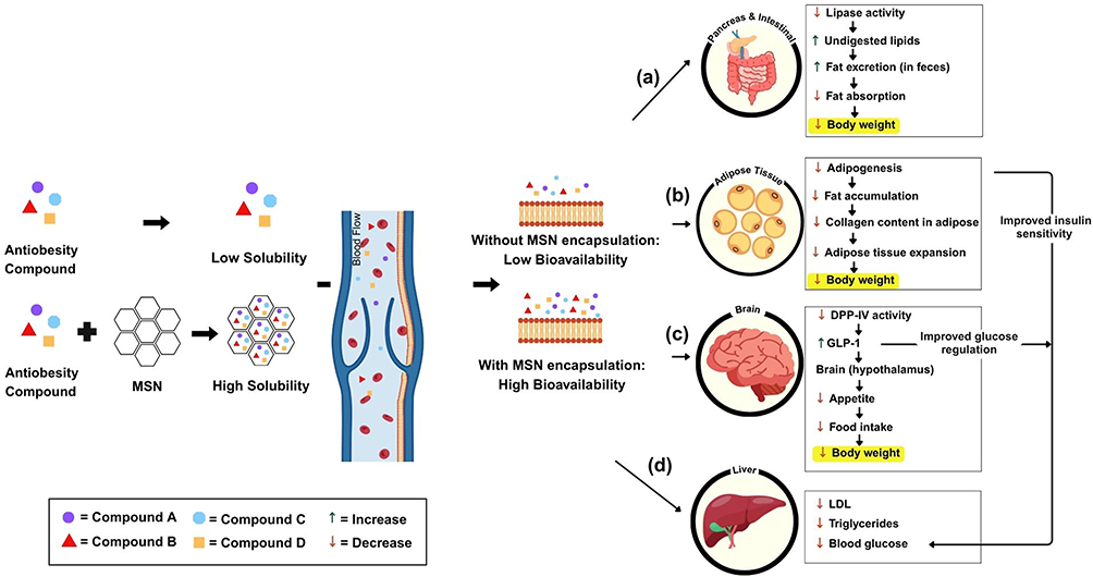 |
Figure 4 The speculated mechanism of the improvement of the antiobesity activity of MSN-encapsulated compounds. MSN loading significantly improves the solubility, cellular uptake, and systemic absorption of the compounds, resulting in enhanced antiobesity effects at multiple physiological targets: (a) Pancreas and intestinal tract: MSN-encapsulated compounds inhibit lipase activity, leading to reduced fat absorption, increased lipid excretion, and a consequent decrease in body weight. (b) Adipose tissue: These compounds downregulate adipogenesis and fat accumulation, reduce collagen content in adipose tissue, and limit tissue expansion, collectively contributing to weight reduction. (c) Brain (hypothalamus): Encapsulation increases GLP-1 secretion and decreases DPP-IV activity, promoting appetite suppression and improved glycemic control, resulting in reduced food intake and body weight. (d) Liver: The compounds lower LDL, triglyceride, and blood glucose levels, contributing to improved metabolic profiles and enhanced insulin sensitivity. |
In Figure 4(b), the antiobesity compounds primarily act by inhibiting the adipogenesis process. Adipogenesis is the differentiation of preadipocytes into mature adipocytes, contributing to fat storage. The inhibition of this process results in a reduced formation of new fat cells and a decrease in lipid accumulation within existing adipocytes.87 Furthermore, some studies reported that treated animals exhibited reduced collagen content associated with adipose tissue, suggesting an improvement in tissue remodeling and possibly mitigating fibrosis that often accompanies obesity. By directly targeting adipose tissue expansion, these compounds help to limit fat storage and contribute to overall weight reduction.88
The brain, particularly the hypothalamus, is central to the regulation of appetite and energy homeostasis. The Figure 4(c), show the compounds indirectly influence the hypothalamic signaling pathways through DPP-4 inhibition and the subsequent increase in GLP-1 levels. GLP-1 acts on the brain to promote satiety and reduce hunger, leading to a significant decrease in food intake.89 By modulating the hormonal signals that control appetite, the compounds can create a negative energy balance, supporting long-term weight management. This brain-mediated pathway highlights the importance of hormonal crosstalk between peripheral organs and the central nervous system in achieving antiobesity effects.90 By modulating the hormonal signals that control appetite, the compounds can create a negative energy balance, supporting long-term weight management. This brain-mediated pathway highlights the importance of hormonal crosstalk between peripheral organs and the central nervous system in achieving antiobesity effects.90
The liver is critically involved in lipid and glucose metabolism, and its function is often impaired during obesity. As shown in the Figure 4(d), several studies indicated that treatment with the antiobesity compounds resulted in improved blood lipid profiles, including reductions in low-density lipoprotein (LDL) cholesterol and triglycerides, as well as better regulation of blood glucose levels. These improvements suggest that the compounds enhance hepatic lipid metabolism and potentially alleviate insulin resistance.91 By the liver is critically involved in lipid and glucose metabolism, and its function is often impaired during obesity. Several studies indicated that treatment with the antiobesity compounds resulted in improved blood lipid profiles, including reductions in low-density lipoprotein (LDL) cholesterol and triglycerides, as well as better regulation of blood glucose levels. These improvements suggest that the compounds enhance hepatic lipid metabolism and potentially alleviate insulin resistance. By reducing the metabolic burden on the liver and preventing lipid accumulation, these interventions may also lower the risk of developing non-alcoholic fatty liver disease (NAFLD), a common complication associated with obesity.92,93
After confirming that the compound was already loaded into the MSNs through characterization, the dissolution study was used to analyze the release. The outcome demonstrated that, in comparison to their pure forms, MSNs have a considerable potential to greatly increase the dissolving rates of different compounds. The unique characteristics of mesoporous silica, such as its large surface area and tunable pore size, are responsible for this enhancement. Compounds dispersed monomolecularly within the MSNs. The dissolution medium was added immediately after the compound was dispersed in the MSNs. After being divided into monomolecules, the substance dissolves rapidly and releases into the solvent used for bulk dissolution. A high concentration or supersaturation level formed in the dissolution media as a result of the compound’s quick dissolution. These characteristics aid in the improved release and dispersion of compounds that are encapsulated. The more soluble form of a compound can be confined in MSNs, most likely because of the nanostructure of the compound’s constituents. This study demonstrates how compound-loaded MSNs with mesoporous morphologies can load a complicated multicomponent mixture in a more soluble state, ideally increasing the bioavailability. Additionally, the medications’ adsorption onto the MSNs improves wettability and expands the surface area that can dissolve, which speeds up drug release. Strong connections between certain chemicals and the mesoporous silica structure lead to this effect, which may cause some of the chemicals to get partially trapped inside the tiny holes.
Although the use of mesoporous silica nanoparticles (MSNs) offers significant advantages in enhancing drug solubility and bioavailability, several limitations remain. In orlistat study, fat excretion in mice that were administered orlistat-loaded MSN was significantly decreased compared to the control group, but not in comparison with groups administered raw orlistat and the commercial product. In some studies, formulations were evaluated following a single administration, limiting therapeutic efficacy regarding their long-term efficacy and safety under repeated dosing. The limited drug-loading capacity of compounds such as 16-hydroxycleroda-3,13-dine-16,15-olide may also hinder optimal dosing strategies. Other concerns include the preliminary safety evaluations, the lack of long-term stability data; potential systemic and organ-specific toxicities of MSN, insufficient studies on biodegradation kinetics and biodistribution, and the risk of interactions with concurrently administered agents. The mechanistic pathways underlying the observed therapeutic effects have not been fully elucidated and require further validation. Moreover, challenges in purification and the accurate quantification of in vivo performance persist. These gaps call for more detailed pharmacokinetic, mechanistic, and toxicological investigations to support future therapeutic applications.
Conclusion and Future Perspective
The encapsulation of antiobesity compounds within mesoporous silica nanoparticles (MSNs) effectively enhances their solubility, dissolution rate, and stability, addressing key challenges associated with poor water solubility. Characterization studies using DSC, XRD, FTIR, TGA, SEM, and nitrogen adsorption–desorption confirm the successful loading of these compounds into MSNs, with transformations from crystalline to amorphous states contributing to improved dissolution. Furthermore, dissolution studies demonstrate that MSN-loaded formulations significantly accelerate compound release, leading to increased dispersion and prolonged retention in solution. The improved pharmacological activity of MSN-loaded antiobesity compounds is attributed to enhanced dissolution and controlled release mechanisms. Studies on various compounds, including orlistat, quercetin, EGCG, and statins, indicate greater enzyme inhibition, lipid metabolism regulation, and glucose homeostasis when formulated with MSNs. These findings highlight the potential of MSNs as an advanced drug delivery system to optimize the therapeutic effects of antiobesity drugs by improving their physicochemical properties and sustained activity. These improved therapeutic effects can help treat obesity by improving drug availability at target sites, assuring regulated release, sustaining pharmacological activity, and minimizing systemic side effects. MSNs strengthen the efficacy and durability of antiobesity interventions in preclinical models by modulating key physiological pathways, such as lipid absorption, adipogenesis, and insulin sensitivity. These underscore the translational potential of MSN-based systems as a viable strategy to address the complex pathophysiology of obesity, including metabolic dysregulation and its associated comorbidities.
Despite these promising results, additional research is important to facilitate the transition to clinical implementation. Future research should investigate the long-term efficacy and safety of MSN-based formulations with repeated dose, evaluate pharmacokinetics, biodistribution, and potential organ-specific toxicity, and confirm mechanisms of action in advanced animal models or clinical trials. Challenges such as formulation scalability, biodegradation behavior, and potential drug–drug interactions must also be considered. Advancing these investigations will be essential to facilitate the practical application of MSNs and fully exploit their promise in obesity treatment.
Data Sharing Statement
The data generated in the present study may be requested from the first author upon reasonable request.
Acknowledgments
The authors would like to thank the National Research and Innovation Agency (BRIN, RIIM3) and the Indonesia Endowment Funds for Education (LPDP) for supporting this work to Diah Lia Aulifa and to Universitas Padjadjaran for the APC. The authors thank the other members of pharmaceutical analysis and medicinal chemistry for their help throughout this work; in particular, Prof. Mutakin provided many useful discussions.
Funding
The authors thank Universitas Padjadjaran for supporting this work (Hibah review No. 2076/UN6.O/TU.00/2025 to Diah Lia Aulifa).
Disclosure
The authors report no conflicts of interest in this work.
References
1. Müller MJ, Geisler C. Defining obesity as a disease. Eur J Clin Nutr. 2017;71(11):1256–1258. doi:10.1038/ejcn.2017.155
2. Nimptsch K, Konigorski S, Pischon T. Diagnosis of obesity and use of obesity biomarkers in science and clinical medicine. Metabolism. 2019;92:61–70. doi:10.1016/j.metabol.2018.12.006
3. Rosenbaum M, Foster G. Differential mechanisms affecting weight loss and weight loss maintenance. Nat Metab. 2023;5(8):1266–1274. doi:10.1038/s42255-023-00864-1
4. Abdi Beshir S, Ahmed Elnour A, Soorya A, et al. A narrative review of approved and emerging anti-obesity medications. Saudi Pharm J. 2023;31(10):101757. doi:10.1016/j.jsps.2023.101757
5. Ferrulli A, Terruzzi I, Senesi P, Succi M, Cannavaro D, Luzi L. Turning the clock forward: new pharmacological and non pharmacological targets for the treatment of obesity. Nutr Metab Cardiovasc Dis. 2022;32(6):1320–1334. doi:10.1016/j.numecd.2022.02.016
6. Gudzune KA, Kushner RF. Medications for obesity: a review. JAMA. 2024;332(7):571–584. doi:10.1001/jama.2024.10816
7. Gadde KM, Atkins KD. The limits and challenges of antiobesity pharmacotherapy.
8. Panuganti K, Nguyen M, Kshirsagar RK. Obesity. Treasure Island (FL): StatPearls Publishing; 2023.
9. Payghan S, Payghan V, Nangare K, Dahiwade L, Khavane K, Phalke R. Preparation and characterization of orlistat bionanocomposites using natural carriers. Turkish J Pharm Sci. 2022;19(2):168–179. doi:10.4274/tjps.galenos.2021.71363
10. Budiman A, Aulifa DL. Encapsulation of drug into mesoporous silica by solvent evaporation: a comparative study of drug characterization in mesoporous silica with various molecular weights. Heliyon. 2021;7(12):e08627. doi:10.1016/j.heliyon.2021.e08627
11. Mccarthy CA, Ahern RJ, Dontireddy R, Ryan KB, Crean AM. Mesoporous silica formulation strategies for drug dissolution enhancement: a review. Expert Opin Drug Deliv. 2016;13(1):93–108. doi:10.1517/17425247.2016.1100165
12. Budiman A, Aulifa DL. A comparative study of the pharmaceutical properties between amorphous drugs loaded-mesoporous silica and pure amorphous drugs prepared by solvent evaporation. Pharmaceuticals. 2022;15(6):730. doi:10.3390/ph15060730
13. Bouchoucha M, Côté M-F, C.-Gaudreault R, Fortin M-A, Kleitz F. Size-controlled functionalized mesoporous silica nanoparticles for tunable drug release and enhanced anti-tumoral activity. Chem Mater. 2016;28(12):4234–4258. doi:10.1021/acs.chemmater.6b00877
14. Islam S, Ahmed MMS, Islam MA, Hossain N, Chowdhury MA. Advances in nanoparticles in targeted drug delivery–A review. Results Surfaces Interfaces. 2025;19:100529. doi:10.1016/j.rsurfi.2025.100529
15. Santhamoorthy M, Asaithambi P, Ramkumar V, Elangovan N, Perumal I, Kim SC. A review on the recent advancements of polymer-modified mesoporous silica nanoparticles for drug delivery under. Polymers. 2025;17(12):1640. doi:10.3390/polym17121640
16. Liu B, Liu W, Xu M, et al. Drug delivery systems based on mesoporous silica nanoparticles for the management of hepatic diseases. Acta Pharm Sin B. 2024;15(2):809. doi:10.1016/j.apsb.2024.12.015
17. Lam YY, Ravussin E. Analysis of energy metabolism in humans: a review of methodologies. Mol Metab. 2016;5(11):1057–1071. doi:10.1016/j.molmet.2016.09.005
18. Musaiger AO. Overweight and obesity in eastern mediterranean region: prevalence and possible causes. J Obes. 2011;2011(January 1990):1–17. doi:10.1155/2011/407237
19. Shatwan IM, Almoraie NM. Correlation between dietary intake and obesity risk factors among healthy adults. Clin Nutr Open Sci. 2022;45:32–41. doi:10.1016/j.nutos.2022.08.007
20. Jin X, Qiu T, Li L, et al. Pathophysiology of obesity and its associated diseases. Acta Pharm Sin B. 2023;13(6):2403–2424. doi:10.1016/j.apsb.2023.01.012
21. Airaodion AI, Ogbuagu U, Oloruntoba AP, et al. Biochemical mechanisms involved in the regulation of appetite and weight - review. Int J Res. 2019;6(2):397–409.
22. Unger RH, Clark GO, Scherer PE, Orci L. Lipid homeostasis, lipotoxicity and the metabolic syndrome. Biochim Biophys Acta - Mol Cell Biol Lipids. 2010;1801(3):209–214. doi:10.1016/j.bbalip.2009.10.006
23. Crooks B, Stamataki NS, McLaughlin JT. Conference on diet and digestive disease symposium 3: extreme bmi, the regulation of intake and impairments of uptake: appetite, the enteroendocrine system, gastrointestinal disease and obesity. Proc Nutr Soc. 2021;80(1):50–58. doi:10.1017/S0029665120006965
24. Basil B, Myke-Mbata BK, Eze OE, Akubue AU. From adiposity to steatosis: metabolic dysfunction-associated steatotic liver disease, a hepatic expression of metabolic syndrome - current insights and future directions. Clin Diabetes Endocrinol. 2024;10(1):39. doi:10.1186/s40842-024-00187-4
25. Lean MEJ, Malkova D. Altered gut and adipose tissue hormones in overweight and obese individuals: cause or consequence. Int J Obes. 2016;40(4):622–632. doi:10.1038/ijo.2015.220
26. Stewart JE, Feinle-Bisset C, Keast RSJ. Fatty acid detection during food consumption and digestion: associations with ingestive behavior and obesity. Prog Lipid Res. 2011;50(3):225–233. doi:10.1016/j.plipres.2011.02.002
27. Mechanick JI, Zhao S, Garvey WT. Leptin, an adipokine with central importance in the global obesity problem. Glob Heart. 2018;13(2):113–127. doi:10.1016/j.gheart.2017.10.003
28. Perakakis N, Mantzoros CS. Evidence from clinical studies of leptin: current and future clinical applications in humans. Metabolism. 2024;161(September):156053. doi:10.1016/j.metabol.2024.156053
29. Steiner BM, Berry DC. The regulation of adipose tissue health by estrogens. Front Endocrinol. 2022;13(May):1–20. doi:10.3389/fendo.2022.889923
30. Small L, Brandon AE, Turner N, Cooney GJ. Modeling insulin resistance in rodents by alterations in diet: what have high-fat and high-calorie diets revealed? Am J Physiol - Endocrinol Metab. 2018;314(3):E251–65. doi:10.1152/ajpendo.00337.2017
31. Li X, Liu Q, Pan Y, Chen S, Zhao Y, Hu Y. New insights into the role of dietary triglyceride absorption in obesity and metabolic diseases. Front Pharmacol. 2023;14(February):1–18.
32. Monteiro R, Azevedo I. Chronic inflammation in obesity and the metabolic syndrome. Mediators Inflamm. 2010;2010(Atp Iii):1–10. doi:10.1155/2010/289645
33. Zatterale F, Longo M, Naderi J, et al. Chronic adipose tissue inflammation linking obesity to insulin resistance and type 2 diabetes. Front Physiol. 2020;10(January):1–20. doi:10.3389/fphys.2019.01607
34. Henning RJ. Obesity and obesity-induced inflammatory disease contribute to atherosclerosis: a review of the pathophysiology and treatment of obesity. Am J Cardiovasc Dis. 2021;11(4):504–529.
35. Westbury S, Oyebode O, van Rens T, Barber TM. Obesity stigma: causes, consequences, and potential solutions. Curr Obes Rep. 2023;12(1):10–23. doi:10.1007/s13679-023-00495-3
36. Kalepu S, Nekkanti V. Insoluble drug delivery strategies: review of recent advances and business prospects. Acta Pharm Sin B. 2015;5(5):442–453. doi:10.1016/j.apsb.2015.07.003
37. Khadka P, Ro J, Kim H, et al. Pharmaceutical particle technologies: an approach to improve drug solubility, dissolution and bioavailability. Asian J Pharm Sci. 2014;9(6):304–316.
38. Trzeciak K, Chotera‐ouda A, Bak‐sypien II, Potrzebowski MJ. Mesoporous silica particles as drug delivery systems—the state of the art in loading methods and the recent progress in analytical techniques for monitoring these processes. Pharmaceutics. 2021;13(7):950. doi:10.3390/pharmaceutics13070950
39. Li T, Shi S, Goel S, et al. Recent advancements in mesoporous silica nanoparticles towards therapeutic applications for cancer. Acta Biomater. 2019;89:1–13. doi:10.1016/j.actbio.2019.02.031
40. Alyassin Y, Sayed EG, Mehta P, et al. Application of mesoporous silica nanoparticles as drug delivery carriers for chemotherapeutic agents. Drug Discov Today. 2020;25(8):1513–1520. doi:10.1016/j.drudis.2020.06.006
41. Siddiqui B, Rehman AU, Haq IU, Al-Dossary AA, Elaissari A, Ahmed N. Exploiting recent trends for the synthesis and surface functionalization of mesoporous silica nanoparticles towards biomedical applications. Int J Pharm X. 2022;4:100116. doi:10.1016/j.ijpx.2022.100116
42. Khalbas AH, Albayati TM, Saady NMC, Zendehboudi S, Salih IK, Tofah ML. Insights into drug loading techniques with mesoporous silica nanoparticles: optimization of operating conditions and assessment of drug stability. J Drug Deliv Sci Technol. 2024;96:105698. doi:10.1016/j.jddst.2024.105698
43. Žid L, Zeleňák V, Almáši M, et al. Mesoporous silica as a drug delivery system for naproxen: influence of surface functionalization. Mol. 2020;25(20):4722. doi:10.3390/molecules25204722
44. Zhou Y, Quan G, Wu Q, et al. Mesoporous silica nanoparticles for drug and gene delivery. Acta Pharm Sin B. 2018;8(2):165–177. doi:10.1016/j.apsb.2018.01.007
45. Budiman A, Anastasya G, Handini AL, Lestari IN, Subra L, Aulifa DL. Characterization of drug with good glass-forming ability loaded mesoporous silica nanoparticles and its impact toward in vitro and in vivo studies. Int J Nanomed. 2024;19:2199–2225. doi:10.2147/IJN.S453873
46. Bukara K, Schueller L, Rosier J, et al. Ordered mesoporous silica to enhance the bioavailability of poorly water-soluble drugs: proof of concept in man. Eur J Pharm Biopharm. 2016;108:220–225. doi:10.1016/j.ejpb.2016.08.020
47. Lainé AL, Price D, Davis J, et al. Enhanced oral delivery of celecoxib via the development of a supersaturable amorphous formulation utilising mesoporous silica and co-loaded HPMCAS. Int J Pharm. 2016;512(1):118–125. doi:10.1016/j.ijpharm.2016.08.034
48. Elbialy NS, Aboushoushah SF, Sofi BF, Noorwali A. Multifunctional curcumin-loaded mesoporous silica nanoparticles for cancer chemoprevention and therapy. Microporous Mesoporous Mater. 2020;291:109540. doi:10.1016/j.micromeso.2019.06.002
49. Park H, Cha KH, Hong SH, et al. Pharmaceutical characterization and in vivo evaluation of orlistat formulations prepared by the supercritical melt-adsorption method using carbon dioxide: effects of mesoporous silica type. Pharmaceutics. 2020;12(4):333. doi:10.3390/pharmaceutics12040333
50. Kim T, Cho AY, Lee SW, Lee HJ. Controlled quercetin release by fluorescent mesoporous nanocarriers for effective anti-adipogenesis. Int J Nanomed. 2024;19:5441–5458. doi:10.2147/IJN.S463765
51. El-Kady DS, Mabrouk M, Kotob SE, et al. Evaluation of mesoporous silica nanoparticles as delivery vehicles for novel hybrid steroids in management of metabolic syndrome in mice. Egypt J Chem. 2021;64(10):5813–5830.
52. Kim T, Cho AY, Lee SW, Lee HJ. Optimized epigallocatechin gallate delivery and adipogenesis inhibition through fluorescent mesoporous nanocarriers. Biomater Res. 2024;28. doi:10.34133/bmr.0053
53. Geng S, Qin L, He Y, et al. Effective and safe delivery of GLP-1AR and FGF-21 plasmids using amino-functionalized dual-mesoporous silica nanoparticles in vitro and in vivo. Biomaterials. 2021;271:120763. doi:10.1016/j.biomaterials.2021.120763
54. Yu XW, Liu T, Lin R. Development and characterization of a glimepiride-loaded gelatin-coated mesoporous hollow silica nanoparticle formulation and evaluation of its hypoglycemic effect on type-2 diabetes model rats. Assay Drug Dev Technol. 2020;18(8):369–378. doi:10.1089/adt.2020.987
55. Huang PK, Lin SX, Tsai MJ, et al. Encapsulation of 16-hydroxycleroda-3,13-dine-16,15-olide in mesoporous silica nanoparticles as a natural dipeptidyl peptidase-4 inhibitor potentiated hypoglycemia in diabetic mice. Nanomaterials. 2017;7(5):1–17. doi:10.3390/nano7050112
56. Sayadi K, Rahdar A, Hajinezhad MR, Nikazar S, Susan MABH. Atorvastatin-loaded SBA-16 nanostructures: synthesis, physical characterization, and biochemical alterations in hyperlipidemic rats. J Mol Struct. 2020;1202:127296.
57. Jin H, Lu W, Zhang Y, et al. Functionalized periodic mesoporous silica nanoparticles for inhibiting the progression of atherosclerosis by targeting low-density lipoprotein cholesterol. Pharmaceutics. 2024;16(1):74. doi:10.3390/pharmaceutics16010074
58. Song K, Tang Z, Song Z, et al. Hyaluronic acid-functionalized mesoporous silica nanoparticles loading simvastatin for targeted therapy of atherosclerosis. Pharmaceutics. 2022;14(6):1265. doi:10.3390/pharmaceutics14061265
59. Pham LM, Kim EC, Ou W, et al. Targeting and clearance of senescent foamy macrophages and senescent endothelial cells by antibody-functionalized mesoporous silica nanoparticles for alleviating aorta atherosclerosis. Biomaterials. 2021;269(January):120677. doi:10.1016/j.biomaterials.2021.120677
60. Costa JAS, de Jesus RA, Santos DO, Mano JF, Romão LPC, Paranhos CM. Recent progresses in the adsorption of organic, inorganic, and gas compounds by MCM-41-based mesoporous materials. Microporous Mesoporous Mater. 2020;291:109698.
61. Zou J, Fan C, Jiang Y, et al. A preliminary study on assessing the brunauer-emmett-teller analysis for disordered carbonaceous materials. Microporous Mesoporous Mater. 2021;327:111411. doi:10.1016/j.micromeso.2021.111411
62. Atwood JL. Thermal Analysis. In: Comprehensive Supramolecular Chemistry II, Molecular Sciences and Chemical Engineering.
63. Eid MM. Characterization of Nanoparticles by FTIR and FTIR-Microscopy. In: Handbook Consumer Nanoproducts. Springer Singapore; 2022:645–673.
64. Xue B, Huang L, Li X, et al. Effect of clay mineralogy and soil organic carbon in aggregates under straw incorporation. Agronomy. 2022;12(2):534. doi:10.3390/agronomy12020534
65. Inkson BJ. Scanning electron microscopy (SEM) and transmission electron microscopy (TEM) for materials characterization. In: Materials Characterization Using Nondestructive Evaluation (NDE) Methods. Elsevier Ltd; 2016:17–43. doi:10.1016/B978-0-08-100040-3.00002-X
66. Tang CY, Yang Z. Transmission Electron Microscopy (TEM). In: Membrane Characterization. Elsevier Inc.; 2017:145–159.
67. Rodriguez-Loya J, Lerma M, Gardea-Torresdey JL. Dynamic light scattering and its application to control nanoparticle aggregation in colloidal systems: a review. Micromachines. 2023;15(1):24. doi:10.3390/mi15010024
68. Jia Z, Li J, Gao L, Yang D, Kanaev A. Dynamic light scattering: a powerful tool for in situ nanoparticle sizing. Colloids Interfaces. 2023;7(1):15. doi:10.3390/colloids7010015
69. Vazquez NI, Gonzalez Z, Ferrari B, Castro Y. Synthesis of mesoporous silica nanoparticles by sol-gel as nanocontainer for future drug delivery applications. Bol la Soc Esp Ceram y Vidr. 2017;56(3):139–145. doi:10.1016/j.bsecv.2017.03.002
70. Stevie FA, Donley CL. Introduction to x-ray photoelectron spectroscopy. J Vac Sci Technol a Vac Surf Film. 2020;38(6).
71. Saputra OA, Safitriono WN, Istiqomah A, Kumalasari R, Irmawan M, Wibowo R. Advances in mesoporous silica nanoparticles: synthesis, characterization, and biomedical uses indonesian journal of chemical analysis. Ind J Chem Anal. 2024;07(02):203–226. doi:10.20885/ijca.vol7.iss2.art9
72. Shaik Mohamed Sayed UF, Moshawih S, Goh HP, et al. Natural products as novel anti-obesity agents: insights into mechanisms of action and potential for therapeutic management. Front Pharmacol. 2023;14(June):1–30.
73. Liu TT, Liu XT, Chen QX, Shi Y. Lipase inhibitors for obesity: a review. Biomed Pharmacother. 2020;128(November 2019):110314. doi:10.1016/j.biopha.2020.110314
74. Lunagariya NA, Patel NK, Jagtap SC, Bhutani KK. Inhibitors of pancreatic lipase: state of the art and clinical perspectives. Excli J. 2014;13:897–921.
75. Subramaniyan V, Hanim YU. Role of pancreatic lipase inhibition in obesity treatment: mechanisms and challenges towards current insights and future directions: physiology and biochemistry. Int J Obes. 2025;49(September 2024):492–506. doi:10.1038/s41366-025-01729-1
76. Jaradat N, Zaid A, Hussein F, Zaqzouq M, Aljammal H, Ayesh O. Anti-lipase potential of the organic and aqueous extracts of ten traditional edible and medicinal plants in palestine; a comparison study with orlistat. Medicines. 2017;4(4):89. doi:10.3390/medicines4040089
77. Kupferschmidt N, Csikasz RI, Ballell L, Bengtsson T, Garcia-Bennett AE. Large pore mesoporous silica induced weight loss in obese mice. Nanomedicine. 2014;9(9):1353–1362. doi:10.2217/nnm.13.138
78. Coutinho W. The first decade of sibutramine and orlistat: a reappraisal of their expanding roles in the treatment of obesity and associated conditions. Arq Bras Endocrinol Metabol. 2009;53(2):262–270. doi:10.1590/S0004-27302009000200018
79. Feingold K. Introduction to lipids and lipoproteins. 2024.
80. Tantu R, Dunggio T, Arwati N. Description of blood glucose levels in obesity patients in. 2020;23–34.
81. Piqueras P, Ballester A, Durá-Gil JV, Martinez-Hervas S, Redón J, Real JT. Anthropometric indicators as a tool for diagnosis of obesity and other health risk factors: a literature review. Front Psychol. 2021;12. doi:10.3389/fpsyg.2021.631179
82. de Moura e Dias M, Dos Reis SA, da Conceição LL, et al. Diet-induced obesity in animal models: points to consider and influence on metabolic markers. Diabetol Metab Syndr. 2021;13(1). doi:10.1186/s13098-021-00647-2
83. Kübeck R, Bonet-Ripoll C, Hoffmann C, et al. Dietary fat and gut microbiota interactions determine diet-induced obesity in mice. Mol Metab. 2016;5(12):1162–1174. doi:10.1016/j.molmet.2016.10.001
84. Liu X, Zhao L, Chen Y, et al. Obesity induces adipose fibrosis and collagen cross-linking through suppressing AMPK and enhancing lysyl oxidase expression. Biochim Biophys Acta - Mol Basis Dis. 2022;1868(9):166454. doi:10.1016/j.bbadis.2022.166454
85. Gutiérrez‐cuevas J, Sandoval‐rodriguez A, Meza‐rios A, et al. Molecular mechanisms of obesity‐linked cardiac dysfunction: an up‐date on current knowledge. Cells. 2021;10(3):1–28. doi:10.3390/cells10030629
86. Rajan L, Palaniswamy D, Mohankumar SK. Targeting obesity with plant-derived pancreatic lipase inhibitors: a comprehensive review. Pharmacol Res. 2020;155:104681. doi:10.1016/j.phrs.2020.104681
87. Mantzoros CS, Magkos CS, Brinkoetter F, et al. Leptin in human physiology and pathophysiology. Am J Physiol Endocrinol Metab. 2011;301(4):567–584. doi:10.1152/ajpendo.00315.2011
88. Sun K, Kusminski CM, Scherer PE. Adipose tissue remodeling and obesity. J Clin Investig. 2011;121(6):2094–2101. doi:10.1172/JCI45887
89. Baggio LL, Drucker DJ. Glucagon-like peptide-1 receptors in the brain: controlling food intake and body weight. J Clin Investig. 2014;124(10):4223–4226. doi:10.1172/JCI78371
90. Trapp S, Brierley DI. Brain GLP-1 and the regulation of food intake: GLP-1 action in the brain and its implications for GLP-1 receptor agonists in obesity treatment. British J Pharmacol. 2021;179(4):557–570.
91. Siavashani AZ, Nazarpak MH, Bakhsh FF, Toliyat T, Solati-Hashjin M. Preparation of mesoporous silica nanoparticles for insulin drug delivery. Adv Mat Res. 2014;829:251–257.
92. Rinaldi L, Pafundi PC, Galiero R, et al. Mechanisms of non-alcoholic fatty liver disease in the metabolic syndrome. A narrative review. Antioxidants. 2021;10(2):1–25. doi:10.3390/antiox10020270
93. El-Kassas M, Awad A, Méndez-Sánchez N. Recent updates in the prevention of nonalcoholic fatty liver disease. Vol. 22. In: Gene Expression the Journal of Liver Research. Xia and He Publishing Inc.; 2023:19–27.
 © 2025 The Author(s). This work is published and licensed by Dove Medical Press Limited. The
full terms of this license are available at https://www.dovepress.com/terms
and incorporate the Creative Commons Attribution
- Non Commercial (unported, 4.0) License.
By accessing the work you hereby accept the Terms. Non-commercial uses of the work are permitted
without any further permission from Dove Medical Press Limited, provided the work is properly
attributed. For permission for commercial use of this work, please see paragraphs 4.2 and 5 of our Terms.
© 2025 The Author(s). This work is published and licensed by Dove Medical Press Limited. The
full terms of this license are available at https://www.dovepress.com/terms
and incorporate the Creative Commons Attribution
- Non Commercial (unported, 4.0) License.
By accessing the work you hereby accept the Terms. Non-commercial uses of the work are permitted
without any further permission from Dove Medical Press Limited, provided the work is properly
attributed. For permission for commercial use of this work, please see paragraphs 4.2 and 5 of our Terms.