Back to Journals » International Journal of Nanomedicine » Volume 20
Mesenchymal Stem Cell-Derived Exosomes for Ocular Diseases: Therapeutic Mechanisms and Clinical Perspectives
Authors Cheng S, Ma Y, Huang F, Luo R, Han L, He L, Yuan ZX ![]()
Received 23 July 2025
Accepted for publication 23 November 2025
Published 6 December 2025 Volume 2025:20 Pages 14521—14550
DOI https://doi.org/10.2147/IJN.S555771
Checked for plagiarism Yes
Review by Single anonymous peer review
Peer reviewer comments 4
Editor who approved publication: Dr Krishna Nune
Shuangjiang Cheng,1 Yafei Ma,1 Fenglan Huang,2 Rong Luo,2 Lu Han,1 Lili He,1 Zhi-Xiang Yuan1
1Key Laboratory of Research and Application of Ethnic Medicine Processing and Preparation on the Qinghai Tibet Plateau, College of Pharmacy and Food, Southwest Minzu University, Chengdu, Sichuan, 610041, People’s Republic of China; 2West China School of Public Health and West China Fourth Hospital, Sichuan University, Chengdu, Sichuan, 610041, People’s Republic of China
Correspondence: Lili He; Zhi-Xiang Yuan, College of Pharmacy and Food, Southwest Minzu University, No. 168, Wenxing Section, Dajian Road, Shuangliu District, Chengdu, Sichuan, 610041, People’s Republic of China, Tel +86-18628270031 ; +86-18683618863, Email [email protected]; [email protected]
Abstract: Ocular diseases represent a major and increasing public health concern. Although current treatment options are available, the management of complex cases, such as corneal diseases, diabetic retinopathy, glaucoma, age-related macular degeneration, and uveitis, remains inadequate. Recent studies have demonstrated that mesenchymal stem cell-derived exosomes (MSC-Exos), obtained from bone marrow, adipose tissue, and umbilical cord, have emerged as a promising cell-free therapeutic platform for various ocular diseases. These nanovesicles can be delivered via systems such as topical eye drops and intravitreal injection, targeting ocular tissues to exert anti-inflammatory, anti-apoptotic, and tissue-repairing effects. This review systematically synthesizes recent advances and the molecular mechanisms underlying the use of MSC-Exos in treating ocular diseases. Moreover, it provides an in-depth discussion of the challenges in the clinical application of MSC-Exos in ophthalmology, including standardized production, dosage optimization, delivery system improvement, and targeting enhancement, and proposes engineered targeting strategies based on surface modification and carrier optimization. Overall, this work establishes a rigorous framework for advancing MSC-Exos from experimental models to clinical implementation, offering novel therapeutic strategies through these innovative biopharmaceuticals for previously untreatable ocular conditions.
Keywords: mesenchymal stem cells, targeted therapy, exosomes, ocular diseases, therapeutic mechanisms
Introduction
Ocular diseases are a major and growing global health concern, and the primary causes of vision impairment worldwide are uncorrected refractive error, cataracts, age-related macular degeneration, glaucoma, and diabetic retinopathy.1 It is estimated that by 2020, out of the global population of 7.79 billion, approximately 43.3 million people were blind, and an additional 295 million people suffered from moderate to severe visual impairment, resulting in a visual impairment prevalence rate of 4.34%.2 Ocular diseases significantly disrupt daily life, often presenting with subtle early symptoms that can go unnoticed. Additionally, the intricate anatomical structure of the eye and the presence of physiological barriers pose significant challenges to the early diagnosis and treatment of ocular diseases.3 Traditional small-molecule drugs and steroids often face fundamental challenges, including low bioavailability and off-target effects, in treating ocular diseases. Furthermore, due to the eye’s physiological barriers (eg, the cornea and blood-retinal barrier), these agents struggle to reach the lesion site and maintain effective therapeutic concentrations, often necessitating frequent administration. This increases the risk of systemic side effects, such as elevated intraocular pressure and cataract formation associated with long-term steroid use.4,5 In contrast, while most of the therapeutic proteins and peptides exhibit superior targeting specificity, they are plagued by inherent limitations, including poor stability, high susceptibility to degradation, and short half-life.6 Given the significant shortcomings and limitations of current prevention and treatment methods, which fail to effectively prevent or halt the progression of ocular surface and retinal diseases, exploring more effective alternative therapeutic approaches becomes crucial.7,8
Mesenchymal stem cells (MSCs) are multipotent stem cells that can differentiate into multilineage cells, such as osteoblasts, chondrocytes, and adipocytes,9 which are extensively distributed in various tissues and organs of the human body, including bone marrow, adipose tissue, umbilical cord, and dental pulp.10 In recent years, MSCs have been widely used in stem cell therapies for the treatment of various diseases, for instance, retinal diseases, due to their remarkable immunosuppressive capabilities and differentiation potential.11–13 However, MSCs also have their shortcomings, such as poor quality control, tumorigenicity, lack of standardized management, and heterogeneity, which limit their clinical development.14,15 To circumvent these challenges, an increasing number of studies are focusing on a new aspect of MSCs: their exosomes, which are nanosized extracellular vesicles (EVs) with a diameter ranging from 30 to 150 nm that are secreted by almost all cells and are believed to play a more critical role in intercellular communication.16,17 Notably, although challenges related to scalable production, storage stability, and batch-to-batch standardization still need to be addressed in clinical applications, MSC-Exos possess significant advantages compared to emerging alternative therapies (such as viral vectors for gene therapy or synthetic nanoparticles): they exhibit innate biocompatibility, demonstrate low immunogenicity, possess natural tissue-homing capabilities, and circumvent the mutagenic risks associated with gene therapies.18,19 As an innovative treatment modality, MSC-Exos demonstrate the potential to integrate multiple advantages. Firstly, as natural nanocarriers, their excellent biocompatibility and low immunogenicity enable them to effectively cross physiological barriers and deliver protective bioactive cargo (eg, nucleic acids, proteins) to otherwise hard-to-reach areas within the eye.20 Secondly, unlike synthetic nanoparticles, MSC-Exos inherently carry therapeutic functions from their parent cells, including anti-inflammatory, anti-fibrotic, and neuroprotective activities, thereby unifying the “drug” and the “delivery system”.21 Beyond their role as innate biological vehicles, they can be strategically engineered into precision therapeutic platforms. Their membranes can be functionally modified with targeting ligands (eg, peptides), and their lumens can be loaded with exogenous therapeutic agents (eg, small-molecule drugs, nucleic acids), thereby enhancing their specificity and efficacy in treating ocular diseases.17,22,23 This engineering flexibility provides MSC-Exos with a distinct advantage over traditional small-molecule drugs and steroids, enabling a targeted, multi-mechanistic therapeutic approach for complex pathologies.
In addition, engineered MSC-Exos can also enhance their targeting ability and become a precise drug delivery system, especially in the treatment of inflammatory and autoimmune diseases.24–26 Studies have shown that subconjunctival injection of human umbilical cord MSC-Exos (hucMSC-Exos) can improve the proliferation and migration of corneal epithelial cells, thereby promoting corneal wound healing and regeneration.27,28 In addition, adipose mesenchymal stem cell-derived exosomes (ADSC-Exos) can regulate microglia-mediated neuroinflammation to alleviate retinal ganglion cells (RGCs) damage caused by high intraocular pressure, thereby assisting in the treatment of glaucoma.29 These results indicate that MSC-Exos therapeutic strategies have shown multifaceted potential in the treatment of ocular diseases and bring new hope for many intractable ophthalmic diseases.
In this review, to comprehensively cover MSC-Exos research in ocular diseases, we searched PubMed, Google Scholar, Scopus, and Web of Science. Keywords like “mesenchymal stem cell exosomes”, “retinal diseases”, “corneal wound healing”, “glaucoma”, and “age-related macular degeneration” were used. We focused on English-language studies published from 2019 to the present, encompassing research articles and reviews regarding MSC-Exos in the aspects of ocular diseases. We then concentrated on discussing the therapeutic potential and mechanism of MSC-Exos in ocular diseases such as retinal diseases, corneal diseases, uveitis, and glaucoma (Figure 1). Furthermore, the limitations and challenges hindering clinical translation of MSC-Exos, along with strategies to overcome them, are discussed. The purpose of this review is to facilitate the clinical translation of MSC-Exos, spur in-depth research in related fields, and accelerate the development and application of new therapies.
 |
Figure 1 Schematic illustration of mesenchymal stem cell-derived exosomes (MSC-Exos) in ocular disease therapy. Notes: MSC-Exos-based delivery systems, categorized into natural exosomes, engineered exosomes, and biomaterial-loaded exosomes (BML-Exos), enable targeted therapeutic interventions for ocular pathologies. MSC-Exos-based therapies are applied via diverse delivery routes (eg, topical, intravitreal) to manage anterior segment disorders (eg, corneal injury, uveitis) and posterior segment pathologies (eg, glaucoma, diabetic retinopathy, age-related macular degeneration). The illustration further highlights the anatomical complexity of ocular tissues, particularly the cornea and retina, to contextualize disease-specific therapeutic targeting. RPEs, retinal pigment epithelium cells. |
Ocular Diseases
Eyes, as vital sensory organs, are fundamental for vision and play a pivotal role in human perception, which are composed of critical structures, including the cornea, iris, and lens, which work together to facilitate light perception, shape recognition, and color differentiation.30 However, ocular diseases often pose a significant threat to this organ.31 The symptoms of ocular diseases are diverse and multifaceted. Visually, patients may experience blurred, decreased, or distorted vision, as well as night blindness, which are closely linked to lesions in various parts of the eye.32 For instance, opacity and ulceration in the cornea can disrupt the normal refraction and entry of light, leading to vision problems, while retinal disorders result in distorted or diminished vision.33,34 Eye discomfort may manifest as pain, swelling, photophobia, tearing, and itching, often accompanied by abnormal visual phenomena, such as floaters and flashes of light.35 Structurally, glaucoma can lead to optic nerve atrophy and visual field defects, while uveitis is associated with characteristic changes, including iris congestion, adhesions, and vitreous opacities.36,37
Ocular diseases encompass a broad spectrum, ranging from corneal and conjunctival disorders in the anterior segment of the eye, to retinal and optic nerve diseases in the posterior segment, with each category further subdivided into numerous specific conditions.38 These complexities require clinicians to possess extensive expertise and substantial clinical experience, making accurate diagnosis and differentiation of ocular diseases particularly challenging. Many ocular diseases progress insidiously during the early stages with subtle or undetectable symptoms. For example, age-related macular degeneration and retinitis pigmentosa develop gradually over a prolonged course, resulting in significant and lasting damage to vision and eye health.39,40 In contrast, acute infectious keratitis presents with rapid onset and pronounced symptoms.41 Additionally, the eye’s structure is highly sensitive to genetic variations and vulnerable to adverse environmental factors. The intricate interplay of internal and external influences critically determines the health and functionality of the visual system.42,43 A variety of pathologic conditions, including corneal diseases, glaucoma, diabetic retinopathy, age-related macular degeneration, and uveitis, frequently result in visual impairment. These disorders are further complicated by systemic factors, such as diabetes or autoimmune diseases, which exacerbate progression and complicate treatment.44,45 Structural or functional damage often accumulates over time, culminating in chronic damage that is frequently irreversible, even with advanced therapeutic interventions.46 Thus, timely diagnosis and treatment are imperative to safeguarding eye health and preserving vision.
MSC-Exos
Characteristics and Functions of MSC-Exos
According to the size and cell origin, EVs can be divided into three main categories, namely exosomes (30–150nm in diameter), microvesicles (100–1000nm), and apoptotic bodies (>1000nm).47 Exosomes originate from the endocytic pathway, forming early endosomes in the cell membrane, further evolving into late endosomes, and finally transforming into multivesicular bodies (MVB). MVB contain internal vesicles (future exosomes). Ultimately, MVB can fuse with lysosomes or autophagosomes and then be degraded, or fuse with the cell membrane and release exosomes to the cellular outside through exocytosis (Figure 2A).48–50 Exosomes can be released from various types of cells, including but not limited to macrophages, dendritic cells, lymphocytes, and even tumor cells, among others.51 Among these cell types, MSCs are notable for their ability to secrete a higher quantity of exosomes compared to many others.17 MSC-Exos have common surface markers like CD81, CD9, and CD63, which are also found in exosomes from other sources (Figure 2B). Moreover, they specifically express the surface markers of MSCs such as CD90, CD44, and CD73, convenient for confirming the identity of MSC-Exos and distinguishing them from other types of exosomes.52,53 As a signaling molecule, MSC-Exos not only exert the same effects as parent cells, but also have a more stable membrane structure than MSCs, providing broader prospects for disease treatment.
 |
Figure 2 Biogenesis, contents, and mechanisms of action of mesenchymal stem cell-derived exosomes (MSC-Exos) in ocular diseases. Abbreviations: IL-1β, interleukin-1 beta; TNF-α, tumor necrosis factor-alpha; PI3K, phosphatidylinositol 3-kinase; AKT, protein kinase B; PTEN, phosphatase and tensin homolog; STAT1, signal transducer and activator of transcription 1; PKC, protein kinase C; NF-κB, nuclear factor kappa B; HG, high glucose; VEGF, vascular endothelial growth factor; ICAM-1, intercellular adhesion molecule-1; VCAM-1, vascular cell adhesion molecule-1; PLCG1, phospholipase Cγ1; FBXW7, F-box and WD repeat domain-containing protein 7; EZH2, enhancer of zeste homolog 2; IRAK1, interleukin-1 receptor-associated kinase 1; TAB2, TGF-Beta activated kinase 1 (MAP3K7) binding protein 2; PDCD4, programmed cell death 4; HOXC6, homeobox protein Hox-C6; EMT, epithelial-mesenchymal transition. Notes: (A) MSC-Exos originate from the endocytic pathway, form early endosomes within the cell membrane, further evolve into late endosomes, and finally transform into multivesicular bodies (MVB). MVB can fuse with lysosomes for degradation, or fuse with the cell membrane and release exosomes to the outside of the cell through exocytosis. (B) MSC-Exos are rich in a variety of typical contents, including CD9, CD63, CD81 and other iconic membrane proteins, which belong to the tetraspanins family; Alix, TSG101 and other exosome-related proteins, which participate in the formation and secretion regulation of multivesicular bodies; mRNA, DNA, RNA, which can mediate the transmission of genetic information between cells; functional proteins include Integrins that can regulate target cell adhesion, and TLR (Toll-like receptors) that participate in immune regulation. (C) These molecules together give exosomes functional characteristics such as regulating intercellular communication, tissue repair and immune regulation. |
MSC-Exos have a wide range of biological functions, and gaining insight into these functions is essential for understanding their role in health and disease. 1) Signal transduction: As a medium for intercellular communication, MSC-Exos can transport their cargo to receptor cells in a variety of ways, such as directly binding to cell surface receptors, fusing with the plasma membrane, or internalizing, thereby interacting with the receptor cells and regulating various physiological and pathological processes (Figure 2C).54,55 2) Inflammation regulation: On the one hand, MSC-Exos may carry and transmit inflammatory mediators, such as cytokines and miRNAs, and then promote the activation of inflammasomes and the aggravation of inflammatory responses. On the other hand, they can reduce the production of inflammatory factors by transmitting specific miRNAs, such as miR-181c, which can reduce TLR4 expression and NF-κB activation.56 3) Immunomodulation: MSCs affect the polarization state of immune cells through MSC-Exos. For example, MSC-Exos can promote the generation of anti-inflammatory M2 macrophages or regulate the immune response by affecting the differentiation and function of T cells.57 4) Repair and regeneration: MSC-Exos exhibit protective and therapeutic properties in wound healing and tissue regeneration by promoting the repair of damaged tissues, highlighting their significant potential in regenerative medicine.58–60 5) Disease diagnosis: Proteins, nucleic acids and other molecules contained in MSC-Exos can be used as biomarkers for early disease diagnosis and monitoring.61,62 The functional molecules found in exosomes differ depending on the cells from which they are secreted and are affected by the surrounding cellular environment. This diversity in molecular composition enables MSC-Exos to perform various biological functions.63
Preparation and Modification of MSC-Exos
To develop a rapid, simple isolation method with high purity and high recovery is crucial for advancing MSC-Exos research and their large-scale application in medical practice.64 Currently, commonly used exosome extraction and isolation methods primarily include ultracentrifugation, size exclusion chromatography, ultrafiltration, polymer precipitation, and immunoaffinity. Ultracentrifugation is widely used, but time-consuming, labor-intensive, and may produce impurities (Figure 3a).65,66 Size exclusion chromatography is another promising method for MSC-Exos separation.67,68 The separation principle is that a solution containing exosomes passes through a stationary phase composed of a porous polymer. Particle molecules then pass through in order of size (large particles are eluted first, and small particles are eluted later), thus achieving material separation (Figure 3b).69 Similar to the principle of size exclusion chromatography, ultrafiltration is a method for screening exosomes by using filter membranes with different pore sizes or molecular weight cut-offs (Figure 3c).70 Currently, commercial exosome isolation kits based on polymer precipitation are commercially available (Figure 3d).71,72 The immunoaffinity technique involves the isolation of exosomes through the tagging of exosome membrane proteins, enabling them to bind specifically to specific antibodies (Figure 3e).73 Despite the array of techniques accessible for MSC-Exos isolation, none have proven entirely efficacious to date. Each method is accompanied by its own distinct constraints.74,75 In recent years, the integration of multiple separation and purification techniques has emerged as a dependable approach for the effective isolation of exosomes.76–78 For example, methods combining tangential flow filtration and size exclusion chromatography can improve the efficiency of separation and the purity of MSC-Exos.79 In addition, the development of microfluidic technology based on physical and biological properties provides new possibilities for MSC-Exos isolation.80 This technology utilizes the characteristics of microscale fluids to capture and analyze exosomes, featuring high-throughput, low sample consumption, and high purity.81,82
 |
Figure 3 Schematic diagram of the isolation, characterization, and analysis of functional properties of mesenchymal stem cell-derived exosomes (MSC-Exos). Notes: (a) Ultracentrifugation is to centrifuge the biological fluid at different speeds and times in sequence, remove dead cells and cell debris in turn, and finally separate the exosomes by centrifugation at a high speed of 100000g for 70 minutes. (b) Size exclusion chromatography is to let the biological fluid pass through the chromatographic column, separate according to molecular size, and then the exosomes with small particles will flow out. (c) Ultrafiltration is to let the biological fluid pass through filter membranes of different pore sizes in sequence, first remove dead cells and debris, and then retain exosomes. (d) Polymer precipitation is to add polyethylene glycol (PEG) to the biological fluid and incubate overnight to form a hydrophobic microenvironment around it, thereby causing exosome precipitation, and then further purifying the exosomes. (e) Immunoaffinity is to mix the biological fluid with magnetic beads carrying antibodies to capture particles containing specific antigens, wash impurities and elute exosomes. (f) Common characterization methods for exosomes include electron microscopy, Western blot (WB), nanoparticle tracking analysis (NTA), and dynamic light scattering (DLS). (g) Common analytical methods for exosomes include quantitative real-time polymerase chain reaction (qPCR), microarrays, next-generation gene sequencing, and electrochemical sensing technology. |
MSC-Exos are progressively undergoing modifications to enhance their functionality, stability, and targeting capabilities owing to their natural biocompatibility.83 A diverse array of methodologies, encompassing biological, chemical, and physical approaches, is utilized in the generation of modified exosomes.84 Biological modification of MSC-Exos usually involves genetic engineering of parent cells to direct the fusion of the gene sequence of a protein or peptide with the gene sequence of a selected exosomal membrane protein. Yang et al introduced cancer cell-specific peptides CDX (FKESWREARGTRIERG) and CREKA (Cys-Arg-Glu-Lys-Ala) into the N-terminus of CD47 (a transmembrane protein abundant in exosomes) and transfected parental cells with plasmids encoding peptide-CD47 fusions, and the surface-modified exosomes showed significant targeting ability in brain tumors in vivo.85 Modification of exosome surface with targeting peptides through covalent and noncovalent modification to improve the specificity of exosomes for their targets is another effective chemical strategy for engineering MSC-Exos.86,87 Xing et al proposed a strategy to add nitrogen groups to MSC-Exos via metabolic glycoengineering and then used click chemistry to anchor these exosomes to dibenzocyclooctyne (DBCO)-modified collagen hydrogels, thus slowing down the in vivo clearance of MSC-Exos. After subcutaneous implantation, the release of MSC-Exos contents from this composite system promoted angiogenesis.88 By modifying MSC-Exos using physical factors such as ultrasound, electrical stimulation, magnetic field, light radiation, and temperature, engineered exosomes can achieve more precise targeting capabilities.89,90 In addition, in order to efficiently load therapeutic molecules to enhance the therapeutic potential of MSC-Exos, a variety of techniques have been developed, including ultrasonic treatment, electroporation, mechanical extrusion, freeze-thaw cycles, cell membrane permeabilization, and hypotonic pressure dialysis.91–93 Yerneni et al incorporated albumin and curcumin into exosomes sequentially using sonication.94 This process produced MSC-Exos as carriers with remarkable stability and anti-inflammatory properties. Therefore, the modification of MSC-Exos has become a key area of biomedical research. With the continuous advancement and optimization of drug loading strategies and modification technologies, the clinical application prospects of MSC-Exos will become broader.
Rigorous characterization of isolated MSC-Exos is crucial for verifying their quality and authenticity. This process typically requires a combination of techniques to analyze their physical properties and biological composition (Figure 3f and g). Electron microscopy, such as transmission electron microscopy (TEM) or scanning electron microscopy (SEM), is regarded as the gold standard for directly observing the cup-shaped morphology and integrity of vesicles.95 It provides direct evidence of the presence, size, and morphology of exosomes. Nanoparticle tracking analysis (NTA) and dynamic light scattering (DLS) are commonly used techniques for determining particle size distribution and concentration.96 NTA allows for tracking the Brownian motion of individual nanoparticles in liquid suspension and measures exosome movement by monitoring each particle through image analysis.97,98 This motion can then be correlated with particle size. In contrast, DLS is a technique used to determine the average hydrodynamic diameter of isolated particles with a relatively uniform size distribution.98 It calculates the average particle size in a sample by analyzing light scattering patterns. Additionally, Western blot (WB) remains a core method for detecting specific exosomal marker proteins (eg, CD9, CD63, CD81, TSG101, Alix).99
Furthermore, a comprehensive analysis of the molecular cargo of MSC-Exos is essential. Several molecular methods are available for exosome analysis. Microarray technology enables high-throughput screening of nucleic acid expression profiles within exosomes, facilitating the rapid identification of differentially expressed genes or miRNAs.100 However, microarrays are limited to detecting sequences homologous to those on the array.99 Quantitative real-time polymerase chain reaction (qPCR) is the gold standard for validating the expression levels of specific nucleic acids, offering high sensitivity and specificity, though its throughput is limited.99 Next-generation sequencing is a hypothesis-free approach that does not rely on prior sequence information. It offers the capability to detect novel genes with good sensitivity to quantify rare variants and transcripts.97 Recently, electrochemical sensing has emerged as a promising technology. It utilizes specific recognition elements (eg, antibodies, aptamers) to generate detectable electrical signals, enabling ultrasensitive and rapid quantification of exosomal surface markers or cargo.101 Although further optimization of specificity and standardization are still needed, this approach holds great potential for point-of-care diagnostics. In summary, these complementary techniques allow for rigorous characterization of exosomes in terms of their physical properties, molecular composition, and functional features.
Applications of MSC-Exos in Ocular Diseases
Corneal Diseases
The cornea, one of the most important components, is the outermost transparent refractive medium of the eye, serving not only to focus light but also to shield the internal ocular structures from potential environmental hazards.102 From a structural perspective, the human cornea consists of five distinct layers, which include the epithelium, Bowman’s layer (anterior elastic layer), stroma (also known as the substantia propria), Descemet’s membrane (posterior elastic layer), and endothelium. The epithelium, stroma, and endothelium are regarded as the most pivotal ones among the corneal structure, because their respective functional roles are fundamental to maintaining the cornea’s normal physiological functions and its ability to repair when damaged.103 The etiology of corneal diseases is multifaceted, encompassing external environmental oxidative stress, injury-induced stimuli, microbial infections, autoimmune conditions, inflammation due to medical interventions, neovascularization, scarring, and ulceration, which seriously impair patients’ life quality and psychosocial well-being.104–106 The recovery from corneal conditions involves multiple mechanisms including the survival, proliferation, and migration of corneal epithelial cells.107 Additionally, neuroinflammation within the corneal microenvironment and the remodeling of the extracellular matrix (ECM) are essential aspects of the healing process.108,109 The management of corneal injuries currently incorporates a spectrum of therapeutic strategies, ranging from topical applications (comprising antibiotics, artificial tear substitutes, immunomodulatory agents, and anti-inflammatory drops) to surgical procedures (including laser treatments and corneal transplant surgeries).110,111 Due to the invasiveness of surgical procedures and the constraints associated with donor corneal availability, MSC-Exos therapy is garnering significant research interest for the treatment of corneal diseases, attributed to its low immunogenicity and high biocompatibility.112,113
Studies have shown that MSC-Exos exhibit excellent therapeutic effects when applied to animal models of corneal burns. The study found that in both in vitro and in vivo experiments, ADSC-Exos can promote the proliferation and migration of corneal endothelial cells (CECs), inhibit cell senescence and endothelial-mesenchymal transition, improve mitochondrial function and regulate related signaling pathways, thereby repairing and regenerating corneal damage.114 Further investigations have clarified the mechanisms behind the therapeutic effects of exosome-based therapies. Specifically, miR-21 delivered by MSC-Exos promotes CECs proliferation and migration to accelerate corneal wound healing by specifically targeting the 3’ untranslated region of PTEN, thereby suppressing PTEN expression, activating the PTEN/PI3K/Akt signaling pathway, and ultimately facilitating the repair of corneal injuries.27 Moreover, MSC-Exos seem to reduce inflammation and apoptosis after corneal injury, demonstrated by an increase in the expression of the anti-inflammatory factor IL-10 and a decrease in the RNA levels of pro-inflammatory cytokines, including IL-13, IL-5, and TNF-α, along with the pro-apoptotic protein Caspase-8.115 Beyond their anti-inflammatory actions, MSC-Exos also promote the restoration and viability of injured neurons.116 In addition, corneal stromal MSC-Exos (CSSC-MSC-Exos) were found to decrease the expression of Acta2, a fibrotic gene. These exosomes also inhibited neutrophil infiltration, minimized scarring, and helped restore the normal morphology of the cornea by transferring microRNAs to keratocytes.117 Likewise, MSC-Exos exerted a favorable effect on corneal epithelial wound healing by reducing the levels of angiogenesis-related matrix metalloproteinases (MMP-2 and MMP-9) and downregulating pro-angiogenic factor (VEGF).115,118 Furthermore, miR-21-5p, delivered by MSC-Exos, effectively inhibited scar formation by targeting the programmed cell death protein 4 (PDCD4) gene. This action reduced the expression of genes related to fibrosis and collagen, thereby promoting the preservation of corneal transparency and maintaining retinal structural integrity.119 Taken together, MSC-Exos exhibit significant effects in the treatment of corneal diseases, effectively improving the healing process after corneal injury by regulating immune responses and promoting corneal epithelial cell proliferation and migration.
Various engineering methods have been used to enhance the ability of MSC-Exos to deliver drugs or bioactive molecules to corneal lesions, bringing new hope for corneal damage repair. Electroporation technology was used to load miRNA 24–3p into ADSC-Exos (Exos-miRNA 24–3p), which was then incorporated into a thermosensitive hyaluronic acid hydrogel-controlled release system. In a corneal alkali burn model, this system effectively regulated the release of Exos-miRNA 24–3p, thereby accelerating corneal epithelial defect healing, reducing stromal fibrosis and macrophage activation, and significantly promoting the regeneration of damaged corneal tissue.22 A thermosensitive chitosan-based hydrogel (CHI) was developed and enriched with induced pluripotent stem cell-derived MSC-Exos (iPSC-MSC-Exos) containing miR-432-5p which can target translocation-associated membrane protein 2 (TRAM2), thereby prevent ECM deposition and enhancing regeneration of the corneal epithelium and stroma (Figure 4A–E).113 Similarly, by employing a peptide linker cleavable by matrix metalloproteinases (MMPs), anti-tumor necrosis factor-α antibody (aT) was precisely anchored onto ADSC-Exos to create a surface-modified exosome complex (aT-Exos). Compared to aT alone, natural exosomes, or their simple mixture, aT-Exo displayed superior performance in reducing inflammation and promoting corneal repair in a corneal injury model. Notably, the elevated expression of MMPs in the corneal injury microenvironment facilitated the release of aT from the exosome surface, significantly increasing its local concentration at the injury site. This precise targeted delivery not only enhanced the anti-inflammatory effects but also markedly promoted corneal tissue repair and regeneration.120 Taken together, current evidence demonstrates that MSC-Exos offer unique advantages in the treatment of corneal diseases. Engineering technologies enable precise loading and targeted delivery of bioactive molecules, coupled with controlled release systems to precisely regulate the release and extend the duration of therapeutic efficacy. However, there remains a limited body of literature on combined therapeutic strategies in Exos-based corneal wound healing treatments. Therefore, future research should focus on developing and integrating these strategies, including the rational selection of specific targeting ligands and optimization of controlled release systems, to better enhance the therapeutic potential of MSC-Exos.
 |
Figure 4 Application of iPSC-MSC-Exos hydrogels for corneal regeneration. Notes: (A) Comparison of healing effects among the control group, CHI hydrogel group, and iPSC-MSC-Exos/CHI hydrogel group on day 3 and day 7 after corneal anterior lamellar injury. (B) Comparison of corneal epithelial healing rate after transplantation. Corneal wound area stained using a fluorescent dye and quantified under cobalt blue light. **P < 0.01 vs the control wound group. (C) The remaining CHI hydrogel (white asterisks) was shown in the newly synthesized corneal stroma, and the H&E staining in the iPSC-MSC-Exos/CHI hydrogel group exhibited more regular organization. (D) The epithelium regenerated to almost its normal thickness in the iPSC-MSC-Exos/CHI hydrogel treatment. *P < 0.05. **P < 0.01. (E) The interaction between corneal stromal stem cells and iPSC-MSC-Exos during the ECM remodeling after corneal anterior lamellar injury. Reproduced with permission from Tang Q, Lu B, He J et al. Exosomes-loaded thermosensitive hydrogels for corneal epithelium and stroma regeneration. Biomaterials. 2022;280:121320. Copyright © 2022 Elsevier.113 |
Diabetic Retinopathy
Diabetic retinopathy (DR), a common microvascular complication of diabetes and the leading cause of blindness in adults, can be divided into two main stages based on the pathogenesis: non-proliferative diabetic retinopathy (NPDR) and proliferative diabetic retinopathy (PDR).121,122 NPDR is often considered an early form of DR, characterized by the increase of vascular permeability and blockage, leading to retinal microaneurysms, cotton-wool spots, hemorrhages, and exudates. This stage progresses slowly, and patients may not notice the gradual worsening of the disease. PDR is a late stage of DR, characterized by the appearance of new but fragile blood vessels that are prone to rupture and cause bleeding.123 Current treatments include laser photocoagulation, surgical ablation of neovascularization, and injection of anti-angiogenic drugs. However, the efficacy of these methods is limited, and there is a potential risk of disease recurrence and serious adverse reactions.124,125
Multiple preclinical investigations have examined the potential of MSC-Exos as a treatment for DR and its related complications.126 Intravitreal administration of MSC-Exos suppressed neuroinflammation and apoptosis in a rat retinal ischemia model.127 Inflammation is a core pathological process in the development of ocular diseases, particularly in conditions such as DR, AMD, and glaucoma.128,129 MSC-Exos have been shown to possess potent anti-inflammatory properties and reduce ocular inflammation through multiple pathways. Firstly, MSC-Exos are rich in anti-inflammatory cytokines (eg, TNF-α, IL-6, and IL-1β), which can dampen the activation of pro-inflammatory pathways.130 By delivering these molecules directly to target cells in the inflamed ocular tissue, MSC-Exos can promote the resolution of inflammation.131 Additionally, MSC-Exos are equipped with a variety of microRNAs that can target and regulate the expression of genes involved in inflammation. For instance, microRNA-17-3p from hucMSC-Exos was found to specifically target signal transducer and activator of transcription 1 (STAT1), effectively mitigating inflammation and oxidative stress in DR mouse models.132 By suppressing STAT1 activity, exosomal miR-17-3p alleviated oxidative damage, regulated blood glucose levels, decreased the levels of inflammatory mediators and VEGF, and protected retinal cells from apoptosis in DR mice (Figure 5A–K).
 |
Figure 5 Therapeutic potential of hucMSC-Exos overexpressing miR-17-3p (Exo-miR-17-3p agomir) in diabetic retinopathy. Notes: (A) HE staining showed that Exo-miR-17-3p alleviated the pathological changes of retinal tissues. (B) TUNEL staining revealed that Exo or Exo-miR-17-3p agomir inhibited retinal cell apoptosis. (C) Apoptosis rate of retinal cells in mice treated with exosomes. (D) Contents of TNF-α, IL-1β and IL-6 in serum of mice treated with exosomes. (E) Contents of TNF-α, IL-1β and IL-6 in retinal tissues of mice treated with exosomes. (F) Contents of MDA and VEGF in serum of mice treated with exosomes. (G) Contents of MDA and VEGF in retinal tissues of mice treated with exosomes. (H) Contents of ROS, SOD and GSH-Px in serum of mice treated with exosomes. (I) Contents of ROS, SOD and GSH-Px in retinal tissues of mice treated with exosomes. (J) The bioinformatics website predicted the presence of binding sites between miR-17-3p and STAT1. (K) Detection of the targeting relationship between miR-17-3p and STAT1 by dual luciferase reporter gene assay. *P < 0.05 vs the DR group. #P < 0.05 vs the Exo group. Reproduced with permission from Li W, Jin L yu, Cui Y bo, Xie N. Human umbilical cord mesenchymal stem cells-derived exosomal microRNA-17-3p ameliorates inflammatory reaction and antioxidant injury of mice with diabetic retinopathy via targeting STAT1. International Immunopharmacology. 2021;90:107010. Copyright © 2021 Elsevier.132 |
Subsequent studies have shown that in a streptozotocin (STZ)-induced diabetic retinopathy rat model, injection of MSC-Exos can inhibit retinal cell apoptosis and oxidative stress, protect retinal pigment epithelial cells (RPEs) from apoptosis and oxidative damage caused by high glucose (HG) conditions, and thus alleviate the development of diabetic retinopathy.133 Mathew et al conducted a study evaluating the effects of MSC-Exos in a rat retinal ischemia model and found that retinal function was restored and neuroinflammation and apoptosis were reduced within 24 h after injection compared to injured but untreated controls.127 In addition, after treatment with MSC-Exos, a significant improvement in apoptosis was observed in all layers of the retina, especially in the ganglion cell layer, where ischemia-induced apoptosis was significantly reduced. Moreover, MSC-Exos treatment also reduced the activation level of microglia and the expression of pro-inflammatory cytokines TNF-α and IL-6. Recent research has also unveiled the potential therapeutic role of hucMSC-Exos in the treatment of DR.134 In a rat model of diabetes induced by STZ, the administration of hucMSC-Exos significantly suppressed the activation of the NLRP3 inflammasome, thereby alleviating retinal inflammation.135 Histological examinations demonstrated that hucMSC-Exos improved the morphological structure of the retina, and concurrently, the functionality of the blood-retinal barrier (BRB) was enhanced, contributing to the mitigation of DR progression. Comparable findings were noted in the STZ-induced diabetic rat model, where BM-MSC-Exos mitigated retinal damage by suppressing the Wnt/β-catenin signaling pathway, leading to reduced oxidative stress and inflammation, effectively inhibiting pathological angiogenesis and attenuating vascular hyperpermeability.136
The study revealed that engineered MSC-Exos specifically targeted the HIF-1α/EZH2/PGC-1α signaling pathway by delivering miR-5068 and miR-10228, exhibiting excellent efficacy in improving retinal function.137 Similarly, hucMSC-Exos delivered miR-30c-5p to target cell phospholipase Cγ1 (PLCG1), downregulated its expression in the DR model, blocked the PKC/NF-κB pathway, reduced the release of inflammatory factors (IL-1β, IL-18, Caspase-1), decreased the levels of intercellular adhesion molecule-1 (ICAM-1) and vascular cell adhesion molecule-1 (VCAM-1), and alleviated inflammation.138 HucMSCs-Exos are also thought to improve diabetic retinal damage by delaying inflammatory response and angiogenesis in DR by targeting miR-18b of the MAP3K1/NF-κB axis.139 MSC-Exos containing high levels of miR-133b-3p negatively regulate the expression of the fibrillin-1 gene (FBN1) for the treatment of DR.140 BM-MSC-Exos enriched in miR-486-3p also suppressed oxidative stress, inflammation, and apoptosis and promoted the proliferation of Müller cells under HG conditions.141 In addition, the researchers found that loading bevacizumab (an anti-VEGF drug widely used in the treatment of DR) into BM-MSC-Exos successfully achieved a sustained neuroprotective effect on retinal cells. Specifically, this combined application mode significantly prolonged the duration of the therapeutic effect, from only one month when used alone to two months, and remarkably reduced the frequency of intravitreal injections required for DR treatment.142 Nevertheless, neither the sole use of bevacizumab alone nor its combination with BM-MSC-Exos effectively alleviated retinal inflammation. In a word, in animal models of DR, MSC-Exos including natural Exos and engineered Exos, exert anti-apoptosis and neuroprotective effects, which help maintain the structure and function of the retina. This, in turn, paves the way for the future clinical application of exosomes. Nonetheless, when it comes to the treatment of DR with MSC-Exos, in-depth and detailed research is still lacking in three key aspects: determining the appropriate dosage, selecting the optimal injection time, and optimizing the injection method. Conducting further research is of utmost significance as it enables us to comprehensively explore and precisely understand the applicability of MSC-Exos in the clinical treatment of DR.
Uveitis
Uveitis, a broad term referring to inflammation of the iris, ciliary body, vitreous body, retina and/or choroid, is a leading cause of visual impairment globally, which can be classified into five categories based on its cause: 1) pure ophthalmological entities, 2) infectious disease, 3) inflammatory disease, 4) masquerade syndrome, and 5) drug-related uveitis.143 Corticosteroids, including dexamethasone and triamcinolone acetonide, are commonly used in systemic or topical treatments and are effective in controlling early-stage inflammation, while combination immunosuppressants (eg, methotrexate, cyclosporine) are employed as follow-up adjunctive therapies to address long-term chronic inflammation.144–147 Although effective, long-term administration of topical or intraocular corticosteroids can lead to glaucoma or cataract formation, as well as other complications, such as vitreous hemorrhage, elevated intraocular pressure (IOP), and retinal toxicity.144,145 Consequently, the search for effective treatments has emerged as a key area of research in uveitis.
Extensive experimental evidence indicates that MSC-Exos have a beneficial effect on inflammatory eye diseases such as experimental autoimmune uveitis (EAU).148,149 EAU is frequently employed as a model for autoimmune ocular inflammation in humans, which is orchestrated by T-lymphocytes. Using the established EAU mouse model, MSC-Exos were administered systemically or locally for treatment and their therapeutic efficacy was evaluated. Using the adoptive transfer model of EAU, the researchers demonstrated that MSC-Exos can inhibit the infiltration of retinal antigen-reactive T cells into the eye.8 Specifically, after the injection of MSC-Exos into mice that received the adoptive transfer of inter-photoreceptor retinal binding protein (IRBP)-reactive T cells, MSC-Exos significantly reduced the expression of IFN-γ, IL-17f, and TNF-α in the eyes of recipient mice, while effectively inhibiting the inflammatory response and preventing the development of EAU. To further enhance the therapeutic effects, Li and his team extracted MSC-Exos overexpressing IL-10, a key anti-inflammatory cytokine responsible for the immunosuppressive properties of MSCs.150 Their findings revealed that IL-10-enriched MSC-Exos were more efficient than normal MSC-Exos in inhibiting the proliferation and differentiation of T-helper cells (Th1 and Th17). Subsequent studies showed that a delivery system, prepared by exogenously loading IL-10 into MSC-Exos, exhibited similar therapeutic effects and higher drug loading efficiency.151 In addition, lentiviral transduction of MSC-Exos with high expression of CD73 significantly inhibited inflammation and tissue damage in EAU mice.152 In vitro experiments further showed that MSC-Exos-CD73 enhanced their immunosuppressive effect in EAU. Further investigation by Li et al explored the therapeutic effects of rapamycin-loaded MSC-Exos (Rapa-MSC-Exos) on EAU.153 Traditionally, rapamycin is administered to patients with uveitis via intravitreal injection, which can lead to serious complications such as intraocular hemorrhage and retinal detachment due to the high frequency of injections. By using drug-loaded exosomes that can be delivered to the retina via subconjunctival injection, the risk of complications associated with intravitreal administration is significantly reduced. Furthermore, Rapa-MSC-Exos showed efficacy in reducing inflammatory cell infiltration and alleviating retinal damage in an experimental autoimmune uveitis model, highlighting their promising potential in the treatment of uveitis. Crucially, Rapa-MSC-Exos enhanced the delivery of Rapamycin to the eye within 24 h after a subconjunctival injection. The modifications and localized drug delivery strategies enhance the therapeutic efficacy and reduce the risks associated with traditional treatment methods, thus highlighting the promising role of MSC-Exos in optimizing pharmaceutical delivery and effectiveness for ocular treatments.
Glaucoma
Glaucoma is a chronic progressive optic neuropathy characterized by the gradual loss of RGCs, which ultimately results in irreversible vision loss.154 It is the leading cause of permanent blindness worldwide and is projected to affect around 112 million people by 2040, placing a significant financial burden on both individuals and healthcare systems.155 Reducing IOP with eye drops or surgical procedures can effectively treat glaucoma, but these approaches only slow the progression of the disease and help maintain some degree of vision.156 Additionally, while glaucoma medications are primarily delivered through topical application, this method faces several challenges such as poor patient adherence, difficulty in administering the drops, toxicity to the ocular surface, and fluctuating efficacy over time.157 Certain medications, such as corticosteroids, anticholinergics, and topiramate, have been reported to increase the risk of angle-closure glaucoma.158 Additionally, glaucoma surgery often relies on traditional filtering procedures, such as trabeculectomy, which can be associated with complications, and more complex glaucoma cases require the use of intubation or anti-fibrotic drugs to improve surgical success rates.159
Given the above-mentioned shortcomings of local and surgical treatments for glaucoma, MSC-Exos, with long-term stability in the ocular microenvironment, have become the most widely used new therapeutic agent in glaucoma cell therapy.160,161 Intravitreal injection of MSC-Exos promoted sustained neuroprotection and regeneration of RGCs after optic nerve injury in a rat model of optic nerve crush.162 Retinal ischemia-reperfusion injury (IRI) is one of the main pathogenic mechanisms of glaucoma. Yu et al showed that intravitreal injection of gingival MSC-Exos (GMSC-Exos) could significantly reduce the thickness of the inner retinal plexiform layer (IPL) induced by IRI.163 Mechanistically, the GMSC-Exos target and inhibit PDCD4, and this inhibition effectively mitigates the activation of caspase-8/3 and the release of inflammatory factors, consequently safeguarding the survival of RGCs and improving retinal function. In sum, this study presents a novel neuroprotective approach for the treatment of IRI. In addition, Seong et al extracted exosomes rich in growth factors (GF) and neurotrophic factors (NF) from human amniotic stromal stem cells (AMMSCs) and amniotic epithelial stem cells (AMESCs) and explored the effects of these exosomes in a rat model of acute glaucoma induced by high IOP. The results showed that intravitreal injection of the exosomes significantly restored IOP, promoted RPEs proliferation, protected RGCs and reversed the contraction of the retinal layer.164 Similar effects were also observed in chronic glaucoma rats by transplanting cultured conjunctival fibroblasts (CFs) into the anterior chamber of the rat eye to simulate glaucoma-induced optic nerve damage. HucMSC-Exos were able to significantly reduce retinal damage, increase the number of retinal ganglion cells, and inhibit the activation of caspase-3 protein associated with apoptosis.165 Besides, intravitreal injection of hucMSC-Exos had no significant effect on IOP, indicating that it is safe in controlling IOP. MSC-Exos facilitate the transfer of neuroprotective factors to RGCs, offering potential support for preserving optic nerve function in patients with glaucoma.165–167 Nevertheless, the relatively short-lived nature of exosome-mediated effects persists as a significant concern. This might be ascribed to the clearance of MSC-Exos and the degradation of their cargo. In the context of glaucoma patients, this issue is particularly pronounced. Glaucoma, being a chronic and progressive condition, necessitates long-term treatment strategies for patients to regulate intraocular pressure and safeguard their retinas. In animal models, any therapeutic impact of MSC-Exos must be enduring. Specifically, the duration of the neuroprotective effects exerted on damaged neuronal tissues requires more in-depth scrutiny. It was reported that MSC-Exos had limited neuroprotective effects against optic nerve injury and had no significant effect on axonal regeneration 21 days after treatment,168 suggesting that improvements may be needed to engineer them for sustained and long-term effects. Another crucial consideration of MSC-Exos-based therapies lies in the limited knowledge regarding the precise pharmacokinetics and therapeutic efficacy of MSC-Exos post-intravitreal injection, especially when considering the eyes of larger animals such as non-human primates. Therefore, based on the current evidence, the appropriate time interval between injections for patients remains unknown.
Age-Related Macular Degeneration
Age-related macular degeneration (AMD) is a degenerative disease caused by the focal accumulation of drusen, lipid-rich membrane fragments, and is one of the leading causes of blindness and visual impairment worldwide.169 AMD primarily affects those aged 55 and older, compromising the sharp central vision needed for crucial activities such as reading, driving, and facial recognition. In clinical classification, AMD is mainly divided into two types: dry AMD, also known as geographic atrophy (GA), and wet AMD, also known as choroidal neovascularization (CNV).170 Although anti-VEGF therapy is widely used, not every patient experiences optimal results, and some may face serious complications, including endophthalmitis and retinal detachment, which can threaten vision. Additionally, the need for frequent intravitreal injections can create challenges for patient adherence to the treatment regimen.171,172 MSC-Exos offer a dual advantage for AMD therapy: their biocompatibility and barrier-penetrating capacity enable sustained drug delivery to target tissues (eg, choroidal neovascularization), reducing reliance on invasive intravitreal injections while enhancing therapeutic precision.65,173 A key factor in the pathogenesis of AMD is dysfunction of the RPE, whose structural and metabolic failures accelerate the accumulation of macular sclerosis and photoreceptor degeneration.174,175
RPEs interact with photoreceptors at the apical side and contact Bruch’s membrane and choroidal capillaries at the basal side. With aging and accumulation of environmental stress, RPEs may malfunction or even die, leading to impaired retinal function.176,177 Therefore, maintaining the function and structural integrity of RPEs is crucial for treating AMD. In order to solve the above problems, researchers are actively exploring the therapeutic potential of MSCs and MSC-Exos.11 Specifically, MSCs can protect RPEs from sodium iodate-induced death by inhibiting the NF-kB pathway that activates the NLRP3 inflammasome, a mechanism that helps maintain mitochondrial integrity. Moreover, MSC-Exos protect RPEs from apoptosis, which is crucial in AMD development (Figure 6A–G).178 They regulate the expression of apoptosis-related proteins Bax and Bcl-2, increasing the Bcl-2/Bax ratio in vitro and in vivo. Since Bcl-2 inhibits apoptosis and Bax promotes it, this ratio increase reduces RPEs apoptosis induced by oxidative stress and other factors, maintaining the RPEs integrity and function to aid in AMD treatment.
 |
Figure 6 MSC-Exos protect RPEs from apoptosis in vitro and in vivo. Notes: (A) Morphological changes in the nuclei of RPEs were analyzed with DAPI staining and the red arrows indicated damaged cells. (B) The percentages of early and late apoptotic retinal pigment epithelial cells (RPEs) after treatment with MSC-exosomes were measured by flow cytometry based on Annexin V and PI binding. (C) Histogram of the percentage of early apoptotic, late apoptotic and total apoptotic cells. (D) The expression of in vitro apoptosis-related proteins were determined by Western blotting. (E) Quantification of in vitro apoptosis-related proteins. (F) The expression of in vivo apoptosis-related proteins were determined by Western blotting. (G) Quantification of in vivo apoptosis-related proteins. *p < 0.05, **p < 0.01, ***p < 0.001, ****p < 0.0001. ns, not significant. Reproduced with permission from Tang Y, Kang Y, Zhang X, Cheng C. Mesenchymal stem cell exosomes as nanotherapeutics for dry age-related macular degeneration. Journal of Controlled Release. 2023;357:356–370. Copyright © 2023 Elsevier.178 |
In addition, subretinal fibrosis is a late feature of wet AMD, and treatment at this stage is particularly critical. Studies have shown that in laser-induced CNV and subretinal fibrosis models, hucMSC-Exos administered intravitreally can effectively alleviate the symptoms of subretinal fibrosis.179 Moreover, hucMSC-Exos can not only effectively inhibit the migration of RPEs through the cargo miR-27b-3p, but also reverse the epithelial-mesenchymal transition (EMT) process induced by transforming growth factor-β2 (TGF-β2) by downregulating the expression of homeobox protein Hox-C6 (HOXC6). These discoveries highlight the pivotal role of MSC-Exos in the treatment of AMD, as they possess the unique dual capability to simultaneously inhibit neovascularization and mitigate RPE dysfunction, thereby playing an indispensable role in restoring the retina to its normal physiological state. In recent years, MSC-Exos from diverse sources have emerged as a promising therapeutic modality for AMD.180 These exosomes have demonstrated not only beneficial effects in anti-inflammatory, anti-angiogenic, and antioxidant therapies but also significant potential as drug carriers for retinal drug delivery, thereby facilitating the development of a novel and highly promising treatment strategy for AMD. However, several critical aspects of this approach remain inadequately understood. Due to the complex and highly specialized architecture of the retina, the mechanisms underlying the ability of MSC-Exos to traverse the BRB and their specific penetration capabilities remain unclear. Furthermore, following intravitreal injection, the distribution patterns and elimination kinetics of MSC-Exos within the ocular environment have yet to be fully characterized. Therefore, it is crucial that future research endeavors address these unresolved questions. Only through such investigations can the practical application of MSC-Exos in the treatment of AMD be effectively optimized and translated into clinical practice.
Other Ocular Diseases
Retinitis pigmentosa (RP) is an inherited retinal disease characterized by genetic mutations or abnormalities in the photoreceptors and retinal pigment epithelium, leading to degeneration of photoreceptors (rods and cones) and ultimately progressive vision loss.181,182 RP is more common in young patients.183 The initial clinical feature of most RP patients is the loss of rod photoreceptors, followed by the degeneration and death of cone photoreceptors. This progressive degeneration of the cones leads to the subsequent atrophy of RPEs, causing a gradual decline in visual field function and, ultimately, blindness.184 Interestingly, recent studies have revealed the remarkable role of MSC-Exos in neuroprotection, especially showing great promise in preventing photoreceptor damage.185 By using intravitreal MSCs transplantation (MSCT) into a mouse model of N-methyl-N-nitrosourea (MNU)-induced photoreceptor loss, the generated MSC-Exos effectively counteracted photoreceptor cell apoptosis and alleviated retinal morphology and functional degradation. Further experiments showed that the MSC-Exos cargo miR-21 effectively maintained the viability of photoreceptors in MNU-driven retinal injury by targeting PDCD4, and this effect could be maintained for up to 1–2 months. Another study has identified that the intravitreal administration of MSC-Exos into the rd10 mouse model of RP can significantly enhance the survival of photoreceptors, preserve their structural integrity, and ameliorate visual function in the mice, underscoring the potential of MSC-Exos as a therapeutic option for RP.186 In addition, exosomes derived from tonsil-derived MSC (T-MSC-Exos) also showed excellent effects in preventing and treating RPEs death. Transcriptome sequencing results revealed that T-MSC-Exos can regulate intracellular oxidative stress responses and effectively protect RPEs from oxidative damage.187 These findings highlight the potential of MSC-Exos to enhance RPEs replacement therapies in treating patients with RP. However, as these preclinical findings are primarily derived from animal models, additional clinical trials are necessary to confirm whether similar outcomes can be achieved in human subjects.
Sjögren’s syndrome dry eye (SSDE), an autoimmune disease characterized by focal inflammation of exocrine glands, commonly causes dry and tired eyes and is a leading cause of severe dry eye disease (DED).188 Effectively suppressing ocular inflammatory responses is pivotal for treating SSDE and DED.189,190 Although eye drops or eye ointments containing components like anti-inflammatory drugs and immunosuppressants have been utilized for the management of dry eye disease, their therapeutic efficacy is substantially hindered by the tear film and corneal barrier.191,192 In fact, less than 5% of the drugs can be absorbed by the ocular tissues, and the proportion of drugs reaching the lacrimal gland is even lower, which severely undermines the treatment effect. The role and mechanisms of MSC-Exos in the management of SSDE have been the subject of various studies.193 Intravenous infusion of Olfactory ecto-MSC-Exos (OE-MSC-Exos) has been found to upregulate arginase expression in SSDE mice, while increasing the levels of ROS and NO, thereby significantly improving the restrictive effect of myeloid suppressor cells (MDSCs).194 Mechanistically, IL-6 secreted by OE-MSC-Exos increased the immunosuppressive capacity of MDSCs by activating the JAK2/STAT3 pathway. In addition, multiple miRNAs (such as miR-125b and miR-6873) were significantly enriched in MSC-Exos, which are associated with immunosuppression.195 MiRNAs contained in MSC-Exos can restore the homeostasis of the ocular surface by inhibiting the activation of the IRAK1/TAB2/NF-κB pathway through multi-targeting, regulating the inflammatory cytokines (IL-10, IL-13, IL-17 and TNF-α) of tears and ocular surface. Another study explored the therapeutic effect of miR-223-3p of MSC-Exos in a dry eye model, showing that miR-223-3p of MSC-Exos directly targeted Fbxw7 expression in mouse corneal epithelial cells (MCEC) to inhibit the degradation of EZH2, thereby inhibiting the inflammatory response in the dry eye model.196 Furthermore, MSC-Exos alleviated the symptoms of SSDE and promoted the repair, regeneration, and functional recovery of salivary and lacrimal glands in mice.193,197 In conclusion, MSC-Exos exhibit substantial potential in mitigating inflammation across various subtypes of DED and promoting corneal healing in the context of ocular surface disorders.198 However, the precise molecular mechanisms underlying the therapeutic effects of MSC-Exos remain poorly understood. Building on the current findings, further research is warranted to elucidate the detailed mechanisms by which MSC-Exos exert their beneficial effects in the treatment of DED.
The mechanisms and applications of MSC-Exos therapy in recent years are summarized in Table 1.
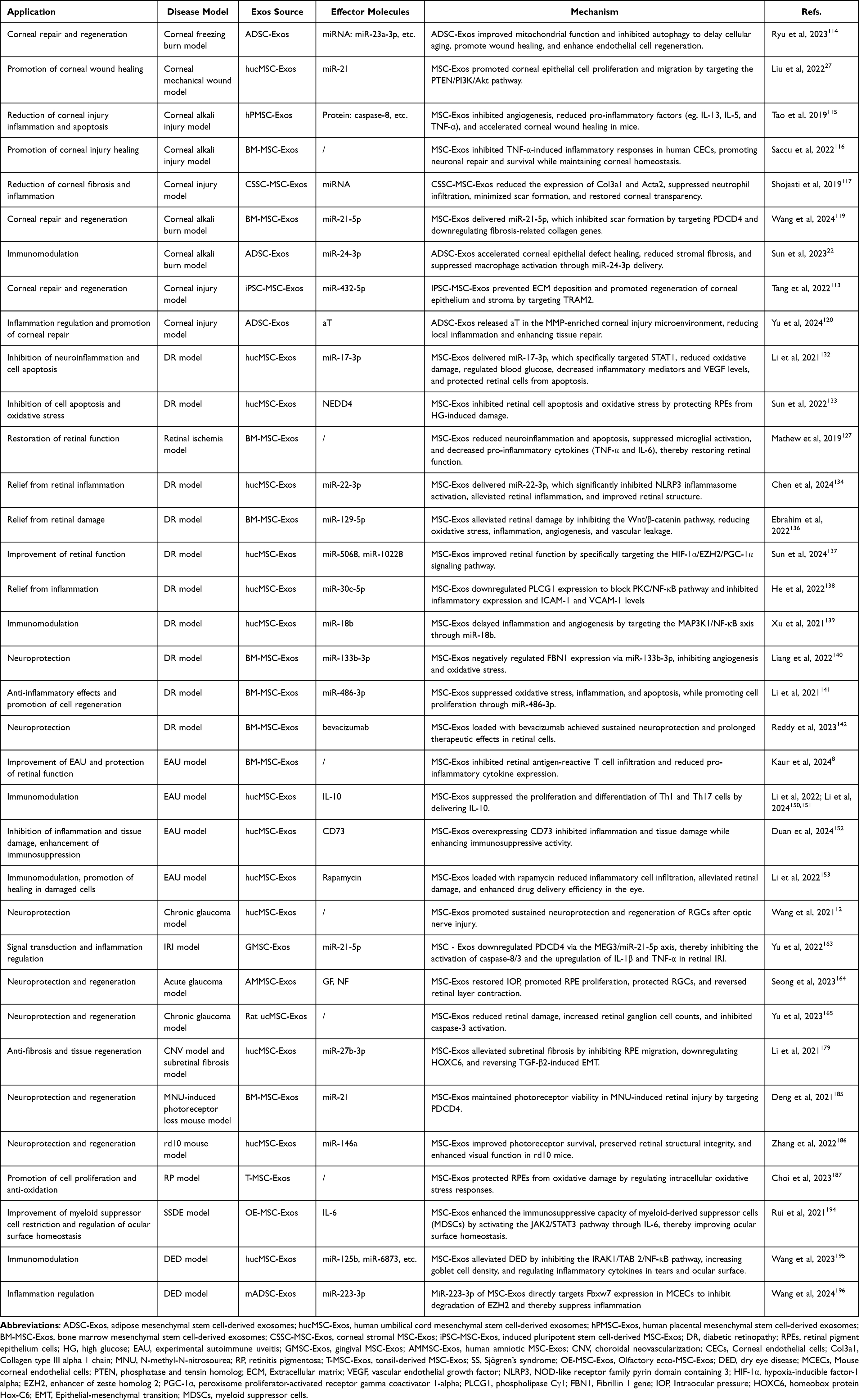 |
Table 1 Summary of the Mechanisms and Applications of MSC-Exos Therapy |
Clinical Trials
Although substantial research has been conducted in laboratory and animal studies, the clinical application of MSCs and their exosomes remains limited. While numerous clinical trials have been conducted, only five studies involving MSC-Exos for ocular disease treatment have been reported on clinicaltrials.gov (Table 2). Notably, among these registered trials, only one (NCT06242379) is currently listed as “Recruiting”, which is insufficient to assess the safety and efficacy of MSC-Exos-based therapies. This highlights the need for further research to validate their efficacy in clinical settings.
 |
Table 2 Summary of Clinical Trials of MSC-Exos for the Treatment of Ocular Diseases (from ClinicalTrials.gov) |
Here, we summarize the possible reasons for the limited number of clinical trials involving MSC-Exos in ocular diseases: MSC-Exos exhibit marked functional and compositional variability depending on their parental cell origin.199 For example, hucMSC-Exos are rich in immune-related proteins and miRNAs such as miR-125b-5p, which may be more beneficial for the treatment of immune-related diseases like acute kidney injury.200 Additionally, MSC-Exos derived from dental pulp contain more proteins related to the nervous system.199 Fetal-derived MSC-Exos exhibit superior osteogenic differentiation capacity compared to adult sources, indicating advantages for developmental tissue repair.201 Therefore, this heterogeneity necessitates rigorous characterization of exosomal cargo and therapeutic mechanisms to optimize donor cell selection for specific ocular pathologies. Secondly, MSC-Exos obtained by different methods may have differences in purity, size, and content, which can lead to inconsistent experimental results and make it difficult to compare data from different studies.202 Undoubtedly, uniform quality control standards, with specific surface markers or RNA content as prime examples, stand as the next formidable hurdle in the way of designing and conducting relevant clinical studies.202 Laboratory-level production is difficult to meet clinical needs, and a large-scale production process that meets Good Manufacturing Practice (GMP) standards needs to be developed.203 In addition, it is reported that only approximately 100 µg of MSC-Exos can be obtained from 500 mL of MSCs conditioned media using commercial separation kits.204 However, numerous published studies have used more than 100 µg of MSC-Exos in in vitro and in vivo models of ocular diseases.115,132,152 This highlights the significant challenges in producing sufficient quantities of MSC-Exos to meet the dosing requirements for future clinical trials, necessitating substantial financial and time investments. Finally, storing MSC-Exos can be challenging, with common methods including short-term storage at 4°C or room temperature, and long-term storage at −20°C or −80°C.205,206 However, MSC-Exos may become unstable during long-term storage, and repeated freeze-thaw cycles at −80°C can lead to structural damage or reduced activity.207 To address this, suitable storage conditions need to be identified, such as the use of cryoprotectants or determining the optimal storage temperature.207,208 Additionally, the high cost of cold-chain transportation and temperature fluctuations during transport can further compromise the quality of MSC-Exos, presenting a challenge for multicenter clinical trials conducted globally.209
Limitations, Perspectives and Challenges
Although robust animal studies have demonstrated that MSC-Exos, whether administered topically or systemically, can reach therapeutic tissue targets (eg, the eye), their clinical applicability remains relatively marginalized. A comprehensive understanding of MSC-Exos remains lacking, compounded by a myriad of challenges and limitations. There is a lot of exciting potential for MSC-Exos therapy, but translating this potential into a true treatment will depend on continued progress in several key areas. As previously noted, the foremost obstacle lies in addressing the heterogeneity of MSC-Exos composition, which is influenced by both the donor source and the method of isolation. While MSC-Exos can be extracted from various tissues, their therapeutic suitability is contingent upon factors such as their biochemical composition and functional properties. MSC-Exos obtained through differing isolation and enrichment techniques may exhibit variable compositions and functionalities, depending on the tissue of origin. For instance, ADSC-Exos contain neutral lysins at levels four times greater than those found in BM-MSC-Exos. Conversely, BM-MSC-Exos demonstrate markedly higher expression of proteins implicated in the Notch signaling pathway, rendering them more suitable for applications in bone and cartilage regeneration, as well as angiogenesis.210 Accordingly, selecting the appropriate MSC-Exos tissue source based on specific therapeutic objectives is essential to achieving optimal treatment outcomes. Variations in isolation methods further exacerbate inter-batch discrepancies: under identical MSC culture conditions, PEG precipitation yields six to seven times more particles and protein compared to ultracentrifugation. Nevertheless, ultracentrifugation produces MSC-Exos with miR-146a-6p levels two to three times higher, which may significantly influence their therapeutic efficacy.211 Ideally, the production method should maximize the therapeutic payload of the resulting MSC-Exos. Moreover, the development of stringent quality standards is imperative to reduce batch-to-batch variability and to safeguard the reproducibility, consistency, safety, and efficacy of MSC-Exos. Realizing these standards will necessitate ongoing research efforts and collaborative engagement across the field.
During the clinical translation process, the relatively short duration of action of MSC-Exos therapy poses another significant obstacle that must be addressed. Although a single administration has demonstrated efficacy in acute injury models, the chronic and progressive nature of many blinding diseases, such as diabetic retinopathy and glaucoma, often requires sustained therapeutic effects to control the condition.212 This transient effect may arise from rapid clearance by the immune system, dilution within the vitreous cavity, or the intrinsic biological turnover mechanisms of exosomes. To overcome this limitation, future research must shift towards developing strategies that extend the duration of therapeutic action and efficacy.213 Key directions include optimizing dosing regimens through systematic studies to determine the optimal administration frequency, concentration, and route, as well as designing advanced delivery systems that function as sustained-release reservoirs. For instance, encapsulating MSC-Exos within biocompatible hydrogels or microparticles can significantly prolong their retention at the target site, thereby reducing the frequency of intravitreal injections or topical administrations and improving patient compliance.
Another notable limitation pertains to scalability challenges in achieving production that complies with Good Manufacturing Practice (GMP) standards. Clinical translation of MSC-Exos mandates adherence to rigorous GMP protocols, particularly with respect to standardizing large-scale production. This imposes stringent demands on equipment, personnel, and procedural integrity, significantly inflating costs and posing further obstacles to clinical implementation.214 Conventional 2D culture methods are low-cost and straightforward to operate, remaining appropriate for fundamental research applications. However, 3D culture—particularly via bioreactor systems—surpasses 2D methods in terms of yield, efficiency, and therapeutic efficacy, rendering it more aligned with clinical application requirements. Nonetheless, it necessitates specialized equipment such as hollow fiber bioreactors, and entails a higher technical threshold and initial investment cost compared to 2D culture.215,216 The development of scalable, cost-efficient, and GMP-compliant 3D bioreactor platforms constitutes a pivotal future direction to surmount the scalability barrier in MSC-Exos production. In addition, refining standard operating procedures for MSC-Exos isolation and storage will aid in mitigating variability and ensuring consistent, high-quality production. Breakthroughs in the above direction will also help address patient concerns regarding immunogenicity, stability, and long-term administration of MSC-Exos. Furthermore, cross-sector collaboration between academia, industry, and regulatory authorities is vital for establishing universal benchmarks governing the quality, safety, and efficacy of MSC-Exos production. It is imperative to formulate guidelines for the standardized production of MSC-Exos as therapeutic agents, thereby facilitating their transition from the laboratory to clinical application.217
Although lipid nanomolecular therapy and gene therapy have been explored as alternative strategies, their limited targeting accuracy within the intricate ocular microenvironment continues to constrain their utility in precision medicine. By contrast, MSC-Exos, owing to their nanoscale dimensions and inherent biocompatibility, can be engineered to achieve targeting specificity, thereby surmounting biological barriers that conventional drugs and gene therapies often fail to penetrate. Notably, MSC-Exos possess the capacity to traverse formidable biological barriers, including the blood-brain barrier and blood-ocular barrier, an attribute rarely achievable with traditional delivery vectors such as liposomes and nanoparticles. Engineering strategies aimed at enhancing the targeting capability, drug-loading efficiency, and intelligent responsiveness of MSC-Exos are pivotal to overcoming existing bottlenecks and enabling precision therapeutics. Key engineering approaches currently under investigation include: (i) CRISPR-mediated modification of parental cells to enrich therapeutic miRNAs;218,219 (ii) Insertion of targeting ligands (peptides, antibodies, or aptamers) into the exosomal lipid bilayer or membrane-anchored proteins, enabling specific homing to diseased tissues/cells;83,220 (iii) biomaterials-loaded MSC-Exos (eg, stimuli-responsive hydrogels) to create “smart” therapeutics capable of dynamically responding to pathological microenvironments (eg, pH, enzymatic activity, or ROS), thereby enhancing cellular uptake and controlled cargo release.113,120 Concurrently, with a better understanding of the pathological characteristics of various ocular disease in progression stages, more precise and targeted MSC-Exos treatment plans are expected to be developed. By addressing these critical challenges and harnessing the power of engineering, MSC-Exos hold immense promise to usher in a new era of precision ophthalmology. In the future, targeted and long-lasting MSC-Exos therapies may well supplant invasive interventions, offering renewed hope for vision preservation on a global scale.
Conclusions
MSC-Exos have emerged as a transformative acellular platform for treating a spectrum of ocular diseases. This review has synthesized evidence demonstrating their efficacy in facilitating tissue repair (eg, in corneal wounds), mitigating neuroinflammation (eg, in diabetic retinopathy and glaucoma), and restoring immune homeostasis (eg, in uveitis), benefits largely attributed to their innate biocompatibility, ability to traverse biological barriers, and delivery of a multifaceted cargo of bioactive molecules. Collectively, these attributes position MSC-Exos as a paradigm shift in ocular therapeutics, with significant potential to supplant conventional invasive treatments and advance the field of precision ophthalmology. To fully realize this potential, future efforts must prioritize establishing standardized GMP production protocols, advancing the clinical translation of engineered exosomes, and optimizing dosing regimens to achieve sustained therapeutic effects. The ongoing integration of deeper pathological insights with bioengineering innovations is therefore crucial to propel MSC-Exos from promising experimental agents to mainstream clinical solutions for global vision preservation.
Abbreviations
ADSC-Exos, Adipose mesenchymal stem cell-derived exosomes; AMD, Age-related macular degeneration; AMESCs, Human amniotic epithelial stem cells; AMMSCs, Human amniotic stromal stem cells; Acta2, Alpha-smooth muscle actin 2; aT, Anti-tumor necrosis factor-α antibody; BM-MSC-Exos, Bone marrow mesenchymal stem cell-derived exosomes; BRB, Blood-retinal barrier; Bax, Bcl-2-associated X protein; Bcl-2, B-cell lymphoma 2; CFs, Conjunctival fibroblasts; CRISPR, Clustered Regularly Interspaced Short Palindromic Repeats; CECs, Corneal endothelial cells; CHI, Chitosan-based hydrogel; CNV, Choroidal neovascularization; CSSC-MSC-Exos, Corneal stromal MSC-Exos; GA, Geographic atrophy; GMP, Good Manufacturing Practice; DBCO, Dibenzocyclooctyne; DED, Dry eye disease; DR, Diabetic retinopathy; EAU, Experimental autoimmune uveitis; ECM, Extracellular matrix; EMT, Epithelial-mesenchymal transition; EVs, Extracellular vesicles; EZH2, Enhancer of zeste homolog 2; FBN1, Fibrillin-1 gene; GF, Growth factors; GMSC-Exos, Gingival MSC-Exos; HG, High glucose; HIF-1α, Hypoxia-inducible factor-1 alpha; HOXC6, Homeobox protein Hox-C6; hPMSC-Exos, Human placental mesenchymal stem cell-derived exosomes; hucMSC-Exos, Human umbilical cord mesenchymal stem cell-derived exosomes; ICAM-1, Intercellular adhesion molecule-1; IL-10, Interleukin-10; IL-5, Interleukin-5; IL-13, Interleukin-13; IPL, Inner plexiform layer; IRBP, Inter-photoreceptor retinal binding protein; IOP, Intraocular pressure; IRI, Retinal ischemia-reperfusion injury; iPSC-MSC-Exos, Induced pluripotent stem cell-derived MSC-exosomes; MDSCs, Myeloid suppressor cells; MMPs, Matrix metalloproteinases; MAP3K1, Mitogen-activated protein kinase kinase kinase 1; MMP-2, Matrix metalloproteinase-2; MMP-9, Matrix metalloproteinase-9; MVB, Multivesicular bodies; MNU, N-methyl-N-nitrosourea; MSC-Exos, Mesenchymal stem cell-derived exosomes; MSCs, Mesenchymal stem cells; MSCT, MSCs transplantation; NF, Neurotrophic factors; NF-κB, Nuclear factor kappa B; NTA, Nanoparticle tracking analysis; NLRP3, NOD-like receptor family pyrin domain containing 3; NPDR, Non-proliferative diabetic retinopathy; NO, Nitric oxide; OE-MSC-Exos, Olfactory ecto-MSC-Exos; PDCD4, Programmed cell death 4; PDR, Proliferative diabetic retinopathy; PEG, Polyethylene glycol; PGC-1α, Peroxisome proliferator-activated receptor gamma coactivator 1-alpha; PLCG1, Phospholipase Cγ1; PTEN, Phosphatase and tensin homolog; qPCR, Quantitative real-time polymerase chain reaction; Rapa-MSC-Exos, Rapamycin-loaded MSC-Exos; ROS, Reactive oxygen species; RGCs, Retinal ganglion cells; RP, Retinitis pigmentosa; RPEs, Retinal pigment epithelium cells; SEM, Scanning electron microscopy; SSDE, Sjögren’s syndrome dry eye; STAT1, Signal transducer and activator of transcription 1; STAT3, Signal transducer and activator of transcription 3; STZ, Streptozotocin; T-MSC-Exos, Tonsil-derived MSC-Exos; TGF-β, Transforming growth factor-beta; TNF-α, Tumor necrosis factor alpha; TRAM2, Translocation-associated membrane protein 2; Th1, T helper 1 cells; Th17, T helper 17 cells; TLR4, Toll-like receptor 4; TEM, Transmission electron microscopy; VCAM-1, Vascular cell adhesion molecule-1; VEGF, Vascular endothelial growth factor; Wnt/β-catenin, Wnt/β-catenin signaling pathway.
Funding
This work was supported by the National Natural Science Foundation of China (81973265), National Forestry and Grassland Administration (CGF2024002), the Miaozi Project in Science and Technology Innovation Program of Sichuan Province (2024JDRC0035), Program of the Chengdu Research Base of Giant Panda Breeding (2024CPB-B17), the Fundamental Research Funds for the Central Universities, Southwest Minzu University (ZYN2025233), and the Open Project Program of Guangxi Key Laboratory of Brain and Cognitive Neuroscience, Guilin Medical University (GKLBCN-202509-02).
Disclosure
The authors report no conflicts of interest in this work.
References
1. Xu T, Wang B, Liu H, et al. Prevalence and causes of vision loss in China from 1990 to 2019: findings from the global burden of disease study 2019. Lancet Public Health. 2020;5(12):e682–e691. doi:10.1016/S2468-2667(20)30254-1
2. Bourne R, Steinmetz JD, Flaxman S, et al. Trends in prevalence of blindness and distance and near vision impairment over 30 years: an analysis for the global burden of disease study. Lancet Glob Health. 2021;9(2):e130–e143. doi:10.1016/S2214-109X(20)30425-3
3. Feng X, Peng Z, Yuan L, et al. Research progress of exosomes in pathogenesis, diagnosis, and treatment of ocular diseases. Front Bioeng Biotechnol. 2023;11:1100310. doi:10.3389/fbioe.2023.1100310
4. Bai S, Bai S, Luo T, et al. Chemiluminescent afterglow material for enhanced tumor diagnosis and photodynamic therapy. Adv Funct Mater. 2024;34. doi: 10.1002/adfm.202400842
5. Ehlers JP, Yeh S, Maguire MG, et al. Intravitreal pharmacotherapies for diabetic macular edema. Ophthalmology. 2022;129(1):88–99. doi:10.1016/j.ophtha.2021.07.009
6. Mandal A, Pal D, Agrahari V, Trinh HM, Joseph M, Mitra AK. Ocular delivery of proteins and peptides: challenges and novel formulation approaches. Adv Drug Deliv Rev. 2018;126:67–95. doi:10.1016/j.addr.2018.01.008
7. Ma S, Liu X, Yin J, Hao L, Diao Y, Zhong J. Exosomes and autophagy in ocular surface and retinal diseases: new insights into pathophysiology and treatment. Stem Cell Res Ther. 2022;13(1):174. doi:10.1186/s13287-022-02854-8
8. Kaur G, Bae E, Zhang Y, et al. Biopotency and surrogate assays to validate the immunomodulatory potency of extracellular vesicles derived from mesenchymal stem/stromal cells for the treatment of experimental autoimmune uveitis. J Extracellular Vesicle. 2024;13(8):e12497. doi:10.1002/jev2.12497
9. Radu M, Brănișteanu DC, Pirvulescu RA, Dumitrescu OM, Ionescu MA, Zemba M. Exploring stem-cell-based therapies for retinal regeneration. Life. 2024;14(6):668. doi:10.3390/life14060668
10. Hoang DM, Pham PT, Bach TQ, et al. Stem cell-based therapy for human diseases. Sig Transduct Target Ther. 2022;7(1):272.
11. Holan V, Palacka K, Hermankova B. Mesenchymal stem cell-based therapy for retinal degenerative diseases: experimental models and clinical trials. Cells. 2021;10(3):588. doi:10.3390/cells10030588
12. Wang Y, Lv J, Huang C, et al. Human umbilical cord-mesenchymal stem cells survive and migrate within the vitreous cavity and ameliorate retinal damage in a novel rat model of chronic glaucoma. Stem Cells Int. 2021;2021:1–11. doi:10.1155/2021/7625134
13. Gugliandolo A, Mazzon E. Dental mesenchymal stem cell secretome: an intriguing approach for neuroprotection and neuroregeneration. IJMS. 2021;23(1):456. doi:10.3390/ijms23010456
14. Zhou T, Yuan Z, Weng J, et al. Challenges and advances in clinical applications of mesenchymal stromal cells. J Hematol Oncol. 2021;14(1):24. doi:10.1186/s13045-021-01037-x
15. Ouzin M, Kogler G. Mesenchymal stromal cells: heterogeneity and therapeutical applications. Cells. 2023;12(16):2039.
16. Abdulmalek OAAY, Husain KH, AlKhalifa HKAA, Alturani MMAB, Butler AE, Moin ASM. Therapeutic applications of stem cell-derived exosomes. IJMS. 2024;25(6):3562.
17. Wang Y, Liu X, Wang B, Sun H, Ren Y, Zhang H. Compounding engineered mesenchymal stem cell-derived exosomes: a potential rescue strategy for retinal degeneration. Biomed Pharmacother. 2024;173:116424. doi:10.1016/j.biopha.2024.116424
18. Song Y, Yin C, Kong N. Stem cell-derived exosomes: natural intercellular messengers with versatile mechanisms for the treatment of diabetic retinopathy. Int J Nanomed. 2024;19:10767–10784. doi:10.2147/IJN.S475234
19. Hakim A, Guido B, Narsineni L, Chen DW, Foldvari M. Gene therapy strategies for glaucoma from IOP reduction to retinal neuroprotection: progress towards non-viral systems. Adv Drug Deliv Rev. 2023;196:114781. doi:10.1016/j.addr.2023.114781
20. Khorrami-Nejad M, Hashemian H, Majdi A, Jadidi K, Aghamollaei H, Hadi A. Application of stem cell-derived exosomes in anterior segment eye diseases: a comprehensive update review. Ocul Surf. 2025;36:209–219. doi:10.1016/j.jtos.2025.01.012
21. Liu J, Gao J, Lu P, et al. Mesenchymal stem cell-derived exosomes as drug carriers for delivering miRNA-29b to ameliorate inflammation in corneal injury via activating autophagy. Invest Ophthalmol Vis Sci. 2024;65(6):16. doi:10.1167/iovs.65.6.16
22. Sun X, Song W, Teng L, et al. MiRNA 24-3p-rich exosomes functionalized DEGMA-modified hyaluronic acid hydrogels for corneal epithelial healing. Bioact Mater. 2023;25:640–656. doi:10.1016/j.bioactmat.2022.07.011
23. Mondal J, Pillarisetti S, Junnuthula V, et al. Hybrid exosomes, exosome-like nanovesicles and engineered exosomes for therapeutic applications. J Control Release. 2023;353:1127–1149. doi:10.1016/j.jconrel.2022.12.027
24. Zhu X, Ma D, Yang B, et al. Research progress of engineered mesenchymal stem cells and their derived exosomes and their application in autoimmune/inflammatory diseases. Stem Cell Res Ther. 2023;14(1):71. doi:10.1186/s13287-023-03295-7
25. Yang L, Li W, Zhao Y, Shang L. Magnetic polysaccharide mesenchymal stem cells exosomes delivery microcarriers for synergistic therapy of osteoarthritis. ACS Nano. 2024;18(31):20101–20110. doi:10.1021/acsnano.4c01406
26. Chen G, Tong K, Li S, et al. Extracellular vesicles released by transforming growth factor-beta 1-preconditional mesenchymal stem cells promote recovery in mice with spinal cord injury. Bioact Mater. 2024;35:135–149. doi:10.1016/j.bioactmat.2024.01.013
27. Liu X, Li X, Wu G, et al. Umbilical cord mesenchymal stem cell-derived small extracellular vesicles deliver mir-21 to promote corneal epithelial wound healing through PTEN/PI3K/Akt pathway. Stem Cells Int. 2022;2022:1–15.
28. Liu Y, Xia P, Yan F, et al. Engineered extracellular vesicles for delivery of an IL‐1 receptor antagonist promote targeted repair of retinal degeneration. Small. 2023;19(46):2302962. doi:10.1002/smll.202302962
29. Ji S, Peng Y, Liu J, Xu P, Tang S. Human adipose tissue-derived stem cell extracellular vesicles attenuate ocular hypertension-induced retinal ganglion cell damage by inhibiting microglia- TLR4/MAPK/NF-κB proinflammatory cascade signaling. Acta Neuropathol Commun. 2024;12(1):44. doi:10.1186/s40478-024-01753-8
30. Long Z, Qiu X, Chan CLJ, et al. A neuromorphic bionic eye with filter-free color vision using hemispherical perovskite nanowire array retina. Nat Commun. 2023;14(1):1972. doi:10.1038/s41467-023-37581-y
31. Ma Y, Zhang Z, Yu Y, et al. Nanomaterials in the diagnosis and treatment of ophthalmic diseases. Nano Today. 2024;54:102117.
32. Kuniyoshi K, Hayashi T, Kameya S, et al. Clinical course and electron microscopic findings in lymphocytes of patients with DRAM2-associated retinopathy. IJMS. 2020;21(4):1331. doi:10.3390/ijms21041331
33. Singh RB, Dohlman TH, Ivanov A, et al. Corneal opacity in the United States. Ophthalmology. 2024;S0161642024004160.
34. McAnany JJ, Park JC, Lim JI. Visual field abnormalities in early-stage diabetic retinopathy assessed by chromatic perimetry. Invest Ophthalmol Vis Sci. 2023;64(2):8. doi:10.1167/iovs.64.2.8
35. Jackson TL, Haller J, Blot KH, Duchateau L, Lescrauwaet B. Ocriplasmin for treatment of vitreomacular traction and macular hole: a systematic literature review and individual participant data meta-analysis of randomized, controlled, double-masked trials. Surv Ophthalmol. 2022;67(3):697–711. doi:10.1016/j.survophthal.2021.08.003
36. Sugisaki K, Inoue T, Yoshikawa K, et al. Factors threatening central visual function of patients with advanced glaucoma. Ophthalmology. 2022;129(5):488–497. doi:10.1016/j.ophtha.2021.11.025
37. Maghsoudlou P, Epps SJ, Guly CM, Dick AD. Uveitis in adults. JAMA. 2025;334(5):419. doi:10.1001/jama.2025.4358
38. Su Y, Fan X, Pang Y. Nano-based ocular drug delivery systems: an insight into the preclinical/clinical studies and their potential in the treatment of posterior ocular diseases. Biomater Sci. 2023;11(13):4490–4507. doi:10.1039/D3BM00505D
39. Jayaram H, Kolko M, Friedman DS, Gazzard G. Glaucoma: now and beyond. Lancet. 2023;402(10414):1788–1801. doi:10.1016/S0140-6736(23)01289-8
40. Becker S, L’Ecuyer Z, Jones BW, Zouache MA, McDonnell FS, Vinberg F. Modeling complex age-related eye disease. Prog Retin Eye Res. 2024;100:101247. doi:10.1016/j.preteyeres.2024.101247
41. Said DG, Rallis KI, Al-Aqaba MA, Ting DSJ, Dua HS. Surgical management of infectious keratitis. Ocul Surf. 2023;28:401–412. doi:10.1016/j.jtos.2021.09.005
42. Wu Y, Li X, Fu X, et al. Innovative nanotechnology in drug delivery systems for advanced treatment of posterior segment ocular diseases. Adv Sci. 2024;11(32):2403399. doi:10.1002/advs.202403399
43. Reis LM, Seese SE, Costakos D, Semina EV. Congenital anterior segment ocular disorders: genotype-phenotype correlations and emerging novel mechanisms. Prog Retin Eye Res. 2024;102:101288. doi:10.1016/j.preteyeres.2024.101288
44. Pedersen FN, Stokholm L, Andersen N, et al. Risk of diabetic retinopathy according to subtype of type 2 diabetes. Diabetes. 2024;73(6):977–982. doi:10.2337/db24-0016
45. Shan H, Liu W, Li Y, Pang K. The autoimmune rheumatic disease related dry eye and its association with retinopathy. Biomolecules. 2023;13(5):724. doi:10.3390/biom13050724
46. Vallabh NA, Armstrong J, Czanner G, et al. Evidence of impaired mitochondrial cellular bioenergetics in ocular fibroblasts derived from glaucoma patients. Free Radic Biol Med. 2022;189:102–110. doi:10.1016/j.freeradbiomed.2022.07.009
47. Dixson A, Dawson TR, Di Vizio D, Weaver AM. Context-specific regulation of extracellular vesicle biogenesis and cargo selection. Nat Rev Mol Cell Biol. 2023;24(7):454–476. doi:10.1038/s41580-023-00576-0
48. Kalluri R, LeBleu VS. The biology, function, and biomedical applications of exosomes. Science. 2020;367(6478):eaau6977. doi:10.1126/science.aau6977
49. Lee YJ, Shin KJ, Chae YC. Regulation of cargo selection in exosome biogenesis and its biomedical applications in cancer. Exp Mol Med. 2024;56(4):877–889. doi:10.1038/s12276-024-01209-y
50. Xu M, Ji J, Jin D, et al. The biogenesis and secretion of exosomes and multivesicular bodies (MVBs): intercellular shuttles and implications in human diseases. Gen Dis. 2023;10(5):1894–1907. doi:10.1016/j.gendis.2022.03.021
51. Li M, Li S, Du C, et al. Exosomes from different cells: characteristics, modifications, and therapeutic applications. Eur J Med Chem. 2020;207:112784. doi:10.1016/j.ejmech.2020.112784
52. Racchetti G, Meldolesi J. Extracellular vesicles of mesenchymal stem cells: therapeutic properties discovered with extraordinary success. Biomedicines. 2021;9(6):667. doi:10.3390/biomedicines9060667
53. Lin Z, Wu Y, Xu Y, Li G, Li Z, Liu T. Mesenchymal stem cell-derived exosomes in cancer therapy resistance: recent advances and therapeutic potential. Mol Cancer. 2022;21(1):179. doi:10.1186/s12943-022-01650-5
54. Wang Y, Xiao T, Zhao C, Li G. The regulation of exosome generation and function in physiological and pathological processes. Int J Mol Sci. 2024;25(1):255. doi:10.3390/ijms25010255
55. Gurung S, Perocheau D, Touramanidou L, Baruteau J. The exosome journey: from biogenesis to uptake and intracellular signalling. Cell Commun Signal. 2021;19(1):47. doi:10.1186/s12964-021-00730-1
56. Noonin C, Thongboonkerd V. Exosome-inflammasome crosstalk and their roles in inflammatory responses. Theranostics. 2021;11(9):4436–4451. doi:10.7150/thno.54004
57. Kou M, Huang L, Yang J, et al. Mesenchymal stem cell-derived extracellular vesicles for immunomodulation and regeneration: a next generation therapeutic tool? Cell Death Dis. 2022;13(7):580. doi:10.1038/s41419-022-05034-x
58. Hade MD, Suire CN, Suo Z. Mesenchymal stem cell-derived exosomes: applications in regenerative medicine. Cells. 2021;10(8):1959.
59. Huang D, Shen H, Xie F, et al. Role of mesenchymal stem cell-derived exosomes in the regeneration of different tissues. J Biol Eng. 2024;18(1):36. doi:10.1186/s13036-024-00431-6
60. Long R, Wang S. Exosomes from preconditioned mesenchymal stem cells: tissue repair and regeneration. Regen Ther. 2024;25:355–366. doi:10.1016/j.reth.2024.01.009
61. Huda MN, Nafiujjaman M, Deaguero IG, et al. Potential use of exosomes as diagnostic biomarkers and in targeted drug delivery: progress in clinical and preclinical applications. ACS Biomater Sci Eng. 2021;7(6):2106–2149. doi:10.1021/acsbiomaterials.1c00217
62. Lozano V, Martín C, Blanco N, et al. Exosomes released by corneal stromal cells show molecular alterations in keratoconus patients and induce different cellular behavior. Biomedicines. 2022;10(10):2348. doi:10.3390/biomedicines10102348
63. Chen YF, Luh F, Ho YS, Yen Y. Exosomes: a review of biologic function, diagnostic and targeted therapy applications, and clinical trials. J Biomed Sci. 2024;31(1):1–16. doi:10.1186/s12929-024-01055-0
64. Liu W, Ma Z, Kang X. Current status and outlook of advances in exosome isolation. Anal Bioanal Chem. 2022;414(24):7123–7141. doi:10.1007/s00216-022-04253-7
65. Tang Y, Zhou Y, Li HJ. Advances in mesenchymal stem cell exosomes: a review. Stem Cell Res Ther. 2021;12(1):71. doi:10.1186/s13287-021-02138-7
66. Han L, Zhan H, Sun X, Zhang ZR, Deng L. A density-changing centrifugation method for efficient separation of free drugs from drug-loaded particulate delivery systems. AAPS J. 2019;21(3):33. doi:10.1208/s12248-019-0306-1
67. Kaddour H, Tranquille M, Okeoma CM. The past, the present, and the future of the size exclusion chromatography in extracellular vesicles separation. Viruses. 2021;13(11):2272. doi:10.3390/v13112272
68. Skovronova R, Scaccia E, Calcat-i-Cervera S, Bussolati B, O’Brien T, Bieback K. Adipose stromal cells bioproducts as cell-free therapies: manufacturing and therapeutic dose determine in vitro functionality. J Transl Med. 2023;21(1):723. doi:10.1186/s12967-023-04602-9
69. Monguió-Tortajada M, Gálvez-Montón C, Bayes-Genis A, Roura S, Borràs FE. Extracellular vesicle isolation methods: rising impact of size-exclusion chromatography. Cell Mol Life Sci. 2019;76(12):2369–2382. doi:10.1007/s00018-019-03071-y
70. Chen J, Li P, Zhang T, et al. Review on strategies and technologies for exosome isolation and purification. Front Bioeng Biotechnol. 2022;9.
71. Gandham S, Su X, Wood J, et al. Technologies and standardization in research on extracellular vesicles. Trends Biotechnol. 2020;38(10):1066–1098. [Special Issue: Therapeutic Biomanufacturing]. doi:10.1016/j.tibtech.2020.05.012
72. Kamei N, Nishimura H, Matsumoto A, et al. Comparative study of commercial protocols for high recovery of high-purity mesenchymal stem cell-derived extracellular vesicle isolation and their efficient labeling with fluorescent dyes. Nanomedicine. 2021;35:102396. doi:10.1016/j.nano.2021.102396
73. Rahmatinejad F, Kharat Z, Jalili H, Renani MK, Mobasheri H. Comparison of morphology, protein concentration, and size distribution of bone marrow and Wharton’s jelly-derived mesenchymal stem cells exosomes isolated by ultracentrifugation and polymer-based precipitation techniques. Tissue and Cell. 2024;88:102427. doi:10.1016/j.tice.2024.102427
74. Sonbhadra S, Mehak PLM, Pandey LM. Biogenesis, isolation, and detection of exosomes and their potential in therapeutics and diagnostics. Biosensors. 2023;13(8):802. doi:10.3390/bios13080802
75. García-Posadas L, Romero-Castillo I, Brennan K, Mc Gee MM, Blanco-Fernández A, Diebold Y. Isolation and characterization of human conjunctival mesenchymal stromal cells and their extracellular vesicles. Invest Ophthalmol Vis Sci. 2023;64(12):38. doi:10.1167/iovs.64.12.38
76. Franco C, Ghirardello A, Bertazza L, et al. Size-exclusion chromatography combined with ultrafiltration efficiently isolates extracellular vesicles from human blood samples in health and disease. Int J Mol Sci. 2023;24(4):3663. doi:10.3390/ijms24043663
77. Chandrasekera D, Shah R, van Hout I, et al. Combination of precipitation and size exclusion chromatography as an effective method for exosome like extracellular vesicle isolation from pericardial fluids. Nanotheranostics. 2023;7(4):345–352. doi:10.7150/ntno.82939
78. Jang J, Jeong H, Jang E, et al. Isolation of high-purity and high-stability exosomes from ginseng. Front Plant Sci. 2023;13:1064412.
79. Liu W, Wang X, Chen Y, et al. Distinct molecular properties and functions of small EV subpopulations isolated from human umbilical cord MSCs using tangential flow filtration combined with size exclusion chromatography. J Extracellular Vesicle. 2025;14(1):e70029. doi:10.1002/jev2.70029
80. Hao R, Hu S, Zhang H, et al. Mechanical stimulation on a microfluidic device to highly enhance small extracellular vesicle secretion of mesenchymal stem cells. Mater Today Bio. 2023;18:100527. doi:10.1016/j.mtbio.2022.100527
81. Ding L, Yang X, Gao Z, et al. A holistic review of the state‐of‐the‐art microfluidics for exosome separation: an overview of the current status, existing obstacles, and future outlook. Small. 2021;17(29):2007174. doi:10.1002/smll.202007174
82. Xu Y, Qin S, Niu Y, Gong T, Zhang Z, Fu Y. Effect of fluid shear stress on the internalization of kidney-targeted delivery systems in renal tubular epithelial cells. Acta Pharmaceutica Sinica B. 2020;10(4):680–692. doi:10.1016/j.apsb.2019.11.012
83. Meng W, Wang L, Du X, et al. Engineered mesenchymal stem cell-derived extracellular vesicles constitute a versatile platform for targeted drug delivery. J Control Release. 2023;363:235–252. doi:10.1016/j.jconrel.2023.09.037
84. Abbasi R, Alamdari-Mahd G, Maleki-Kakelar H, et al. Recent advances in the application of engineered exosomes from mesenchymal stem cells for regenerative medicine. Eur J Pharmacol. 2025;989:177236. doi:10.1016/j.ejphar.2024.177236
85. Yang Z, Shi J, Xie J, et al. Large-scale generation of functional mRNA-encapsulating exosomes via cellular nanoporation. Nat Biomed Eng. 2019;4(1):69–83. doi:10.1038/s41551-019-0485-1
86. Yu M, Liu W, Li J, et al. Exosomes derived from atorvastatin-pretreated MSC accelerate diabetic wound repair by enhancing angiogenesis via AKT/eNOS pathway. Stem Cell Res Ther. 2020;11(1):350. doi:10.1186/s13287-020-01824-2
87. Akbari A, Nazari-Khanamiri F, Ahmadi M, Shoaran M, Rezaie J. Engineered exosomes for tumor-targeted drug delivery: a focus on genetic and chemical functionalization. Pharmaceutics. 2023;15(1):66. doi:10.3390/pharmaceutics15010066
88. Xing Y, Yerneni SS, Wang W, Taylor RE, Campbell PG, Ren X. Engineering pro-angiogenic biomaterials via chemoselective extracellular vesicle immobilization. Biomaterials. 2022;281:121357. doi:10.1016/j.biomaterials.2021.121357
89. Wu D, Zhao X, Xie J, et al. Physical modulation of mesenchymal stem cell exosomes: a new perspective for regenerative medicine. Cell Proliferation. 2024;57(8):e13630. doi:10.1111/cpr.13630
90. Zhang M, Hu S, Liu L, et al. Engineered exosomes from different sources for cancer-targeted therapy. Sig Transduct Target Ther. 2023;8(1):1–20.
91. Bahadorani M, Nasiri M, Dellinger K, Aravamudhan S, Zadegan R. Engineering exosomes for therapeutic applications: decoding biogenesis, content modification, and cargo loading strategies. IJN. 2024;19:7137–7164. doi:10.2147/IJN.S464249
92. Chen J, Zhang E, Wan Y, Huang T, Wang Y, Jiang H. Engineered hsa‐miR‐455‐3p‐abundant extracellular vesicles derived from 3D‐cultured adipose mesenchymal stem cells for tissue‐engineering hyaline cartilage regeneration. Adv Healthcare Mater. 2024;13(18):2304194. doi:10.1002/adhm.202304194
93. Xiong J, Liu Z, Jia L, et al. Bioinspired engineering ADSC nanovesicles thermosensitive hydrogel enhance autophagy of dermal papilla cells for androgenetic alopecia treatment. Bioact Mater. 2024;36:112–125. doi:10.1016/j.bioactmat.2024.02.023
94. Yerneni SS, Yalcintas EP, Smith JD, Averick S, Campbell PG, Ozdoganlar OB. Skin-targeted delivery of extracellular vesicle-encapsulated curcumin using dissolvable microneedle arrays. Acta Biomaterialia. 2022;149:198–212. doi:10.1016/j.actbio.2022.06.046
95. Lotfi S, Anvari P, Ramezani A, Kazemi P, Ahmadvand D. Celecoxib-loaded mesenchymal stem cell-derived exosomes ameliorate laser-induced choroidal neovascularization in a rabbit model. J Drug Deliv Sci Technol. 2025;114:107514. doi:10.1016/j.jddst.2025.107514
96. Bagrov DV, Senkovenko AM, Nikishin II, Skryabin GO, Kopnin PB, Tchevkina EM. Application of AFM, TEM, and NTA for characterization of exosomes produced by placenta-derived mesenchymal cells. J Phy. 2021;1942(1):012013.
97. Nguyen CM, Sallam M, Islam MS, et al. Placental exosomes as biomarkers for maternal diseases: current advances in isolation, characterization, and detection. ACS Sensors. 2023;8(7):2493–2513. doi:10.1021/acssensors.3c00689
98. Gurunathan S, Kang MH, Jeyaraj M, Qasim M, Kim JH. Review of the isolation, characterization, biological function, and multifarious therapeutic approaches of exosomes. Cells. 2019;8(4):307. doi:10.3390/cells8040307
99. Mukerjee N, Bhattacharya A, Maitra S, et al. Exosome isolation and characterization for advanced diagnostic and therapeutic applications. Mater Today Bio. 2025;31:101613. doi:10.1016/j.mtbio.2025.101613
100. Xu H, Zhao G, Zhang Y, et al. Mesenchymal stem cell-derived exosomal microRNA-133b suppresses glioma progression via Wnt/β-catenin signaling pathway by targeting EZH2. Stem Cell Res Ther. 2019;10(1):381. doi:10.1186/s13287-019-1446-z
101. He L, Piao G, Yin X, et al. A novel electrochemical biosensor based on TiO2 nanotube array films for highly sensitive detection of exosomes. Talanta. 2025;286:127545. doi:10.1016/j.talanta.2025.127545
102. Price MO, Mehta JS, Jurkunas UV, Price FW. Corneal endothelial dysfunction: evolving understanding and treatment options. Prog Retin Eye Res. 2021;82:100904. doi:10.1016/j.preteyeres.2020.100904
103. Kumar R, Sinha NR, Mohan RR. Corneal gene therapy: structural and mechanistic understanding. Ocul Surf. 2023;29:279–297. doi:10.1016/j.jtos.2023.05.007
104. Brown L, Leck AK, Gichangi M, Burton MJ, Denning DW. The global incidence and diagnosis of fungal keratitis. Lancet Infect Dis. 2021;21(3):e49–e57. doi:10.1016/S1473-3099(20)30448-5
105. Somayajulu M, McClellan SA, Wright R, et al. Airborne exposure of the cornea to PM10 induces oxidative stress and disrupts Nrf2 mediated anti-oxidant defenses. IJMS. 2023;24(4):3911. doi:10.3390/ijms24043911
106. Cerván-Martín M, Higueras-Serrano I, González-Muñoz S, et al. Comprehensive evaluation of the genetic basis of keratoconus: new perspectives for clinical translation. Invest Ophthalmol Vis Sci. 2024;65(12):32. doi:10.1167/iovs.65.12.32
107. Xu Y, Liu J, Song W, et al. Biomimetic convex implant for corneal regeneration through 3D printing. Adv Sci. 2023;10(11):2205878. doi:10.1002/advs.202205878
108. Lasagni Vitar RM, Rama P, Ferrari G. The two-faced effects of nerves and neuropeptides in corneal diseases. Prog Retin Eye Res. 2022;86:100974. doi:10.1016/j.preteyeres.2021.100974
109. Li M, Wei R, Liu C, et al. A “T.E.S.T.” hydrogel bioadhesive assisted by corneal cross-linking for in situ sutureless corneal repair. Bioact Mater. 2023;25:333–346. doi:10.1016/j.bioactmat.2023.02.006
110. Dang DH, Riaz KM, Karamichos D. Treatment of non-infectious corneal injury: review of diagnostic agents, therapeutic medications, and future targets. Drugs. 2022;82(2):145–167. doi:10.1007/s40265-021-01660-5
111. Mohan RR, Kempuraj D, D’Souza S, Ghosh A. Corneal stromal repair and regeneration. Prog Retin Eye Res. 2022;91:101090. doi:10.1016/j.preteyeres.2022.101090
112. Zhao L, Shi Z, Qi X, et al. Corneal stromal structure replicating humanized hydrogel patch for sutureless repair of deep anterior-corneal defect. Biomaterials. 2025;313:122754. doi:10.1016/j.biomaterials.2024.122754
113. Tang Q, Lu B, He J, et al. Exosomes-loaded thermosensitive hydrogels for corneal epithelium and stroma regeneration. Biomaterials. 2022;280:121320. doi:10.1016/j.biomaterials.2021.121320
114. Ryu Y, Hwang JS, Bo Noh K, Park SH, Seo JH, Shin YJ. Adipose mesenchymal stem cell-derived exosomes promote the regeneration of corneal endothelium through ameliorating senescence. Invest Ophthalmol Vis Sci. 2023;64(13):29. doi:10.1167/iovs.64.13.29
115. Tao H, Chen X, Cao H, et al. Mesenchymal stem cell-derived extracellular vesicles for corneal wound repair. Stem Cells Int. 2019;2019:1–9. doi:10.1155/2019/5738510
116. Saccu G, Menchise V, Gai C, et al. Bone marrow mesenchymal stromal/stem cell-derived extracellular vesicles promote corneal wound repair by regulating inflammation and angiogenesis. Cells. 2022;11(23):3892. doi:10.3390/cells11233892
117. Shojaati G, Khandaker I, Funderburgh ML, et al. Mesenchymal stem cells reduce corneal fibrosis and inflammation via extracellular vesicle-mediated delivery of miRNA. Stem Cells Transl Med. 2019;8(11):1192–1201. doi:10.1002/sctm.18-0297
118. Yu Z, Hao R, Du J, et al. A human cornea-on-a-chip for the study of epithelial wound healing by extracellular vesicles. Iscience. 2022;25(5):104200. doi:10.1016/j.isci.2022.104200
119. Wang J, Wang X, Ma X, et al. Mesenchymal stem cells derived extracellular vesicles modified PLGA electrospinning nanofibrous scaffolds for corneal and retinal repair. Mater Design. 2024;247:113389. doi:10.1016/j.matdes.2024.113389
120. Yu F, Zhao X, Wang Q, et al. Engineered mesenchymal stromal cell exosomes-loaded microneedles improve corneal healing after chemical injury. ACS Nano. 2024;18(31):20065–20082. doi:10.1021/acsnano.4c00423
121. Dai L, Wu L, Li H, et al. A deep learning system for detecting diabetic retinopathy across the disease spectrum. Nat Commun. 2021;12(1):3242. doi:10.1038/s41467-021-23458-5
122. Huang RS, Naidu SC, Mihalache A, et al. Loss to follow-up in patients with proliferative diabetic retinopathy or diabetic macular edema. JAMA Network Open. 2024;7(12):e2450942. doi:10.1001/jamanetworkopen.2024.50942
123. Wei L, Sun X, Fan C, Li R, Zhou S, Yu H. The pathophysiological mechanisms underlying diabetic retinopathy. Front Cell Dev Biol. 2022;10:963615.
124. Jiang Q, Liu C, Li CP, et al. Circular RNA-ZNF532 regulates diabetes-induced retinal pericyte degeneration and vascular dysfunction. J Clin Invest. 2020;130(7):3833–3847. doi:10.1172/JCI123353
125. Tan GSW, Chakravarthy U, Wong TY. Anti-VEGF therapy or vitrectomy surgery for vitreous hemorrhage from proliferative diabetic retinopathy. JAMA. 2020;324(23):2375. doi:10.1001/jama.2020.22829
126. Yang M, Chen J, Chen L. The roles of mesenchymal stem cell-derived exosomes in diabetes mellitus and its related complications. Front Endocrinol. 2022;13:1027686. doi:10.3389/fendo.2022.1027686
127. Mathew B, Ravindran S, Liu X, et al. Mesenchymal stem cell-derived extracellular vesicles and retinal ischemia-reperfusion. Biomaterials. 2019;197:146–160. doi:10.1016/j.biomaterials.2019.01.016
128. Nashine S, Cohen P, Wan J, Kenney MC. Effect of Humanin G (HNG) on inflammation in age-related macular degeneration (AMD). Aging. 2022;14(10):4247–4269. doi:10.18632/aging.204074
129. Lim RR, Wieser ME, Ganga RR, et al. NOD-like receptors in the eye: uncovering its role in diabetic retinopathy. IJMS. 2020;21(3):899. doi:10.3390/ijms21030899
130. Harrell CR, Jovicic N, Djonov V, Volarevic V. Therapeutic Use of mesenchymal stem cell-derived exosomes: from basic science to clinics. Pharmaceutics. 2020;12(5):474. doi:10.3390/pharmaceutics12050474
131. Wang S, Lei B, Zhang E, et al. Targeted therapy for inflammatory diseases with mesenchymal stem cells and their derived exosomes: from basic to clinics. IJN. 2022;17:1757–1781. doi:10.2147/IJN.S355366
132. Li W, yu JL, Cui Y, Xie N. Human umbilical cord mesenchymal stem cells-derived exosomal microRNA-17-3p ameliorates inflammatory reaction and antioxidant injury of mice with diabetic retinopathy via targeting STAT1. Int Immunopharmacol. 2021;90:107010. doi:10.1016/j.intimp.2020.107010
133. Sun F, Sun Y, Zhu J, et al. Mesenchymal stem cells-derived small extracellular vesicles alleviate diabetic retinopathy by delivering NEDD4. Stem Cell Res Ther. 2022;13(1):293. doi:10.1186/s13287-022-02983-0
134. Chen Y, Yao G, Tong J, et al. MSC-derived small extracellular vesicles alleviate diabetic retinopathy by delivering miR-22-3p to inhibit NLRP3 inflammasome activation. Stem Cells. 2024;42(1):64–75. doi:10.1093/stmcls/sxad078
135. Gao Y. Inflammation and gut microbiota in the alcoholic liver disease. Food Med Homol. 2024;1(2):9420020. doi:10.26599/FMH.2024.9420020
136. Ebrahim N, El-Halim HEA, Helal OK, et al. Effect of bone marrow mesenchymal stem cells-derived exosomes on diabetes-induced retinal injury: implication of Wnt/ b-catenin signaling pathway. Biomed Pharmacother. 2022;154:113554. doi:10.1016/j.biopha.2022.113554
137. Sun F, Sun Y, Wang X, et al. Engineered mesenchymal stem cell-derived small extracellular vesicles for diabetic retinopathy therapy through HIF-1α/EZH2/PGC-1α pathway. Bioact Mater. 2024;33:444–459. doi:10.1016/j.bioactmat.2023.11.008
138. He Y, Zhang Z, Yao T, et al. Extracellular vesicles derived from human umbilical cord mesenchymal stem cells relieves diabetic retinopathy through a microRNA-30c-5p-dependent mechanism. Diabetes Res Clin Pract. 2022;190:109861. doi:10.1016/j.diabres.2022.109861
139. Xu Z, Tian N, Li S, et al. Extracellular vesicles secreted from mesenchymal stem cells exert anti-apoptotic and anti-inflammatory effects via transmitting microRNA-18b in rats with diabetic retinopathy. Int Immunopharmacol. 2021;101:108234. doi:10.1016/j.intimp.2021.108234
140. Liang G, Qin Z, Luo Y, et al. Exosomal microRNA-133b-3p from bone marrow mesenchymal stem cells inhibits angiogenesis and oxidative stress via FBN1 repression in diabetic retinopathy. Gene Ther. 2022;29(12):710–719. doi:10.1038/s41434-021-00310-5
141. Li W, Jin L, Cui Y, Nie A, Xie N, Liang G. Bone marrow mesenchymal stem cells-induced exosomal microRNA-486-3p protects against diabetic retinopathy through TLR4/NF-κB axis repression. J Endocrinol Invest. 2021;44(6):1193–1207. doi:10.1007/s40618-020-01405-3
142. Reddy SK, Ballal AR, Shailaja S, et al. Small extracellular vesicle-loaded bevacizumab reduces the frequency of intravitreal injection required for diabetic retinopathy. Theranostics. 2023;13(7):2241–2255. doi:10.7150/thno.78426
143. Bertrand PJ, Jamilloux Y, Ecochard R, et al. Uveitis: autoimmunity… and beyond. Autoimmun Rev. 2019;18(9):102351. doi:10.1016/j.autrev.2019.102351
144. Valdes LM, Sobrin L. Uveitis therapy: the corticosteroid options. Drugs. 2020;80(8):765–773. doi:10.1007/s40265-020-01314-y
145. Zhang R, Zhou J, Lin D, et al. Dexamethasone-peptide prodrug supramolecular hydrogel effectively alleviates experimental autoimmune uveitis (EAU). Chem Eng J. 2021;421:129623. doi:10.1016/j.cej.2021.129623
146. Zhong Z, Deng D, Gao Y, et al. Combinations of immunomodulatory agents for prevention of uveitis relapse in patients with severe Behçet’s disease already on corticosteroid therapy: a randomised, open-label, head-to-head trial. Lancet Rheumatol. 2024;6(11):e780–e790. doi:10.1016/S2665-9913(24)00194-2
147. Rathinam SR, Gonzales JA, Thundikandy R, et al. Effect of corticosteroid-sparing treatment with mycophenolate mofetil vs methotrexate on inflammation in patients with uveitis: a randomized clinical trial. JAMA. 2019;322(10):936.
148. Harrell CR, Djonov V, Antonijevic A, Volarevic V. NLRP3 inflammasome as a potentially new therapeutic target of mesenchymal stem cells and their exosomes in the treatment of inflammatory eye diseases. Cells. 2023;12(18):2327.
149. Kang M, Choi JK, Jittayasothorn Y, Egwuagu CE. Interleukin 35-producing exosomes suppress neuroinflammation and autoimmune uveitis. Front Immunol. 2020;11:1051. doi:10.3389/fimmu.2020.01051
150. Li Y, Ren X, Zhang Z, et al. Effect of small extracellular vesicles derived from IL-10-overexpressing mesenchymal stem cells on experimental autoimmune uveitis. Stem Cell Res Ther. 2022;13(1):100.
151. Li B, Zhang M, Chen S, Zhao C, Li X, Zhang X. Small extracellular vesicle-based delivery of interleukin-10 improves treatment of experimental autoimmune uveitis. Exp Eye Res. 2024;244:109936. doi:10.1016/j.exer.2024.109936
152. Duan Y, Chen X, Shao H, et al. Enhanced immunosuppressive capability of mesenchymal stem cell-derived small extracellular vesicles with high expression of CD73 in experimental autoimmune uveitis. Stem Cell Res Ther. 2024;15(1):149. doi:10.1186/s13287-024-03764-7
153. Li H, Zhang Z, Li Y, et al. Therapeutic effect of rapamycin-loaded small extracellular vesicles derived from mesenchymal stem cells on experimental autoimmune uveitis. Front Immunol. 2022;13:864956.
154. Aldaas K, Challa P, Weber DJ, Fleischman D. Infections and glaucoma. Surv Ophthalmol. 2022;67(3):637–658.
155. Steinmetz JD, Bourne RRA, Briant PS, et al. Causes of blindness and vision impairment in 2020 and trends over 30 years, and prevalence of avoidable blindness in relation to VISION 2020: the right to sight: an analysis for the global burden of disease study. Lancet Glob Health. 2021;9(2):e144–e160.
156. Xia Q, Zhang D, Su Z, Yan A, Ding P. Apoptosis in glaucoma: a new direction for the treatment of glaucoma (Review). Mol Med Rep. 2024;29(5):1–20. doi:10.3892/mmr.2023.13124
157. Casson RJ. Medical therapy for glaucoma: a review. Clin Exp Ophthalmol. 2022;50(2):198–212. doi:10.1111/ceo.13989
158. Wu A, Khawaja AP, Pasquale LR, Stein JD. A review of systemic medications that may modulate the risk of glaucoma. Eye. 2020;34(1):12–28.
159. Sunaric Megevand G, Bron AM. Personalising surgical treatments for glaucoma patients. Prog Retin Eye Res. 2021;81:100879.
160. Liang Y, Duan L, Lu J, Xia J. Engineering exosomes for targeted drug delivery. Theranostics. 2021;11(7):3183–3195.
161. Harrell CR, Fellabaum C, Arsenijevic A, Markovic BS, Djonov V, Volarevic V. Therapeutic potential of mesenchymal stem cells and their secretome in the treatment of glaucoma. Stem Cells Int. 2019;2019(1):7869130. doi:10.1155/2019/7869130
162. da Silva-Junior AJ, Mesentier-Louro LA, Nascimento-dos-Santos G, et al. Human mesenchymal stem cell therapy promotes retinal ganglion cell survival and target reconnection after optic nerve crush in adult rats. Stem Cell Res Ther. 2021;12(1):69. doi:10.1186/s13287-020-02130-7
163. Yu Z, Wen Y, Jiang N, et al. TNF-α stimulation enhances the neuroprotective effects of gingival MSCs derived exosomes in retinal ischemia-reperfusion injury via the MEG3/miR-21a-5p axis. Biomaterials. 2022;284:121484.
164. Seong HR, Noh CH, Park S, et al. Intraocular pressure-lowering and retina-protective effects of exosome-rich conditioned media from human amniotic membrane stem cells in a rat model of glaucoma. IJMS. 2023;24(9):8073.
165. Yu F, Wang Y, Huang CQ, Lin SJ, Gao RX, Wu RY. Neuroprotective effect of mesenchymal stem cell-derived extracellular vesicles on optic nerve injury in chronic ocular hypertension. Neural Regen Res. 2023;18(10):2301. doi:10.4103/1673-5374.369121
166. Park M, Shin HA, Duong VA, Lee H, Lew H. The role of extracellular vesicles in optic nerve injury: neuroprotection and mitochondrial homeostasis. Cells. 2022;11(23):3720. doi:10.3390/cells11233720
167. Ren C, Chen M, Ren B, et al. Mesenchymal stem cell-derived small extracellular vesicles enhance the therapeutic effect of retinal progenitor cells in retinal degenerative disease rats. Neural Regen Res. 2024;21(2):821–832. doi:10.4103/NRR.NRR-D-23-02108
168. Pan D, Chang X, Xu M, et al. UMSC-derived exosomes promote retinal ganglion cells survival in a rat model of optic nerve crush. J Chem Neuroanat. 2019;96:134–139. doi:10.1016/j.jchemneu.2019.01.006
169. Voelker R. What is age-related macular degeneration? JAMA. 2024;331(24):2142. doi:10.1001/jama.2024.4281
170. Sarkar A, Jayesh Sodha S, Junnuthula V, Kolimi P, Dyawanapelly S. Novel and investigational therapies for wet and dry age-related macular degeneration. Drug Discov Today. 2022;27(8):2322–2332. doi:10.1016/j.drudis.2022.04.013
171. Ke X, Jiang H, Li Q, et al. Preclinical evaluation of KH631, a novel rAAV8 gene therapy product for neovascular age-related macular degeneration. Mol Ther. 2023;31(11):3308–3321. doi:10.1016/j.ymthe.2023.09.019
172. Chacin Ruiz EA, Swindle-Reilly KE, Ford Versypt AN. Experimental and mathematical approaches for drug delivery for the treatment of wet age-related macular degeneration. J Control Release. 2023;363:464–483. doi:10.1016/j.jconrel.2023.09.021
173. Lotfy A, AboQuella NM, Wang H. Mesenchymal stromal/stem cell (MSC)-derived exosomes in clinical trials. Stem Cell Res Ther. 2023;14(1):66. doi:10.1186/s13287-023-03287-7
174. Miura M, Makita S, Yasuno Y, et al. Evaluation of retinal pigment epithelium changes in serous pigment epithelial detachment in age-related macular degeneration. Sci Rep. 2021;11(1):2764. doi:10.1038/s41598-021-82563-z
175. Dörschmann P, Thalenhorst T, Seeba C, et al. Comparison of fucoidans from saccharina latissima regarding age-related macular degeneration relevant pathomechanisms in retinal pigment epithelium. IJMS. 2023;24(9):7939. doi:10.3390/ijms24097939
176. Yang YC, Chien Y, Yarmishyn AA, et al. Inhibition of oxidative stress-induced epithelial-mesenchymal transition in retinal pigment epithelial cells of age-related macular degeneration model by suppressing ERK activation. J Adv Res. 2024;60:141–157. doi:10.1016/j.jare.2023.06.004
177. Kaufmann M, Han Z. RPE melanin and its influence on the progression of AMD. Ageing Res Rev. 2024;99:102358. doi:10.1016/j.arr.2024.102358
178. Tang Y, Kang Y, Zhang X, Cheng C. Mesenchymal stem cell exosomes as nanotherapeutics for dry age-related macular degeneration. J Control Release. 2023;357:356–370. doi:10.1016/j.jconrel.2023.04.003
179. Li D, Zhang J, Liu Z, Gong Y, Zheng Z. Human umbilical cord mesenchymal stem cell-derived exosomal miR-27b attenuates subretinal fibrosis via suppressing epithelial–mesenchymal transition by targeting HOXC6. Stem Cell Res Ther. 2021;12(1):24. doi:10.1186/s13287-020-02064-0
180. Qarawani A, Naaman E, Elimelech RBZ, et al. Mesenchymal stem cell-derived exosomes mitigate amyloid β-induced retinal toxicity: insights from rat model and cellular studies. J Ex Bio. 2025;4(1):e70024. doi:10.1002/jex2.70024
181. Scalabrino ML, Thapa M, Wang T, Sampath AP, Chen J, Field GD. Late gene therapy limits the restoration of retinal function in a mouse model of retinitis pigmentosa. Nat Commun. 2023;14(1):8256. doi:10.1038/s41467-023-44063-8
182. Vingolo EM, Casillo L, Contento L, Toja F, Florido A. Retinitis Pigmentosa (RP): the role of oxidative stress in the degenerative process progression. Biomedicines. 2022;10(3):582. doi:10.3390/biomedicines10030582
183. Cross N, Steen van C, Zegaoui Y, Satherley A, Angelillo L. Retinitis pigmentosa: burden of disease and current unmet needs. OPTH. 2022;16:1993–2010. doi:10.2147/OPTH.S365486
184. Ellis EM, Paniagua AE, Scalabrino ML, et al. Cones and cone pathways remain functional in advanced retinal degeneration. Curr Biol. 2023;33(8):1513–1522.e4. doi:10.1016/j.cub.2023.03.007
185. Deng CL, Hu CB, Ling ST, et al. Photoreceptor protection by mesenchymal stem cell transplantation identifies exosomal MiR-21 as a therapeutic for retinal degeneration. Cell Death Differ. 2021;28(3):1041–1061. doi:10.1038/s41418-020-00636-4
186. Zhang J, Li P, Zhao G, et al. Mesenchymal stem cell-derived extracellular vesicles protect retina in a mouse model of retinitis pigmentosa by anti-inflammation through miR-146a-Nr4a3 axis. Stem Cell Res Ther. 2022;13(1):394. doi:10.1186/s13287-022-03100-x
187. Choi SW, Seo S, Hong HK, et al. Therapeutic extracellular vesicles from tonsil-derived mesenchymal stem cells for the treatment of retinal degenerative disease. Tissue Eng Regen Med. 2023;20(6):951–964. doi:10.1007/s13770-023-00555-8
188. Baldini C, Fulvio G, La Rocca G, Ferro F. Update on the pathophysiology and treatment of primary Sjögren syndrome. Nat Rev Rheumatol. 2024;20(8):473–491. doi:10.1038/s41584-024-01135-3
189. Tsubota K, Pflugfelder SC, Liu Z, et al. Defining Dry Eye from a Clinical Perspective. IJMS. 2020;21(23):9271. doi:10.3390/ijms21239271
190. Mu J, Ding X, Song Y, et al. ROS‐responsive microneedle patches enable peri‐lacrimal gland therapeutic administration for long‐acting therapy of sjögren’s syndrome‐related dry eye. Adv Sci. 2025;12(16):2409562. doi:10.1002/advs.202409562
191. Tong L, Sun CC, Yoon KC, Lim Bon Siong R, Puangsricharern V, Baudouin C. Cyclosporine anionic and cationic ophthalmic emulsions in dry eye disease: a literature review. Ocul Immunol Inflamm. 2021;29(7–8):1606–1615. doi:10.1080/09273948.2020.1757121
192. Li S, Lu Z, Huang Y, et al. Anti‐oxidative and anti‐inflammatory micelles: break the dry eye vicious cycle. Adv Sci. 2022;9(17):2200435. doi:10.1002/advs.202200435
193. Zhao J, An Q, Zhu X, et al. Research status and future prospects of extracellular vesicles in primary Sjögren’s syndrome. Stem Cell Res Ther. 2022;13(1):230. doi:10.1186/s13287-022-02912-1
194. Rui K, Hong Y, Zhu Q, et al. Olfactory ecto-mesenchymal stem cell-derived exosomes ameliorate murine Sjögren’s syndrome by modulating the function of myeloid-derived suppressor cells. Cell Mol Immunol. 2021;18(2):440–451. doi:10.1038/s41423-020-00587-3
195. Wang L, Wang X, Chen Q, et al. MicroRNAs of extracellular vesicles derived from mesenchymal stromal cells alleviate inflammation in dry eye disease by targeting the IRAK1/TAB2/NF-κB pathway. Ocul Surf. 2023;28:131–140. doi:10.1016/j.jtos.2023.03.002
196. Wang G, Zhu Y, Liu Y, Yang M, Zeng L. Mesenchymal stem cells-derived exosomal miR-223-3p alleviates ocular surface damage and inflammation by downregulating Fbxw7 in dry eye models. Invest Ophthalmol Vis Sci. 2024;65(12):1. doi:10.1167/iovs.65.12.1
197. Habibi A, Khosravi A, Soleimani M, Nejabat M, Dara M, Azarpira N. Efficacy of topical mesenchymal stem cell exosome in Sjögren’s syndrome-related dry eye: a randomized clinical trial. BMC Ophthalmol. 2025;25(1). doi:10.1186/s12886-025-04078-9
198. Chan SM, Tsai C, Lee TP, Huang ZR, Huang WH, Lin CT. Therapeutic potential of umbilical cord MSC-derived exosomes in a severe dry eye rat model: enhancing corneal protection and modulating inflammation. Biomedicines. 2025;13(5):1174. doi:10.3390/biomedicines13051174
199. Li Y, Chen Y, Liu B, et al. Deciphering the heterogeneity landscape of mesenchymal stem/stromal cell‐derived extracellular vesicles for precise selection in translational medicine. Adv Healthcare Mater. 2023;12(15):2202453. doi:10.1002/adhm.202202453
200. Cao JY, Wang B, Tang TT, et al. Exosomal miR-125b-5p deriving from mesenchymal stem cells promotes tubular repair by suppression of p53 in ischemic acute kidney injury. Theranostics. 2021;11(11):5248–5266. doi:10.7150/thno.54550
201. Gençer EB, Lor YK, Abomaray F, et al. Transcriptomic and proteomic profiles of fetal versus adult mesenchymal stromal cells and mesenchymal stromal cell-derived extracellular vesicles. Stem Cell Res Ther. 2024;15(1):77. doi:10.1186/s13287-024-03683-7
202. Huang L, Rau C, Wu S, et al. Identification and characterization of hADSC‐derived exosome proteins from different isolation methods. J Cellular Molecular Medi. 2021;25(15):7436–7450. doi:10.1111/jcmm.16775
203. Mansoor H, Ong HS, Riau AK, Stanzel TP, Mehta JS, Yam GHF. Current trends and future perspective of mesenchymal stem cells and exosomes in corneal diseases. IJMS. 2019;20(12):2853. doi:10.3390/ijms20122853
204. Park H, Chugh RM, El Andaloussi A, et al. Human BM-MSC secretome enhances human granulosa cell proliferation and steroidogenesis and restores ovarian function in primary ovarian insufficiency mouse model. Sci Rep. 2021;11(1):4525. doi:10.1038/s41598-021-84216-7
205. Klymiuk MC, Balz N, Elashry MI, Wenisch S, Arnhold S. Effect of storage conditions on the quality of equine and canine mesenchymal stem cell derived nanoparticles including extracellular vesicles for research and therapy. Discover Nano. 2024;19(1):80. doi:10.1186/s11671-024-04026-4
206. Rogulska O, Vackova I, Prazak S, et al. Storage conditions affect the composition of the lyophilized secretome of multipotent mesenchymal stromal cells. Sci Rep. 2024;14(1):10243. doi:10.1038/s41598-024-60787-z
207. Levy D, Jeyaram A, Born LJ, et al. Impact of storage conditions and duration on function of native and cargo-loaded mesenchymal stromal cell extracellular vesicles. Cytotherapy. 2023;25(5):502–509. doi:10.1016/j.jcyt.2022.11.006
208. Driscoll J, Yan IK, Patel T. Development of a lyophilized off-the-shelf mesenchymal stem cell-derived acellular therapeutic. Pharmaceutics. 2022;14(4):849. doi:10.3390/pharmaceutics14040849
209. Maumus M, Rozier P, Boulestreau J, Jorgensen C, Noël D. Mesenchymal stem cell-derived extracellular vesicles: opportunities and challenges for clinical translation. Front Bioeng Biotechnol. 2020;8:997. doi:10.3389/fbioe.2020.00997
210. Wang Z, He Z, Liang S, Yang Q, Cheng P, Chen A. Comprehensive proteomic analysis of exosomes derived from human bone marrow, adipose tissue, and umbilical cord mesenchymal stem cells. Stem Cell Res Ther. 2020;11(1):511. doi:10.1186/s13287-020-02032-8
211. Jia L, Li B, Fang C, et al. Extracellular vesicles of mesenchymal stem cells are more effectively accessed through polyethylene glycol-based precipitation than by ultracentrifugation. Stem Cells Int. 2022;2022:1–12. doi:10.1155/2022/3577015
212. Elsherbiny SA, El-Kamel AH, Bakr BA, Heikal LA. Microneedle loaded with luteolin-colostrum-derived exosomes: a dropless approach for treatment of glaucoma. Drug Deliv Transl Res. 2025. doi:10.1007/s13346-025-01914-9
213. Ferreira D, Moreira JN, Rodrigues LR. New advances in exosome-based targeted drug delivery systems. Crit Rev Oncol Hematol. 2022;172:103628. doi:10.1016/j.critrevonc.2022.103628
214. Larey AM, Spoerer TM, Daga KR, et al. High throughput screening of mesenchymal stromal cell morphological response to inflammatory signals for bioreactor-based manufacturing of extracellular vesicles that modulate microglia. Bioact Mater. 2024;37:153–171. doi:10.1016/j.bioactmat.2024.03.009
215. Han M, Yang H, Lu X, et al. Three-dimensional-cultured MSC-derived exosome-hydrogel hybrid microneedle array patch for spinal cord repair. Nano Lett. 2022;22(15):6391–6401. doi:10.1021/acs.nanolett.2c02259
216. Cao J, Wang B, Tang T, et al. Three-dimensional culture of MSCs produces exosomes with improved yield and enhanced therapeutic efficacy for cisplatin-induced acute kidney injury. Stem Cell Res Ther. 2020;11(1):206. doi:10.1186/s13287-020-01719-2
217. Koh HB, Kim HJ, Kang SW, Yoo TH. Exosome-based drug delivery: translation from bench to clinic. Pharmaceutics. 2023;15(8):2042. doi:10.3390/pharmaceutics15082042
218. Hazrati A, Malekpour K, Soudi S, Hashemi SM. CRISPR/Cas9-engineered mesenchymal stromal/stem cells and their extracellular vesicles: a new approach to overcoming cell therapy limitations. Biomed Pharmacother. 2022;156:113943. doi:10.1016/j.biopha.2022.113943
219. Zhao J, Cui X, Zhan Q, et al. CRISPR-Cas9 library screening combined with an exosome-targeted delivery system addresses tumorigenesis/TMZ resistance in the mesenchymal subtype of glioblastoma. Theranostics. 2024;14(7):2835–2855. doi:10.7150/thno.92703
220. Chen X, Ouyang H, Zhang Y, et al. Antigen-specific T cell activation through targeted delivery of in-situ generated antigen and calcium ionophore to enhance antitumor immunotherapy. J Control Release. 2024;365:544–557. doi:10.1016/j.jconrel.2023.12.002
 © 2025 The Author(s). This work is published and licensed by Dove Medical Press Limited. The
full terms of this license are available at https://www.dovepress.com/terms
and incorporate the Creative Commons Attribution
- Non Commercial (unported, 4.0) License.
By accessing the work you hereby accept the Terms. Non-commercial uses of the work are permitted
without any further permission from Dove Medical Press Limited, provided the work is properly
attributed. For permission for commercial use of this work, please see paragraphs 4.2 and 5 of our Terms.
© 2025 The Author(s). This work is published and licensed by Dove Medical Press Limited. The
full terms of this license are available at https://www.dovepress.com/terms
and incorporate the Creative Commons Attribution
- Non Commercial (unported, 4.0) License.
By accessing the work you hereby accept the Terms. Non-commercial uses of the work are permitted
without any further permission from Dove Medical Press Limited, provided the work is properly
attributed. For permission for commercial use of this work, please see paragraphs 4.2 and 5 of our Terms.
Recommended articles
Activation of Dynamin-Related Protein 1 and Induction of Mitochondrial Apoptosis by Exosome-Rifampicin Nanoparticles Exerts Anti-Osteosarcoma Effect

Chen W, Lin W, Yu N, Zhang L, Wu Z, Chen Y, Li Z, Gong F, Li N, Chen X, He X, Wu Y, Zeng X, Yueh Y, Xu R, Ji G
International Journal of Nanomedicine 2022, 17:5431-5446
Published Date: 18 November 2022
Mesenchymal Stem Cell–Derived Exosomes in Various Chronic Liver Diseases: Hype or Hope?
Zhu L, Wang Q, Guo M, Fang H, Li T, Zhu Y, Jiang H, Xiao P, Hu M
Journal of Inflammation Research 2024, 17:171-189
Published Date: 10 January 2024
Exosomes Derived from Antler Mesenchymal Stem Cells Promote Wound Healing by miR-21-5p/STAT3 Axis
Meng D, Li Y, Chen Z, Guo J, Yang M, Peng Y
International Journal of Nanomedicine 2024, 19:11257-11273
Published Date: 4 November 2024
Exosomes Derived from Apelin-Pretreated Mesenchymal Stem Cells Ameliorate Sepsis-Induced Myocardial Dysfunction by Alleviating Cardiomyocyte Pyroptosis via Delivery of miR-34a-5p

Li T, Zhao Y, Cao Z, Shen Y, Chen J, Huang X, Shao Z, Zeng Y, Chen Q, Yan X, Li X, Zhang Y, Hu B
International Journal of Nanomedicine 2025, 20:687-703
Published Date: 17 January 2025
Exosomes as Emerging Therapeutic Strategies in Primary Osteoporosis: A Narrative Review
Chen G, Li H, Chen R, Chen G
Drug Design, Development and Therapy 2025, 19:9099-9115
Published Date: 7 October 2025