
Back to Journals » International Journal of Nanomedicine » Volume 20
Membrane-Camouflaged Biomimetic Nanoparticles for Effective Ischemic Stroke Therapeutics? A Comprehensive Review of the Literature
Authors Cheng F, Wang J, Sun F
Received 25 August 2025
Accepted for publication 13 December 2025
Published 21 December 2025 Volume 2025:20 Pages 15461—15477
DOI https://doi.org/10.2147/IJN.S563061
Checked for plagiarism Yes
Review by Single anonymous peer review
Peer reviewer comments 3
Editor who approved publication: Professor Farooq A. Shiekh
Fangshuo Cheng,1 Jinghua Wang,2 Fen Sun1,2
1College of Basic Medicine, Zhejiang Key Laboratory of Medical Epigenetics, Hangzhou Normal University, Hangzhou, Zhejiang, 311121, People’s Republic of China; 2Department of Neurology, The Affiliated Hospital of Hangzhou Normal University, Hangzhou, Zhejiang, 310015, People’s Republic of China
Correspondence: Fen Sun, College of Basic Medicine, Hangzhou Normal University, Hangzhou, Zhejiang, 311121, People’s Republic of China, Email [email protected] Jinghua Wang, Department of Neurology, The Affiliated Hospital of Hangzhou Normal University, Hangzhou, Zhejiang, 310015, People’s Republic of China, Email [email protected]
Abstract: Ischemic stroke (IS) poses a significant global health burden, with treatment efficacy often limited by the blood-brain barrier (BBB) and narrow therapeutic windows. Cell membrane-camouflaged biomimetic nanoparticles (CMC@NPs) represent an advanced drug delivery platform that integrates the versatility of synthetic nanocarriers with the biological functionality of natural cell membranes, thereby enhancing targeted delivery and immune evasion. However, a systematic assessment of their biosafety remains incomplete. This review critically evaluates both the safety profile and therapeutic efficacy of CMC@NPs in the context of IS, with a specific focus on the structure-activity relationships between their physicochemical properties and toxicological outcomes. We further explore their biosafety within the unique pathological microenvironment of IS. Key findings demonstrate that optimal particle size and surface functionalization critically determine biodistribution, enabling superior tissue penetration and prolonged circulation. Furthermore, naturally derived or engineered membrane proteins facilitate precise targeting to ischemic lesions, thereby enhancing drug accumulation and therapeutic efficacy. Concurrently, a mildly negative surface charge mitigates the risk of cerebral microvascular embolism, and targeted delivery significantly reduces systemic toxicity. The pivotal role of cell-specific uptake and clearance mechanisms in governing neurotoxicity and long-term accumulation is also emphasized. This review provides a foundational framework for the development of safer and more effective biomimetic nanomedicines for IS.
Keywords: biomimetic nanoparticles, ischemic stroke, safety, neurotoxicity, biocompatibility, cell membrane
Introduction
Ischemic stroke (IS), which constitutes approximately 80% of all stroke cases, is the second leading cause of mortality worldwide.1,2 The pathological process begins with the interruption of oxygen supply and energy metabolism failure, subsequently triggering a cascade of reactions that includes a burst of reactive oxygen species (ROS), increased levels of inflammatory factors, and overactivation of neuroglial cells.3 This process not only disrupts the integrity of the blood-brain barrier (BBB),but also worsens the infiltration of immune cells, ultimately resulting in a vicious cycle of neuroinflammation and secondary neuronal death.3 However, in the chronic phase, immune cells such as macrophages can also participate in neural repair processes.3
The current therapeutic strategy for IS combines early revascularization via thrombolysis or thrombectomy with neuroprotective agents.4 However, the clinical outcomes of this approach remain suboptimal. In addition to the bleeding risk associated with systemic thrombolysis, the application of neuroprotective agents poses challenges due to inadequate BBB penetration, a narrow therapeutic time window, and substantial individual heterogeneity.5 Nanomedicine represents a promising strategy for advancing IS treatment.6 It enhances therapeutic delivery by allowing for targeted accumulation through surface modifications and ensuring sustained release by protecting encapsulated drugs, thereby potentially improving outcomes.7 Cell membrane-camouflaged nanoparticles (CMC@NPs) represent a significant advancement in nanomedicine for IS. Retaining the beneficial properties of traditional nanocarriers whilst incorporating the biological functions of source cells, CMC@NPs achieve immune evasion,7 targeted delivery, and immunomodulation,8 making them a cutting-edge research direction.
While research on CMC@NPs for IS is increasingly focused on optimizing therapeutic efficacy—particularly in enhancing BBB penetration and ischemic targeting3,9—a systematic evaluation of their safety remains inadequate. Critical issues, including the intracranial fate of nanoparticles (NPs)10 and their long-term biosafety,3 are particularly understudied. This review, informed by a literature search from the past five years on topics such as “nanoparticles”, “membrane-camouflaged”, “biomimetic”, “ischemic stroke”, and “neurotoxicity”, critically assesses the safety profile and mechanic basis for the efficacy of CMC@NPs in IS. We specifically analyze the structure-activity relationship between their physicochemical properties, effective delivery and toxicological outcomes and explore their biosafety within the stroke pathological microenvironment. Our aim is to provide a theoretical foundation for advancing the clinical translation of safe and effective biomimetic nanomedicines.
What Do We Understand About CMC@NPs?
CMC@NPs represent an advanced drug delivery platform, constructed by coating synthetic NPs with natural cell membranes (eg, derived from red blood cells, platelets, or leukocytes).3 By fusing the camouflage ability of natural membranes with the drug-carrying properties of artificial NPs, this platform merges biological functions with therapeutic efficacy.3
However, the therapeutic potential of any nanoplatform is contingent upon its safety profile. The in vivo fate and toxicological profile of NPs are governed by their physicochemical characteristics, including size, shape, surface chemistry, and aggregation state.11 Therefore, the safety evaluation of CMC@NPs must first elucidate how the cell membrane camouflage technology modulates these core properties. Current evidence indicates that different types of natural cell membranes significantly alter the size and surface potential of NPs (Table 1).12,13 Critically, the preserved functionality of membrane proteins is a key determinant for the efficacy of their biomimetic functions.
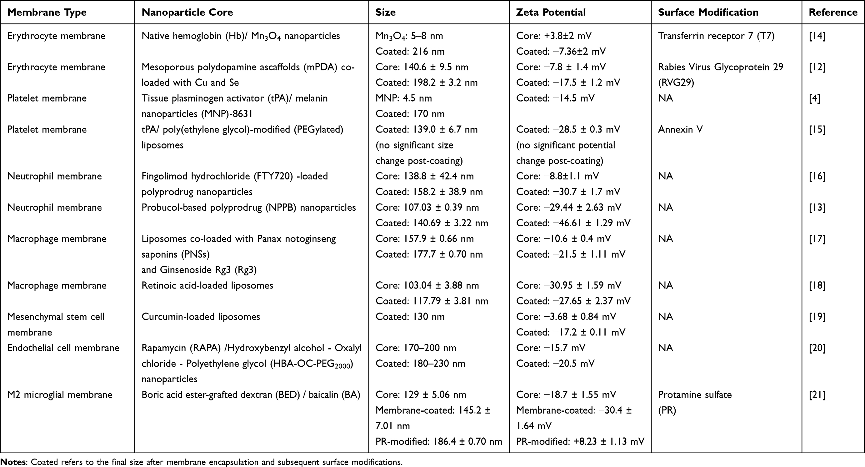 |
Table 1 Physicochemical Characteristics of Membrane-Camouflaged Nanoparticles for Ischemic Stroke Therapy |
CMC@NPs Size Determines Circulation and Brain Accumulation
The size of NPs is a pivotal determinant of their pharmacokinetics, critically influencing blood circulation half-life and tissue accumulation.11 Tang et al demonstrated that, across 30~200 nm size range, smaller micelles displayed enhanced tissue permeability and therapeutic efficacy, whereas larger counterparts possessed a greater tissue accumulation.22 Smaller NPs penetrate tissues more effectively but are cleared rapidly, while larger NPs have limited penetration but longer tissue retention.11 These principles are further supported by studies on poly(lactic-co-glycolic acid) (PLGA) NPs, which indicate an optimal size range of 10~100 nm for evading hepatic and renal clearance.23 Notably, some systems up to 200 nm can remain effective,24 underscoring that size is not the sole determinant. The CMC@NPs platform ingeniously integrates these insights. Its larger membrane shell facilitates long circulation and targeted accumulation, while the smaller core, released at the lesion site, enables deep penetration.4 Furthermore, the versatile drug loading strategies—encapsulation within the core or conjugation onto the membrane—endow this system with great flexibility for multifunctional synergistic therapy.13,18,25
Furthermore, particle size is closely related to the therapeutic performance of NPs. For example, in the platelet membrane-camouflaged NPs system, designated as tPA/MNP@PM (where tPA: tissue plasminogen activator; MNP: melanin nanoparticles; PM: platelet membrane vesicle), the 4.5 nm natural MNP core offers a large specific surface area, conferring high photothermal conversion efficiency and potent radical scavenging activity.4 Crucially, its minimal size enables superior BBB penetration compared to its 20 nm and 100 nm counterparts.4 The therapeutic action involves a dynamic size transformation: the intact 180 nm “pseudo-cell” first targets the thrombus.4 Upon near-infrared irradiation, the carrier disassembles, releasing the 4.5 nm MNP core and tPA. This facilitates local thrombolysis by tPA, followed by MNP-4.5 crossing the BBB to access the ischemic region (Figure 1).4
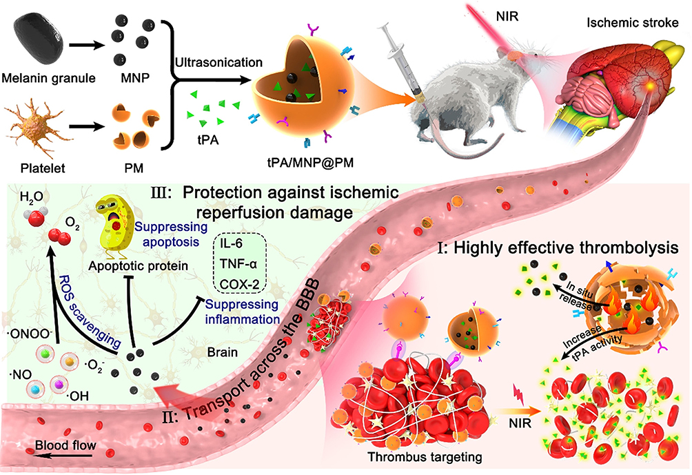 |
Figure 1 Schematic illustration of the fabrication of tMP and its application for cascaded thrombolysis and neuroprotection mechanisms in ischemic stroke, including (I) highly effective thrombolysis, (II) transport across the BBB, and (III) protection against ischemic reperfusion injury. Reprinted from Acta Biomaterialia, Vol 140, Yu W, Yin N, Yang Y, Xuan C, Liu X, Liu W, Zhang Z, Zhang K, Liu J, Shi J, Rescuing ischemic stroke by biomimetic nanovesicles through accelerated thrombolysis and sequential ischemia-reperfusion protection, Pages No. 625–640, Copyright (2022), with permission from Elsevier.4 Abbreviations: tMP, tissue plasminogen activator-loaded melanin nanoparticle-platelet membrane vesicle; BBB, surface charge on blood-brain barrier. |
The Mildly Negative Surface Charge Confers Biocompatibility and Mitigates Microembolism Risk in CMC@NPs
The brain microvascular endothelial cells exhibit a uniquely high negative surface charge, rendering the BBB inherently less permeable to anionic solutes while favoring the passage of cationic ones.26 This electrostatic preference underpins adsorptive-mediated transcytosis, wherein cationic biomolecules are initially concentrated on the endothelial surface prior to internalization.26 While this principle suggests a clear advantage for cationic NPs in BBB engagement, their positive charge also promotes non-specific interactions with plasma proteins and the extracellular matrix, leading to protein corona formation, compromised colloidal stability, and accelerated clearance by the mononuclear phagocyte system.24,27,28 Furthermore, studies by Lockman et al demonstrated that both cationic and high-concentration anionic NPs can compromise BBB integrity, whereas neutral and low-concentration anionic NPs are devoid of such detrimental effects (Figure 2).29 Consequently, near-neutral or moderately negative surface charges are generally preferred, as they confer superior biocompatibility, prolonged circulation, enhanced stealth properties, and ultimately, a higher in vivo safety profile.30,31
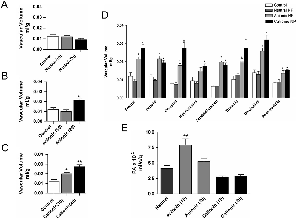 |
Figure 2 The impact of NPs surface charge on BBB integrity and permeability. An increase in cortical cerebrovascular volume implies BBB disruption and the subsequent distribution of vascular markers into brain parenchyma. In the control group, the frontal lobe cortex exhibited stable vascular volume. (A) Neutral NPs did not alter BBB integrity at either concentration. (B) Anionic NPs induced significant disruption only at 20 mg/mL. (C) Cationic NPs caused substantial BBB disruption at both concentrations. (D) Regional cerebrovascular volume after perfusion with NPs (20 mg/mL). (E) Blood to brain permeability coefficients, showing enhanced anionic NP permeability at 10 mg/mL that was abolished at 20 mg/mL; cationic NPs showed no significant change. *P<0.05, **P<0.01. Reprinted with permission from Lockman et al. Nanoparticle surface charges alter blood-brain barrier integrity and permeability. J Drug Target. 2004;12(9–10):635–41. Copyright 2004 Taylor & Francis.29 Abbreviation: NPs, nanoparticle. |
Under physiological conditions, the vascular endothelium and circulating blood cells maintain a stable anionic surface charge.32 In the pathological context of IS, however, vascular damage diminishes this negative charge density and can even reverse the charge polarity, a change frequently associated with thrombus formation.32 Positively charged materials are known to promote thrombosis, whereas negatively charged surfaces generally confer antithrombotic properties, with performance improving as the density and uniformity of the negative charge increase.32 CMC@NPs typically exhibit a physiologically weak negative charge (Table 1), a property dictated by the integrated contributions of the synthetic core, the native cell membrane, and any engineered surface ligands.21 This inherent anionic character significantly mitigates the risk of CMC@NP-induced microthrombosis in the cerebral microvasculature.
A key parameter in process optimization is the ratio of the cell membrane to the core NPs, which directly determines the surface potential of the final construct. Tuning this ratio to match the zeta potential of native membrane vesicles enables an ideal biomimetic effect.33 This is exemplified by the macrophage-disguised FTY-loaded MnO2 NPs system (Ma@(MnO2+FTY), where MnO2: Manganese dioxide; FTY: Fingolimod), where a membrane-to-core protein mass ratio of 2:1 yields a nanocomplex with a zeta potential of −21.43 mV, closely mirroring that of natural vesicles (−22.00 mV), and confers excellent stability.33 This biomimetic surface potential is critical for preserving the topological structure and bioactivity of native membrane proteins, thereby providing the structural basis for subsequent targeted delivery.34
Native Membrane Proteins and Engineered Modifications Enable Advanced Biomimetic Functions
Cell Membrane Camouflage Enhances Brain Targeting and Immunocompatibility in CMC@NPs
Polyethylene glycolylation (PEGylation) represents a widely employed strategy for NPs surface functionalization.35 It prolongs systemic circulation and improves biodistribution by forming a hydrophilic layer and steric shield around the NPs, which minimizes opsonization, enzymatic degradation, and clearance by the mononuclear phagocyte system and reticuloendothelial organs.36–38 However, a significant drawback is the potential for polyethylene glycol (PEG) to act as a hapten, inducing anti-PEG antibodies39 that can accelerate blood clearance, induce hypersensitivity reactions, and compromise carrier integrity.40,41 In contrast, NPs coated with natural cell membranes carry specific membrane proteins or engineered membrane components on their surfaces, which can endow CMC@NPs with unique biomimetic functions and serve as the molecular basis for their interactions with biological systems (Table 2).
 |
Table 2 A Comparative Analysis of Cell Membrane Types for Ischemic Stroke Therapy |
The targeted functionality of CMC@NPs is underpinned by specific membrane proteins that confer distinct biological capabilities. The cluster of differentiation 47 (CD47), a widely expressed transmembrane glycoprotein, delivers a “don’t eat me” signal via the CD47-signal-regulatory protein α (SIRPα) axis to evade phagocytosis.20,44 The cluster of differentiation 62P (CD62P), overexpressed on activated platelet membranes, mediates the adhesion of platelets to leukocytes and endothelial cells, thereby targeting thrombus sites.15,44 Neutrophil-derived chemokine receptors (eg, Cysteine-X-Cysteine chemokine receptor type 4 (CXCR4), Cysteine-X-Cysteine chemokine receptor type 2 (CXCR2)) guide navigation toward ischemic chemokine gradients,43 while β2 integrins (eg, lymphocyte function-associated antigen-1 (LFA-1), macrophage-1 antigen (Mac-1)) facilitate firm adhesion to the endothelium via intracellular adhesion molecule-1 (ICAM-1) and complement recognition, which is critical for BBB penetration.48 This transmigration is further aided by platelet endothelial cell adhesion molecule (PECAM-1).49 Collectively, these proteins constitute the molecular basis for the precise targeting of CMC@NPs to pathological sites. This principle can be leveraged therapeutically; for instance, genetic engineering to upregulate CXCR4 expression enhances the targeting capability of neural stem cells to stromal cell-derived factor-1 (SDF-1)-overexpressing ischemic regions.20 Consequently, extracting cell membranes with overexpressed CXCR4 allows for the preparation of CMC@NPs with enhanced chemotactic affinity for stroke lesions.
CMC@NPs Reduce Systemic Toxicity by Confining Drug Distribution
Although recombinant tPA is a cornerstone therapy for acute IS, its short half-life necessitates high clinical doses, paradoxically increasing the risk of devastating hemorrhagic transformation.4 To address this issue, Yu W et al developed tPA-loaded platelet membrane-biomimetic NPs (referred to as tMP).4 By leveraging the innate targeting ability of platelet membranes, the tMP system markedly enhanced drug accumulation at the thrombotic site.4 This targeted delivery allowed for a ten-fold reduction in the effective recombinant tPA dose (to 1 mg/kg), substantially mitigating the risk of cerebral hemorrhage.4 In vivo fluorescence imaging corroborated these findings, demonstrating intense signal localization at the thrombus and diminished non-specific accumulation in off-target organs, underscoring the enhanced treatment safety.4 Fingolimod hydrochloride (FTY720), an Food and Drug Administration (FDA)-approved sphingosine-1-phosphate receptor agonist, shows neuroprotective promise in IS but paradoxically induces dose-limiting bradycardia and immunosuppression at effective concentrations.50–52 To overcome this, Zhao et al developed a neutrophil membrane-camouflaged nanoparticle (NRN) for targeted and stimulus-responsive delivery.16 The NRN system selectively accumulated in ischemic brain tissue and released FTY720 in response to elevated ROS.16 This strategy boosted brain drug delivery by 15.2-fold compared to the free drug and, crucially, markedly attenuated its cardiotoxicity and infection risks (Figure 3), thereby decoupling therapeutic efficacy from systemic adverse effects.16
 |
Figure 3 Schematic illustration of the formation and anti-inflammation effects of NRNs. (A) Preparation of NRNs. (B) BBB penetration and ischemic brain targeting of NRNs. (C) ROS-responsive release of FTY720 from NRNs. (D) Reduced cardiotoxicity and infection risks of FTY720 by NRNs. (E) scRNA-seq to reveal the mechanism of the anti-inflammatory effects of NRNs. Reproduced from Advanced Materials, Volume 36, Zhao Y, Li Q, Niu J, Guo E, Zhao C, Zhang J, Liu X, Wang L, Rao L, Chen X, Yang K. Neutrophil Membrane-Camouflaged Polyprodrug Nanomedicine for Inflammation Suppression in Ischemic Stroke Therapy, Copyright (2024), John Wiley & Sons, Inc.16 Abbreviations: NRNs, neutrophil membrane-coated, reactive oxygen species-responsive polyprodrug nanoparticle; FTY720, Fingolimod hydrochloride; scRNA-seq, single-cell RNA sequencing; ROS, Reactive oxygen species; BBB, surface charge on blood-brain barrier. |
How Effective Are CMC@NPs as Therapeutics?
CMC@NPs Traverse the BBB Employing Transcytosis and Sensory Neural Pathways
The BBB poses a major challenge for stroke therapy by efficiently excluding most therapeutics and nano-delivery systems.8 To enter the brain parenchyma, substances must traverse the cerebrovascular endothelium, primarily via transcellular pathways involving various endocytic mechanisms.42 The specific endocytic route is strongly influenced by NPs size: typically, sub-150 nm NPs enter through clathrin- or caveolae-mediated endocytosis, whereas larger particles (250 nm~3 μm) are internalized via macropinocytosis or phagocytosis.53 For instance, macrophage membrane-camouflaged NPs, MM-Lip-Rg3/PNS (where MM: macrophage membrane; Lip: Liposome; Rg3: Ginsenoside Rg3; PNS: Panax notoginseng saponins), are effectively taken up by human cerebral microvascular endothelial cells via clathrin-mediated endocytosis and micropinocytosis.17 However, successful brain delivery requires NPs to subsequently escape endosomal entrapment; failure to do so results in lysosomal degradation and loss of therapeutic efficacy.54
The pathological alterations of the BBB during IS present unique opportunities for drug delivery.55 The disruption of tight junctions opens a paracellular route, permitting the passive diffusion of therapeutics into the brain.42 Beyond this passive mechanism, active targeting strategies are widely explored. NPs functionalized with glucose derivatives, for instance, can exploit specific transporter proteins for transcytosis, although this pathway is susceptible to competitive inhibition by endogenous substrates and efflux pumps.42 Receptor-mediated transcytosis provides an efficient cellular uptake pathway for macromolecular drugs and nanocarriers.42 CMC@NPs are particularly advantageous, as their natural or engineered surface receptors mediate precise recognition and internalization by brain endothelial cells, significantly enhancing targeting and brain entry.17 Nevertheless, ligand-mediated targeting carries off-target risks due to receptor expression in peripheral tissues.42 Alternatively, modulating surface charge—such as with positively charged polysaccharides or cationic albumin—can promote endothelial uptake via electrostatic adsorption.42,56 However, this non-specific interaction often leads to significant sequestration by the mononuclear phagocyte system, reducing brain accumulation and potentially increasing systemic toxicity.42
Intranasal administration offers a direct route to the brain by bypassing the BBB via sensory neural pathways, primarily the olfactory and trigeminal nerves.57 The olfactory nerve provides a short pathway from the nasal epithelium to the olfactory bulb, while the widely distributed trigeminal nerve projects to the brainstem, serving as another critical delivery conduit.58 Supporting this, Li et al demonstrated that PEGylated polycaprolactone nanoparticles entered the brainstem via the trigeminal nerve, with both intact nanoparticles and their curcumin payload detected within two hours post-administration.57,59 Despite this promise, the clinical translation of intranasal delivery is hampered by challenges in precise dosing and a delivery scope largely confined to small molecules.5
Core Material and Cell Type Orchestrate the Retention and Clearance of CMC@NPs
The in vivo clearance pathway of NPs governs their therapeutic applicability and long-term safety.10 Although CMC@NPs achieve targeted brain accumulation, studies confirm their predominant distribution in peripheral organs, particularly the liver, spleen, and kidneys,4,14,43 where they are efficiently cleared via hepatic and renal routes.60,61 In contrast, NPs reaching the brain are distributed in the surrounding microenvironment or taken up by brain parenchymal cells.62 They can be degraded by extracellular enzymes (eg, aminopeptidases and hyaluronidases), or effluxed via transporters like P-glycoprotein and low-density lipoprotein receptor-related protein 1 (LRP1) on the BBB.63 Following neuronal or glial uptake, NPs may undergo intracellular transport and efflux,63 lysosomal degradation, and subsequent clearance from the brain via the glymphatic system.54
A paradox exists in nanomedicine where strategies to prolong intracellular retention of drug-loaded NPs for enhanced efficacy54 may conversely promote toxic responses due to persistent, non-degradable accumulation.64 Gu et al demonstrated that after parenchymal injection, approximately 80% of poly(ethylene glycol)-poly(lactic acid) copolymer (PEG-PLA) NPs were rapidly cleared via the paravascular glymphatic pathway, with a half-life of less than 5 hours.65 This process relies on the phagocytic activity of microglia and the migratory function of nanomaterials.66,67 Unlike organic NPs, inorganic NPs such as metal- and silica-based particles remain trapped within microglia for extended periods, thereby disrupting the microglia-mediated paravascular transport pathway and leading to inefficient clearance.10 Investigations in Blasi Ventura-2 (BV-2) microglial cells revealed that while both organic and inorganic NPs initially colocalized with lysosomes, the inorganic NPs exhibited significantly prolonged lysosomal retention.10 The impaired efflux of inorganic NPs is potentially attributed to their inhibition of the extracellular signal-regulated kinase 1/2 (ERK1/2) signaling pathway, which disrupts microglial exosome synthesis—a crucial mechanism for clearance.10
The intracellular retention and clearance of NPs exhibit significant cell-type dependence, meaning that NPs with identical physicochemical properties can show notable differences in excretion rates across different cell types.54 Furthermore, studies on the release behavior of mesoporous silica NPs (MSNs) in various mammalian cell lines indicate that the unique properties of different cell lines in retaining and clearing MSNs may lead to an uneven distribution of MSNs among cells.68 Therefore, a precise evaluation of the retention and excretion pathways of NPs in specific brain cell types is crucial for reliably predicting neurotoxicity, assessing accumulation potential, and defining a safe therapeutic window.54 Overlooking such cell-specific characteristics may lead to misjudgments regarding the biosafety and therapeutic utility of nanomaterials. Furthermore, NP characteristics such as size, surface chemistry, and intra-/extracellular concentrations collectively regulate their metabolic behavior.54,69,70
The Retained Inorganic Nanocore Underlies Multiple Neurotoxic Mechanisms
Although the enhanced biocompatibility conferred by membrane camouflage, the intrinsic physicochemical properties of the core particle—including its chemical composition, size, shape, and charge density—can still induce neurotoxicity.23 These cores are broadly categorized as polymeric or inorganic. Biodegradable polymeric materials generally exhibit favorable safety profiles with no significant neurotoxicity reported.71 In contrast, inorganic nanoparticles (especially metal-based materials) carry clear neurotoxicity risks.57 Common inorganic nanoparticles used for treating ischemic stroke include cerium oxide,72,73 graphene oxide,74 manganese oxide,14,33 platinum,75,76 and selenium.77 These nanoparticles offer advantages such as high chemical stability and large drug-loading capacity.78 While valued for their high stability and drug-loading capacity, their limited biodegradability promotes accumulation in vital organs, including the brain.57 Notably, particles smaller than 1 nm can readily cross the BBB and persist in brain tissue, posing a long-term safety concern.79
Metal nanoparticles elicit neurotoxicity through multiple mechanisms, involving oxidative stress, apoptosis and autophagy activation, neuroinflammation, and disruption of sensorimotor signaling pathways.57 A principal mechanism is oxidative stress, driven by mitochondrial dysfunction.80 This process impairs neuronal integrity via lipid peroxidation, reduces mitochondrial membrane potential, and compromises adenosine triphosphate (ATP) production, ultimately triggering apoptosis.81 The central nervous system is particularly vulnerable to such insult due to its high metabolic rate, limited antioxidant defenses, and post-mitotic nature.82 The high reactivity and specific surface area of nanoparticles potently induce ROS generation,81 which can be further amplified by surface functional groups, as demonstrated with graphene oxide.83 Notably, ROS can inflict broad damage, including biomembrane degradation, deoxyribonucleic acid (DNA) conformational changes, and protein dysfunction, with mitochondrial DNA being especially susceptible.57 NPs can also trigger inflammatory responses, as evidenced in studies involving carbon nanotubes and fullerene derivatives.84
Cerium dioxide (CeO2) NPs function as efficient oxygen buffers, exerting therapeutic potential through free radical scavenging via oxygen vacancy generation,85 inhibition of macrophage M1 polarization, and superoxide dismutase-mimetic activity.86 Despite these beneficial roles, evidence indicates that this material can paradoxically exhibit pro-oxidant cytotoxicity both in vitro and in vivo (Figure 4).87 Its toxicity, closely associated with surface Ce3⁺ content, involves oxidative stress and inflammatory responses.85 For instance, CeO2 NPs have been shown to inhibit neural stem cell differentiation and impede growth cone development.88 Furthermore, toxicological studies suggest a potential risk for multi-organ toxicity, underscoring the need for a comprehensive toxicological assessment.85
 |
Figure 4 Major mechanisms of ceria nanoparticles toxicity in mammalian cells. Reproduced from Journal of Zhejiang University Science B, Vol 25, Fu X, Li P, Chen X, Ma Y, Wang R, Ji W, Gu J, Sheng B, Wang Y, Zhang Z, Ceria nanoparticles: biomedica applications and toxicity, Page 361–388, Copyright 2024, with permission from Springer Nature.85 Abbreviations: ROS, reactive oxygen species; ER, endoplasmic reticulum. |
While cell membrane camouflage endows NPs with advantageous biomimetic functions like specific targeting and immune evasion, it can also introduce unique safety risks. The functional molecules responsible for targeting may inadvertently interfere with physiological processes. For instance, the CD47 protein on erythrocyte membranes, though enabling immune evasion, may also suppress the phagocytic clearance of pathological proteins by microglia, potentially exacerbating neuroinflammation.89 Similarly, platelet membrane proteins glycoprotein VI (GPVI) and CD62P, which direct NPs to vascular injuries, carry the risk of inadvertently activating circulating platelets and promoting microthrombosis.90 Furthermore, neutrophil membranes utilizing Mac-1 for ischemic lesion site targeting may retain pro-inflammatory signals that could activate pathways like NF-κB.48 To mitigate these risks, membrane protein engineering emerges as a promising strategy. For example, engineering neutrophil membranes with incorporated Resolvin D2 was shown to inhibit post-reperfusion neutrophil infiltration while preserving targeting capability, offering a novel approach to balancing efficacy and safety.49
The Future Prospective
The safety profile of CMC@NPs has been preliminarily established through standard in vitro cytotoxicity experiments and short-term rodent toxicity studies (Table 3). A diverse array of cell membranes has been successfully employed for camouflage, encompassing blood cells (eg, erythrocytes, platelets, neutrophils, macrophages), stem cells, vascular endothelial cells, engineered hybrid membranes, and exosomes (Table 3). These membrane materials, mostly derived from animal blood cells or human cancer cell lines, generally exhibit good biocompatibility at therapeutic doses. Nevertheless, xenogeneic or allogeneic membrane materials may trigger recognition and rapid clearance by the host immune system.91 Therefore, sourcing membranes from primary human cells with high immune compatibility is critical for clinical translation, a goal that remains challenging due to limited cell sources, human leukocyte antigen (HLA) matching complexities, and ethical considerations.92,93
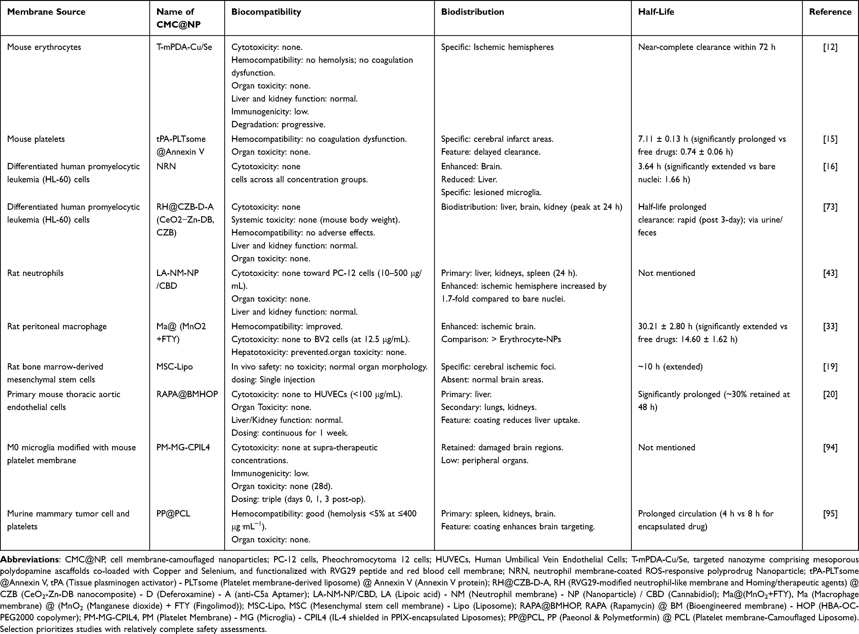 |
Table 3 Summary of Membrane-Camouflaged Nanoparticles Platforms for Ischemic Stroke Therapy |
A systematic safety evaluation of CMC@NPs should encompass key mechanisms such as induced oxidative stress, cell death, and genotoxicity, focusing on the resultant cytotoxicity, organ toxicity, and hemocompatibility.11 Current cytotoxicity assessments are often oversimplified, with some studies relying on a single high concentration (far beyond the therapeutic range) to claim no significant toxicity.4,49 Such an approach, lacking systematic concentration gradients, fails to reveal crucial toxicological parameters like the safe concentration window and toxicity threshold. In contrast, the study on gold nanoclusters provides a more rigorous paradigm: by employing concentration gradients and multiple time points, it systematically monitored microglial and neuroblastoma cells, clearly defining a safe window (≤5 μg/mL) and a growth inhibition threshold (>30 μg/mL).96 It further revealed a time-dependent metabolic decline not apparent at 24 hours.96 For organ toxicity, evaluation methods remain highly inconsistent across studies, and genotoxicity data are generally scarce (Table 3). Positively, hemocompatibility tests for platelet membrane-camouflaged systems showed no hemolysis or coagulation abnormalities.4,45 Regarding in vivo distribution, while membrane camouflage significantly improves pharmacokinetics—prolonging circulation half-life (eg, extending tPA half-life from 0.74 h to 7.11 h)15 and enhancing targeted accumulation in infarcted brain tissue97—metabolic organs (liver, kidneys, spleen, lungs) remain the primary deposition sites (Table 3), largely due to non-specific uptake by the reticuloendothelial systems.20
The current safety assessment framework for nanomedicines remains constrained by several critical limitations. Preclinical models predominantly rely on oversimplified two-dimensional monocultures of immortalized cell lines (eg, BV2 microglia), which fail to recapitulate the complexity of the BBB and overlook cell-type-specific responses. Furthermore, in vivo studies are often hampered by short-term observational windows (typically 24–72 hours), which are misaligned with clinical dosing regimens and insufficient for detecting chronic toxicity. The evaluation endpoints are also incomplete, lacking data under physiologically vulnerable conditions (eg, aging or metabolic abnormalities), and often neglecting immunogenicity (eg, complement activation and the risk of cytokine storms). Technologically, the field is hindered by traditional endpoint assays that cannot resolve dynamic toxicity mechanisms, a notable underutilization of high-throughput omics technologies, and the absence of a standardized Good Laboratory Practice (GLP)-compliant nanotoxicity testing platform.
The safety assessment of CMC@NPs must extend beyond conventional standards due to their unique biological characteristics. These particularities include: 1) The inherent bioactivity of surface membrane proteins, which may retain functions in immune regulation and receptor signaling; 2) Batch-to-batch variations in membrane composition that can lead to unpredictable biological outcomes; and 3) The dynamic nature of the membrane structure, which influences in vivo stability and degradation kinetics. Critically, immune risk must be evaluated for the complete delivery system rather than individual components. For instance, while the cyclic arginine-glycine-aspartic acid (RGD) peptide (eg, c(RGDyK)) alone is non-immunogenic, its conjugation onto PEGylated liposomes can induce acute hypersensitivity upon repeated administration.98 This phenomenon underscores that the combination of PEG and RGD creates a novel entity with emergent immunogenic properties through physicochemical interplay, rather than merely additive functions.
IS represents a substantial global health burden with limited treatment options, creating a pressing need for innovative therapeutic strategies. Nanotechnology, particularly CMC@NPs, has emerged as a promising platform to overcome the BBB and enhance therapeutic efficacy through superior biocompatibility and precise targeting capabilities. However, their clinical translation hinges on a rigorous safety assessment, which currently faces significant challenges. This review elucidates the intrinsic relationships between the physicochemical properties of CMC@NPs and their in vivo fate and toxicity, providing a comprehensive analysis of their potential neurotoxic risks.
Despite preliminary verification of CMC@NPs safety through standard assays, this review underscores persistent and critical gaps, particularly concerning their intracellular degradation kinetics, excretion pathways, and chronic toxicity profiles.10 These knowledge gaps represent a major barrier to clinical translation. Future research should undergo a paradigm shift from a singular focus on delivery efficiency to a balanced consideration of efficacy and safety. Key priorities should encompass: 1) establishing evaluation models that better mimic human disease states; 2) implementing advanced in vivo dynamic imaging for real-time NPs tracking; 3) strengthening targeted assessment modules for comprehensive immunogenicity profiling, long-term biodistribution, and degradation product identification; and 4) ultimately establishing a unified, GLP-compliant safety evaluation framework.
Abbreviations
ATP, Adenosine Triphosphate; BA, baicalin; BBB, blood-brain barrier; BED, boric acid ester-grafted dextran; BV-2, Blasi Ventura-2; CD47, Cluster of Differentiation 47; CD62P, Cluster of Differentiation 62P; CeO2, Cerium dioxide; CMC@NP, cell membrane-camouflaged nanoparticles; CLSM, Confocal laser scanning microscopy; CPIL4, IL-4 shielded in PPIX-encapsulated Liposomes; CXCR2, Cysteine-X-Cysteine chemokine receptor type 2; CXCR4, Cysteine-X-Cysteine chemokine receptor type 4; CZB, CeO2-Zn-DB nanocomposite; DNA, deoxyribonucleic acid; ERK1/2, Extracellular Signal-Regulated Kinase 1/2; FDA, Food and Drug Administration; FTY, Fingolimod; FTY720, Fingolimod hydrochloride; Hb, native hemoglobin; HOP, HBA-OC-PEG2000 copolymer; HUVECs, Human Umbilical Vein Endothelial Cells; ICAM-1, Intracellular adhesion molecule-1; IS, ischemic stroke; LA-NM-NP/CBD, Lipoic acid-Neutrophil membrane-Nanoparticle/Cannabidiol; LFA-1, Lymphocyte function-associated antigen-1; Lip, Liposome; LRP1, Low-density lipoprotein Receptor-related Protein 1; Ma@(MnO2+FTY), Macrophage membrane-camouflaged Manganese dioxide and Fingolimod nanoparticles; Mac-1, Macrophage-1 antigen; MM, Macrophage membrane; mPDA, mesoporous polydopamine scaffolds; MNP, melanin nanoparticle; MnO2, Manganese dioxide; MSC-Lipo, Mesenchymal stem cell membrane-camouflaged Liposome; MSNs, mesoporous silica nanoparticles; NIR, Near-Infrared; NPPB, probucol-based polyprodrug; NRN, neutrophil membrane-coated ROS-responsive polyprodrug Nanoparticle; NPs, nanoparticles; PCL, Platelet membrane-Camouflaged Liposome; PC-12 cells, Pheochromocytoma 12 cells; PECAM-1, platelet endothelial cell adhesion molecule; PEG, Polyethylene Glycol; PEG-PLA, Poly(ethylene glycol)-poly(lactic acid) copolymer; PEGylation, Polyethylene Glycolylation; PLTsome, Platelet membrane-derived liposome; PM, Platelet membrane vesicle; PM-MG, Platelet Membrane-camouflaged Microglia; PNS, Panax notoginseng saponins; PP@PCL, Paeonol and Polymetformin loaded in Platelet membrane-Camouflaged Liposome; PR, protamine sulfate; RAPA@BMHOP, Rapamycin loaded in Bioengineered membrane-camouflaged HBA-OC-PEG2000 copolymer; RGD, Arginine-Glycine-Aspartic acid; Rg3, Ginsenoside Rg3; RH@CZB-D-A, RVG29-modified neutrophil-like membrane-camouflaged CeO2-Zn-DB nanocomposite loaded with Deferoxamine and anti-C5a Aptamer; ROS, reactive oxygen species; rtPA, recombinant tissue plasminogen activator; RVG29, Rabies Virus Glycoprotein 29; scRNA-seq, single-cell RNA sequencing; SDF-1, Stromal cell-Derived Factor-1; SIRPα, Signal-regulatory protein α; T7, Transferrin receptor; T-mPDA-Cu/Se, Targeted mesoporous polydopamine scaffolds co-loaded with Copper and Selenium; tPA, tissue plasminogen activator.
Acknowledgments
We acknowledge that the figures in this review were adapted from previous publications with permission from the copyright holders. Additionally, the graphical abstract was created using BioRender.com.
Funding
This work was supported by the Start-up Funding of Hangzhou Normal University [4255C50221204123], the Hangzhou Youth Innovation Team Project [TD2023020], Ministry of Education’s China University Industry Research Innovation Fund [2022BC025], Hangzhou Biomedical and Health Industry Development Support Science and Technology Special Project [2021WJCY136, 2021WJCY285, 2022WJC199], Zhejiang Medical Association Clinical Medical Research Special Fund Project [2022ZYC-Z29], Hangzhou Normal University Affiliated Hospital Pei Yuan Plan [PYJH202305], Key Scientific Research Project in the Field of Agriculture and Social Development of Hangzhou Science and Technology Bureau [20241203A12], and the Construction Fund of Key Medical Disciplines of Hangzhou [2025HZGF02].
Disclosure
The authors report no conflicts of interest in this work.
References
1. Yu H, Yu J, Yao G. Recent advances in aptamers-based nanosystems for diagnosis and therapy of cardiovascular diseases: an updated review. Int J Nanomed. 2025;20:2427–2443. doi:10.2147/IJN.S507715
2. Kim HY, Kim TJ, Kang L, et al. Mesenchymal stem cell-derived magnetic extracellular nanovesicles for targeting and treatment of ischemic stroke. Biomaterials. 2020;243:119942. doi:10.1016/j.biomaterials.2020.119942
3. Li Y, Wu C, Yang R, et al. Application and development of cell membrane functionalized biomimetic nanoparticles in the treatment of acute ischemic stroke. Int J Mol Sci. 2024;25(15):8539. doi:10.3390/ijms25158539
4. Yu W, Yin N, Yang Y, et al. Rescuing ischemic stroke by biomimetic nanovesicles through accelerated thrombolysis and sequential ischemia-reperfusion protection. Acta Biomater. 2022;140:625–640. doi:10.1016/j.actbio.2021.12.009
5. Parvez S, Kaushik M, Ali M, et al. Dodging blood brain barrier with “nano” warriors: novel strategy against ischemic stroke. Theranostics. 2022;12(2):689–719. doi:10.7150/thno.64806
6. Islam Y, Leach AG, Smith J, et al. Physiological and pathological factors affecting drug delivery to the brain by nanoparticles. Adv Sci. 2021;8(11):2002085. doi:10.1002/advs.202002085
7. Lv W, Liu Y, Li S, Lv L, Lu H, Xin H. Advances of nano drug delivery system for the theranostics of ischemic stroke. J Nanobiotechnol. 2022;20(1):248. doi:10.1186/s12951-022-01450-5
8. Thakur N, Kumar T, Singh C, Kumar R, Kumar A. Cell membrane-coated nanoparticles for neurodegenerative disorders management. Int J Pharm. 2025;681:125875. doi:10.1016/j.ijpharm.2025.125875
9. Wang G, Li Z, Wang G, et al. Advances in engineered nanoparticles for the treatment of ischemic stroke by enhancing angiogenesis. Int J Nanomed. 2024;19:4377–4409. doi:10.2147/IJN.S463333
10. Gao J, Song Q, Gu X, et al. Intracerebral fate of organic and inorganic nanoparticles is dependent on microglial extracellular vesicle function. Nat Nanotechnol. 2024;19(3):376–386. doi:10.1038/s41565-023-01551-8
11. Domb AJ, Sharifzadeh G, Nahum V, Hosseinkhani H. Safety evaluation of nanotechnology products. Pharmaceutics. 2021;13(10):1615. doi:10.3390/pharmaceutics13101615
12. Wu Y, Xu J, Wang X, et al. Active transport of biomimetic cascaded nanozymes across blood–brain barrier to scavenge ROS and alleviate neuroinflammation against cerebral ischemia reperfusion injury. Adv Funct Mater. 2025:e20000. doi:10.1002/adfm.202520000
13. Jia M, Miao W, Li Y, et al. A polymerized probucol nanoformulation with neutrophil extracellular vesicle camouflage for cerebral ischemia-reperfusion injury therapy. Innovation. 2025;6(4):100761. doi:10.1016/j.xinn.2024.100761
14. Shi J, Yu W, Xu L, et al. Bioinspired nanosponge for salvaging ischemic stroke via free radical scavenging and self-adapted oxygen regulating. Nano Lett. 2020;20(1):780–789. doi:10.1021/acs.nanolett.9b04974
15. Quan X, Han Y, Lu P, et al. Annexin V-modified platelet-biomimetic nanomedicine for targeted therapy of acute ischemic stroke. Adv Healthc Mater. 2022;11(16):e2200416. doi:10.1002/adhm.202200416
16. Zhao Y, Li Q, Niu J, et al. Neutrophil membrane-camouflaged polyprodrug nanomedicine for inflammation suppression in ischemic stroke therapy. Adv Mater. 2024;36(21):e2311803. doi:10.1002/adma.202311803
17. Liu T, Wang Y, Zhang M, et al. The optimization design of macrophage membrane camouflaging liposomes for alleviating ischemic stroke injury through intranasal delivery. Int J Mol Sci. 2024;25(5):2927. doi:10.3390/ijms25052927
18. Cai S, Gao J, Weng X, et al. Synergistic enhancement of efferocytosis and cholesterol efflux via macrophage biomimetic nanoparticle to attenuate atherosclerosis progression. Bioact Mater. 2026;55:131–143. doi:10.1016/j.bioactmat.2025.09.022
19. Wu H, Jiang X, Li Y, et al. Engineering stem cell derived biomimetic vesicles for versatility and effective targeted delivery. Adv Funct Mater. 2020;30(49):2006169. doi:10.1002/adfm.202006169
20. Luo L, Zang G, Liu B, et al. Bioengineering CXCR4-overexpressing cell membrane functionalized ROS-responsive nanotherapeutics for targeting cerebral ischemia-reperfusion injury. Theranostics. 2021;11(16):8043–8056. doi:10.7150/thno.60785
21. Wu Y, Feng H, Gao L, et al. Functionalized biomimetic nanoparticles are delivered from the nose to the brain for the synergistic targeted treatment of cerebral ischemia/reperfusion injury. Regen Biomater. 2025:
22. Wang J, Mao W, Lock LL, et al. The role of micelle size in tumor accumulation, penetration, and treatment. ACS Nano. 2015;9(7):7195–7206. doi:10.1021/acsnano.5b02017
23. Gong JY, Holt MG, Hoet PHM, Ghosh M. Neurotoxicity of four frequently used nanoparticles: a systematic review to reveal the missing data. Arch Toxicol. 2022;96(5):1141–1212. doi:10.1007/s00204-022-03233-1
24. Pinto M, Silva V, Barreiro S, et al. Brain drug delivery and neurodegenerative diseases: polymeric PLGA-based nanoparticles as a forefront platform. Ageing Res Rev. 2022;79:101658. doi:10.1016/j.arr.2022.101658
25. Vankayala R, Corber SR, Mac JT, Rao MP, Shafie M, Anvari B. Erythrocyte-derived nanoparticles as a theranostic agent for near-infrared fluorescence imaging and thrombolysis of blood clots. Macromol Biosci. 2018;18(4):e1700379. doi:10.1002/mabi.201700379
26. Wu Y, Li L, Ning Z, et al. Autophagy-modulating biomaterials: multifunctional weapons to promote tissue regeneration. Cell Commun Signal. 2024;22(1):124. doi:10.1186/s12964-023-01346-3
27. Machtakova M, Thérien-Aubin H, Landfester K. Polymer nano-systems for the encapsulation and delivery of active biomacromolecular therapeutic agents. Chem Soc Rev. 2022;51(1):128–152. doi:10.1039/D1CS00686J
28. Xiao QQ, Zoulikha M, Qiu M, et al. The effects of protein Corona on in vivo fate of nanocarriers. Adv Drug Deliv Rev. 2022;186:114356. doi:10.1016/j.addr.2022.114356
29. Lockman PR, Koziara JM, Mumper RJ, Allen DD. Nanoparticle surface charges alter blood-brain barrier integrity and permeability. J Drug Targeting. 2004;12(9–10):635–641. doi:10.1080/10611860400015936
30. Xiao K, Li Y, Luo J, et al. The effect of surface charge on in vivo biodistribution of PEG-oligocholic acid based micellar nanoparticles. Biomaterials. 2011;32(13):3435–3446. doi:10.1016/j.biomaterials.2011.01.021
31. Liu S, Wang L, Zhang M, et al. Tumor microenvironment-responsive nanoshuttles with sodium citrate modification for hierarchical targeting and improved tumor theranostics. ACS Appl Mater Interfaces. 2019;11(29):25730–25739. doi:10.1021/acsami.9b07957
32. Srinivasan S, Sawyer PN. Role of surface charge of the blood vessel wall, blood cells, and prosthetic materials in intravascular thrombosis. J Colloid Interface Sci. 1970;32(3):456–463. doi:10.1016/0021-9797(70)90131-1
33. Li C, Zhao Z, Luo Y, et al. Macrophage-disguised manganese dioxide nanoparticles for neuroprotection by reducing oxidative stress and modulating inflammatory microenvironment in acute ischemic stroke. Adv Sci. 2021;8(20):e2101526. doi:10.1002/advs.202101526
34. Bombarda E, Becker T, Ullmann GM. Influence of the membrane potential on the protonation of bacteriorhodopsin: insights from electrostatic calculations into the regulation of proton pumping. J Am Chem Soc. 2006;128(37):12129–12139. doi:10.1021/ja0619657
35. Jokerst JV, Lobovkina T, Zare RN, Gambhir SS. Nanoparticle PEGylation for imaging and therapy. Nanomedicine. 2011;6(4):715–728. doi:10.2217/nnm.11.19
36. Kolate A, Baradia D, Patil S, Vhora I, Kore G, Misra A. PEG — a versatile conjugating ligand for drugs and drug delivery systems. J Control Release. 2014;192:67–81. doi:10.1016/j.jconrel.2014.06.046
37. Kozlowski A, Milton Harris J. Improvements in protein PEGylation: pegylated interferons for treatment of hepatitis C. J Control Release. 2001;72(1):217–224. doi:10.1016/S0168-3659(01)00277-2
38. Swierczewska M, Lee KC, Lee S. What is the future of PEGylated therapies?. Expert Opin Emerg Drugs. 2015;20(4):531–536. doi:10.1517/14728214.2015.1113254
39. Sroda K, Rydlewski J, Langner M, Kozubek A, Grzybek M, Sikorski AF. Repeated injections of PEG-PE liposomes generate anti-PEG antibodies. Cell Mol Biol Lett. 2005;10(1):37–47.
40. Sellaturay P, Nasser S, Islam S, Gurugama P, Ewan PW. Polyethylene glycol (PEG) is a cause of anaphylaxis to the pfizer/BioNTech mRNA COVID-19 vaccine. Clin Exp Allergy J Br Soc Allergy Clin Immunol. 2021;51(6):861–863. doi:10.1111/cea.13874
41. Castells MC, Phillips EJ. Maintaining safety with SARS-CoV-2 vaccines. N Engl J Med. 2021;384(7):643–649. doi:10.1056/NEJMra2035343
42. Qi M, Sun Y, Zhao Z, et al. Cell membrane-coated nanoparticles: a biomimetic strategy for effective treatment of ischemic stroke. J Drug Delivery Sci Technol. 2025;108:106966. doi:10.1016/j.jddst.2025.106966
43. Liu S, Xu J, Liu Y, et al. Neutrophil-biomimetic “nanobuffer” for remodeling the microenvironment in the infarct core and protecting neurons in the penumbra via neutralization of detrimental factors to treat ischemic stroke. ACS Appl Mater Interfaces. 2022;14(24):27743–27761. doi:10.1021/acsami.2c09020
44. Yuan S, Hu D, Gao D, et al. Recent advances of engineering cell membranes for nanomedicine delivery across the blood-brain barrier. J Nanobiotechnol. 2025;23(1):493. doi:10.1186/s12951-025-03572-y
45. Xu J, Wang X, Yin H, et al. Sequentially site-specific delivery of thrombolytics and neuroprotectant for enhanced treatment of ischemic stroke. ACS Nano. 2019;13(8):8577–8588. doi:10.1021/acsnano.9b01798
46. Shi J, Yang Y, Yin N, et al. Engineering CXCL12 biomimetic decoy-integrated versatile immunosuppressive nanoparticle for ischemic stroke therapy with management of overactivated brain immune microenvironment. Small Methods. 2022;6(1):e2101158. doi:10.1002/smtd.202101158
47. Dehaini D, Wei X, Fang RH, et al. Erythrocyte-platelet hybrid membrane coating for enhanced nanoparticle functionalization. Adv Mater. 2017;29(16). doi:10.1002/adma.201606209
48. Jickling GC, Liu D, Ander BP, Stamova B, Zhan X, Sharp FR. Targeting neutrophils in ischemic stroke: translational insights from experimental studies. J Cereb Blood Flow Metab. 2015;35(6):888–901. doi:10.1038/jcbfm.2015.45
49. Dong X, Gao J, Zhang CY, Hayworth C, Frank M, Wang Z. Neutrophil membrane-derived nanovesicles alleviate inflammation to protect mouse brain injury from ischemic stroke. ACS Nano. 2019;13(2):1272–1283. doi:10.1021/acsnano.8b06572
50. Racca V, Di Rienzo M, Cavarretta R, et al. Fingolimod effects on left ventricular function in multiple sclerosis. Mult Scler Houndmills Basingstoke Eng. 2016;22(2):201–211. doi:10.1177/1352458515587753
51. Brinkmann V, Billich A, Baumruker T, et al. Fingolimod (FTY720): discovery and development of an oral drug to treat multiple sclerosis. Nat Rev Drug Discov. 2010;9(11):883–897. doi:10.1038/nrd3248
52. Naseh M, Vatanparast J, Rafati A, Bayat M, Haghani M. The emerging role of FTY720 as a sphingosine 1-phosphate analog for the treatment of ischemic stroke: the cellular and molecular mechanisms. Brain Behav. 2021;11(6):e02179. doi:10.1002/brb3.2179
53. Foroozandeh P, Aziz AA. Insight into cellular uptake and intracellular trafficking of nanoparticles. Nanoscale Res Lett. 2018;13(1):339. doi:10.1186/s11671-018-2728-6
54. Peng Y, Yang Z, Sun H, Li J, Lan X, Liu S. Nanomaterials in medicine: understanding cellular uptake, localization, and retention for enhanced disease diagnosis and therapy. Aging Dis. 2025;16(1):168. doi:10.14336/AD.2024.0206-1
55. Jiao H, Wang Z, Liu Y, Wang P, Xue Y. Specific role of tight junction proteins claudin-5, occludin, and ZO-1 of the blood-brain barrier in a focal cerebral ischemic insult. J Mol Neurosci. 2011;44:130–139.
56. Huang S, Gao Y, Li H, et al. Manganese@albumin nanocomplex and its assembled nanowire activate TLR4-dependent signaling cascades of macrophages. Adv Mater. 2024;36:e2310979.
57. Zia S, Islam Aqib A, Muneer A, et al. Insights into nanoparticles-induced neurotoxicity and cope up strategies. Front Neurosci. 2023;17:1127460. doi:10.3389/fnins.2023.1127460
58. Moseman EA, Blanchard AC, Nayak D, McGavern DB. T cell engagement of cross-presenting microglia protects the brain from a nasal virus infection. Sci Immunol. 2020;5(48):eabb1817. doi:10.1126/sciimmunol.abb1817
59. Li Y, Wang C, Zong S, et al. The trigeminal pathway dominates the nose-to-brain transportation of intact polymeric nanoparticles: evidence from aggregation-caused quenching probes. J Biomed Nanotechnol. 2019;15(4):686–702. doi:10.1166/jbn.2019.2724
60. Wei YC, Quan L, Zhou C, Zhan QQ. Factors relating to the biodistribution & clearance of nanoparticles & their effects on in vivo application. Nanomedicine. 2018;13:1495–1512.
61. Yang G, Phua SZ, Lim WQ, et al. A hypoxia-responsive albumin-based nanosystem for deep tumor penetration and excellent therapeutic efficacy. Adv Mater. 2019;31:e1901513.
62. Tosi G, Vilella A, Chhabra R, et al. Insight on the fate of CNS-targeted nanoparticles. Part II: intercellular neuronal cell-to-cell transport. J Control Release. 2014;177:96–107.
63. Li P, Chen Y, Gao X, Jiang G. A review on brain-targeting nano-based drug delivery. Chin Sci Bull. 2025. doi:10.1360/TB-2024-1147
64. Hawkins SJ, Crompton LA, Sood A, et al. Nanoparticle-induced neuronal toxicity across placental barriers is mediated by autophagy and dependent on astrocytes. Nat Nanotechnol. 2018;13:427–433.
65. Gu X, Song Q, Zhang Q, et al. Clearance of two organic nanoparticles from the brain via the paravascular pathway. J Control Release. 2020;322:31–41. doi:10.1016/j.jconrel.2020.03.009
66. Borst K, Dumas AA, Prinz M. Microglia: immune and non-immune functions. Immunity. 2021;54:2194–2208.
67. Bourquin J, Milosevic A, Hauser D, et al. Biodistribution, clearance, and long-term fate of clinically relevant nanomaterials. Adv Mater. 2018;30:e1704307.
68. Slowing I, Vivero-Escoto J, Zhao Y, et al. Exocytosis of mesoporous silica nanoparticles from mammalian cells: from asymmetric cell-to-cell transfer to protein harvesting. Small. 2011;7:1526–1532.
69. Serda RE, Mack A, van de Ven AL, et al. Logic-embedded vectors for intracellular partitioning, endosomal escape, and exocytosis of nanoparticles. Small. 2010;6:2691–2700.
70. Bartczak D, Nitti S, Millar TM, Kanaras AG. Exocytosis of peptide functionalized gold nanoparticles in endothelial cells. Nanoscale. 2012;4:4470–4472.
71. Zeng J, Martin A, Han X, Shirihai OS, Grinstaff MW. Biodegradable PLGA nanoparticles restore lysosomal acidity and protect neural PC-12 cells against mitochondrial toxicity. Ind Eng Chem Res. 2019;58(31):13910–13917. doi:10.1021/acs.iecr.9b02003
72. Li X, Han Z, Wang T, et al. Cerium oxide nanoparticles with antioxidative neurorestoration for ischemic stroke. Biomaterials. 2022;291:121904. doi:10.1016/j.biomaterials.2022.121904
73. Li XS, Li HY, Peng D, et al. A biomimetic multifunctional nanoplatform with precise targeting-penetration and efficient synergistic therapy for ischemic stroke. ACS Appl Mater Interfaces. 2025;17(28):39925–39954. doi:10.1021/acsami.5c01878
74. Wang P, Li J, Li S, et al. Palladium-reduced graphene oxide nanocomposites enhance neurite outgrowth and protect neurons from ishemic stroke. Mater Today Bio. 2024;28:101184. doi:10.1016/j.mtbio.2024.101184
75. Takamiya M, Miyamoto Y, Yamashita T, et al. Neurological and pathological improvements of cerebral infarction in mice with platinum nanoparticles. J Neurosci Res. 2011;89(7):1125–1133. doi:10.1002/jnr.22622
76. Takamiya M, Miyamoto Y, Yamashita T, Deguchi K, Ohta Y, Abe K. Strong neuroprotection with a novel platinum nanoparticle against ischemic stroke- and tissue plasminogen activator-related brain damages in mice. Neuroscience. 2012;221:47–55. doi:10.1016/j.neuroscience.2012.06.060
77. Amani H, Habibey R, Shokri F, et al. Selenium nanoparticles for targeted stroke therapy through modulation of inflammatory and metabolic signaling. Sci Rep. 2019;9(1):6044. doi:10.1038/s41598-019-42633-9
78. Liao J, Li Y, Luo Y, et al. Recent advances in targeted nanotherapies for ischemic stroke. Mol Pharm. 2022;19(9):3026–3041. doi:10.1021/acs.molpharmaceut.2c00383
79. Caito S, Aschner M. Neurotoxicity of metals. Handb Clin Neurol. 2015;131,169–189. doi:10.1016/B978-0-444-62627-1.00011-1
80. Sharma A, Liaw K, Sharma R, Zhang Z, Kannan S, Kannan RM. Targeting mitochondrial dysfunction and oxidative stress in activated microglia using dendrimer-based therapeutics. Theranostics. 2018;8(20):5529–5547. doi:10.7150/thno.29039
81. Tripathy DB, Pradhan S, Gupta A, Agarwal P. Nanoparticles induced neurotoxicity. Nanotoxicology. 2025;19(3):325–352. doi:10.1080/17435390.2025.2488310
82. Li J, O W, Li W, Jiang ZG, Ghanbari HA. Oxidative stress and neurodegenerative disorders. Int J Mol Sci. 2013;14:24438–24475. doi:10.3390/ijms141224438
83. Zhang J, Cao HY, Wang JQ, Wu GD, Wang L. Graphene oxide and reduced graphene oxide exhibit cardiotoxicity through the regulation of lipid peroxidation, oxidative stress, and mitochondrial dysfunction. Front Cell Dev Biol. 2021;9:616888. doi:10.3389/fcell.2021.616888
84. Baktur R, Patel H, Kwon S. Effect of exposure conditions on SWCNT-induced inflammatory response in human alveolar epithelial cells. Toxicol Vitro. 2011;25(5):1153–1160. doi:10.1016/j.tiv.2011.04.001
85. Fu X, Li P, Chen X, et al. Ceria nanoparticles: biomedical applications and toxicity. J Zhejiang Univ Sci B. 2024;25(5):361–388. doi:10.1631/jzus.B2300854
86. Sun Y, Sun X, Li X, et al. A versatile nanocomposite based on nanoceria for antibacterial enhancement and protection from aPDT-aggravated inflammation via modulation of macrophage polarization. Biomaterials. 2021;268:120614. doi:10.1016/j.biomaterials.2020.120614
87. Naidi SN, Harunsani MH, Tan AL, Khan MM. Green-synthesized CeO2 nanoparticles for photocatalytic, antimicrobial, antioxidant and cytotoxicity activities. J Mater Chem B. 2021;9(28):5599–5620. doi:10.1039/d1tb00248a
88. Gliga AR, Edoff K, Caputo F, et al. Cerium oxide nanoparticles inhibit differentiation of neural stem cells. Sci Rep. 2017;7:9284. doi:10.1038/s41598-017-09430-8
89. Lehrman EK, Wilton DK, Litvina EY, et al. CD47 protects synapses from excess microglia-mediated pruning during development. Neuron. 2018;100(1):120–134.e6. doi:10.1016/j.neuron.2018.09.017
90. Fröhlich E. Action of nanoparticles on platelet activation and plasmatic coagulation. Curr Med Chem. 2016;23(5):408–430. doi:10.2174/0929867323666160106151428
91. Shi Y, Qian H, Rao P, et al. Bioinspired membrane-based nanomodulators for immunotherapy of autoimmune and infectious diseases. Acta Pharm Sin B. 2022;12(3):1126–1147. doi:10.1016/j.apsb.2021.09.025
92. Fang RH, Kroll AV, Gao W, Zhang L. Cell membrane coating nanotechnology. Adv Mater. 2018;30(23):e1706759. doi:10.1002/adma.201706759
93. Achón Buil B, Rentsch NH, Weber RZ, et al. Beneath the radar: immune-evasive cell sources for stroke therapy. Trends Mol Med. 2024;30(3):223–238. doi:10.1016/j.molmed.2023.12.004
94. Li Y, Teng X, Yang C, et al. Ultrasound controlled anti-inflammatory polarization of platelet decorated microglia for targeted ischemic stroke therapy. Angew Chem Int Ed Engl. 2021;60(10):5083–5090. doi:10.1002/anie.202010391
95. Tang L, Yin Y, Liu H, et al. Blood-brain barrier-penetrating and lesion-targeting nanoplatforms inspired by the pathophysiological features for synergistic ischemic stroke therapy. Adv Mater. 2024;36(21):e2312897. doi:10.1002/adma.202312897
96. Xiao L, Wei F, Zhou Y, et al. Dihydrolipoic acid-gold nanoclusters regulate microglial polarization and have the potential to alter neurogenesis. Nano Lett. 2020;20(1):478–495. doi:10.1021/acs.nanolett.9b04216
97. Feng L, Dou C, Xia Y, et al. Neutrophil-like cell-membrane-coated nanozyme therapy for ischemic brain damage and long-term neurological functional recovery. ACS Nano. 2021;15(2):2263–2280. doi:10.1021/acsnano.0c07973
98. Wang X, Wang H, Jiang K, et al. Liposomes with cyclic RGD peptide motif triggers acute immune response in mice. J Control Release. 2019;293:201–214. doi:10.1016/j.jconrel.2018.12.003
 © 2025 The Author(s). This work is published and licensed by Dove Medical Press Limited. The
full terms of this license are available at https://www.dovepress.com/terms
and incorporate the Creative Commons Attribution
- Non Commercial (unported, 4.0) License.
By accessing the work you hereby accept the Terms. Non-commercial uses of the work are permitted
without any further permission from Dove Medical Press Limited, provided the work is properly
attributed. For permission for commercial use of this work, please see paragraphs 4.2 and 5 of our Terms.
© 2025 The Author(s). This work is published and licensed by Dove Medical Press Limited. The
full terms of this license are available at https://www.dovepress.com/terms
and incorporate the Creative Commons Attribution
- Non Commercial (unported, 4.0) License.
By accessing the work you hereby accept the Terms. Non-commercial uses of the work are permitted
without any further permission from Dove Medical Press Limited, provided the work is properly
attributed. For permission for commercial use of this work, please see paragraphs 4.2 and 5 of our Terms.