Back to Journals » Infection and Drug Resistance » Volume 14
Magnitude of Antibody Cross-Reactivity in Medically Important Mosquito-Borne Flaviviruses: A Systematic Review
Authors Endale A , Medhin G, Darfiro K, Kebede N, Legesse M
Received 27 August 2021
Accepted for publication 7 October 2021
Published 19 October 2021 Volume 2021:14 Pages 4291—4299
DOI https://doi.org/10.2147/IDR.S336351
Checked for plagiarism Yes
Review by Single anonymous peer review
Peer reviewer comments 2
Editor who approved publication: Prof. Dr. Héctor Mora-Montes
Adugna Endale,1,2 Girmay Medhin,1 Koyamo Darfiro,3 Nigatu Kebede,1 Mengistu Legesse1
1Aklilu Lemma Institute of Pathobiology, Addis Ababa University, Addis Ababa, Ethiopia; 2School of Medicine, College of Medicine and Health Sciences, Dire Dawa University, Dire Dawa, Ethiopia; 3Department of External Quality Assessment, Hossaena Public Health Institute Laboratory, Hossaena, Ethiopia
Correspondence: Adugna Endale Email [email protected]
Introduction: Flaviviruses are a genus of enveloped single-stranded RNA viruses that include dengue virus (DENV), yellow fever virus, West Nile virus (WNV), Japanese encephalitis virus, and Zika virus. Nowadays, diverse serological assays are available to diagnose flaviviruses. However, infection with flaviviruses induces cross-reactive antibodies, which are a challenge for serological diagnosis.
Objective: This systematic review aimed to assess the magnitude of medically important mosquito-borne flavivirus–induced antibody cross-reactivity and its influence on serological test outcomes.
Methods: This study was designed based on the PRISMA guidelines. It includes original research articles published between 1994 and 2019 that reported serological cross-reactions between medically important mosquito-borne flaviviruses. Articles were searched on PubMed using controlled vocabulary. Eligibility was assessed by title, abstract, and finally by reading the full paper. The articles included are compared, evaluated, and summarized narratively.
Results: A total of 2,911 articles were identified, and finally 14 were included. About 15.4%– 84% of antibodies produced against non-DENV flaviviruses were cross-reactive with DENV on different assays. Up to 30% IgM and up to 60% IgG antibodies produced against non-WNV flaviviruses were cross-reactive with WNV on EIA assays. The magnitude of antibodies produced against flaviviruses that are cross-reactive with chikungunya virus (Alphavirus) was minimal (only about 7%). The highest antibody cross-reactivity of flaviviruses was reported in IgG-based assays compared to IgM-based assays and assays based on E-specific immunoglobulin compared to NS1-specific immunoglobulin. It was found that preexisting immunity due to vaccination or prior flavivirus exposure to antigenetically related species enhanced the cross-reactive antibody titer.
Conclusion: This review found the highest cross-reaction between DENV and non-DENV flaviviruses, especially yellow fever virus, and the least between chikungunya virus and DENV. Moreover, cross-reaction was higher on IgG assays than IgM ones and assays based on Eprotein compared to NS1protein. This implies that the reliability of serological test results in areas where more than one flavivirus exists is questionable. Therefore, interpretation of the existing serological assays should be undertaken with a great caution. Furthermore, research on novel diagnostic signatures for differential diagnosis of flaviviruses is needed.
Keywords: antibodies, cross-reaction, mosquito-borne flaviviruses, serological diagnosis
Introduction
Flaviviruses are enveloped single-stranded RNA viruses belonging to the genus Flavivirus and family Flaviviridae.1 There are 53 recognized Flavivirus spp., of which 40 are known to cause disease in humans.2 The major human pathogenic viruses under this genera include dengue virus (DENV), yellow fever virus (YFV), West Nile virus (WNV), Japanese encephalitis virus (JEV), Zika virus (ZKV), and others that may cause hemorrhagic fever and encephalitis.3 These viruses are considered arboviruses, and are transmitted via mosquito bites.1,4 The term “flavivirus” originates from YFV, the prototype virus for the family. The Latin word flavus means “yellow,” and YFV in turn is so named because of its propensity to cause jaundice in victims.1 Infections due to flaviviruses represent a severe global public health problem with major individual, social, and economic consequences,5 especially in tropical and subtropical countries.4 DENV alone infects >100 million people annually, and 500,000 people suffer from dengue fever.3 While many flavivirus infections are asymptomatic, they may begin as an aspecific febrile illness and develop into a severe and life-threatening disease.1
Flaviviruses have a worldwide distribution, but individual species are restricted to specific endemic or epidemic areas. For example, YFV prevails in tropical and subtropical regions of Africa and South America, DENV in tropical areas of Asia, Oceania, Africa, and the Americas, and JEV in Southeast Asia. In the last five decades, many flaviviruses, such as DENV, WNV, and YFV, have exhibited dramatic increases in incidence, disease severity, and/or geographic range.6,7
The flavivirus genome encodes three structural proteins (capsid [C], premembrane/membrane [prM/M], and envelope [E]) required for the formation of virus particles and 7 nonstructural (NS) proteins (NS1, NS2A, NS2B, NS3, NS4A, NS4B, and NS5) that are not part of infectious virus particles, but are critical for replication of viral RNA by suppressing antiviral defense responses mounted by the host after expression in infected cells.8 In most flaviviruses the immunodominant antigens are the E, prM, and NS1 proteins, and most serological tools rely on the detection of anti-E and/or anti-NS1 antibodies. The major neutralizing determinants are present in the E protein.9 Upon folding, each flavivirus E protein monomer is organized into three structurally distinct envelope domains: EDI, EDII, and EDIII.10,11 Domain III peptides of flavivirus envelope proteins are useful antigens for serological diagnosis and targets for immunization,12,13 because they contain important antigenic epitopes with strong antigenicity that directly interact with potent neutralizing antibodies.13 In addition, these epitopes are the main target cell receptor–binding sites that assist viral entry into host cells.14
Flaviviruses can be diagnosed using virological, molecular and serological techniques. Virus isolation (virological technique) and/or detection of viral RNA by PCR (molecular technique) are the methods of choice during the acute phase of the infection. However, the virological and molecular techniques are seldom possible, since flaviviruses have a short viremic period and patients mostlyshow clinical symptoms after they have passed the viremic phase. On top of this, patients with flavivirus infections often present similar clinical features, and co-occurrence15 of multiple flaviviruses in several geographic areas is common. Therefore, by taking the nature of flaviviruses and the technical infeasibility of virological and molecular techniques into consideration, diagnosis of infection with a flavivirus largely relies on serological assays.16
Nowadays, diverse serological assays are available to diagnose infections with flaviviruses: the plaque-reduction neutralization test (PRNT), microvirus-neutralization test, immunofluorescence assay (IFA), ELISA, and microsphere immunoassay.17 Currently, the PRNT is considered the gold standard for detecting and quantifying circulating levels of neutralizing antibodies against flaviviruses.18 Each serological method has its own advantages and drawbacks over the others. Since infections with flavivirus induce cross-reactive antibodies in addition to species-specific antibodies,9 there is growing concern about the reliability of serological assays for the diagnosis of flaviviruses. Therefore, this systematic review aimed to assess the magnitude of medically important mosquito-borne flavivirus–induced antibodiy cross-reactivity and its influence on serological test outcomes.
Methods
Eligibility Criteria
This systematic review conducted on peer-reviewed original research articles published in English, regardless of date of study (or publication), that met the PICOS (participants, intervention/exposure, comparator, outcomes, and setting/design) criteria. Studies that involved human participants of any age and reported magnitude of antibody cross-reactivity between mosquito-borne flaviviruses, ie, DENV, YFV, ZKV, and WNV, and one alphavirus (chikungunya virus[CHIKV]) irrespective of study design and assay types were included. Studies done on nonhuman primates and articles without full text were excluded. The researchers independently evaluated the eligibility of all retrieved articles.
Design, Information Sources, and Search Strategies
This systematic review was designed based on the Preferred Reporting Items for Systematic Reviews and Meta-Analyses (PRISMA) guidelines.19 We searched the PubMed database for articles. EndNote X7 reference-management software was used to download, organize, review, and cite the related articles. A comprehensive search was performed using the search terms “serological [MeSH] OR serological [MeSH] AND cross-reaction [MeSH] OR cross neutralization [MeSH] AND flavivirus [MeSH]” OR dengue [MeSH] OR yellow-fever [MeSH] OR chikungunya [MeSH] OR Zika [MeSH] OR West-Nile [MeSH] AND antibody [MeSH]. Additional relevant articles were manually searched using backward and forward search strategies.
Study Selection
Eligibility assessment of the studies was performed by the investigators first by title, then by reading abstracts, and finally by reading the full papers.
Data-Collection Process and Data Items
After the screening had been completed, relevant data from each included article were extracted using a prepiloted data-extraction format prepared using a Microsoft Excel spreadsheet. The pilot test was performed on two randomly selected papers from the 14 eligible articles and refinements made accordingly. Finally, from each included article, data on name of target flavivirus, source of clinical sample (either flavivirus-infected patient or flavivirus-vaccinated), study design, sample size, target antibody detected, lab method, magnitude of cross-reaction, and factors boosting flavivirus cross-reaction were extracted.
Data Analysis
The articles included in this systematic review were compared, evaluated, and summarized narratively. Due to the heterogeneity of outcome-measurement tools (lab methods) employed in the studies, a meta-analysis was not conducted.
Results
Search Results
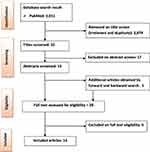
The search of PubMed yielded 2,911 records. After removal of irrelevant and duplicate records by title (2,879) and abstract (17) screening, 15 remained. An additional five were identified by forward and backward searches, which made 20 papers eligibile for full-text assessment. Finally, 14 articles published between 1994 and 2019 were included.16,20–32 Figure 1 is adapted from the PRISMA guidelines19 and summarizes the search process and results.
 |
Figure 1 Study-selection flowchart. |
Characteristics of Included Studies
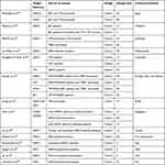
As shown in Table 1, the number of samples recruited in the studies included in this systematic review ranged from three to 77. Four target flaviviruses — DENV, YFV, WNV, and ZIKV — and one alphavirus were included. The studies had recruited participants from Okinawa,20 Thailand,25 Germany,21 the Netherlands,22 Colombia,23 different countries in Europe, Asia and Africa,16 Thailand,32 Singapore,24 Taiwan,27 Yap,28,33 and the US.27,30 All the articles were cohort studies.
 |
Table 1 Characteristics of included studies |
Result of Individual Studies
Cross-Reaction of DENV with Other Flaviviruses
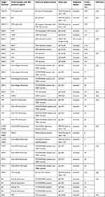
Of the 14 papers included, six16,21–23,25,32 reported cross-reactivity of DENV with other non-dengue flaviviruses, ie, YFV, ZIKV, JEV, and TBEV. The magnitude of cross-reactivity varied within species of flavivirus, type of assay used, and the target-immunoglobulin class or target-protein type. About 15.4%–84% of antibodies produced against non-dengue flaviviruses were reported as cross-reactive with dengue using different assays (lab methods). Cross-reactivity ranged up to 76.9% with antibodies produced against YFV using IgG ELISA23 and up to 84% with antibodies produced against one or multiple non-dengue flaviviruses (YFV, WNV, JEV, and TBEV) using IgG EIA assays.16 With respect to the assay methods employed, the highest cross-reactivity of DENV was reported using IgG-capture ELISA/IFA/EIA over IgM ELISA/IFA/EIA or PRNT and assays based on E-specific immunoglobulin over NS1-specific immunoglobulin. NS1-specific IgG/M-capture ELISA for DENV showed no cross-reaction with ZIKV, unlike E-specific IgG/M-capture ELISA (Table 2).
 |
Table 2 Magnitude of cross-reaction reported in individual studies |
Cross-Reaction of YFV with Other Flaviviruses
Two papers16,23 reported cross-reaction of YFV with DENV and sera from DENV/WN/JE patients and TBEV vaccines. Up to 80% antibodies produced against DENV infection were reported as cross-reactive using the PRNT. Relatively higher cross-reaction was reported using IgG-based assays than IgM assays (Table 2).
Cross-Reaction of WNV with Other Flaviviruses
One paper indicated WNV cross-reactivity with non-WNV flaviviruses to be 10% and 50% using IgM-based IFA and IgG-based IFA, and 30% and 60% using IgM-based EIA and IgG-based EIA respectively.16 The highest cross-reactivity reported was with IgG-based EIA over IgM EIA and IgG-based IFA over IgM-based IFA. Another study20 reported 32.1% cross-reaction with antibodies from YFV and JEV vaccines (Table 2).
Cross-Reaction of JEV with Other Flaviviruses
As reported in one paper,16 4% and 16% of antibodies induced against non-JEV flaviviruses (sera from YFV and/or WNV and/or JEV patients) were cross-reactive with JEV using IgG-based IFA and IgM-based IFA respectively. Cross-reaction rose to 32% and 74% using IgG-based EIA and IgM-based EIA, respectively (Table 2).
Cross-Reaction of CHIKV (Alphavirus) with Flaviviruses
Unlike cross-reaction within the genus Flavivurus, very minimal cross-reaction was reported between Flavivurus and Alphavirus. As reported in one paper24 only 6% of antibodies produced against DENV and 7% of antibodies produced against non-DENV flaviviruses were cross-reactive with CHIKV (Alphavirus), but up to 58% cross-reaction of CHIKV with other non-CHIKV alphaviruses was reported using the PRNT5024 (Table 2).
Factors Boosting Flavivirus Cross-Reactivity
Studies showed that preexisting immunity due to vaccination or prior flavivirus exposure to antigenetically similar species enhanced serological cross-reactivity.26,27,29–31 In one study, secondary flavivirus–infected patients showed a high degree of serological cross-reactivity with other flaviviruses compared to primary flavivirus–infected patients.28
Discussion
A number of studies revealed extensive serological cross-reactions between flaviviruses on different assays. This provides a challenge for accurate diagnosis of flavivuruses, especially in areas where multiple species circulate. The magnitude of cross-reaction varied among species, serological assay used, and target antibody for detection.
Within the flaviviruses, the highest cross-reactions were observed between YFV and DENV and between DENV and ZIKV. The magnitude of cross-reactivity for target flaviviruses increased (eg, up to 84% for DENV) in cases of sera taken from participants with a history of infections and/or vaccination with multiple serotypes. The lowest cross-reaction was reported from CHIKV with DENV and non-DENV flaviviruses (only up to 7%). This variation in the magnitude of cross-reaction might largely depend on the range of antigenic similarities among species. DENV, ZKV, YFV, WNV, and JEV are in the same family (Flaviviridae) and genus (Flavivirus) with common antigenic determinants, while CHIKV is categorized under another family (Togaviridae) and genus (Alphavirus).35
With respect to lab methods, the highest cross-reactivity was demonstrated with IgG-capture assays (ELISA/IFA/EIA) compared to IgM-capture assays (ELISA/IFA/EIA). Animal-model studies on closely related flaviviruses also demonstrated that IgG-based assays were less specific than IgM-based assays for homologous viruses.16,36 It was revealed that assays based on the E protein compared to those based on the NS1 protein led to higher cross-reactivity. This variation in the degree of cross-reaction might be due to differences in specificity of methods, which in turn relies on the nature of target-flavivirus proteins used for diagnosis. It was found that the E protein elicited flavivirus cross-reactive neutralization antibodies, while the NS1 protein induced a nonneutralizing virus-specific antibody response.31 In another flavivirus study, it was also suggested that antibodies to NS1 can be used as diagnostic markers of a flavivirus species–specific infection.37 In contrast to early studies, which found NS1 to be a species-specific marker in flavivirus serology, one recent study on ZIKV NS1 IgM and IgG revealed significant cross-reactivity with DENV.38 This raises a question on the specificity of the NS1 marker, and hopefully future research can solve this dilemma. Although currently, the PRNT is considered the gold standard for detecting and measuring antibodies that can neutralize viruses,18,39 the results of this review indicate it tends to be subject to cross-reactivity, especially in patients with prior flavivirus infection or immunization history. This finding is incongruent with one study that suggested that the PRNT does not accurately discriminate flavivirus infection in cases of subsequent infection.40
The findings of this study showed that preexisting immunity due to vaccination or natural infection to antigenetically related species enhances the serological cross-reaction titer. This evidence is supported by studies that demonstrated relatively higher cross-reactions to ZKV observed in patients with secondary DENV exposure than patients with primary DENV exposure.22,34 This might be due to reactivation of preexisting memory B cells that target conserved epitopes.34
Limitations
This systematic review basically focused on medically important mosquito-borne flaviviruses only, but did not fully address serological cross-reactions within and across all flaviviruses. Despite the endemicity of mosquito-borne flaviviruses in African, Caribbean, and Southeast Asian countries, the review findings do not reflect the situations in this countries, due to a paucity of research in these regions. Furthermore, the sera used for the studies included in this review were not standardized, which might have interfered with patient histories of different flavivirus infections, including tick-borne flavivirus.
Conclusion
The findings of this review revealed that the magnitude of cross-reactivity varies within the species of flavivirus, type of serological assay, and target biological marker. Cross-reactivity was higher between DENV and non-DENV flaviviruses, especially YFV, and the lowest cross-reactivity was observed in CHIKV with DENV and non-DENV flaviviruses. Similarly, cross-reactivity was higher for IgG assays than IgM assays and assays based on the E protein than the NS1 protein. Furthermore, preexisting immunity to antigenetically similar species enhanced the serological cross-reactivity. This can ultimately affect the reliability of serological test outcomes due to false-positive results. Therefore, test outcomes should be interpreted with great caution. Otherwise, it is advisable to use a combination of virological and molecular techniques together with serological investigations to boost the reliability of test results. Researchers in this arena are urged to search for novel diagnostic markers for accurate differential diagnosis of flaviviruses.
Acknowledgments
The authors would like to acknowledge Addis Ababa University for providing systematic review–protocol training to the authors to technically support the review process.
Disclosure
The authors declare no conflicts of interest for this work.
References
1. Huang YJ, Higgs S, Horne KM, Vanlandingham DL. Flavivirus-mosquito interactions. Viruses. 2014;6(11):4703–4730. doi:10.3390/v6114703
2. Gubler DJ, Markoff L. 2007 flaviviruses. In: Knipe DM, Howley PM, editors. Fields Virology.
3. Simmonds P, Becher P, Bukh J, et al. ICTV virus taxonomy profile: flaviviridae. J Gen Virol. 2017;98(1):2–3. doi:10.1099/jgv.0.000672
4. Mota MT, Terzian AC, Silva ML, Estofolete C, Nogueira ML. Mosquito-transmitted viruses - the great Brazilian challenge. Braz J Microbiol. 2016;47(Suppl 1):38–50. doi:10.1016/j.bjm.2016.10.008
5. Diosa-Toro M, Urcuqui-Inchima S, Smit JM. Arthropod-borne flaviviruses and RNA interference: seeking new approaches for antiviral therapy. Adv Virus Res. 2013;85:91–111.
6. Calzolari M, Ze-Ze L, Vazquez A, Sanchez Seco MP, Amaro F, Dottori M. Insect-specific flaviviruses, a worldwide widespread group of viruses only detected in insects. Infect Genet Evol. 2016;40:381–388. doi:10.1016/j.meegid.2015.07.032
7. Holbrook MR. Historical perspectives on flavivirus research. Viruses. 2017;9(5):97. doi:10.3390/v9050097
8. Chambers TJ, Hahn CS, Galler R, Rice CM. Flavivirus genome organization, expression, and replication. Annu Rev Microbiol. 1990;44:649–688. doi:10.1146/annurev.mi.44.100190.003245
9. Crill WD, Chang GJ. Localization and characterization of flavivirus envelope glycoprotein cross-reactive epitopes. J Virol. 2004;78(24):13975–13986. doi:10.1128/JVI.78.24.13975-13986.2004
10. Volk DE, May FJ, Gandham SH, et al. Structure of yellow fever virus envelope protein domain III. Virology. 2009;394(1):12–18. doi:10.1016/j.virol.2009.09.001
11. Cherrier MV, Kaufmann B, Nybakken GE, et al. Structural basis for the preferential recognition of immature flaviviruses by a fusion-loop antibody. EMBO J. 2009;28(20):3269–3276. doi:10.1038/emboj.2009.245
12. Chavez JH, Silva JR, Amarilla AA, Moraes Figueiredo LT. Domain III peptides from flavivirus envelope protein are useful antigens for serologic diagnosis and targets for immunization. Biologicals. 2010;38(6):613–618. doi:10.1016/j.biologicals.2010.07.004
13. Matsui K, Gromowski GD, Li L, Barrett AD. Characterization of a dengue type-specific epitope on dengue 3 virus envelope protein domain III. J Gen Virol. 2010;91(Pt 9):2249–2253. doi:10.1099/vir.0.021220-0
14. Roehrig JT, Butrapet S, Liss NM, et al. Mutation of the dengue virus type 2 envelope protein heparan sulfate binding sites or the domain III lateral ridge blocks replication in Vero cells prior to membrane fusion. Virology. 2013;441(2):114–125. doi:10.1016/j.virol.2013.03.011
15. Heinz FX, Stiasny K. Flaviviruses and flavivirus vaccines. Vaccine. 2012;30(29):4301–4306. doi:10.1016/j.vaccine.2011.09.114
16. Koraka P, Zeller H, Niedrig M, Osterhaus AD, Groen J. Reactivity of serum samples from patients with a flavivirus infection measured by immunofluorescence assay and ELISA. Microbes Infect. 2002;4(12):1209–1215. doi:10.1016/S1286-4579(02)01647-7
17. Hobson-Peters J. Approaches for the development of rapid serological assays for surveillance and diagnosis of infections caused by zoonotic flaviviruses of the Japanese encephalitis virus serocomplex. J Biomed Biotechnol. 2012;2012:379738. doi:10.1155/2012/379738
18. Thomas SJ, Nisalak A, Anderson KB, et al. Dengue plaque reduction neutralization test (PRNT) in primary and secondary dengue virus infections: how alterations in assay conditions impact performance. Am J Trop Med Hyg. 2009;81(5):825–833. doi:10.4269/ajtmh.2009.08-0625
19. Liberati A, Altman DG, Tetzlaff J, et al. The PRISMA statement for reporting systematic reviews and meta-analyses of studies that evaluate health care interventions: explanation and elaboration. PLoS Med. 2009;6(7):e1000100. doi:10.1371/journal.pmed.1000100
20. Mansfield KL, Horton DL, Johnson N, et al. Flavivirus-induced antibody cross-reactivity. J Gen Virol. 2011;92(Pt 12):2821–2829. doi:10.1099/vir.0.031641-0
21. Allwinn R, Doerr HW, Emmerich P, Schmitz H, Preiser W. Cross-reactivity in flavivirus serology: new implications of an old finding? Med Microbiol Immunol. 2002;190(4):199–202. doi:10.1007/s00430-001-0107-9
22. van Meer MPA, Mogling R, Klaasse J, et al. Re-evaluation of routine dengue virus serology in travelers in the era of Zika virus emergence. J Clin Virol. 2017;92:25–31. doi:10.1016/j.jcv.2017.05.001
23. Houghton-Trivino N, Montana D, Castellanos J. Dengue-yellow fever sera cross-reactivity; challenges for diagnosis. Revista de salud publica. 2008;10(2):299–307. doi:10.1590/S0124-00642008000200010
24. Kam YW, Pok KY, Eng KE, et al. Sero-prevalence and cross-reactivity of chikungunya virus specific anti-E2EP3 antibodies in arbovirus-infected patients. PLoS Negl Trop Dis. 2015;9(1):e3445. doi:10.1371/journal.pntd.0003445
25. Makino Y, Tadano M, Saito M, et al. Studies on serological cross-reaction in sequential flavivirus infections. Microbiol Immunol. 1994;38(12):951–955. doi:10.1111/j.1348-0421.1994.tb02152.x
26. Lai CY, Tsai WY, Lin SR, et al. Antibodies to envelope glycoprotein of dengue virus during the natural course of infection are predominantly cross-reactive and recognize epitopes containing highly conserved residues at the fusion loop of domain II. J Virol. 2008;82(13):6631–6643. doi:10.1128/JVI.00316-08
27. Lai L, Rouphael N, Xu Y, et al. Innate, T-, and B-cell responses in acute human zika patients. Clin Infect Dis. 2018;66(1):1–10. doi:10.1093/cid/cix732
28. Lanciotti RS, Kosoy OL, Laven JJ, et al. Genetic and serologic properties of Zika virus associated with an epidemic, Yap State, Micronesia, 2007. Emerg Infect Dis. 2008;14(8):1232–1239. doi:10.3201/eid1408.080287
29. Priyamvada L, Suthar MS, Ahmed R, Wrammert J. Humoral immune responses against zika virus infection and the importance of preexisting flavivirus immunity. J Infect Dis. 2017;216(suppl_10):S906–S911. doi:10.1093/infdis/jix513
30. Rogers TF, Goodwin EC, Briney B, et al. Zika virus activates de novo and cross-reactive memory B cell responses in dengue-experienced donors. Sci Immunol. 2017;2(14):eaan6809. doi:10.1126/sciimmunol.aan6809
31. Stettler K, Beltramello M, Espinosa DA, et al. Specificity, cross-reactivity, and function of antibodies elicited by Zika virus infection. Science. 2016;353(6301):823–826. doi:10.1126/science.aaf8505
32. Souza N, Felix AC, de Paula AV, Levi JE, Pannuti CS, Romano CM. Evaluation of serological cross-reactivity between yellow fever and other flaviviruses. IJID. 2019;81:4–5.
33. Priyamvada L, Quicke KM, Hudson WH, et al. Human antibody responses after dengue virus infection are highly cross-reactive to Zika virus. Proc Natl Acad Sci U S A. 2016;113(28):7852–7857. doi:10.1073/pnas.1607931113
34. Priyamvada L, Cho A, Onlamoon N, et al. B cell responses during secondary dengue virus infection are dominated by highly cross-reactive, memory-derived plasmablasts. J Virol. 2016;90(12):5574–5585. doi:10.1128/JVI.03203-15
35. Robert E, Shope PW, Amelia Travassosd AR. The Arboviruses: Epidemiology and Ecology. CRC Press; 1988:38.
36. Vazquez S, Valdes O, Pupo M, et al. MAC-ELISA and ELISA inhibition methods for detection of antibodies after yellow fever vaccination. J Virol Methods. 2003;110(2):179–184. doi:10.1016/S0166-0934(03)00128-9
37. Rastogi M, Sharma N, Singh SK. Flavivirus NS1: a multifaceted enigmatic viral protein. Virol J. 2016;13:131. doi:10.1186/s12985-016-0590-7
38. Simmons G, Stone M, Busch MP. Arbovirus diagnostics: from bad to worse due to expanding dengue virus vaccination and zika virus epidemics. Clin Infect Dis. 2018;66(8):1181–1183. doi:10.1093/cid/cix972
39. Ratnam S, Gadag V, West R, et al. Comparison of commercial enzyme immunoassay kits with plaque reduction neutralization test for detection of measles virus antibody. J Clin Microbiol. 1995;33(4):811–815. doi:10.1128/jcm.33.4.811-815.1995
40. Sirivichayakul C, Sabchareon A, Limkittikul K, Yoksan S. Plaque reduction neutralization antibody test does not accurately predict protection against dengue infection in Ratchaburi cohort, Thailand. Virol J. 2014;11:48. doi:10.1186/1743-422X-11-48
 © 2021 The Author(s). This work is published and licensed by Dove Medical Press Limited. The full terms of this license are available at https://www.dovepress.com/terms.php and incorporate the Creative Commons Attribution - Non Commercial (unported, v3.0) License.
By accessing the work you hereby accept the Terms. Non-commercial uses of the work are permitted without any further permission from Dove Medical Press Limited, provided the work is properly attributed. For permission for commercial use of this work, please see paragraphs 4.2 and 5 of our Terms.
