")
Back to Journals » International Journal of Nanomedicine » Volume 19
Macrophage-Targeting DNA Nanomaterials: A Future Direction of Biological Therapy
Authors Tu YC , Wang YM, Yao LJ
Received 12 January 2024
Accepted for publication 28 March 2024
Published 23 April 2024 Volume 2024:19 Pages 3641—3655
DOI https://doi.org/10.2147/IJN.S459288
Checked for plagiarism Yes
Review by Single anonymous peer review
Peer reviewer comments 2
Editor who approved publication: Professor Lijie Grace Zhang
Yu-Chi Tu,* Yu-Mei Wang,* Li-Jun Yao
Department of Nephrology, Union Hospital, Tongji Medical College, Huazhong University of Science and Technology, Wuhan, People’s Republic of China
*These authors contributed equally to this work
Correspondence: Li-Jun Yao, Email [email protected]
Abstract: DNA can be used for precise construction of complex and flexible micro-nanostructures, including DNA origami, frame nucleic acids, and DNA hydrogels. DNA nanomaterials have good biocompatibility and can enter macrophages via scavenger receptor-mediated endocytosis. DNA nanomaterials can be uniquely and flexibly designed to ensure efficient uptake by macrophages, which represents a novel strategy to regulate macrophage function. With the development of nanotechnology, major advances have been made in the design and manufacturing of DNA nanomaterials for clinical therapy. In diseases accompanied by macrophage disturbances including tumor, infectious diseases, arthritis, fibrosis, acute lung injury, and atherosclerosis, DNA nanomaterials received considerable attention as potential treatments. However, we lack sufficient information to guarantee precise targeting of macrophages by DNA nanomaterials, which precludes their therapeutic applications. In this review, we summarize recent studies of macrophage-targeting DNA nanomaterials and discuss the limitations and challenges of this approach with regard to its potential use as a biological therapy.
Keywords: macrophages, DNA nanomaterials, therapy, macrophage-related diseases
Introduction
Macrophages are abundant immune cells that differentiate from monocytes and play a vital role in immune responses. Macrophages specifically recognize targets through different surface receptors, and this recognition is necessary for their phagocytic function.1 Phagocytosis by macrophages is a key innate immune system mechanism to remove pathogens, microorganisms, and harmful foreign particles from the body.2 Macrophages also present antigens to recruit and activate other immune cells (such as T and NK cells) to initiate adaptive immunity processes.3
In addition to their involvement in immune responses, macrophages also regulate inflammatory reactions. Depending on the local environment, macrophages acquire different activation states. Traditionally, they are mainly divided into a “classically activated” phenotype (also referred to as M1) and an “alternatively activated” phenotype (also referred to as M2). Under stimulation with IFN-γ and lipopolysaccharide(LPS), macrophages mainly present the M1 macrophage phenotype (characterized by MHC-II and costimulatory molecules such as CD40, CD81, CD86, and intracellular iNOS expression), known as “classically activated” phenotype. CD68+ M1 macrophages promote inflammatory responses through the secretion of pro-inflammatory factors, such as tumor necrosis factor α (TNF-α), interleukin 6 (IL-6), and IL-12. Under stimulation with IL-4 and IL-13, macrophages present the M2 macrophage phenotype (characterized by CD206, CD163, arginase-1 expression), also known as “alternatively activated” phenotype. The anti-inflammatory effect of CD206+ M2 macrophages is mediated by the secretion of anti-inflammatory cytokines, such as transforming growth factor β (TGF-β) and IL-10, which inhibit inflammation.4 Changes in macrophage polarization phenotype occur in many pathological states and play an important role in cancer, fibrosis, and infectious diseases.5,6 Therefore, the regulation of macrophage function is an essential target for the treatment of diseases accompanied by disturbances of macrophage properties.
DNA nanomaterials with flexible structures and high biocompatibility are an emerging nanotechnology that can be used to target macrophages. DNA nanomaterials can be engineered to enter macrophages through scavenger receptor-mediated endocytosis,7 and various structural surface and shape modifications can increase the rate of uptake of these substances by macrophages.8,9 Unlike conventional nanomaterials,10 DNA nanomaterials are not overly toxic and have minor side effects and good biological safety after the internalization by macrophages.11 These properties provide a unique advantage for macrophage targeting. The high loading capacity of DNA nanomaterials is also a useful feature that can be exploited when creating therapeutic agents targeting macrophages.
In this review, we provide a brief overview of the biological functions of macrophages in diseases, describe different types of DNA nanostructures, and consider the necessary characteristics of macrophage-targeting DNA nanomaterials. Next, we discuss the regulation of macrophages by DNA nanomaterials and their possible applications in various diseases.
Rationale for Macrophages as Therapeutic Targets in Diseases
Macrophages are present in almost all body tissues. Upon exposure to harmful stimuli (such as pathogens, microorganisms, etc.), macrophages undergo a series of reactions and participate in the inflammatory response, a primary defense mechanism in the body.12 During the early inflammatory stages or late stages of chronic inflammation, macrophages have the pro-inflammatory phenotype. In contrast, macrophages show the anti-inflammatory phenotype during the late stages of acute inflammation.13 This shift makes them key to multiple disorders (Figure 1).
 |
Figure 1 Rationale for using macrophages as therapeutic targets in diseases. |
Macrophages have two phenotypes: M1 macrophages secrete pro-inflammatory factors, initiating the creation of nitric oxide (NO) and reactive oxygen species (ROS), which can be used to resist pathogen invasion. In contrast, M2 macrophages have an anti-inflammatory phenotype and secrete various cytokines, such as C-C motif chemokine ligand 22, and anti-inflammatory mediators, such as arginase-1, to prevent excessive inflammatory damage to tissues.14,15 Phenotype transformation in macrophages is critical in multiple pathophysiological states. The enhanced antigen-presenting activity and pro-inflammatory phenotypes of M1 macrophages eliminate infections caused by bacteria and viruses.16,17
Changes in macrophage polarization accompany tissue damage and repair.18 When acute injury occurs, tissue-resident and monocyte-derived macrophages act as receptors and effectors of inflammation and tissue damage.19 M1 macrophages secrete inflammatory factors that play a classical inflammatory role, whereas M2 macrophages drive the repair process following tissue damage.20 M2 macrophages have a profibrotic phenotype, which is a possible cause of scar formation and fibrosis in various pathological states.21
Tumor-associated macrophages (TAMs) are a crucial component of the tumor microenvironment. Differences in TAM phenotype are essential for tumor development and progression. M1 TAMs recognize tumor cells and initiate the subsequent immune response, thereby inhibiting tumor progression.22 M2 TAMs enhance immunosuppression by secreting tumor growth-promoting cytokines, such as the epidermal growth factor and C-C motif chemokine ligand 18, factors that promote tumor metastasis, such as matrix metalloproteinases and urokinases, and tumor-promoting substances (such as IL-10 and prostaglandin E2).23 Thus, TAMs have become an attractive target for tumor therapy. For example, targeting M2 macrophages was shown to suppress tumor growth and metastasis.24
The key role of macrophages in inflammatory responses suggests that they cannot be ignored in diseases involving inflammation. Unbalanced macrophage numbers during the inflammatory phase can lead to uncontrolled inflammatory responses. At the onset of rheumatoid arthritis, macrophage infiltration is a manifestation of disease activity.25 M1 macrophages contribute to the release of inflammatory factors in the synovial fluid of joints, which worsens joint erosion.26 Thus, modulation of macrophage phenotypes and inhibition of their activation are important therapeutic strategies for rheumatoid arthritis (RA) inflammation.
In addition to their role in inflammation-related diseases, macrophages are involved in the development of atherosclerosis. Macrophage aggregation in the vessel wall is a major pathological feature of atherosclerosis. Macrophages internalize lipids and retain them in the arterial wall, which is the basis of atherogenic plaque formation.27 Both macrophage numbers and phenotype affect this process. Oxidized cholesterol is taken up by macrophage scavenger receptors. Oxidized cholesterol stored in the cells inhibits cytotoxicity.28 Abnormal apoptosis of macrophages, which is a manifestation of clearance failure, exacerbates atherogenic plaque formation. The pro-inflammatory effect of M1 macrophages is also crucial for plaque formation and contributes to vulnerable plaques.29 Given their critical role in atherosclerosis, macrophages are attractive therapeutic targets.
M2 macrophages are key regulators of damage repair and play crucial roles in the initiation, maintenance, and resolution of damage.30–32 Tissue macrophages are important chemokine producers that recruit T-cells and fibroblasts to coordinate the development of fibrosis.33 Abnormal wound repair can lead to fibrosis and scar formation. M2 macrophages play an essential role in fibrosis of various etiologies.34 Targeting M2 macrophages may be used in antifibrotic therapy.
In-depth exploration of macrophage roles in inflammation and repair led to the realization that targeted regulation of macrophage polarity and activation may help achieving a therapeutic effect by delaying disease progression and/or reversing the disease occurrence.
DNA Nanomaterials
Overview of Functional DNA Nanostructures
DNA is a general polymer material that is widely used to construct complex nanostructures, as flexible conformation of its deoxynucleotide chain can be achieved by precise design. DNA nanotechnology has enabled the development of complex, convenient, and diverse DNA nanomaterials. With the development of DNA manipulation technology, DNA stiffness-based tile and nanodynamic mechanical components can be produced,35,36 which are used as raw materials for constructing nanostructures of different sizes.37,38 Specific base arrangement and superposition of bases affect DNA flexibility.
DNA nanostructures can have single-layer, wireframe single-layer, and multilayer designs39 and be assembled into one-dimensional or two-dimensional arrays, as well as three-dimensional DNA lattices. A three-dimensional DNA crystal based on the bottom-up DNA assembly was reported in 2009.40 Subsequently, the concept of DNA origami (DO) has brought new ideas and possibilities for the advancement of DNA nanotechnology.DO utilizes numerous short DNA oligonucleotides to create various designer nanoscale architectures by folding long single-stranded DNA.41 DO significantly improves the scalability and complexity of DNA nanostructures, offering great potential for the development of highly controlled nanomaterials.
Frame nucleic acid (FNA) structures, especially tetrahedral DNA nanostructures (TDNs)42 that have been created based on the DO technology, as well as other functional DNA nanomaterials are currently a focus of intensive research. Various DNA self-assembled structures, such as DNA nanoflowers for immunotherapy and DNA nanotubes as drug carrier platforms, have been used in preclinical therapy.43,44 DNA hydrogels have been also proposed as functional DNA materials. Hydrogels can be divided into hybrid and pure DNA hydrogels.39 Owing to the three-dimensional network structure formed by hydrophilic DNA cross-linking, DNA hydrogels have predictable structures and adjustable mechanical strength.45 The programmability of DNA hydrogels may have great therapeutic potential as their properties enable modulation of drug release. Functional DNA nanomaterials also include mixtures of proteins or nanoparticles, such as DNA oligonucleotides attached to nanoparticles on poly(ethylene glycol)-coated superparamagnetic iron oxide nanoparticles (PEG-SPIONs).46 In addition, various chemical, fluorescent, and fluorescence-quenching groups can be modified at both ends or in the middle of the DNA chain to enrich DNA function.47,48 For example, DNA nanomaterials with a flat solid disk shape modified by the Cy5 dye can be efficiently taken up by macrophages.49 Mn2+-incorporating tetrahedral DNA nanostructures can exert synergistic therapeutic effects.50
Biological Properties of DNA Nanomaterials
Biological properties of DNA nanomaterials are closely related to their structure. As a therapeutic technology with great potential, DNA nanotechnology has shown excellent structure stability, high load capacity, good biocompatibility and biological safety, and high selectivity (Figure 2). These biological features render DNA nanotechnology a very important modality in disease treatment.
 |
Figure 2 Biological properties of DNA nanomaterials. DNA- SPIONs, DNA superparamagnetic iron oxide nanoparticles. Abbreviations: DNA nanoflowers; TDN, tetrahedral DNA nanostructures; DNA-based disk-shaped NPs, DNA-based disk-shaped nanoparticles. Note: DNA-SPIONs is adapted from Zhang L, Tian XY, Chan CKW, et al. Promoting the Delivery of Nanoparticles to Atherosclerotic Plaques by DNA Coating. ACS Appl Mater Interfaces. 2019;11:13888–13904. Copyright {2019} American Chemical Society.46 |
The advantages of structural stability, high yield, versatility, and purity, are an important basis for the use of DNA nanomaterials in biological applications. Most DNA nanostructures rely on hydrogen bonds to maintain their stability. Recently, Wang et al used molecular docking tools based on a DNA framework to rationally design DNA nanomaterials with higher stability.51 A method that utilizes topoisomerases to maintain the stability of DNA nanomaterials has been reported recently.52 Topology isomerase transforms the original DNA topology into a nano-reticular structure, which strengthens DNA nanomaterial and increases its stability.53 Schipperkes et al developed multifunctional DNA nanomaterials consisting of silica nanoparticles and carbon nanotubes functionalized with DNA.54
Because their characteristics can be programmed and modified, DNA nanomaterials can be used as substance carriers for therapeutic purposes. DNA nanomaterials loaded with pharmacological compounds, ribonucleic acids, and antibiotics show high load capacities and can have different effects. Such DNA nanostructures have been made with the use of DNA origami technology, DNA monocytes, nanocapsules, DNA self-assembly, and other approaches.
Good biocompatibility and biological safety are key advantages of DNA nanomaterials. DNA nanomaterials are nontoxic and have been shown to have no side effects in several animal models.55 Hu et al reported high biocompatibility of silicon nanoparticle/carbon nanotube/DNA nanocomposites that had minimal adverse effects on the human body, which enhanced the efficacy of the carried drug.56
DNA nanomaterials show high selectivity. This feature, combined with the high loading capacity is particularly useful for targeted therapy. Advances in research have enabled targeting specific cells and even subcellular structures by DNA nanomaterials. DNA nanomaterials, such as DNA tetrahedron, spherical nucleic acids, and nanoparticle-templated DNA nanostructures, can be taken up by cells through endocytosis to enable targeted intracellular delivery of functional substances, such as small molecules, aptamers, and proteins.57,58 DNA nanostructures target specific cells in a ligand-receptor interaction-like fashion. The precise specificity of such cellular targeting is a landmark development in therapy. Owing to its high specificity, precise targeted administration can reduce drug side effects and increase treatment utility.
The distinctive biological properties of DNA nanomaterials make them promising candidates for medical applications. The high selectivity of DNA nanomaterials gives them a unique advantage in targeted therapy.
Characteristics of DNA Nanomaterials Internalized by Macrophages
DNA nanomaterials exert their characteristic therapeutic effects by various mechanisms, including macrophage targeting via scavenger receptor-mediated endocytosis (Figure 3).7 Scavenger receptors are specifically expressed on the surface of macrophages, where they take part in pathophysiological processes, such as inflammation. In Caenorhabditis elegans and zebrafish, DNA nanomaterials are taken up by macrophages through scavenger receptor-mediated endocytosis.59,60 Cui et al reported that the DNA nanodevice E64-DNA was preferentially localized to mouse TAMs through endocytosis mediated by the specific scavenger receptors SCARB1 (class B scavenger receptor type 1) and MSR1 (macrophage scavenger receptor 1).61 The novel scavenger receptor inhibitor PEGylated poly-lysine peptide has been shown to inhibit the uptake of DNA nanoparticles by Kupffer cells in the liver.62
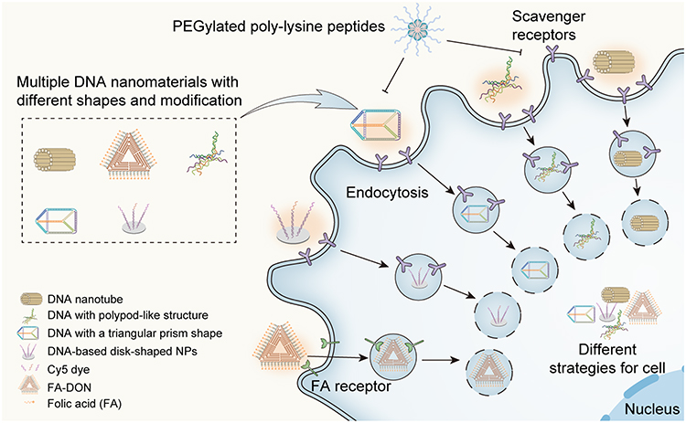 |
Figure 3 Various mechanisms of entry of DNA nanomaterials into cells. FA, folic acid; FA-DON, folic acid/DNA origami nanostructure. Abbreviation: NPs, nanoparticles. Note: FA-DON is adapted from Ma Y, Lu Z, Jia B, et al. DNA Origami as a Nanomedicine for Targeted Rheumatoid Arthritis Therapy through Reactive Oxygen Species and Nitric Oxide Scavenging. ACS Nano. 2022;16(8):12520–1253. Copyright {2022} American Chemical Society.63 |
Structural properties of DNA nanomaterials and modifications of the material surface significantly affect uptake efficiency (Table 1). DNA nanostructure density and shape affect internalization by mouse macrophage-like cell line RAW264.7 with high-density DNA structures having higher cellular uptake rates.8 Ohtsuki et al used DO technology to design rectangular DNA nanostructures with different rigidities.64 They found that single-stranded circular DNA folded into rectangular DNA nanostructure, and the number of staples positively correlated with the degree of uptake by RAW264.7 cells. In contrast, the distance between DNA helices negatively correlated with the uptake rate. DNA with polypod-like structures can be efficiently taken up by macrophages. Mohri et al reported that uptake in RAW264.7, cells increased with an increase in pod numbers.65,66
 |
Table 1 Influence of Physiochemical Properties of DNA Nanomaterials on Their Interaction with Macrophages |
The differences in cellular uptake are related not only to the structural properties but also to functional modifications of DNA nanomaterials. DNA nanostructures can be edited to increase macrophage targeting. The modification by adding functional groups such as peptides, folate, and dyes can promote the internalization by macrophages. In pathological states, some glycoproteins, such as VCAM-1, are highly expressed on the surface of macrophages. Wang et al designed DNA tetrahedron decoy oligodeoxynucleotides (TDN-dODNs) carrying a peptide that bound specifically to VCAM-1, which enabled targeting macrophages in a pathological state.67 Folic acid receptor is overexpressed in inflammation-activated macrophages,9 which provides theoretical support for the creation of DNA nanomaterials targeting macrophages via folic acid modifications. Ma et al developed a triangular origami nanostructure (FA-tDON), that targeted M1 macrophages via the added folate group.63 Koga et al used DO technique to design flat solid disks functionalized with different dyes and showed that mouse macrophage-like RAW264.7 cells preferentially accumulated disks containing fluorophores conjugated with the handle/anti-handle modification of the Cy5 dye.49
The unique structure and endocytic mode of DNA nanomaterials give them the advantage of selective macrophage targeting, thereby providing new therapeutic strategies for pathological responses in which macrophages participate.
Strategies for DNA Nanomaterials Targeting Macrophages
Regulation of Macrophage Polarization
The phenotypic transformation of macrophages depends primarily on the environmental signals. M1 macrophages have a pro-inflammatory phenotype and secrete various inflammatory factors, such as TNF-α, IL-1β, and IL-6. M2 macrophages secrete large amounts of TGF-β and IL-10 to promote repair and angiogenesis.68 DNA nanomaterials can modulate macrophage polarity (Figure 4A). Wang et al reported that tetrahedral frame nucleic acids (tFNAs) inhibited M2 macrophage polarization.69 Zhang et al showed that tetrahedral DNA nanostructures induced M1 polarization of RAW264.7 cells.70 The effects of DNA nanomaterials on macrophage polarization may enable them to have therapeutic effects in diseases associated with dysregulation of macrophage properties.
 |
Figure 4 Strategies for targeting macrophages by using DNA nanomaterials. (A) Tetrahedral frame nucleic acid regulates macrophage polarization. (B) DNA nanomaterials activate pattern recognition receptors in macrophages. (C) DNA nanomaterials act as carriers of drugs modulating macrophage functions. Abbreviations: Dex, dexamethasone; dODN, decoy oligodeoxynucleotides; ER, endoplasmic reticulum; IFN, interferon; IL, interleukin; siRNA, small interfering RNA; TLR, toll-like receptor. |
Activation of Pattern Recognition Receptors in Macrophages
Pattern recognition receptors (PRRs) act as the frontline of immune response.71 According to their cellular location, PRRs are classified into endosomal Toll-like receptors (TLRs) and cytosolic PRRs (such as STING signaling receptors).72 The CpG motif is a well-characterized agonist of TLR9.73 Li et al designed a stable and non-toxic TDN structure loaded with immunoreactive methylated cytosine-phosphate-guanosine (CpG motif) oligodeoxynucleotides.74 Macrophage-like RAW264.7 cells internalized this TDN, which was then recognized by TLR9, leading to macrophage activation. In addition to TDN, multiple DNA nanostructures with loaded CpG motifs can also activate TLRs, including self-assembled DNA immunonanoflowers, Y-shaped DNA nanostructures, DNA-based disk-shaped nanoparticles, and polypod-like DNA nanostructures (Figure 4B).43,66,75,76 Meanwhile, the expression of co-stimulatory signals such as CD40 and CD83 in macrophages was significantly increased, resulting from the activation of TLR9 by DNA nanomaterials.76
The STING/type I interferon pathway is essential for autoimmunity. Liang et al reported that TDNs nanostructures activate the STING/type I interferon pathway in macrophages,50 which induces the production of IFN-β and NF-κB-dependent pro-inflammatory cytokines.77
DNA nanomaterials regulate autoimmunity in macrophages by activating PRRs. This provides a new direction for treating autoimmune-related diseases.
DNA Nanomaterials as Carriers of Substances Acting on Macrophages
DNA nanomaterials loaded with glucocorticoids, small interfering RNAs (siRNAs), and deoxynucleotides show a high load capacity and could play critical roles in the treatment of conditions accompanied by disturbances in macrophage properties (Figure 4C). Sellner et al designed a DNA nanotube that enabled targeted delivery of dexamethasone to macrophages. The anti-inflammatory strategy utilizing such drug delivery platform can reduce the off-target effects of the delivered drugs.44 Zhang et al used tFNA as a vector to deliver siRNA that targeted macrophages to downregulate TLR2 expression.78 The NF-κB dODNs specifically inhibited the expression of genes encoding pro-inflammatory cytokines by acting on the NF-κB signaling pathway. Wang et al developed a self-assembled TDN loaded with NF-κB bait dODN that decreased the inflammation response in macrophages.67
Application of DNA Nanomaterials for Treatment of Diseases Accompanied by Macrophage Disturbances
Owing to their ability to target macrophages, DNA nanomaterials have received considerable attention as potential treatments of pathological states associated with dysregulation of these cells. In particular, applications of DNA nanomaterials to treat tumor, infectious diseases, arthritis, fibrosis, acute lung injury, and atherosclerosis may be particularly promising (Table 2). The effects of DNA nanomaterials in these diseases based on macrophage targeting are described in detail below.
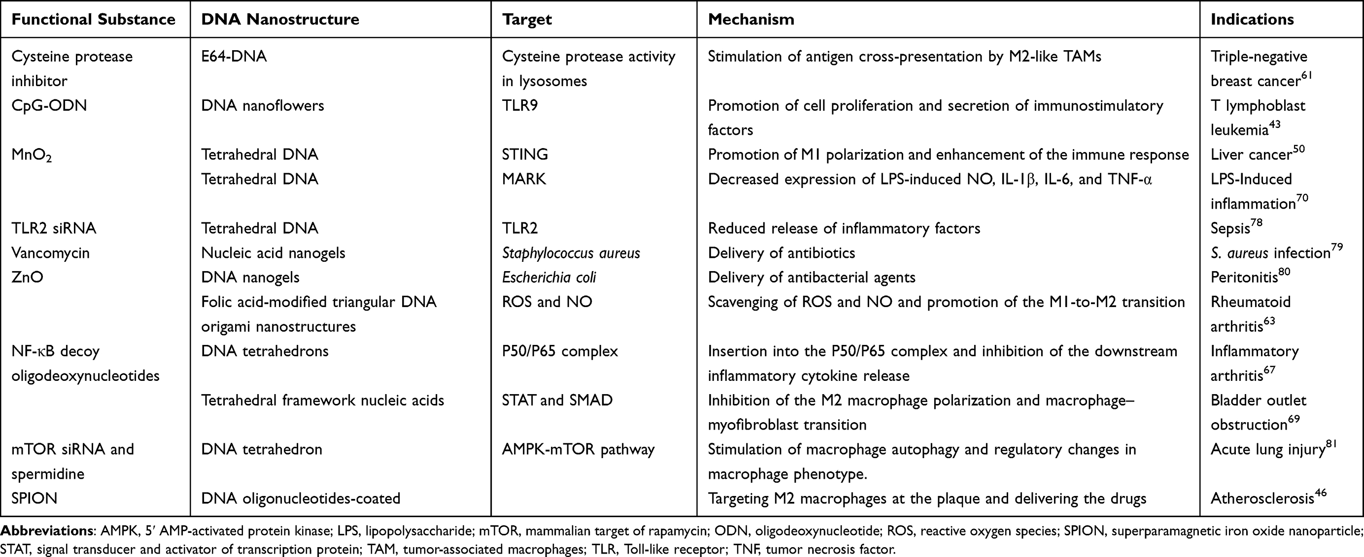 |
Table 2 DNA Nanostructures and Functional Substances They Carry for Treatment of Diseases with Macrophage Dysregulation |
Tumor
Chronic inflammation increases the risk of tumor. Continuous production of various growth factors and chemokines in the tumor microenvironment promotes the proliferation and survival of tumor cells, leading to tumor invasion and progression.82 Therefore, inhibition of persistent inflammation can effectively prevent and delay the occurrence and development of tumors.83 Tumor-associated inflammation involves complex interactions between multiple types of cells. TAMs play a key role in this process.84 Pro-inflammatory M1 macrophages secrete antitumor factors, such as TNF-α, IL-6, and others, whereas anti-inflammatory M2 macrophages release cytokines such as TGF-β, IL-10, and others, which promote tumor progression.85
TAMs are a promising target for tumor therapy. In recent years, researchers have developed various DNA nanomaterials to treat tumors by regulating macrophage function (Figure 5A). DNA nanodevice E64-DNA designed by Cui et al targets TAMs, particularly M2 macrophages.61 The cysteine protease inhibitor E64 affects the localization of macrophage lysosomes benefiting from scavenger receptor-mediated specific uptake. Excessive activity of cysteine proteases in TAM inhibits CD8+ T-cell activation by hindering antigen cross-presentation. The anti-tumor effect of E64-DNA was achieved by reducing the expression of the lysosomal cysteine protease. A combination of E64-DNA and cyclophosphamide significantly reduced the proliferation of E0771 carcinoma cells.61 Liang et al found TDNs can enter and activate macrophages.50 TDNs mainly exert antitumor effects by promoting the activation of the cytosolic immune recognition receptor STING and polarization of M1 macrophages. Synergizing with Mn2+, TDNs inhibited the growth of mouse Hepa 1–6 hepatoma cells by upregulating the levels of IFN-β, iNOS, and various co-stimulatory molecules for antigen presentation.50 Zhang et al designed self-assembled DNA nanoflowers integrated with a CpG motif by the rolling circle replication.43 These DNA nanoflowers effectively delivered immune stimulation, triggering macrophage proliferation.43 Tumor cells undergo apoptosis and necrosis resulting from the secretion of inflammatory factors by activated macrophages DO nanostructures were also shown to bind to CpG motifs by complementary hybridization to activate TLR9 in RAW264.7 macrophages.76
 |
Figure 5 Application of DNA nanomaterials in diseases associated with disturbances of macrophage properties. (A) E64-DNA (modified from ref.50), CpG DNA nanoflowers (NFs), and MnO2 tetrahedral DNA nanostructures (TDNs) in the cancer therapy. (B) TDNs, Van- DNA nanogels (DNL) and ZnO-DNL in the anti-infective therapy (Image B Van-DNL is adapted from Obuobi S, Julin K, Fredheim EGA, Johannessen M, Škalko-Basnet N. Liposomal delivery of antibiotic loaded nucleic acid nanogels with enhanced drug loading and synergistic anti-inflammatory activity against S. aureus intracellular infections. J Control Release. 2020;324:620–632. Creative Commons).79 (C) TDNs and triangular DNA origami nanostructures (tDONs) in the arthritis therapy. (D) Application of tetrahedral frame nucleic acids (tFNAs) in the bladder outlet obstruction (BOO) therapy. (E) Application of TDNs in the therapy of acute lung injury. (F) DNA superparamagnetic iron oxide nanoparticles (SPIONs) in the therapy of atherosclerotic plaques. |
DNA nanostructures have been shown to inhibit tumor growth and proliferation by promoting antigen cross-presentation, proliferation, and immune activation of macrophages.
Infectious Diseases
When pathogens invade the body, various immune cells, including macrophages, elicit an immune response.The occurrence of infectious diseases is closely linked to the activation of immune cells and the production of various inflammatory factors. French et al observed the co-localization of the DNA flower structure in macrophages infected with Mycobacterium tuberculosis and Leishmania infantum.86 This suggests that targeting macrophages by DNA nanostructures may remarkably facilitate treatment of infectious diseases.
To better explore their application prospects, various DNA nanomaterials have been tested in infectious disease models (Figure 5B). Zhang et al studied the effects of TDNs on lipopolysaccharide (LPS)-induced macrophages.70 They observed that TDNs attenuated the production of various cytokines, including IL-1β, IL-6, and NO in LPS-induced RAW264.7 cells by inhibiting the phosphorylation of MAPK. In addition, TDNs inhibited ROS production and cell apoptosis by activating the transcription of the antioxidative enzyme heme oxygenase-1 gene.70
DNA nanostructures can also serve as a transport platform to carry “cargo” that regulates the ability of macrophages to counteract infections. Zhang et al designed a TDN to deliver siRNA that blocks the LPS-induced expression of TLR2, which inhibits the synthesis and release of inflammatory factors in macrophages.78 tFNAs represent a new approach for the prevention and treatment of sepsis. DNA nanomaterials provide a better platform for future antimicrobial therapies. Obuobi et al fabricated a DNA nanogel coated with liposomal vesicles that relied on noncovalent electrostatic interactions to control the release of loaded antibiotics.79 Macrophages infected with Staphylococcus aureus showed strong anti-inflammatory activity in synergy with loaded vancomycin.
DNA nanomaterials are potential therapeutic agents for treating various bacterial infections. Chen et al designed neutrophil extracellular trap-like structures in which ZnO was adsorbed to DNA-HCl nanogels.80 These DNA nanogels decreased the expression of TNF-α, IL-6, iNOS, and COX-2 in LPS-stimulated Raw264.7 cells. In a mouse infection model, these DNA hydrogels alleviated peritonitis symptoms and inhibited the entry of Escherichia coli into the circulatory system.
DNA nanomaterials are continuously being developed for therapy because of their superior biological safety, programmability, and high load capacity. These substances have high specificity and can be particularly useful for future targeted antimicrobial therapies.
Arthritis
Rheumatoid arthritis (RA) is an autoimmune disease characterized by the chronic inflammation of the joints and synovium. RA severity is closely related to the number of macrophages present in the arthritic synovium.87 Macrophages secrete pro-inflammatory cytokines, chemokines, and growth factors that destroy the bone and synovial tissue.88 Thus, macrophages are important targets for the treatment of RA.
As a potential strategy for RA treatment, Wang et al created TDN-P-dODN, a DNA nanodrug that consists of an NF-κB decoy oligodeoxynucleotide and VCAM-1 targeting peptide (P) bound to self-assembled DNA tetrahedrons.67 TDN-P-dODNs are taken up by macrophages and inserted across the P50/P65 complex in the cytoplasm. TDN-P-dODNs reduced inflammation by inhibiting the downstream activity of the P50/P65 pathway. Pro-inflammatory M1 macrophages express ROS and NO, leading to the development of synovitis and bone damage, which cause RA. FA-tDON developed by Ma et al that targets M1 macrophages owing to folate modification reduced the production of ROS and NO.63 Furthermore, FA-tDON promoted the transformation of M1 macrophages into M2 macrophages, which may be a useful therapeutic strategy for RA (Figure 5C).
Thus, the unique biological properties of DNA nanostructures make them an emerging platform for RA therapy, and targeting macrophages with DNA nanostructures may be a promising therapeutic approach.
Fibrotic Disease
Macrophages play a key role in inflammatory responses, tissue remodeling, and homeostasis. Of the two different phenotypes of macrophages mentioned above, M2 macrophages (activated by IL-4, IL-10, IL-13, or TGF-β) inhibit inflammation and have a pro-fibrotic phenotype that drives repair during tissue damage. However, they can also cause scarring in pathological situations.21 Effective inhibition of macrophage polarization may be a viable strategy for anti-fibrotic therapy.
Targeting macrophages using DNA nanotechnology may be an important modality for anti-fibrotic therapy. Wang et al found that the extent of M2 macrophage infiltration in the bladder wall was related closely to the degree of fibrosis in the bladder outlet obstruction (BOO).69 tFNA has been shown to inhibit the polarization of M2 macrophage.70 Thus Wang et al constructed a tFNA that inhibited both M2 macrophage polarization and macrophage–myofibroblast transition process in BOO (Figure 5D). The inhibition of M2 polarization mediated by tFNA can be attributed to the inactivation of the transcriptional activator STAT3/6. Moreover, tFNAs inhibited the macrophage–myofibroblast transition by the inhibition of SMAD2/3 signaling.
Owing to the effect on macrophages, tFNA attenuated urethral orifice fibrosis. tFNA can also promote wound healing and reduce skin fibrosis,89,90 suggesting that it may be an ideal and promising candidate for anti-fibrotic therapy.
Acute Lung Injury
Acute lung injury (ALI) is characterized by severe inflammation and damage to the lung tissue. Macrophages are among the most critical effector cells in the pulmonary innate immune system that play an essential role in the initiation, development, and outcome of ALI.91
Targeting macrophages using DNA nanomaterials is a new therapeutic strategy for treating ALI. Intercellular adhesion molecule-1 (ICAM-1) is overexpressed in many lung diseases, and 3D nanomaterials are specifically targeted to the lungs using antibodies against ICAM-1.41 This also provides a possibility of specific targeting of DNA nanomaterials in lung diseases. Huang et al designed a DNA nanoplatform loaded with mTOR siRNA and spermidine that exerted an anti-inflammatory effect in ALI by regulating the macrophage phenotype.81 Spermidine inhibits mTOR through adenosine 5′-monophosphate-activated protein kinase. The siRNA-mediated inhibition of mTOR expression enhanced autophagy in macrophages, shifting their transition to the M2 phenotype (Figure 5E).
Targeting macrophages in the lung makes delivery of DNA nanomaterials more efficient. As safe and specific carriers, DNA nanomaterials deserve to be systematically explored as part of ALI treatment.
Atherosclerosis
During the development of atherosclerosis, macrophages internalize lipids and deposit them in the arterial wall to form atherosclerotic plaques.28 The delivery of therapeutic agents specifically to the atherosclerotic plaques has been the focus of many studies. DNA nanomaterials have also been used in atherosclerosis models owing to their biocompatibility and flexible editing properties.
Zhang et al developed SPIONs coated with DNA oligonucleotides as efficient atherosclerotic plaque delivery carriers.46 DNA-SPIONs enter RAW 264.7 cells via class A scavenger receptor and lipid rafts. In a mouse model of atherosclerosis, DNA-SPIONs were delivered rapidly and specifically to M2 macrophages of atherosclerotic plaques in vivo (Figure 5F). This demonstrates the potential of delivering therapeutics targeting atherosclerotic plaques.
Conclusions
Macrophages play an essential role in the immune defense against invasion. The regulation of the polarization and immune activity of macrophages makes them a target in diseases characterized by dysregulation of macrophage properties. The unique structural and functional advantages of DNA nanomaterials have made them an active subject of study in biological therapy. DNA nanomaterials can enter macrophages through scavenger receptor-mediated endocytosis, and their shapes and surface modifications (eg, by folic acid, VCAM1-targeting peptide, Cy5 dye, and other functional moieties) have been designed to promote the internalization by macrophages. DNA nanomaterials have been shown to regulate macrophage polarization, activate PRRs, and serve as carrier systems to deliver drugs, siRNAs, and dODNs. DNA nanomaterials have excellent biocompatibility and can function as delivery platforms to improve the efficiency of drug delivery. This makes DNA nanomaterials an attractive option for the treatment of diseases accompanied by macrophage dysregulation. In particular, DNA nanomaterials, such as tFNAs or TDNs, regulate macrophage polarity and inhibit inflammatory factor production. DNA nanomaterials can also be designed into a variety of structures (such as DNA nanoflowers, DNA hydrogels, and DNA tetrahedrons) for the treatment of tumors, infections, arthritis, BOO, ALI, and atherosclerosis.
However, the current studies of DNA nanomaterials are still in their infancy. The structural and material superiority of DNA nanotechnology and its therapeutic effectiveness have been verified so far only using animal models. The specific mechanism of targeting macrophages and their effects on the diseases in humans requires further exploration.
Acknowledgments
The authors acknowledge Editage for English language editing.
Funding
This research was supported by the National Natural Science Foundation of China (NO.81570657, No. 81974102).
Disclosure
The authors declare no conflicts of interest in this work.
References
1. Figueiredo Borgognoni C, Kim JH, Zucolotto V, Fuchs H, Riehemann K. Human macrophage responses to metal-oxide nanoparticles: a review. Artif Cells Nanomed Biotechnol. 2018;46(sup2):694–703. doi:10.1080/21691401.2018.1468767
2. Zhu M, Nie G, Meng H, Xia T, Nel A, Zhao Y. Physicochemical properties determine nanomaterial cellular uptake, transport, and fate. Acc Chem Res. 2013;46(3):622–631. doi:10.1021/ar300031y
3. Elsabahy M, Wooley KL. Cytokines as biomarkers of nanoparticle immunotoxicity. Chem Soc Rev. 2013;42:5552–5576.
4. Martin KE, García AJ. Macrophage phenotypes in tissue repair and the foreign body response: implications for biomaterial-based regenerative medicine strategies. Acta Biomater. 2021;133.
5. Buechler MB, Fu W, Turley SJ. Fibroblast-macrophage reciprocal interactions in health, fibrosis, and cancer. Immunity. 2021;54:903–915.
6. Dukhinova M, Kokinos E, Kuchur P, Komissarov A, Shtro A. Macrophage-derived cytokines in pneumonia: linking cellular immunology and genetics. Cytokine Growth Factor Rev. 2021;59:46–61. doi:10.1016/j.cytogfr.2020.11.003
7. Sellner S, Kocabey S, Nekolla K, Krombach F, Liedl T, Rehberg M. DNA nanotubes as intracellular delivery vehicles in vivo. Biomaterials. 2015;53:453–463. doi:10.1016/j.biomaterials.2015.02.099
8. Maezawa T, Ohtsuki S, Hidaka K, et al. DNA density-dependent uptake of DNA origami-based two-or three-dimensional nanostructures by immune cells. Nanoscale. 2020;12(27):14818–14824. doi:10.1039/D0NR02361B
9. Steinz MM, Ezdoglian A, Khodadust F, et al. Folate Receptor Beta for Macrophage Imaging in Rheumatoid Arthritis. Front Immunol. 2022;13:819163. doi:10.3389/fimmu.2022.819163
10. Pandey RK, Prajapati VK. Molecular and immunological toxic effects of nanoparticles. Int J Biol Macromol. 2018;107:1278–1293. doi:10.1016/j.ijbiomac.2017.09.110
11. Xia K, Kong H, Cui Y, et al. Systematic Study in Mammalian Cells Showing No Adverse Response to Tetrahedral DNA Nanostructure. ACS Appl Mater Interfaces. 2018;10(18):15442–15448. doi:10.1021/acsami.8b02626
12. Mosser DM, Hamidzadeh K, Goncalves R. Macrophages and the maintenance of homeostasis. Cell Mol Immunol. 2021;18(3):579–587. doi:10.1038/s41423-020-00541-3
13. Braga TT, Moura IC, Lepique AP, Camara NOS. Editorial: macrophages Role in Integrating Tissue Signals and Biological Processes in Chronic Inflammation and Fibrosis. Front Immunol. 2017;8:845. doi:10.3389/fimmu.2017.00845
14. Sun J, Sun J, Song B, et al. Fucoidan inhibits CCL22 production through NF-κB pathway in M2 macrophages: a potential therapeutic strategy for cancer. Sci Rep. 2016;6(1):35855. doi:10.1038/srep35855
15. Chen W, Zhang F, Ju Y, Hong J, Ding Y. Gold Nanomaterial Engineering for Macrophage-Mediated Inflammation and Tumor Treatment. Adv Healthc Mater. 2021;10(5):e2000818. doi:10.1002/adhm.202000818
16. Varol C, Mildner A, Jung S. Macrophages: development and tissue specialization. Annu Rev Immunol. 2015;33:643–675.
17. Hu Q, Lyon CJ, Fletcher JK, Tang W, Wan M, Hu TY. Extracellular vesicle activities regulating macrophage- and tissue-mediated injury and repair responses. Acta Pharm Sin B. 2021;11:1493–1512.
18. Placek K, Schultze JL, Aschenbrenner AC. Epigenetic reprogramming of immune cells in injury, repair, and resolution. J Clin Invest. 2019;129(8):2994–3005. doi:10.1172/JCI124619
19. Singer BD, Chandel NS. Immunometabolism of pro-repair cells. J Clin Invest. 2019;129(7):2597–2607. doi:10.1172/JCI124613
20. Watanabe S, Alexander M, Misharin AV, Budinger GRS. The role of macrophages in the resolution of inflammation. J Clin Invest. 2019;129(7):2619–2628. doi:10.1172/JCI124615
21. Conte E. Targeting monocytes/macrophages in fibrosis and cancer diseases: therapeutic approaches. Pharmacol Ther. 2022;234:108031.
22. Yang Q, Guo N, Zhou Y, Chen J, Wei Q, Han M. The role of tumor-associated macrophages (TAMs) in tumor progression and relevant advance in targeted therapy. Acta Pharm Sin B. 2020;10(11):2156–2170. doi:10.1016/j.apsb.2020.04.004
23. D-L S, Z-M L, Shen M-N, Li X, Sun L-Y. Roles of pro- and anti-inflammatory cytokines in the pathogenesis of SLE. J Biomed Biotechnol. 2012;2012:347141.
24. Tang X, Mo C, Wang Y, Wei D, Xiao H. Anti-tumour strategies aiming to target tumour-associated macrophages. Immunology. 2013;138.
25. Kim S-J, Chang HJ, Volin MV, et al. Macrophages are the primary effector cells in IL-7-induced arthritis. Cell Mol Immunol. 2020;17(7):728–740. doi:10.1038/s41423-019-0235-z
26. Udalova IA, Mantovani A, Feldmann M. Macrophage heterogeneity in the context of rheumatoid arthritis. Nat Rev Rheumatol. 2016;12:472–485.
27. Khoury MK, Yang H, Liu B. Macrophage Biology in Cardiovascular Diseases. Arterioscler Thromb Vasc Biol. 2021;41(2):e77–e81. doi:10.1161/ATVBAHA.120.313584
28. Chen W, Schilperoort M, Cao Y, Shi J, Tabas I, Tao W. Macrophage-targeted nanomedicine for the diagnosis and treatment of atherosclerosis. Nat Rev Cardiol. 2022;19:228–249.
29. Koelwyn GJ, Corr EM, Erbay E, Moore KJ. Regulation of macrophage immunometabolism in atherosclerosis. Nat Immunol. 2018;19(6):526–537. doi:10.1038/s41590-018-0113-3
30. Wynn TA, Vannella KM. Macrophages in Tissue Repair, Regeneration, and Fibrosis. Immunity. 2016;44(3):450–462. doi:10.1016/j.immuni.2016.02.015
31. Vannella KM, Wynn TA. Mechanisms of Organ Injury and Repair by Macrophages. Annu Rev Physiol. 2017;79:593–617. doi:10.1093/nar/gkaa341
32. Krenkel O, Tacke F. Liver macrophages in tissue homeostasis and disease. Nat Rev Immunol. 2017;17(5):306–321. doi:10.1038/nri.2017.11
33. Borthwick LA, Barron L, Hart KM, et al. Macrophages are critical to the maintenance of IL-13-dependent lung inflammation and fibrosis. Mucosal Immunol. 2016;9(1):38–55. doi:10.1038/mi.2015.34
34. Henderson NC, Rieder F, Wynn TA. Fibrosis: from mechanisms to medicines. Nature. 2020;587(7835):555–566. doi:10.1038/s41586-020-2938-9
35. Ma W, Zhan Y, Zhang Y, Mao C, Xie X, Lin Y. The biological applications of DNA nanomaterials: current challenges and future directions. Signal Transduct Target Ther. 2021;6:351. doi:10.1038/s41392-021-00727-9
36. Toivari M, Nygård Y, Kumpula E-P, et al. Metabolic engineering of Saccharomyces cerevisiae for bioconversion of D-xylose to D-xylonate. Metab Eng. 2012;14(4):427–436. doi:10.1016/j.ymben.2012.03.002
37. Fu J, Liu M, Liu Y, Yan H. Spatially-interactive biomolecular networks organized by nucleic acid nanostructures. Acc Chem Res. 2012;45(8):1215–1226. doi:10.1021/ar200295q
38. Wang X, Chandrasekaran AR, Shen Z, et al. Paranemic Crossover DNA: there and Back Again. Chem Rev. 2019;119(10):6273–6289. doi:10.1021/acs.chemrev.8b00207
39. Qu Y, Shen F, Zhang Z, et al. Applications of Functional DNA Materials in Immunomodulatory Therapy. ACS Appl Mater Interfaces. 2022;14(40):45079–45095. doi:10.1021/acsami.2c13768
40. Zhang C, Su M, He Y, et al. Conformational flexibility facilitates self-assembly of complex DNA nanostructures. Proc Natl Acad Sci U S A. 2008;105(31):10665–10669. doi:10.1073/pnas.0803841105
41. Hong F, Zhang F, Liu Y, Yan H. DNA Origami: scaffolds for Creating Higher Order Structures. Chem Rev. 2017;117(20):12584–12640. doi:10.1021/acs.chemrev.6b00825
42. Goodman RP, Berry RM, Turberfield AJ. The single-step synthesis of a DNA tetrahedron. Chem Commun. 2004;1372–1373. doi:10.1039/b402293a
43. Zhang L, Zhu G, Mei L, et al. Self-Assembled DNA Immunonanoflowers as Multivalent CpG Nanoagents. ACS Appl Mater Interfaces. 2015;7(43):24069–24074. doi:10.1021/acsami.5b06987
44. Sellner S, Kocabey S, Zhang T, et al. Dexamethasone-conjugated DNA nanotubes as anti-inflammatory agents in vivo. Biomaterials. 2017;134:78–90. doi:10.1016/j.biomaterials.2017.04.031
45. Hu Y, Kahn JS, Guo W, et al. Reversible Modulation of DNA-Based Hydrogel Shapes by Internal Stress Interactions. J Am Chem Soc. 2016;138(49):16112–16119. doi:10.1021/jacs.6b10458
46. Zhang L, Tian XY, Chan CKW, et al. Promoting the Delivery of Nanoparticles to Atherosclerotic Plaques by DNA Coating. ACS Appl Mater Interfaces. 2019;11:13888–13904.
47. Li S, Jiang Q, Liu S, et al. A DNA nanorobot functions as a cancer therapeutic in response to a molecular trigger in vivo. Nat Biotechnol. 2018;36(3):258–264. doi:10.1038/nbt.4071
48. Kwon PS, Ren S, Kwon S-J, et al. Designer DNA architecture offers precise and multivalent spatial pattern-recognition for viral sensing and inhibition. Nat Chem. 2020;12(1):26–35. doi:10.1038/s41557-019-0369-8
49. Koga MM, Comberlato A, Rodríguez-Franco HJ, Bastings MMC. Strategic Insights into Engineering Parameters Affecting Cell Type-Specific Uptake of DNA-Based Nanomaterials. Biomacromolecules. 2022;23:2586–2594. doi:10.1021/acs.biomac.2c00282
50. Liang S, Li J, Zou Z, et al. Tetrahedral DNA nanostructures synergize with MnO2 to enhance antitumor immunity via promoting STING activation and M1 polarization. Acta Pharm Sin B. 2022;12(5):2494–2505. doi:10.1016/j.apsb.2021.12.010
51. Wang C, Yu Y, Irfan M, et al. Rational Design of DNA Framework-Based Hybrid Nanomaterials for Anticancer Drug Delivery. Small. 2020;16(44):e2002578. doi:10.1002/smll.202002578
52. Wang X, Yu J, Lan W, et al. Novel Stable DNA Nanoscale Material and Its Application on Specific Enrichment of DNA. ACS Appl Mater Interfaces. 2020;12(17):19834–19839. doi:10.1021/acsami.0c02242
53. Tian T, Li Y, Lin Y. Prospects and challenges of dynamic DNA nanostructures in biomedical applications. Bone Res. 2022;10(1):40. doi:10.1038/s41413-022-00212-1
54. Schipperges A, Hu Y, Moench S, et al. Formulation of DNA Nanocomposites: towards Functional Materials for Protein Expression. Polymers;2021. 13. doi:10.3390/polym14010013
55. Willem de Vries J, Schnichels S, Hurst J, et al. DNA nanoparticles for ophthalmic drug delivery. Biomaterials;2018. 157. doi:10.1016/j.biomaterials.2018.08.016
56. Hu Y, Niemeyer CM. Designer DNA-silica/carbon nanotube nanocomposites for traceable and targeted drug delivery. J Mater Chem B. 2020;8:2250–2255. doi:10.1039/C9TB02861G
57. Liang L, Li J, Li Q, et al. Single-particle tracking and modulation of cell entry pathways of a tetrahedral DNA nanostructure in live cells. Angew Chem Int Ed Engl. 2014;53(30):7745–7750. doi:10.1002/anie.201403236
58. Choi CHJ, Hao L, Narayan SP, Auyeung E, Mirkin CA. Mechanism for the endocytosis of spherical nucleic acid nanoparticle conjugates. Proc Natl Acad Sci U S A. 2013;110:7625–7630. doi:10.1073/pnas.1305804110
59. Surana S, Bhat JM, Koushika SP, Krishnan Y. An autonomous DNA nanomachine maps spatiotemporal pH changes in a multicellular living organism. Nat Commun. 2011;2(1):340. doi:10.1038/ncomms1340
60. Veetil AT, Zou J, Henderson KW, et al. DNA-based fluorescent probes of NOS2 activity in live brains. Proc Natl Acad Sci U S A. 2020;117(26):14694–14702. doi:10.1073/pnas.2003034117
61. Cui C, Chakraborty K, Tang XA, et al. A lysosome-targeted DNA nanodevice selectively targets macrophages to attenuate tumours. Nat Nanotechnol. 2021;16(12):1394–1402. doi:10.1038/s41565-021-00988-z
62. Allen RJ, Mathew B, Rice KG. PEG-Peptide Inhibition of Scavenger Receptor Uptake of Nanoparticles by the Liver. Mol Pharm. 2018;15(9):3881–3891. doi:10.1021/acs.molpharmaceut.8b00355
63. Ma Y, Lu Z, Jia B, et al. DNA Origami as a Nanomedicine for Targeted Rheumatoid Arthritis Therapy through Reactive Oxygen Species and Nitric Oxide Scavenging. ACS Nano. 2022;16(8):12520–12531. doi:10.1021/acsnano.2c03991
64. Ohtsuki S, Shiba Y, Maezawa T, et al. Folding of single-stranded circular DNA into rigid rectangular DNA accelerates its cellular uptake. Nanoscale. 2019;11(48):23416–23422. doi:10.1039/C9NR08695A
65. Mohri K, Nishikawa M, Takahashi N, et al. Design and development of nanosized DNA assemblies in polypod-like structures as efficient vehicles for immunostimulatory CpG motifs to immune cells. ACS Nano. 2012;6(7):5931–5940. doi:10.1021/nn300727j
66. Takahashi Y, Maezawa T, Araie Y, Takahashi Y, Takakura Y, Nishikawa M. In Vitro and In Vivo Stimulation of Toll-Like Receptor 9 by CpG Oligodeoxynucleotides Incorporated Into Polypod-Like DNA Nanostructures. J Pharm Sci. 2017;106:2457–2462. doi:10.1016/j.xphs.2017.03.028
67. Wang Z, Chu X, Li N, Fu L, Gu H, Zhang N. Engineered DNA nanodrugs alleviate inflammation in inflammatory arthritis. Int J Pharm. 2020;577:119047. doi:10.1016/j.ijpharm.2020.119047
68. Locati M, Curtale G. Diversity, Mechanisms, and Significance of Macrophage Plasticity. Annu Rev Pathol. 2020;15:123–147. doi:10.1146/annurev-pathmechdis-012418-012718
69. Wang W, Xiao D, Lin L, et al. Antifibrotic Effects of Tetrahedral Framework Nucleic Acids by Inhibiting Macrophage Polarization and Macrophage-Myofibroblast Transition in Bladder Remodeling. Adv Healthc Mater;2023. e2203076. doi:10.1002/adhm.202203076
70. Zhang Q, Lin S, Shi S, et al. Anti-inflammatory and Antioxidative Effects of Tetrahedral DNA Nanostructures via the Modulation of Macrophage Responses. ACS Appl Mater Interfaces. 2018;10(4):3421–3430. doi:10.1021/acsami.7b17928
71. Tan X, Sun L, Chen J, Chen ZJ. Detection of Microbial Infections Through Innate Immune Sensing of Nucleic Acids. Annu Rev Microbiol. 2018;72(1):447–478. doi:10.1146/annurev-micro-102215-095605
72. McWhirter SM, Jefferies CA. Nucleic Acid Sensors as Therapeutic Targets for Human Disease. Immunity. 2020;53(1):78–97. doi:10.1016/j.immuni.2020.04.004
73. Krieg AM. Therapeutic potential of Toll-like receptor 9 activation. Nat Rev Drug Discov. 2006;5(6):471–484. doi:10.1038/nrd2059
74. Li J, Pei H, Zhu B, et al. Self-assembled multivalent DNA nanostructures for noninvasive intracellular delivery of immunostimulatory CpG oligonucleotides. ACS Nano. 2011;5(11):8783–8789. doi:10.1021/nn202774x
75. Yang G, Koo JE, Lee HE, Shin SW, Um SH, Lee JY. Immunostimulatory activity of Y-shaped DNA nanostructures mediated through the activation of TLR9. Biomed Pharmacother. 2019;112:108657. doi:10.1016/j.biopha.2019.108657
76. Comberlato A, Koga MM, Nüssing S, Parish IA, Bastings MMC. Spatially Controlled Activation of Toll-like Receptor 9 with DNA-Based Nanomaterials. Nano Lett. 2022;22:2506–2513. doi:10.1021/acs.nanolett.2c00275
77. Woo S-R, Fuertes MB, Corrales L, et al. STING-dependent cytosolic DNA sensing mediates innate immune recognition of immunogenic tumors. Immunity. 2014;41(5):830–842. doi:10.1016/j.immuni.2014.10.017
78. Zhang X, Zhang M, Zhou M, et al. Tetrahedral-Framework Nucleic Acids Carry Small Interfering RNA to Downregulate Toll-Like Receptor 2 Gene Expression for the Treatment of Sepsis. ACS Appl Mater Interfaces. 2022;14(5):6442–6452. doi:10.1021/acsami.1c23708
79. Obuobi S, Julin K, Fredheim EGA, Johannessen M, Škalko-Basnet N. Liposomal delivery of antibiotic loaded nucleic acid nanogels with enhanced drug loading and synergistic anti-inflammatory activity against S. aureus intracellular infections. J Control Release. 2020;324:620–632. doi:10.1016/j.jconrel.2020.06.002
80. Chen Y-F, Chiou Y-H, Chen Y-C, Jiang Y-S, Lee T-Y, Jan J-S. ZnO-loaded DNA nanogels as neutrophil extracellular trap-like structures in the treatment of mouse peritonitis. Mater Sci Eng C Mater Biol Appl. 2021;131:112484. doi:10.1016/j.msec.2021.112484
81. Huang C, You Q, Xu J, et al. An mTOR siRNA-Loaded Spermidine/DNA Tetrahedron Nanoplatform with a Synergistic Anti-Inflammatory Effect on Acute Lung Injury. Adv Healthc Mater. 2022;11(11):e2200008. doi:10.1002/adhm.202200008
82. Greten FR, Grivennikov SI. Inflammation and Cancer: triggers, Mechanisms, and Consequences. Immunity. 2019;51(1):27–41. doi:10.1016/j.immuni.2019.06.025
83. Hou J, Karin M, Sun B. Targeting cancer-promoting inflammation - have anti-inflammatory therapies come of age? Nat Rev Clin Oncol. 2021;18(5):261–279. doi:10.1038/s41571-020-00459-9
84. Mantovani A, Marchesi F, Malesci A, Laghi L, Allavena P. Tumour-associated macrophages as treatment targets in oncology. Nat Rev Clin Oncol. 2017;14(7):399–416. doi:10.1038/nrclinonc.2016.217
85. Xiang X, Wang J, Lu D, Xu X. Targeting tumor-associated macrophages to synergize tumor immunotherapy. Signal Transduct Target Ther. 2021;6(1):75. doi:10.1038/s41392-021-00484-9
86. Franch O, Gutiérrez-Corbo C, Domínguez-Asenjo B, et al. DNA flowerstructure co-localizes with human pathogens in infected macrophages. Nucleic Acids Res. 2020;48:6081–6091.
87. Jain S, Tran T-H, Amiji M. Macrophage repolarization with targeted alginate nanoparticles containing IL-10 plasmid DNA for the treatment of experimental arthritis. Biomaterials. 2015;61:162–177. doi:10.1016/j.biomaterials.2015.05.028
88. Chen Z, Bozec A, Ramming A, Schett G. Anti-inflammatory and immune-regulatory cytokines in rheumatoid arthritis. Nat Rev Rheumatol. 2019;15.
89. Zhu J, Zhang M, Gao Y, et al. Tetrahedral framework nucleic acids promote scarless healing of cutaneous wounds via the AKT-signaling pathway. Signal Transduct Target Ther. 2020;5(1):120. doi:10.1038/s41392-020-0173-3
90. Jiang Y, Li S, Zhang T, et al. Tetrahedral Framework Nucleic Acids Inhibit Skin Fibrosis via the Pyroptosis Pathway. ACS Appl Mater Interfaces. 2022;14:15069–15079. doi:10.1021/acsami.2c02877
91. Lee J-W, Chun W, Lee HJ, et al. The Role of Macrophages in the Development of Acute and Chronic Inflammatory Lung Diseases. Cells;2021. 10. doi:10.3390/cells11010010
 © 2024 The Author(s). This work is published and licensed by Dove Medical Press Limited. The full terms of this license are available at https://www.dovepress.com/terms.php and incorporate the Creative Commons Attribution - Non Commercial (unported, v3.0) License.
By accessing the work you hereby accept the Terms. Non-commercial uses of the work are permitted without any further permission from Dove Medical Press Limited, provided the work is properly attributed. For permission for commercial use of this work, please see paragraphs 4.2 and 5 of our Terms.
© 2024 The Author(s). This work is published and licensed by Dove Medical Press Limited. The full terms of this license are available at https://www.dovepress.com/terms.php and incorporate the Creative Commons Attribution - Non Commercial (unported, v3.0) License.
By accessing the work you hereby accept the Terms. Non-commercial uses of the work are permitted without any further permission from Dove Medical Press Limited, provided the work is properly attributed. For permission for commercial use of this work, please see paragraphs 4.2 and 5 of our Terms.