
Back to Journals » International Journal of Nanomedicine » Volume 20
Macrophage-Derived Extracellular Vesicles: A Novel Therapeutic Alternative for Diabetic Wound
Authors Shi S  , Ou X, Wang Q, Zhang L
Received 21 January 2025
Accepted for publication 25 April 2025
Published 4 May 2025 Volume 2025:20 Pages 5763—5777
DOI https://doi.org/10.2147/IJN.S518655
Checked for plagiarism Yes
Review by Single anonymous peer review
Peer reviewer comments 2
Editor who approved publication: Prof. Dr. RDK Misra
Shaoyan Shi, Xuehai Ou, Qian Wang, Li Zhang
Department of Hand Surgery, Honghui Hospital, Xi’an Jiaotong University, Xi’an Honghui Hospital North District, Xi’an, Shaanxi, 710000, People’s Republic of China
Correspondence: Li Zhang, Department of Hand Surgery, Honghui Hospital, Xi’an Jiaotong University, Xi’an Honghui Hospital North District, Xi’an, Shaanxi, 710000, People’s Republic of China, Email [email protected]
Abstract: Diabetic wounds represent a significant clinical and economic challenge owing to their chronicity and susceptibility to complications. Dysregulated macrophage function is a key factor in delayed wound healing. Recent studies have emphasized the therapeutic potential of macrophage-derived extracellular vesicles (MDEVs), which are enriched with bioactive molecules such as proteins, lipids, and nucleic acids that mirror the state of their parent cells. MDEVs influence immune modulation, angiogenesis, extracellular matrix remodeling, and intercellular communication. In this review, we summarize and discuss the biological properties and therapeutic mechanisms of MDEVs in diabetic wound healing, highlighting strategies to enhance their efficacy through bioengineering and advanced delivery systems. We also explore the integration of MDEVs into innovative wound care technologies. Addressing current limitations and advancing clinical translation of MDEVs could advance diabetic wound management, offering a precise, effective, and versatile therapeutic option.
Keywords: extracellular vesicle, diabetic wound, macrophage, nanomedicine, therapy
Introduction
Diabetic wounds represent a global clinical and economic challenge, characterized by prolonged healing durations, an increased risk of infection, and severe complications, including limb amputation.1 These chronic wounds impose a significant burden on healthcare systems due to frequent hospitalizations, the need for advanced wound care therapies, and extensive long-term patient management.2 Effective therapeutic strategies to mitigate these issues are urgently needed.
Macrophages, critical players in the immune response and tissue repair, exhibit remarkable plasticity, transitioning between pro-inflammatory (M1) and anti-inflammatory (M2) phenotypes during normal wound healing. This dynamic balance is essential for coordinating inflammation resolution and promoting tissue regeneration.3,4 However, in diabetic conditions, this phenotypic switch is impaired, with macrophages often locked in a chronic pro-inflammatory state. This dysfunction contributes to sustained inflammation and delayed wound healing, emphasizing the potential of targeting macrophage polarization as a therapeutic strategy to restore effective wound repair. Advances in understanding the molecular pathways governing macrophage polarization have provided insights into potential therapeutic interventions.5–7 Additionally, the use of extracellular vesicles, particularly macrophage-derived extracellular vesicles (MDEVs), has emerged as a novel approach to deliver pro-reparative signals directly to the wound site.
Extracellular vesicles (EVs) have been demonstrated significant in mediating intercellular communication, involved in a broad spectrum of physiological and pathological processes.8,9 Classified into subtypes such as exosomes and microvesicles based on their size and biogenesis, EVs serve as carriers of bioactive molecules, including proteins, lipids, and nucleic acids. Through the delivery of these molecules to recipient cells, EVs modulate cellular functions and play critical roles in maintaining tissue homeostasis and responding to injury.10 MDEVs are an especially complicated subset of EVs, reflecting the functional state of their parent macrophages and capable of modulating the wound microenvironment.11–13 MDEVs carry a diverse cargo that can influence key processes in wound healing, including immune modulation, angiogenesis, and extracellular matrix remodeling.
In this review, we offer an examination of the biological characteristics of MDEVs and their significant roles in tissue repair, with a particular focus on their application in diabetic wound healing. We explore the molecular and cellular mechanisms through which MDEVs modulate key processes such as immune regulation, angiogenesis, extracellular matrix remodeling, and cellular communication, all of which are essential for effective wound repair. Furthermore, we summarize the latest developments in bioengineering strategies designed to enhance the therapeutic potential of MDEVs. Emerging technologies such as hydrogel-based delivery systems and nano-carriers are examined for their ability to optimize the localized application of MDEVs, thereby maximizing therapeutic outcomes.14,15
Macrophage Polarization in Diabetic Wounds
Macrophages play a central role in orchestrating the wound-healing process, primarily through their ability to adopt distinct functional states, commonly categorized as M1 and M2 phenotypes.13 In the early stages of normal wound healing, macrophages polarize towards the M1 phenotype, which is characterized by a pro-inflammatory profile. This state is crucial for pathogen clearance, removal of necrotic tissue, and the initiation of inflammatory responses. As the healing process progresses, a phenotypic switch occurs, with macrophages transitioning to the M2 phenotype.16,17 M2 macrophages are associated with anti-inflammatory and reparative functions, promoting angiogenesis, extracellular matrix (ECM) remodeling, and tissue regeneration. This dynamic and tightly regulated transition between M1 and M2 states is essential for effective and timely wound closure.
In diabetic wounds, the delicate balance of macrophage polarization is significantly disrupted due to the chronic hyperglycemic and inflammatory conditions inherent to the diabetic microenvironment. Macrophages in diabetic wounds are frequently switched towards a prolonged M1 phenotype, resulting in sustained inflammation and an impaired transition to the M2 reparative state. This imbalance leads to delayed wound healing, inadequate granulation tissue formation, and an increased susceptibility to infections, which are hallmarks of diabetic wound pathology.18,19 EVs derived from macrophages offer a unique perspective into the functional state of these immune cells and their role in wound healing. These vesicles encapsulate a diverse array of bioactive molecules—proteins, lipids, and nucleic acids—that reflect the phenotype of their parent macrophages.20
The dual role of MDEVs in both influencing the wound microenvironment and serving as biomarkers of macrophage function emphasizes their potential as therapeutic agents and diagnostic tools. MDEVs can modulate the wound-healing process by either perpetuating inflammation or fostering repair, depending on their origin and cargo. Consequently, understanding the specific contributions of M1- and M2-derived EVs is critical for developing targeted therapies aimed at correcting the imbalance in macrophage polarization observed in diabetic wounds. This approach could pave the way for precision medicine strategies that leverage MDEVs to restore a balanced inflammatory response and promote effective tissue repair in chronic wounds.
Biological Characteristics of MDEVs
MDEVs are small, membrane-bound particles released through cellular vesicle formation processes. These vesicles play essential roles in cell-to-cell communication and are critical mediators of immune responses, tissue remodeling, and wound healing.21 MDEV biogenesis begins with the inward budding of the endosomal membrane, forming multivesicular bodies (MVBs). These MVBs subsequently fuse with the plasma membrane, releasing exosomes into the extracellular space. In addition to exosomes, MDEVs can also originate as microvesicles, which are directly shed from the macrophage surface through outward membrane budding.22
The molecular composition of MDEVs is highly diverse and reflects the functional state of the parent macrophage. This includes a broad spectrum of bioactive molecules such as lipids, proteins, RNAs, and microRNAs (miRNAs), which collectively enable MDEVs to serve as potent signaling entities.23 These vesicles are involved in various cellular processes, such as inflammation, angiogenesis, and tissue regeneration, making them crucial players in the wound-healing process (Figure 1).
 |
Figure 1 Macrophage Polarization and Small EVs Formation. (A) Macrophages (Mφs) can be classified into two major subtypes, M1 and M2, based on their responses to distinct environmental signals. M1 Mφs are typically activated by IFN-γ or LPS, leading to the production of proinflammatory cytokines like TNF-α, IL-1β, IL-6, IL-12, and IL-23, which amplify inflammatory and cytotoxic reactions. In contrast, M2 Mφs, stimulated by IL-4 or IL-10, secrete small EVs that not only dampen proinflammatory responses but also enhance anti-inflammatory factors like IL-10 and TGF-β, contributing to the resolution of inflammation and tissue repair. (B) The process of small EVs biogenesis begins with the invagination of the macrophage plasma membrane, forming endocytic vesicles. These vesicles then merge to create early sorting endosomes, which mature into late sorting endosomes, or multivesicular bodies. Subsequently, multivesicular bodies fuse with the plasma membrane, releasing their cargo as small EVs into the extracellular milieu. Reproduced from Ye J, Liu X. Macrophage-derived small extracellular vesicles in multiple diseases: biogenesis, function, and therapeutic applications. Front Cell Dev Biol. 2022;10:913110. Copyright © 2022 Ye and Liu. This is an open-access article distributed under the terms of the Creative Commons Attribution License (CC BY).23 |
Surface markers typical of extracellular vesicles, such as CD9, CD63, and CD81, are present on MDEVs. However, they also carry macrophage-specific proteins that provide insights into their origin and functional status.24 For example, pro-inflammatory MDEVs derived from M1 macrophages are enriched with cytokines and enzymes like tumor necrosis factor-alpha (TNF-α) and inducible nitric oxide synthase (iNOS). These vesicles are primarily involved in promoting inflammation and host defense.25 Conversely, MDEVs derived from anti-inflammatory M2 macrophages are characterized by an abundance of anti-inflammatory cytokines and growth factors, including vascular endothelial growth factor (VEGF) and TGF-β, which contribute to tissue repair and resolution of inflammation.26 The distinct cargo composition of M1- and M2-MDEVs suggests their potential for modulated therapeutic applications, where M1-derived vesicles could serve antimicrobial and immunostimulatory roles, whereas M2-MDEVs hold promise for accelerating wound healing. However, the challenge remains in optimizing their therapeutic balance to achieve a controlled immune response that supports effective tissue regeneration.
In addition to proteins, MDEVs are rich in regulatory RNAs, including miRNAs, which play pivotal roles in modulating gene expression within recipient cells. Specific miRNAs such as miR-21 and miR-223 have been shown to influence key aspects of the wound-healing process, including inflammation, angiogenesis, and tissue regeneration.27,28 By delivering these regulatory molecules, MDEVs facilitate intercellular communication, directing the behavior of target cells in the wound microenvironment. This highlights their potential as therapeutic tools for modulating pathological processes, such as chronic wound healing in conditions like diabetes.
The dynamic nature of MDEV cargo, which mirrors the functional plasticity of macrophages, suggests that these vesicles may act as biomarkers for macrophage activation states and provide a means to manipulate immune responses in a controlled manner.14,29 Thus, MDEVs hold great promise not only as diagnostic markers but also as innovative therapeutic agents capable of enhancing wound healing and tissue regeneration in clinical settings.
Mechanisms of MDEVs in Diabetic Wound Healing
Immunomodulation: Regulation of Inflammatory Pathways
MDEVs are crucial regulators of the inflammatory milieu in wound healing, particularly in the context of diabetic wounds, where chronic and dysregulated inflammation presents a major barrier to tissue repair.13,23 MDEVs contribute to the resolution of inflammation by delivering a range of bioactive molecules, including anti-inflammatory cytokines, signaling molecules, and miRNAs, to immune cells. These vesicles modulate macrophage polarization, promoting the transition from the pro-inflammatory M1 phenotype to the anti-inflammatory M2 phenotype, thus attenuating the excessive activation of inflammatory pathways. Key inflammatory mediators such as tumor necrosis factor-alpha (TNF-α) and interleukin-6 (IL-6) are downregulated by MDEVs, creating a more conducive environment for tissue repair.30,31 By restoring immune homeostasis, MDEVs play a pivotal role in overcoming the prolonged inflammatory response that characterizes diabetic wound pathology. For instance, in a prior study, Xia et al demonstrated the critical role of MDEVs of healthy lean mice (ExosLean) in promoting diabetic wound healing.32 ExosLean facilitate the transition of macrophages from the pro-inflammatory M1 phenotype to the anti-inflammatory M2 phenotype by delivering and upregulating miR-222-3p, thereby reducing inflammation and accelerating wound repair. Inhibition of miR-222-3p significantly impaired this effect, highlighting its essential role in macrophage polarization and tissue regeneration. These findings offer a potential new MDEVs -based therapeutic strategy for the management of diabetic wounds (Figure 2).
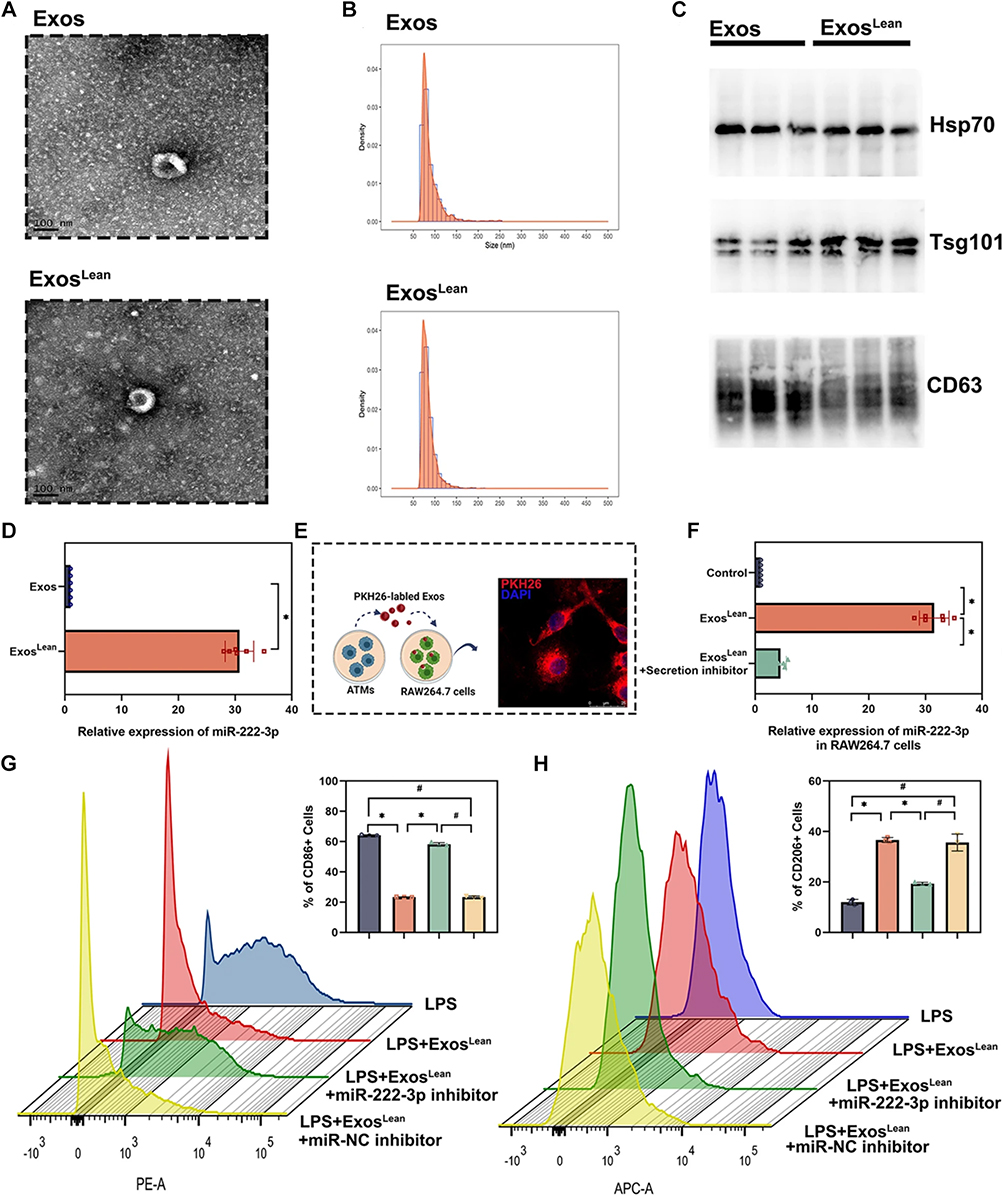 |
Figure 2 (A) Representative TEM image of exosomes. (B) Size distribution of exosomes confirmed by NTA. (C) Western blot analysis demonstrating the presence of exosomal markers HSP70, TSG101, and CD63 in both Exos and ExosLean. (D) MiR-222-3p levels in exosomes verified through qRT-PCR, with statistical significance (*P < 0.05, paired t-test, n = 6). (E) Exosomes labeled with PKH26 were incubated with RAW264.7 macrophages. (F) Impact of an EV secretion inhibitor on miR delivery via exosomes, analyzed using repeated-measures ANOVA (*P < 0.05, n = 6). LPS-stimulated RAW264.7 cells were treated with ExosLean, ExosLean combined with a miR-222-3p inhibitor, or ExosLean plus a miR-NC inhibitor. (G) Flow cytometry analysis quantifying CD86+ cells (*P < 0.05, #P < 0.05, paired t-test, n = 3). (H) Flow cytometry analysis quantifying CD206+ cells (*P < 0.05, #P < 0.05, paired t-test, n = 3). A-H Reproduced from Xia W, Liu Y, Jiang X, et al. Lean adipose tissue macrophage derived exosome confers immunoregulation to improve wound healing in diabetes. J Nanobiotechnology. 2023;21:128. Copyright © 2023, The Author(s).This article is licensed under a Creative Commons Attribution 4.0 International License.32 |
Angiogenesis: Promoting Vascularization Through Cargo Molecules
Angiogenesis is a fundamental process in wound healing, responsible for supplying regenerating tissues with the oxygen and nutrients required for proper repair.33 MDEVs play a significant role in promoting angiogenesis, a process often impaired in diabetic wounds due to endothelial dysfunction and reduced vascularization.25 These vesicles are enriched with pro-angiogenic factors such as vascular endothelial growth factor (VEGF), as well as angiogenesis-related miRNAs, including miR-126 and miR-210.20 These cargo molecules act on endothelial cells, stimulating their proliferation, migration, and tube formation, which are essential steps in new blood vessel formation. The ability of MDEVs to enhance vascularization is critical in diabetic wound healing, where insufficient blood supply contributes to delayed tissue repair and increased risk of infection. For a recent example, Jiang et al highlighted the development of two-dimensional carbide (MXene)-M2 macrophage exosome nanohybrids (FM-Exo) as a novel approach to enhance angiogenesis and promote diabetic wound healing (Figure 3). FM-Exo effectively mitigated high-glucose-induced immune suppression by sustaining the release of M2 macrophage-derived exosomes, which optimized macrophage polarization and activated the PI3K/Akt signaling pathway.34 This modulation led to increased fibroblast proliferation, endothelial cell migration, and angiogenic activity. In a diabetic wound model, FM-Exo reduced inflammation, enhanced VEGF-mediated angiogenesis, and improved collagen deposition, resulting in accelerated wound closure with minimal scarring. These findings offer a promising biomaterial-based strategy to address impaired angiogenesis in diabetic wounds.
 |
Figure 3 Schematic representation of the FM-Exo hydrogel, designed with multifunctional properties to enhance diabetic wound healing and facilitate skin reconstruction. Reproduced rom Jiang X, Ma J, Xue K, et al. Highly bioactive MXene-M2-exosome nanocomposites promote angiogenic diabetic wound repair through reconstructing high glucose-derived immune inhibition. ACS Nano. 2024;18:4269–4286.34 Copyright 2024, American Chemical Society. |
ECM Remodeling and Fibroblast Activation: Enhancing Granulation Tissue Formation
The remodeling of the ECM and fibroblast activation are essential steps in the formation of granulation tissue, which serves as the scaffold for new tissue in the healing process. MDEVs facilitate ECM remodeling by delivering matrix metalloproteinases (MMPs) and their regulators, which are involved in the degradation of damaged ECM components.35 Simultaneously, MDEVs support the synthesis of new ECM proteins, such as collagen, promoting the reconstruction of the tissue matrix.36 Furthermore, MDEVs influence fibroblast behavior by transferring growth factors and miRNAs that enhance fibroblast migration, proliferation, and secretion of ECM components.15 This coordinated action ensures the proper formation of granulation tissue, which is crucial for wound contraction and closure, particularly in the challenging environment of diabetic wounds, where ECM turnover is often impaired. For instance, a recent study introduced a hydrogel system combining functionalized gold nanorods (AuNRs) and M2 macrophage-derived exosomes (M2-Exos) to enhance granulation tissue formation and promote diabetic wound healing.15 The hydrogel, with its unique ion-cross-linked and covalent network structure, offers antiswelling properties and photothermal effects. M2-Exos released from the hydrogel were pivotal in reducing inflammation, promoting angiogenesis, and enhancing granulation tissue formation. The AuNRs, activated by near-infrared (NIR) irradiation, provided antibacterial activity, further supporting tissue regeneration (Figure 4). This integrated approach demonstrates significant potential in accelerating chronic diabetic wound healing and improving tissue repair outcomes.
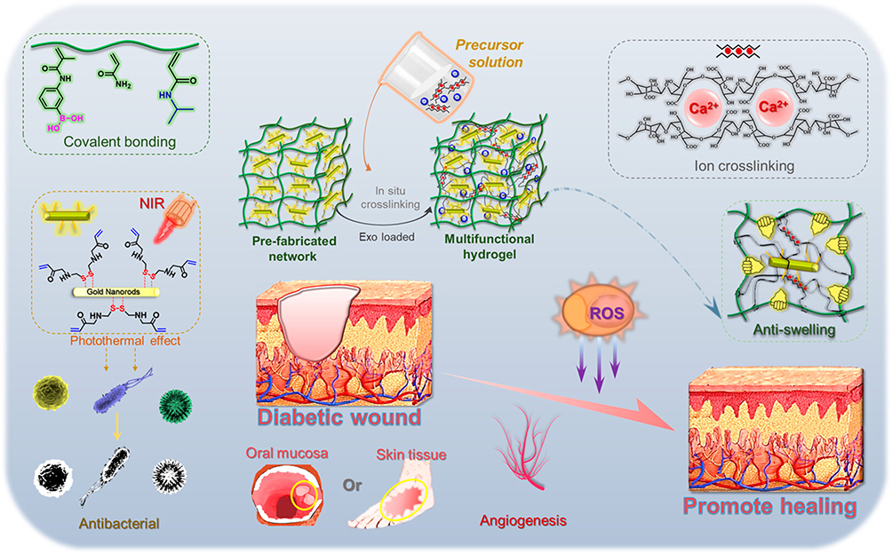 |
Figure 4 Schematic illustration of the hydrogel system integrating functionalized AuNRs and M2-Exos designed to enhance granulation tissue formation and facilitate diabetic wound healing. Reproduced from Li W, Wu S, Ren L, et al. Development of an antiswelling hydrogel system incorporating M2-exosomes and photothermal effect for diabetic wound healing. ACS Nano. 2023;17:22106–22120.15 Copyright 2023, American Chemical Society. |
Crosstalk With Other Cell Types: Interactions With Keratinocytes, Endothelial Cells, and Fibroblasts
MDEVs mediate complex crosstalk between the various cell types involved in wound healing, including keratinocytes, endothelial cells, and fibroblasts. These vesicles promote keratinocyte migration and proliferation, accelerating the process of re-epithelialization, which is crucial for covering the wound and restoring the integrity of the skin barrier.37,38 MDEVs also interact with endothelial cells, enhancing angiogenesis by stimulating endothelial cell proliferation and tube formation. In addition, MDEVs facilitate fibroblast activation, thereby promoting ECM deposition, collagen synthesis, and wound contraction.39 In a prior study, Sharma et al demonstrated the significant role of macrophage-derived exosomes (Exomϕ) in keratinocyte interactions during wound healing, particularly through the transfer of TOMM70, a mitochondrial outer membrane protein.40 Exomϕ, enriched with TOMM70, localize in leading-edge keratinocytes during early wound reepithelialization, compensating for hypoxia-induced depletion of TOMM70 and restoring mitochondrial metabolism. This process supports glycolytic ATP production, crucial for keratinocyte migration. Blocking exosomal uptake disrupts wound closure and sustains proinflammatory macrophages, highlighting the essential bidirectional crosstalk between macrophages and keratinocytes (Figure 5). This mechanism is particularly relevant in nonhealing diabetic foot ulcers, where keratinocyte TOMM70 deficiency is observed.
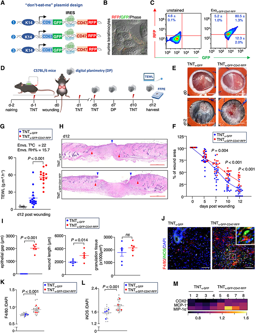 |
Figure 5 Bidirectional communication between wound-edge macrophages (WE mϕ) and resident keratinocytes is essential for effective wound closure. (A) Design of a Krt14-promoter-driven tetraspanin plasmid linked via the IRES element to a “don’t eat me” CD47 sequence, incorporating GFP and RFP reporters. (B) Confocal images illustrating coexpression of RFP (red) and GFP (green) in murine keratinocytes. Scale bar, 20 μm. (C) Flow cytometry of murine Exoκ captured on pan-CD magnetic beads, showing dual GFP and RFP positivity in “don’t eat me” Exoκ-GFP-CD47-RFP. (D) Experimental setup schematic. (E) Digital images of excisional stented punch wounds (6 mm) at day 0 and day 12 post-wounding in C57BL/6 mice treated with either TNTκ-GFP or TNTκ-GFP-CD47-RFP. (F) Wound area quantification via digital planimetry after treatment with TNTκ-GFP or TNTκ-GFP-CD47-RFP. Scale bar, 2 mm (n = 20). (G) TEWL measurements in C57BL/6 mice at day 12 post-wounding following treatment with either TNTκ-GFP or TNTκ-GFP-CD47-RFP, with gray dots representing normal skin (n = 16). (H) H&E staining of wounds in C57BL/6 mice at day 12 post-wounding, showing either TNTκ-GFP or TNTκ-GFP-CD47-RFP treatment. Scale bar, 1000 μm. Blue arrowheads indicate complete reepithelialization; red arrowheads indicate the wound edge (WE). (I) Morphometric analysis of epithelial gap, wound length, and granulation tissue area in C57BL/6 mice at day 12 post-wounding (n = 6). (J) Coimmunofluorescence staining of F4/80 (red) with iNOS (green) and DAPI in WE granulation tissue at day 12 post-wounding, following either TNTκ-GFP or TNTκ-GFP-CD47-RFP treatment. (K and L) Quantification of F4/80 and iNOS intensity in WE tissue, with each dot representing a quantified ROI, and blue/red dots indicating the mean per mouse (n = 4–5, with at least 5 ROI per mouse). (M) Heat map showing expression of proinflammatory markers COX2, MCP-1, and MIP-1α in WE granulation tissue at day 12 post-wounding in C57BL/6 mice, following treatment with TNTκ-GFP or TNTκ-GFP-CD47-RFP. Data in panels (C, F, G, I, K, and L) are presented as mean ± SEM and analyzed using a two-tailed unpaired Student’s t-test. Reproduced from Sharma A, Srivastava R, Gnyawali SC, et al. Mitochondrial bioenergetics of functional wound closure is dependent on macrophage-keratinocyte exosomal crosstalk. ACS Nano. 2024;18:30405–30420. Copyright © 2024 The Authors. Published by American Chemical Society. This publication is licensed under CC-BY 4.0.40 |
Engineering Macrophage-Derived EVs for Enhanced Therapeutic Efficacy
Bioengineering Approaches to Modify EV Cargo
To enhance the therapeutic potential of MDEVs, various bioengineering strategies are being developed to modify their cargo composition.40 These techniques aim to mediate the molecular content of MDEVs to address specific challenges in wound healing, particularly in diabetic conditions where processes like angiogenesis, inflammation resolution, and ECM remodeling are often impaired.41 One approach involves manipulating the parent macrophages to produce EVs enriched with specific proteins, RNAs, or miRNAs that promote tissue repair. For example, transfection of macrophages with nucleic acids encoding pro-healing factors—such as growth factors or anti-inflammatory cytokines—ensures that these beneficial molecules are selectively packaged into the EVs.41 Similarly, the targeted loading of miRNAs that regulate inflammation, tissue regeneration, or angiogenesis, can be achieved through genetic modification or chemical induction.42
Beyond genetic engineering, direct loading methods such as electroporation, sonication, or incubation with therapeutic molecules are employed to further enhance the functionality of MDEVs.43 These techniques enable the efficient encapsulation of drugs, peptides, or small RNAs that might otherwise be challenging to deliver. For example, electroporation facilitates the introduction of larger biomolecules into MDEVs by temporarily permeabilizing the vesicle membrane, thereby increasing cargo uptake.44
Genetic engineering approaches ensure the secretion of EVs with enriched and physiologically relevant therapeutic cargo. This method offers sustained and precise modulation of MDEV composition but poses challenges in terms of safety, scalability, and regulatory approval. In contrast, direct cargo loading methods provide a flexible strategy for incorporating small-molecule drugs, siRNAs, or proteins post-isolation. While this approach allows for rapid adaptation to different therapeutic needs, it may compromise vesicle integrity and functional stability. The selection between these strategies depends on the intended application: genetic modification is ideal for long-term, sustained therapeutic effects, whereas direct loading enables customizable, short-term interventions.
Targeting Strategies for Specific Wound Sites
Efficient and targeted delivery of engineered MDEVs to the wound site is another critical factor in maximizing their therapeutic efficacy. Because MDEVs possess the intrinsic ability to interact with a range of cell types, it is essential to further refine their targeting mechanisms to ensure that their therapeutic cargo is delivered specifically to the site of injury.45 One approach is surface modification, which involves the decoration of MDEVs with ligands, peptides, or antibodies that recognize specific receptors or markers expressed on the surface of damaged or inflamed tissues.46 For example, peptides that bind to integrins or other adhesion molecules overexpressed in diabetic wounds can be used to enhance the localization of MDEVs to the injury site.45,47 Similarly, antibodies targeting cell surface receptors such as E-selectin, which are upregulated in endothelial cells during the inflammatory phase of wound healing, can direct MDEVs specifically to the vascular endothelium at the wound site.45
Another promising strategy is the functionalization of MDEVs with biomaterials, such as polysaccharides, lipids, or synthetic polymers, that can improve vesicle stability, bioavailability, and retention at the target site.26,48,49 By embedding MDEVs into hydrogel-based delivery systems, sustained release of vesicles can be achieved, allowing for prolonged therapeutic effects at the wound site. Hydrogel-based systems, for instance, can provide a 3D matrix that mimics the extracellular environment of tissues, enhancing MDEV adhesion, and promoting the gradual release of their therapeutic cargo over time.50,51 These biomaterial-based delivery platforms can also offer additional advantages, such as improving the mechanical properties of the wound site and aiding in tissue regeneration by providing a supportive scaffold for cell migration and ECM deposition.
Moreover, the combination of MDEVs with advanced drug-delivery technologies, such as nanoparticles or microneedles, can further enhance their targeting precision and therapeutic efficiency.52,53 Such multi-modal approaches not only improve the local concentration of MDEVs at the wound site but also allow for synergistic effects between different therapeutic agents. The refinement of these targeting strategies, combined with the ability to engineer MDEVs with tailored cargo, ensures that these vesicles can effectively address the specific needs of chronic diabetic wounds. By concentrating the therapeutic payload at the site of injury and promoting sustained release, these engineered MDEVs hold great promise for overcoming the limitations of traditional wound-healing therapies and improving outcomes in diabetic patients.
Delivery Strategies for MDEVs
Current Challenges in EV Delivery: Stability, Targeting, and Dosage
The effective therapeutic application of MDEVs faces several critical challenges that should be addressed to fully exploit their potential in clinical settings. A major obstacle is the stability of EVs in biological environments.54 Extracellular vesicles are inherently fragile and susceptible to enzymatic degradation, aggregation, or loss of bioactivity once exposed to bodily fluids, which significantly limits their therapeutic lifespan. Ensuring the preservation of EV structural integrity during storage, handling, and delivery is therefore paramount to maintaining their efficacy. To overcome these stability issues, strategies for improving the robustness of EVs, such as encapsulation in protective materials or the use of stabilizing agents, are being explored.
Targeting efficiency also remains a critical concern.55 For MDEVs to exert their therapeutic effects in wound healing, they should be efficiently delivered to the site of injury, while minimizing off-target interactions that could result in undesired systemic effects. The biological barriers to EV delivery, such as the endothelial lining, extracellular matrix, and immune clearance mechanisms, make the targeted delivery of MDEVs to diabetic wounds a complex challenge.56,57 Surface modifications, such as functionalization with targeting ligands, peptides, or antibodies, can improve the specificity of MDEV localization.20 However, these strategies should be optimized to ensure effective targeting without inducing immune responses or toxicity.
Additionally, determining the optimal dosage of MDEVs for therapeutic efficacy remains an unresolved issue.58 Emerging studies illustrates the inconsistencies in dosing regimens, including variations in EV quantification (particle count vs protein content), administration routes, and treatment frequencies (Table 1).20,35,59 While EVs offer promising benefits in terms of their bioactivity, their optimal concentration and administration schedule vary depending on the condition being treated, the severity of the wound, and the specific therapeutic goals. Excessive delivery of MDEVs may result in immune reactions, while insufficient dosage could lead to suboptimal therapeutic outcomes.24 Establishing standardized protocols for dosing and administration is crucial for the clinical translation of MDEV-based therapies.
 |
Table 1 The Variations in MDEV Dosing Across Different Studies |
Advances in Hydrogel-Based Delivery Systems for Sustained Release
To overcome the challenges associated with EV delivery, particularly those related to stability and targeted release, hydrogel-based delivery systems have gained prominence as an innovative solution.38,60 Hydrogels are three-dimensional, water-swollen networks of biocompatible and biodegradable materials that can encapsulate MDEVs, offering significant advantages in terms of stability, sustained release, and local retention at the wound site. By embedding MDEVs within hydrogel matrices, it is possible to protect these delicate vesicles from premature degradation, allowing for prolonged therapeutic activity.61 The hydrogel matrix serves as a protective scaffold, shielding the EVs from enzymatic breakdown and facilitating their slow, controlled release over time.62 For example, in a prior study, Liu et al introduced a multifunctional hydrogel loaded with M2 macrophage exosomes as a dressing for diabetic wounds.63 The hydrogel exhibits injectable, self-healing, and tissue-adhesive properties, enabling it to rapidly form a protective barrier over the wound, effectively stopping bleeding and creating a favorable healing environment. A key feature of this hydrogel is its sustained release of M2 macrophage exosomes, which facilitates angiogenesis in the wound area, ultimately accelerating the healing process.
Moreover, hydrogels can be engineered to respond to environmental cues such as pH, temperature, or enzymatic activity, enabling the precise control of MDEV release in response to the local wound microenvironment.45,60,61 For example, acidic conditions often present in inflamed or infected tissues can trigger the release of EVs from pH-sensitive hydrogels, ensuring that the therapeutic cargo is delivered precisely where it is most needed.64,65 Similarly, thermosensitive hydrogels can undergo phase transitions in response to changes in temperature, facilitating the controlled release of EVs at the wound site.66
Hydrogels can also be designed to enhance the retention of EVs at the wound site by incorporating adhesion molecules or extracellular matrix components, which help to anchor the hydrogel and its cargo to the tissue.67 This localized retention is particularly important in chronic wound healing, such as in diabetic ulcers, where prolonged therapeutic intervention is often necessary to support tissue regeneration and repair.68 In addition, hydrogel-based systems can be designed to integrate with the surrounding tissue as they degrade, offering a dynamic, supportive scaffold that promotes cell migration, angiogenesis, and ECM remodeling—key processes in wound healing.20,69 For instance, Xiong et al reported an in situ injectable HA@MnO2/FGF-2/Exos hydrogel designed to enhance diabetic wound healing.12 A key focus is its sustained release and tissue retention of M2 macrophage-derived exosomes (M2 Exos). Upon local injection, the hydrogel forms a protective barrier over the wound, offering rapid hemostasis and prolonged antibacterial effects. Crucially, the hydrogel enables the controlled and prolonged release of M2 Exos and FGF-2, promoting angiogenesis and epithelialization. Both in vivo and in vitro results confirm its ability to accelerate diabetic wound repair, making it a promising solution for chronic wound management.
Recent advances in hydrogel formulations, including the incorporation of nanoparticles or biologically active molecules, have further enhanced the versatility and therapeutic potential of these delivery systems. For example, combining MDEVs with nanoparticle carriers within hydrogel matrices may improve the bioavailability of EVs, enhance their tissue penetration, and provide additional synergistic effects through the incorporation of other therapeutic agents.20,69 By addressing the limitations of conventional EVs delivery systems, hydrogel-based platforms offer a promising approach for enhancing the stability, targeting, and sustained release of EVs in wound healing.70 The integration of these advanced materials with EV-based therapies holds significant potential for improving outcomes in the treatment of diabetic wounds, where enhanced tissue regeneration, inflammation resolution, and angiogenesis are essential for effective healing.
Challenges and Future Perspectives
Heterogeneity within EV Populations
A major challenge in utilizing MDEVs for therapeutic applications stems from the inherent heterogeneity within EV populations. Despite originating from a single cell type, EVs exhibit variability in size, composition, and function, complicating their consistent application in clinical settings. This heterogeneity arises due to differences in biogenesis pathways, environmental conditions during EV production, and the dynamic nature of MSCs themselves, which respond to various stimuli by altering their secreted vesicles. Such variability complicates their consistent application in clinical settings, as it becomes difficult to ensure batch-to-batch reproducibility and predict therapeutic outcomes reliably.
To address this challenge, researchers should develop advanced techniques to isolate specific EV subpopulations that possess well-defined therapeutic properties. Ultracentrifugation, size-exclusion chromatography, and density gradient centrifugation are traditional methods used for EV isolation, but these approaches often yield mixed populations with overlapping characteristics. More refined techniques, such as immunoaffinity-based separation using specific surface markers or microfluidic-based sorting, hold promise for achieving greater purity and consistency.
Deficiency of Standardized Protocols
The absence of standardized protocols for isolating and characterizing EVs remains another critical barrier to their widespread use. Current isolation methods, such as ultracentrifugation and size-exclusion chromatography, often yield inconsistent results and may co-purify contaminants.71 These methods may also co-purify contaminants, such as proteins, lipoproteins, or cellular debris, which can interfere with downstream applications and compromise the therapeutic potential of MDEVs. Furthermore, differences in laboratory techniques, equipment, and reagents contribute to variability in EV preparations, making it difficult to compare results across studies and hindering progress toward clinical translation.
To overcome these challenges, it is essential to establish robust, reproducible protocols that ensure high purity, yield, and functional integrity of MDEVs. Advances in microfluidic-based isolation, immunoaffinity capture using EV-specific surface markers, and tangential flow filtration offer promising solutions for improving standardization and scalability. Additionally, clear guidelines for characterizing EVs—such as determining their size distribution through nanoparticle tracking analysis (NTA), identifying surface markers via flow cytometry or Western blotting, and analyzing cargo content using omics technologies—are crucial for ensuring consistent and reproducible outcomes. By implementing standardized protocols and rigorous quality control measures, researchers can enhance the reliability and clinical applicability of MDEV-based therapies, facilitating their integration into regenerative medicine and other biomedical applications.
Scalability of Bioengineering Methods
Current isolation and purification techniques, including ultracentrifugation, size-exclusion chromatography, and microfluidic sorting, are labor-intensive and difficult to scale while maintaining EV integrity and functionality. Moreover, macrophage culture conditions, activation states, and stimulation protocols significantly influence EV composition, creating batch-to-batch variability that challenges reproducibility. Advances in bioreactor systems and continuous-flow microfluidics offer promising solutions, yet standardization and regulatory compliance remain critical hurdles. Additionally, the need for high-yield, cost-effective production strategies necessitates the development of optimized cell expansion platforms and engineered vesicle secretion systems. Ensuring that bioengineered macrophage-derived EVs retain their therapeutic potency and phenotypic fidelity at an industrial scale remains an essential objective for their widespread clinical use.
Immunogenicity of Surface Modifications
Surface modifications of EVs introduce immunogenicity concerns that can alter their pharmacokinetics and therapeutic efficacy. Functionalization strategies, including lipid membrane engineering, protein conjugation, and ligand incorporation, are employed to enhance targeting capabilities and extend circulation time. However, these modifications may trigger immune recognition, leading to rapid clearance or unintended immunostimulatory effects. For instance, synthetic polymers or targeting peptides may disrupt EV biocompatibility, necessitating strategies such as autologous protein coatings or immune-evasive surface modifications. Furthermore, EV heterogeneity complicates precise modification, requiring advanced bioengineering techniques to achieve consistent and predictable surface functionalization. Addressing these challenges requires a balance between enhancing EV targeting efficiency and preserving immune tolerance, which remains a key consideration in developing macrophage-derived EV-based therapeutics.
Long-Term Stability of Hydrogels
Hydrogels serve as promising platforms for controlled EV delivery, yet their long-term stability significantly impacts therapeutic efficacy. The degradation kinetics of hydrogels must be carefully tailored to match the desired EV release profile, ensuring sustained bioavailability while maintaining vesicle functionality. Crosslinking density, polymer composition, and environmental factors such as pH and enzymatic activity influence hydrogel stability and EV retention. Additionally, hydrogel porosity and mechanical properties must be optimized to prevent premature EV degradation while allowing efficient diffusion at the target site. Advanced biomaterials, including stimuli-responsive and self-healing hydrogels, offer innovative solutions to enhance stability and tunability. However, achieving precise control over hydrogel degradation and EV release remains a critical challenge that requires further investigation. Future research should focus on integrating bioinspired materials and dynamic hydrogel systems to optimize macrophage-derived EV delivery for clinical applications.
Future Perspectives
Omics technologies, including proteomics, transcriptomics, and lipidomics, offer powerful tools for dissecting the complex cargo of MDEVs.72,73 These approaches enable the identification of specific molecules that mediate the therapeutic effects of EVs, facilitating the design of more targeted and effective interventions. Integrating these technologies will provide deeper insights into how MDEVs influence wound healing and guide efforts to enhance their therapeutic efficacy.
Innovative wound dressing systems, such as smart hydrogels, nanofiber scaffolds, and electrospun materials, hold great promise as delivery platforms for MDEVs. These systems can provide controlled, sustained release of MDEVs at the wound site, and their responsiveness to environmental cues ensures that therapeutic EVs are released in alignment with the wound’s healing needs.62 The combination of MDEVs with such advanced dressings offers a promising strategy to enhance diabetic wound healing.
Moreover, a growing body of pre-clinical research has demonstrated the efficacy of MDEVs in promoting wound healing in diabetic models. Several animal studies have shown that MDEVs can significantly accelerate wound closure, reduce inflammation, and enhance tissue regeneration. For instance, Liu et al investigated the effect of MDEVs in a streptozotocin-induced diabetic mouse model. The study found that topical application of MDEVs led to increased wound closure rates, enhanced re-epithelialization, and improved collagen deposition compared to control groups.63 Mechanistically, these vesicles modulated macrophage polarization, favoring a shift from the pro-inflammatory M1 phenotype to the anti-inflammatory M2 phenotype, thereby facilitating immune resolution and tissue repair.
In another pre-clinical study, Xiong et al developed a hydrogel-loaded MDEV system to provide sustained release of therapeutic vesicles at the wound site. This system significantly improved angiogenesis and fibroblast activation in diabetic mice, demonstrating enhanced granulation tissue formation and extracellular matrix remodeling.12 The hydrogel-based approach further extended the retention time of MDEVs, maximizing their therapeutic potential.
Conclusions
As MDEVs advance toward clinical application, addressing ethical, regulatory, and quality control challenges will be essential to ensure their safe and effective use. The production of MDEVs should adhere to rigorous quality assurance standards, including Good Manufacturing Practice (GMP) guidelines, to guarantee consistency, purity, and therapeutic efficacy. Ethical considerations, such as the sourcing of donor cells, informed consent, and compliance with international regulatory frameworks, should be carefully managed to foster public trust and facilitate regulatory approval.
Furthermore, establishing clear and standardized guidelines for preclinical and clinical studies will be critical for the successful translation of MDEVs from laboratory research to clinical practice. Robust methodologies for EV isolation, characterization, and functional validation will play a key role in ensuring reproducibility and scalability. Future research should focus on addressing these challenges, optimizing large-scale production strategies, and further elucidating the mechanisms underlying MDEV-mediated therapeutic effects. By overcoming these barriers, MDEVs hold the potential to become a transformative treatment modality for diabetic wound healing and other regenerative medicine applications.
Acknowledgments
This work was supported by the Natural Science Foundation of Shaanxi Province, Grant Number: 2021SF-241.
Disclosure
The author(s) report no conflicts of interest in this work.
References
1. Sen CK. Human wound and its burden: updated 2020 compendium of estimates. Adv Wound Care. 2021;10:281–292. doi:10.1089/wound.2021.0026
2. Malone-Povolny MJ, Maloney SE, Schoenfisch MH. Nitric oxide therapy for diabetic wound healing. Adv Healthc Mater. 2019;8:e1801210. doi:10.1002/adhm.201801210
3. Louiselle AE, Niemiec SM, Zgheib C, Liechty KW. Macrophage polarization and diabetic wound healing. Transl Res. 2021;236:109–116. doi:10.1016/j.trsl.2021.05.006
4. Sharifiaghdam M, Shaabani E, Faridi-Majidi R, De Smedt SC, Braeckmans K, Fraire JC. Macrophages as a therapeutic target to promote diabetic wound healing. Mol Ther. 2022;30:2891–2908. doi:10.1016/j.ymthe.2022.07.016
5. Liu W, Yu M, Xie D, et al. Melatonin-stimulated MSC-derived exosomes improve diabetic wound healing through regulating macrophage M1 and M2 polarization by targeting the PTEN/AKT pathway. Stem Cell Res Ther. 2020;11:259. doi:10.1186/s13287-020-01756-x
6. Silva JC, Pitta MGR, Pitta IR, Koh TJ, Abdalla DSP. New peroxisome proliferator-activated receptor agonist (GQ-11) improves wound healing in diabetic mice. Adv Wound Care. 2019;8:417–428. doi:10.1089/wound.2018.0911
7. Wang P, Theocharidis G, Vlachos IS, et al. Exosomes derived from epidermal stem cells improve diabetic wound healing. J Invest Dermatol. 2022;142:2508–2517e13. doi:10.1016/j.jid.2022.01.030
8. Wang Y, Lin Q, Zhang H, et al. M2 macrophage-derived exosomes promote diabetic fracture healing by acting as an immunomodulator. Bioact Mater. 2023;28:273–283. doi:10.1016/j.bioactmat.2023.05.018
9. Xiong Y, Lin Z, Bu P, et al. A whole-course-repair system based on neurogenesis-angiogenesis crosstalk and macrophage reprogramming promotes diabetic wound healing. Adv Mater. 2023;35:e2212300. doi:10.1002/adma.202212300
10. Fan L, Yao L, Li Z, et al. Exosome-based mitochondrial delivery of circRNA mSCAR alleviates sepsis by orchestrating macrophage activation. Adv Sci. 2023;10:e2205692. doi:10.1002/advs.202205692
11. Wang D, Qiu G, Zhu X, et al. Macrophage-inherited exosome excise tumor immunosuppression to expedite immune-activated ferroptosis. J Immunother Cancer. 2023;11:e006516. doi:10.1136/jitc-2022-006516
12. Xiong Y, Chen L, Liu P, et al. All-in-one: multifunctional hydrogel accelerates oxidative diabetic wound healing through timed-release of exosome and fibroblast growth factor. Small. 2022;18:e2104229. doi:10.1002/smll.202104229
13. Xiong Y, Mi BB, Lin Z, et al. The role of the immune microenvironment in bone, cartilage, and soft tissue regeneration: from mechanism to therapeutic opportunity. Mil Med Res. 2022;9:65. doi:10.1186/s40779-022-00426-8
14. Kwak G, Cheng J, Kim H, et al. Sustained exosome-guided macrophage polarization using hydrolytically degradable PEG hydrogels for cutaneous wound healing: identification of key proteins and MiRNAs, and sustained release formulation. Small. 2022;18:e2200060. doi:10.1002/smll.202200060
15. Li W, Wu S, Ren L, et al. Development of an antiswelling hydrogel system incorporating M2-exosomes and photothermal effect for diabetic wound healing. ACS Nano. 2023;17:22106–22120. doi:10.1021/acsnano.3c09220
16. Lv D, Cao X, Zhong L, et al. Targeting phenylpyruvate restrains excessive NLRP3 inflammasome activation and pathological inflammation in diabetic wound healing. Cell Rep Med. 2023;4:101129. doi:10.1016/j.xcrm.2023.101129
17. Wu X, He W, Mu X, et al. Macrophage polarization in diabetic wound healing. Burns Trauma. 2022;10:tkac051. doi:10.1093/burnst/tkac051
18. Audu CO, Melvin WJ, Joshi AD, et al. Macrophage-specific inhibition of the histone demethylase JMJD3 decreases STING and pathologic inflammation in diabetic wound repair. Cell mol Immunol. 2022;19:1251–1262. doi:10.1038/s41423-022-00919-5
19. Fu YJ, Shi YF, Wang LY, et al. All-natural immunomodulatory bioadhesive hydrogel promotes angiogenesis and diabetic wound healing by regulating macrophage heterogeneity. Adv Sci. 2023;10:e2206771. doi:10.1002/advs.202206771
20. Zeng J, Sun Z, Zeng F, Gu C, Chen X. M2 macrophage-derived exosome-encapsulated microneedles with mild photothermal therapy for accelerated diabetic wound healing. Mater Today Bio. 2023;20:100649. doi:10.1016/j.mtbio.2023.100649
21. Wang Y, Zhao M, Liu S, et al. Macrophage-derived extracellular vesicles: diverse mediators of pathology and therapeutics in multiple diseases. Cell Death Dis. 2020;11:924. doi:10.1038/s41419-020-03127-z
22. Liu N, Dong J, Li L, Liu F. Osteoimmune interactions and therapeutic potential of macrophage-derived small extracellular vesicles in bone-related diseases. Int J Nanomed. 2023;18:2163–2180. doi:10.2147/IJN.S403192
23. Ye J, Liu X. Macrophage-derived small extracellular vesicles in multiple diseases: biogenesis, function, and therapeutic applications. Front Cell Dev Biol. 2022;10:913110. doi:10.3389/fcell.2022.913110
24. Xing Y, Sun X, Dou Y, et al. The immuno-modulation effect of macrophage-derived extracellular vesicles in chronic inflammatory diseases. Front Immunol. 2021;12:785728. doi:10.3389/fimmu.2021.785728
25. Li D, Zhang C, Gao Z, et al. Curcumin-loaded macrophage-derived exosomes effectively improve wound healing. Mol Pharm. 2023;20:4453–4467. doi:10.1021/acs.molpharmaceut.3c00062
26. Yang J, Huang X, Yu Q, et al. Extracellular vesicles derived from M2-like macrophages alleviate acute lung injury in a miR-709-mediated manner. J Extracell Vesicles. 2024;13:e12437. doi:10.1002/jev2.12437
27. Hou X, Yin S, Ren R, et al. Myeloid-cell-specific IL-6 signaling promotes MicroRNA-223-enriched exosome production to attenuate NAFLD-associated fibrosis. Hepatology. 2021;74:116–132. doi:10.1002/hep.31658
28. Xie X, Qu P, Wu H, et al. Circulating exosomal miR-21 mediates HUVEC proliferation and migration through PTEN/PI3K/AKT in Crohn’s disease. Ann Transl Med. 2022;10:258. doi:10.21037/atm-22-475
29. Kim H, Wang SY, Kwak G, Yang Y, Kwon IC, Kim SH. Exosome-guided phenotypic switch of M1 to M2 macrophages for cutaneous wound healing. Adv Sci. 2019;6:1900513. doi:10.1002/advs.201900513
30. Ding J, Zhang Y, Cai X, et al. Yang, Extracellular vesicles derived from M1 macrophages deliver miR-146a-5p and miR-146b-5p to suppress trophoblast migration and invasion by targeting TRAF6 in recurrent spontaneous abortion. Theranostics. 2021;11:5813–5830. doi:10.7150/thno.58731
31. Li L, Cao J, Li S, et al. M2 macrophage-derived sEV regulate pro-inflammatory CCR2(+) macrophage subpopulations to favor post-AMI cardiac repair. Adv Sci. 2023;10:e2202964. doi:10.1002/advs.202202964
32. Xia W, Liu Y, Jiang X, et al. Lean adipose tissue macrophage derived exosome confers immunoregulation to improve wound healing in diabetes. J Nanobiotechnology. 2023;21:128. doi:10.1186/s12951-023-01869-4
33. Ding JY, Chen MJ, Wu LF, et al. Mesenchymal stem cell-derived extracellular vesicles in skin wound healing: roles, opportunities and challenges. Mil Med Res. 2023;10:36. doi:10.1186/s40779-023-00472-w
34. Jiang X, Ma J, Xue K, et al. Highly bioactive MXene-M2-exosome nanocomposites promote angiogenic diabetic wound repair through reconstructing high glucose-derived immune inhibition. ACS Nano. 2024;18:4269–4286. doi:10.1021/acsnano.3c09721
35. Banerjee A, Singh P, Sheikh PA, Kumar A, Koul V, Bhattacharyya J. A multifunctional silk-hyaluronic acid self-healing hydrogel laden with alternatively activated macrophage-derived exosomes reshape microenvironment of diabetic wound and accelerate healing. Int J Biol Macromol. 2024;270:132384. doi:10.1016/j.ijbiomac.2024.132384
36. Wang J, Han Y, Huang F, Tang L, Mu J, Liang Y. Diabetic macrophage small extracellular vesicles-associated miR-503/IGF1R axis regulates endothelial cell function and affects wound healing. Front Immunol. 2023;14:1104890. doi:10.3389/fimmu.2023.1104890
37. Zhu M, Sun X, Qi X, Xia L, Wu Y. Exosomes from high glucose-treated macrophages activate macrophages and induce inflammatory responses via NF-kappaB signaling pathway in vitro and in vivo. Int Immunopharmacol. 2020;84:106551. doi:10.1016/j.intimp.2020.106551
38. Xiong Y, Mi BB, Shahbazi MA, Xia T, Xiao J. Microenvironment-responsive nanomedicines: a promising direction for tissue regeneration. Mil Med Res. 2024;11:69. doi:10.1186/s40779-024-00573-0
39. Lyu L, Cai Y, Zhang G, et al. Exosomes derived from M2 macrophages induce angiogenesis to promote wound healing. Front Mol Biosci. 2022;9:1008802. doi:10.3389/fmolb.2022.1008802
40. Sharma A, Srivastava R, Gnyawali SC, et al. Mitochondrial bioenergetics of functional wound closure is dependent on macrophage-keratinocyte exosomal crosstalk. ACS Nano. 2024;18:30405–30420. doi:10.1021/acsnano.4c07610
41. Song Y, Hu J, Ma C, Liu H, Li Z, Yang Y. Macrophage-derived exosomes as advanced therapeutics for inflammation: current progress and future perspectives. Int J Nanomed. 2024;19:1597–1627. doi:10.2147/IJN.S449388
42. You DG, Lim GT, Kwon S, et al. Metabolically engineered stem cell-derived exosomes to regulate macrophage heterogeneity in rheumatoid arthritis. Sci Adv. 2021;7. doi:10.1126/sciadv.abe0083
43. Zhang H, Mao Y, Nie Z, et al. Iron oxide nanoparticles engineered macrophage-derived exosomes for targeted pathological angiogenesis therapy. ACS Nano. 2024;18:7644–7655. doi:10.1021/acsnano.4c00699
44. Wu G, Zhang J, Zhao Q, et al. Molecularly engineered macrophage-derived exosomes with inflammation tropism and intrinsic heme biosynthesis for atherosclerosis treatment. Angew Chem Int Ed Engl. 2020;59:4068–4074. doi:10.1002/anie.201913700
45. Hade MD, Suire CN, Mossell J, Suo Z. Extracellular vesicles: emerging frontiers in wound healing. Med Res Rev. 2022;42:2102–2125. doi:10.1002/med.21918
46. Zhuang Y, Jiang S, Deng X, et al. Energy metabolism as therapeutic target for aged wound repair by engineered extracellular vesicle. Sci Adv. 2024;10:eadl0372. doi:10.1126/sciadv.adl0372
47. Xiao T, Wei J, Cai D, et al. Extracellular vesicle mediated targeting delivery of growth differentiation factor-15 improves myocardial repair by reprogramming macrophages post myocardial injury. Biomed Pharmacother. 2024;172:116224. doi:10.1016/j.biopha.2024.116224
48. Cianciaruso C, Beltraminelli T, Duval F, et al. Molecular profiling and functional analysis of macrophage-derived tumor extracellular vesicles. Cell Rep. 2019;27:3062–3080e11. doi:10.1016/j.celrep.2019.05.008
49. Xie L, Chen J, Hu H, et al. Engineered M2 macrophage-derived extracellular vesicles with platelet membrane fusion for targeted therapy of atherosclerosis. Bioact Mater. 2024;35:447–460. doi:10.1016/j.bioactmat.2024.02.015
50. Zhang K, Du L, Li Z, et al. M2 macrophage-derived small extracellular vesicles ameliorate pyroptosis and intervertebral disc degeneration. Biomater Res. 2024;28:0047. doi:10.34133/bmr.0047
51. Zhu J, Liu B, Wang Z, et al. Exosomes from nicotine-stimulated macrophages accelerate atherosclerosis through miR-21-3p/PTEN-mediated VSMC migration and proliferation. Theranostics. 2019;9:6901–6919. doi:10.7150/thno.37357
52. Tang L, Yin Y, Cao Y, et al. Extracellular vesicles-derived hybrid nanoplatforms for amplified CD47 blockade-based cancer immunotherapy. Adv Mater. 2023;35:e2303835. doi:10.1002/adma.202303835
53. Wang Y, Li C, Zhao R, et al. CircUbe3a from M2 macrophage-derived small extracellular vesicles mediates myocardial fibrosis after acute myocardial infarction. Theranostics. 2021;11:6315–6333. doi:10.7150/thno.52843
54. Ning J, Hou X, Hao J, et al. METTL3 inhibition induced by M2 macrophage-derived extracellular vesicles drives anti-PD-1 therapy resistance via M6A-CD70-mediated immune suppression in thyroid cancer. Cell Death Differ. 2023;30:2265–2279. doi:10.1038/s41418-023-01217-x
55. Peng W, Xie Y, Liu Y, et al. Hu, Targeted delivery of CD163(+) macrophage-derived small extracellular vesicles via RGD peptides promote vascular regeneration and stabilization after spinal cord injury. J Control Release. 2023;361:750–765. doi:10.1016/j.jconrel.2023.08.025
56. Chen P, Wang L, Fan X, et al. Targeted delivery of extracellular vesicles in heart injury. Theranostics. 2021;11:2263–2277. doi:10.7150/thno.51571
57. Pan K, Zhu Y, Chen P, et al. Biological functions and biomedical applications of extracellular vesicles derived from blood cells. Free Radic Biol Med. 2024;222:43–61. doi:10.1016/j.freeradbiomed.2024.06.002
58. Barone A, d’Avanzo N, Cristiano MC, Paolino D, Fresta M. Macrophage-derived extracellular vesicles: a promising tool for personalized cancer therapy. Biomedicines. 2022;10:1252. doi:10.3390/biomedicines10061252
59. Meng H, Su J, Shen Q, et al. A smart MMP-9-responsive hydrogel releasing M2 macrophage-derived exosomes for diabetic wound healing. Adv Healthc Mater. 2025;14:e2404966. doi:10.1002/adhm.202404966
60. Akbar N, Azzimato V, Choudhury RP, Aouadi M. Extracellular vesicles in metabolic disease. Diabetologia. 2019;62:2179–2187. doi:10.1007/s00125-019-05014-5
61. Lou K, Feng S, Luo H, Zou J, Zhang G, Zou X. Extracellular vesicles derived from macrophages: current applications and prospects in tumors. Front Bioeng Biotechnol. 2022;10:1097074. doi:10.3389/fbioe.2022.1097074
62. Zhou C, Zhang B, Yang Y, et al. Stem cell-derived exosomes: emerging therapeutic opportunities for wound healing. Stem Cell Res Ther. 2023;14:107. doi:10.1186/s13287-023-03345-0
63. Liu P, Xiong Y, Chen L, et al. Angiogenesis-based diabetic skin reconstruction through multifunctional hydrogel with sustained releasing of M2 macrophage-derived exosome. Chem Eng J. 2022;431. doi:10.1016/j.cej.2021.132413
64. Muskan M, Abeysinghe P, Cecchin R, Branscome H, Morris KV, Kashanchi F. Therapeutic potential of RNA-enriched extracellular vesicles: the next generation in RNA delivery via biogenic nanoparticles. Mol Ther. 2024;32:2939–2949. doi:10.1016/j.ymthe.2024.02.025
65. Xiong Y, Mi B, Liu G, Zhao Y. Microenvironment-sensitive nanozymes for tissue regeneration. Biomaterials. 2024;309:122585. doi:10.1016/j.biomaterials.2024.122585
66. Tang Q, Lu B, He J, et al. Exosomes-loaded thermosensitive hydrogels for corneal epithelium and stroma regeneration. Biomaterials. 2022;280:121320. doi:10.1016/j.biomaterials.2021.121320
67. Shen Z, Kuang S, Zhang Y, et al. Chitosan hydrogel incorporated with dental pulp stem cell-derived exosomes alleviates periodontitis in mice via a macrophage-dependent mechanism. Bioact Mater. 2020;5:1113–1126. doi:10.1016/j.bioactmat.2020.07.002
68. Yang Y, Zhang J, Wu S, et al. Exosome/antimicrobial peptide laden hydrogel wound dressings promote scarless wound healing through miR-21-5p-mediated multiple functions. Biomaterials. 2024;308:122558. doi:10.1016/j.biomaterials.2024.122558
69. Shi Y, Wang S, Wang K, et al. Relieving macrophage dysfunction by inhibiting SREBP2 activity: a hypoxic mesenchymal stem cells-derived exosomes loaded multifunctional hydrogel for accelerated diabetic wound healing. Small. 2024;20:e2309276. doi:10.1002/smll.202309276
70. Deng D, Li X, Zhang JJ, et al. Biotin-avidin system-based delivery enhances the therapeutic performance of MSC-derived exosomes. ACS Nano. 2023;17:8530–8550. doi:10.1021/acsnano.3c00839
71. An Y, Lin S, Tan X, et al. Exosomes from adipose-derived stem cells and application to skin wound healing. Cell Prolif. 2021;54:e12993. doi:10.1111/cpr.12993
72. Manzoor T, Farooq N, Sharma A, et al. Exosomes in nanomedicine: a promising cell-free therapeutic intervention in burn wounds. Stem Cell Res Ther. 2024;15:355. doi:10.1186/s13287-024-03970-3
73. Sun T, Li M, Liu Q, et al. Insights into optimizing exosome therapies for acute skin wound healing and other tissue repair. Front Med. 2024;18:258–284. doi:10.1007/s11684-023-1031-9
 © 2025 The Author(s). This work is published and licensed by Dove Medical Press Limited. The
full terms of this license are available at https://www.dovepress.com/terms
and incorporate the Creative Commons Attribution
- Non Commercial (unported, 4.0) License.
By accessing the work you hereby accept the Terms. Non-commercial uses of the work are permitted
without any further permission from Dove Medical Press Limited, provided the work is properly
attributed. For permission for commercial use of this work, please see paragraphs 4.2 and 5 of our Terms.
© 2025 The Author(s). This work is published and licensed by Dove Medical Press Limited. The
full terms of this license are available at https://www.dovepress.com/terms
and incorporate the Creative Commons Attribution
- Non Commercial (unported, 4.0) License.
By accessing the work you hereby accept the Terms. Non-commercial uses of the work are permitted
without any further permission from Dove Medical Press Limited, provided the work is properly
attributed. For permission for commercial use of this work, please see paragraphs 4.2 and 5 of our Terms.
Recommended articles
Research and Application Prospect of Nanomedicine in Kidney Disease: A Bibliometric Analysis From 2003 to 2024

Meng Y, Sui L, Xu T, Zhao H, Yuan Q, Sun L
International Journal of Nanomedicine 2025, 20:3007-3030
Published Date: 12 March 2025
Zeolitic Imidazolate Framework-90 Treats Fungal Keratitis by Promoting Macrophage Apoptosis and Targeting Increased Mitochondrial Reactive Oxygen Species in Aspergillus Fumigatus
Fu X, Zhang L, Lin J, Wang Q, Wang Z, Chi M, Li D, Zhao G, Li C
International Journal of Nanomedicine 2025, 20:4551-4569
Published Date: 12 April 2025
Emerging Insights into the Role of Macrophages in Multiple Sclerosis: Pathogenesis and Potential Therapeutic Strategies
Yang J, Fu Z, Zou Q, Li Z, Xing Y
Journal of Inflammation Research 2025, 18:14131-14146
Published Date: 10 October 2025
Nano-Reagents in Acute Pancreatitis: Diagnostic, Therapeutic, and Theranostic Advances
Yang X, Li Q, Tang L, Huang L, Zhu C, Chen Q
International Journal of Nanomedicine 2026, 21:562220
Published Date: 19 February 2026