Back to Journals » International Journal of Chronic Obstructive Pulmonary Disease » Volume 13
Lung function and CT lung densitometry in 37- to 39-year-old individuals with alpha-1-antitrypsin deficiency
Authors Mostafavi B, Diaz S, Piitulainen E, Stoel BC ![]() , Wollmer P, Tanash HA
, Wollmer P, Tanash HA
Received 7 March 2018
Accepted for publication 28 June 2018
Published 8 November 2018 Volume 2018:13 Pages 3689—3698
DOI https://doi.org/10.2147/COPD.S167497
Checked for plagiarism Yes
Review by Single anonymous peer review
Peer reviewer comments 3
Editor who approved publication: Prof. Dr. Richard Russell
Behrouz Mostafavi,1 Sandra Diaz,2 Eeva Piitulainen,1 Berend C Stoel,3 Per Wollmer,4 Hanan A Tanash1
1Department of Respiratory Medicine and Allergology Malmö, Skåne University Hospital, Lund University, Malmö, Sweden; 2Department of Clinical Physiology Malmö, Skåne University Hospital, Lund University, Malmö, Sweden; 3Division of Image Processing, Department of Radiology, Leiden University Medical, Leiden, the Netherlands; 4Department of Translational Medicine, Lund University, Malmö, Sweden
Background: Alpha-1-antitrypsin (AAT) deficiency is a hereditary disorder that predisposes to emphysema. A cohort of severe (PiZZ) and moderate (PiSZ) AAT-deficient newborn infants was identified by the Swedish national neonatal AAT screening program in 1972–1974 and has been followed-up since birth. Our aim was to study whether the cohort has signs of emphysema in pulmonary function tests (PFTs) and computed tomography (CT) densitometry at 38 years of age in comparison with an age-matched control group, randomly selected from the population registry.
Methods: Forty-one PiZZ, 18 PiSZ, and 61 control subjects (PiMM) underwent complete PFTs, measurement of resistance and reactance in the respiratory system by impulse oscillometry (IOS)/forced oscillation technique (FOT), and CT densitometry. The results were related to self-reported smoking habits.
Results: The total lung capacity (TLC) % of the predicted value was significantly higher in the PiZZ ever-smokers than in the PiZZ never-smokers (P<0.05), PiSZ never-smokers (P=0.01) and the PiMM never-smokers (P=0.01). The residual volume (RV) % of the predicted value was significantly higher in the PiZZ ever-smokers compared to the PiMM never-smokers (P<0.01). The PiZZ ever-smokers had a significantly lower carbon monoxide transfer coefficient (Kco) than the PiSZ never-smokers (P<0.01) and PiMM never-smokers (P<0.01). Respiratory system resistance at 5 Hz (P<0.01), at 20 Hz (P<0.01), and the area of low reactance (Alx; P<0.05) were significantly lower and respiratory system reactance at 5 Hz (P<0.05) was significantly higher in PiZZ subjects compared to the PiMM subjects. No statistically significant differences in the CT densitometry parameters were found between the Pi subgroups.
Conclusion: The physiological parameters in the PiZZ ever-smokers showed evidence of hyperinflation and emphysema before the age of 40 years.
Keywords: alpha-1-antitrypsin deficiency, lung function, lung disease, screening, impulse oscillometry
Introduction
Severe alpha-1-antitrypsin (AAT) deficiency (PiZZ) is a known risk factor for the development of lung emphysema and COPD.1,2 AAT is a 52-kDa protein, a member of the SERPIN (SERine Protease INhibitor) family that inactivates serine proteases such as neutrophil elastase. AAT deficiency is an autosomal codominant hereditary disorder. Z protein is associated with intracellular accumulation in the liver, decreased serum levels, and has shown chemotactic properties in vivo. S protein is not associated with intracellular accumulation and inhibits elastase normally.3–6 S protein is shown to be associated with exaggerated intracellular degradation of newly synthesized AAT in the liver, resulting in reduced secretion of AAT.7
In clinical practice, COPD is diagnosed initially by the primary physician according to the Global Initiative for Chronic Obstructive Lung Disease (GOLD) criteria by compiling complete anamnestic information, including exposure, symptoms, smoking habits, and result of spirometry. For a more comprehensive diagnosis, complete pulmonary function tests (PFTs), including diffusing capacity for carbon monoxide, are employed. Impulse oscillometry (IOS)/forced oscillation technique (FOT) is a noninvasive method for measuring airway resistance and reactance that can be performed during tidal breathing. IOS/FOT is reported to be a good complementary diagnostic tool for the management and monitoring of asthma and COPD.8–11 Computed tomography (CT) with CT densitometry is considered to be more accurate to assess the progression of emphysema than PFTs.12,13
AAT deficiency is most often diagnosed in patients who already have developed clinical signs of lung disease, before being identified as AAT deficient. The natural course and early signs of lung disease are still not fully understood.1,2 The detection of early signs of lung disease is crucial for treatments to inhibit the further development of the disease.14
The Swedish national neonatal AAT screening program was carried out in 1972–1974, when all 200,000 newborn children were screened for AAT deficiency.15 A cohort including 127 PiZZ, 2 PiZnull, 54 PiSZ, and 1 PiSnull children was identified. Five PiZZ children and one PiSZ child died before the age of 8 years. The cohort has been followed up regularly. During the follow-up, five new PiZZ individuals, born abroad during the screening period, have been identified and added to the cohort.
Our aim was to investigate signs of emphysema in this cohort at 38 years of age, in comparison with age-matched control subjects, randomly selected from the population registry, using PFT, IOS/FOT, and CT-densitometry as early detection tools.
Methods
Study population
One PiZZ and three PiSZ individuals had died before the present check-up. The remaining cohort, including 126 PiZZ, 2 PiZnull, 50 PiSZ, and 1 PiSnull individuals, was invited to participate in this follow-up study. A random sample of 300 age-matched individuals, randomly picked from the Swedish population registry, was invited as a control group.
The study was conducted in accordance with the Helsinki Declaration and approved by the Regional Ethical Review Board of Lund, Sweden. All study participants gave their signed informed consent.
Questionnaire
All participants visited the Department of Respiratory Medicine, Skåne University Hospital Malmö, Sweden and completed a questionnaire on smoking habits and symptoms. (Figure S1) The study participants reported whether they had ever smoked. If yes, the date when they began and stopped smoking, if applicable, and the average number of cigarettes per day during the period of smoking were reported in the questionnaire. A smoker was defined as a person who had smoked one cigarette or more per day for at least 1 year.
PFT
Complete PFT were performed with the same equipment and by an experienced technician at the Department of Clinical Physiology, Skåne University Hospital Malmö. The PFT was performed after administration of 1.0 mg inhaled terbutaline. The functions measured by the PFT included forced expiratory volume in 1 second (FEV1), vital capacity (VC), total lung capacity (TLC), functional residual capacity (FRC), residual volume (RV), diffusing capacity for carbon monoxide (DL,CO), and alveolar volume (VA). The carbon monoxide transfer coefficient (Kco) was calculated. FRC, TLC, and RV were measured with body plethysmography. In addition, the RV/TLC ratio and FRC/TLC ratio were calculated as indicators of hyperinflation. The PFT, including the calibration of the equipment, was performed according to the guidelines of the American Thoracic Society/European Respiratory Society.16,17 The PFT results are reported as the percentage of the predicted values, according to the European Coal and Steel Community reference values.18 The FEV1/VC ratios are also expressed as absolute values.
IOS/FOT
IOS/FOT was performed directly after the complete PFT examination, with the same equipment and by the same technicians at the Department of Clinical Physiology, Skåne University Hospital Malmö. IOS (MasterScreen IOS; Jaeger, Hoechbery, Germany) with a broad-frequency wave of 5–35 Hz and a pulse generation interval of 0.2 seconds was employed. Respiratory system resistance at 5 Hz (R5Hz) (kPa/(L/s)) and 20 Hz (R20Hz) (kPa/(L/s)), the resistance in the small airways (R5Hz–R20Hz) (kPa/(L/s)), respiratory system reactance at 5 Hz (X5Hz) (kPa/(L/s)), resonant frequency (Fres) (Hz), and the area of low reactance (Alx) (kPa/L) were measured.8–10,19
CT densitometry
The CT examinations were performed at the Department of Radiology, University Hospital in Malmö, using a Siemens Somatom Sensation 64® (Erlangen, Germany) multidetector scanner at full inspiration (ie, at TLC) after three deep inhalation maneuvers, using the helical technique and acquisition in the caudal–cranial direction to avoid breathing artifacts at the level of the diaphragm. The images were processed with Pulmo-CMS® software (Medis Specials, Leiden, the Netherlands). The same method was used as at the previously published check-ups of the cohort.20 The 15th percentile density (PD15) was calculated as a measure of lung density. PD15 is expressed as g/L by adding 1,000 to the HU values. In addition, the relative area below −910 HU (RA−910) was calculated. Lung volume was determined by summing the volumes of all voxels within the lungs and accounting for overlap between slices. Heterogeneity was calculated as a measure of variation in percentile density between the different vertical partitions.21 The CT densitometry was performed after inhalation of 1.0 mg of terbutaline. An experienced radiologist (SD) processed all the CT images. She was blinded to all clinical data and to the Pi phenotype of the subjects.
Statistical analysis
The Statistical Package for the Social Sciences (SPSS 22.0) software was employed for performing the statistical analysis. The continuous variables were analyzed by ANOVA, and the Tukey’s honest significant difference test was used for multiple comparisons. Categorical values were analyzed by chi-squared test. A P-value of <0.05 was considered significant.
Results
Study participants
Table 1 presents the demographic data of the study participants. The proportion of men was higher among the AAT-deficient subjects than among the controls, but the difference was not statistically significant. Current smoking was less frequent in the cohort than in the control group, but the difference was not statistically significant.
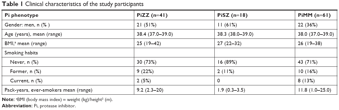 | Table 1 Clinical characteristics of the study participants |
PFT and IOS
The results of PFT and IOS in the Pi subgroups are reported in Table 2. The PiZZ subjects had significantly higher RV than the PiMM subjects (P<0.01) and lower Kco than the PiSZ subjects (P<0.01). They also had lower Kco than the PiMM subjects but the difference was not statistically significant. The RV/TLC ratio (P<0.05) and RV/TLC % of predicted (P<0.05) were significantly higher in the PiZZ subjects compared with the PiMM subjects. The FRC/TLC ratio was significantly higher in the PiZZ than in the PiSZ subjects (P<0.05). The mean Alx (P<0.05), R5Hz (P<0.01), R20Hz (P<0.01) were significantly lower while X5Hz (P<0.05) was significantly higher in the PiZZ group than in the PiMM group (Table 2).
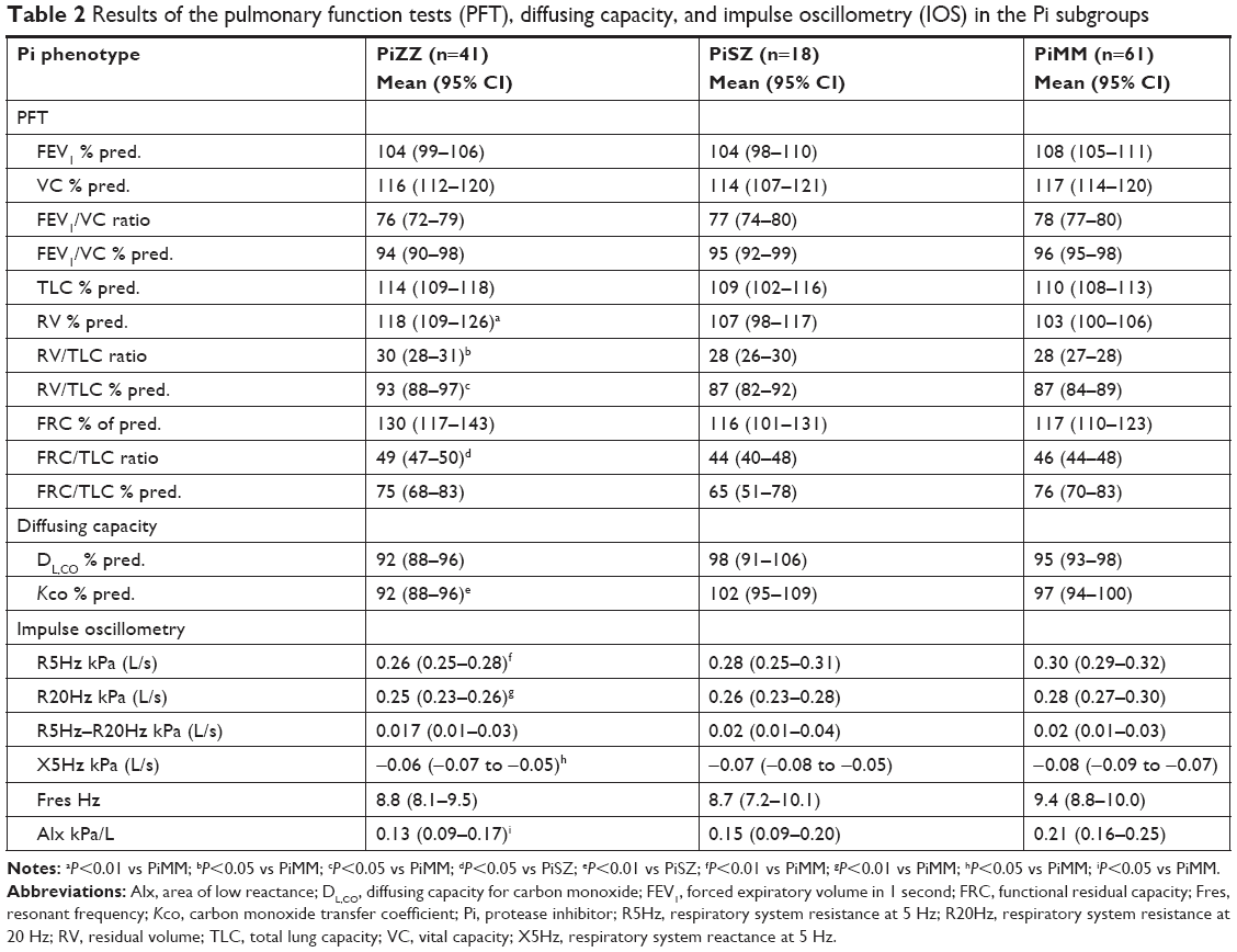 | Table 2 Results of the pulmonary function tests (PFT), diffusing capacity, and impulse oscillometry (IOS) in the Pi subgroups |
The results of PFT in the smoking subgroups are shown in Table 3. Because only two PiZZ current smokers participated in the study, the results of the PFT were compared between the ever- and never-smokers. Because only two PiSZ subjects were former smokers and none was a current smoker, they were excluded from the statistical analysis. The PiZZ ever-smokers had significantly higher TLC than the PiZZ never-smokers (P<0.05), the PiSZ never-smokers (P<0.01) and the PiMM never-smokers (P=0.01). They also had significantly higher RV (P<0.01) (Figure 1), RV/TLC ratios (P=0.01), and RV/TLC % of predicted (P=0.01) than the PiMM never-smokers. FRC was significantly higher in the PiZZ ever-smokers than in the PiZZ never-smokers (P<0.05), PiSZ never-smokers (P=0.01), PiMM ever-smokers (P=0.01), and PiMM never-smokers (P=0.01). The PiZZ ever-smokers had a significantly higher FRC/TLC ratio than the PiSZ never-smokers (P<0.05) and significantly lower Kco than the PiSZ never-smokers (P<0.01) and the PiMM never-smokers (P<0.01) (Figure 2 and Table 3).
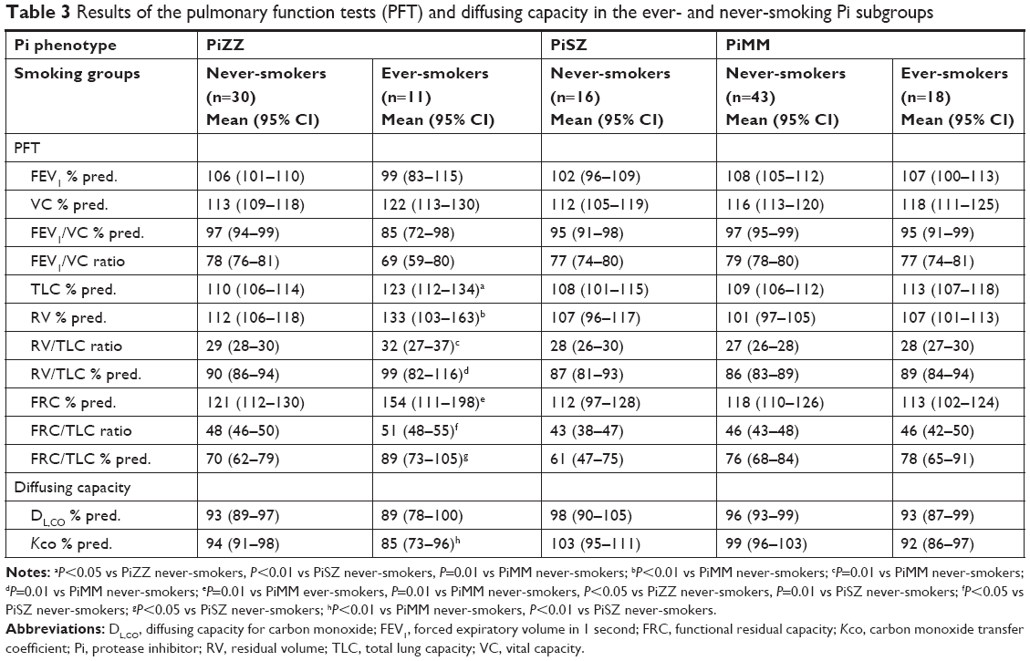 | Table 3 Results of the pulmonary function tests (PFT) and diffusing capacity in the ever- and never-smoking Pi subgroups |
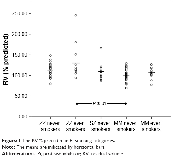 | Figure 1 The RV % predicted in Pi-smoking categories. |
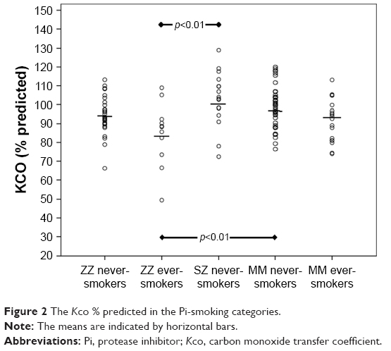 | Figure 2 The Kco % predicted in the Pi-smoking categories. |
Two PiZZ ever-smokers (18%) and none in the other smoking subgroups had FEV1 <80% of the predicted value (NS). Four PiZZ never-smokers (13%), three PiZZ ever-smokers (27%), four PiMM never-smokers (13%), and two PiMM ever-smokers (11%) had a FEV1/VC ratio <0.7 (NS).
The mean resistance and reactance did not reveal any statistically significant differences among the smoking subgroups (data not shown).
CT densitometry
The results of CT densitometry are shown in Tables 4 and 5. The PiZZ ever-smokers had lower mean apical PD15 and higher mean apical RA−910 than the PiZZ never-smokers, PiSZ never-smokers and both PiMM smoking subgroups, but the differences were not statistically significant. Fourteen (47%) PiZZ never-smokers, 8 (73%) PiZZ ever-smokers, 15 (35%) PiMM never-smokers, and 7 (39%) PiMM ever-smokers had whole PD15 (g/L) below the lower limit of the 95% CI (NS). Eleven (37%) PiZZ never-smokers, 8 (73%) PiZZ ever-smokers, 13 (30%) PiMM never-smokers, and 7 (39%) PiMM ever-smokers had the whole RA−910 above the upper limit of the 95% CI (NS).
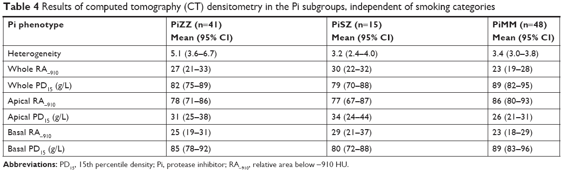 | Table 4 Results of computed tomography (CT) densitometry in the Pi subgroups, independent of smoking categories |
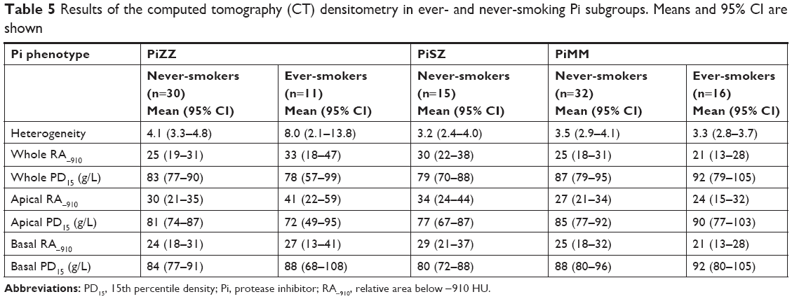 | Table 5 Results of the computed tomography (CT) densitometry in ever- and never-smoking Pi subgroups. Means and 95% CI are shown |
The mean heterogeneity was higher in the PiZZ subjects than other Pi subgroups but the differences were not statistically significant. No signs of emphysema were detected by the visual assessment of the CT images.
The PiZZ current smokers
Two PiZZ current smokers participated in the study, both female. The results of their lung function tests and CT densitometry are shown in Table 6. Both of them had manifest COPD, GOLD stadium II with FEV1/VC ratios below 0.70, high RV, FRC, FRC/RV ratios, and decreased FEV1, Kco, and DL,CO. They had low PD15 and high RA−910. None of them had emphysema as assessed visually on CT images. None of the PiMM current smokers had FEV1/VC ratios below 0.70.
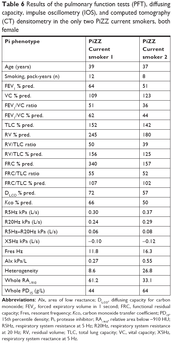 | Table 6 Results of the pulmonary function tests (PFT), diffusing capacity, impulse oscillometry (IOS), and computed tomography (CT) densitometry in the only two PiZZ current smokers, both female |
Discussion
This follow-up, case–control study of AAT-deficient subjects, identified by neonatal screening, shows that ever-smoking PiZZ individuals have, before the age of 40, physiological changes indicating early signs of emphysema. Furthermore, the two PiZZ current smokers had manifest COPD with an FEV1/VC ratio <0.70, high RVs, and low Kco (Table 6). These results are in accordance with our recently published results on symptoms and dynamic spirometry in this cohort.22
AAT deficiency has a quite broad clinical and disease spectrum. Our previously published study from the Swedish AATD Registry has also shown that many never-smoking PiZZ individuals, identified by screening, have normal life expectancy compared with the Swedish general population.23 Thus, individuals with severe AAT deficiency may live a healthy life without any clinical manifestations of disease. Factors such as smoking are the major risk for the development of emphysema.24
This cohort has been followed-up since birth. We have chosen the frequency of check-ups of 4 years in adulthood because of the well-preserved health status of the members of the cohort. With a longer interval, there would be a risk of loosening the contact with the subjects, and a shorter interval would not reveal any clinically relevant changes between the check-ups.
At the 30-year follow-up, only two PiZZ ever-smokers (one PiZZ current smoker) participated in the study; both shown signs of COPD.25 At the 34-year follow-up, the PiZZ ever-smokers had significantly lower Kco in comparison to the PiSZ and PiMM never-smokers.20
IOS showed lower resistance at 5 Hz and 20 Hz in the PiZZ subjects than in the PiMM subjects. Also reactance at 5 Hz was higher (less negative) and Fres lower in the PiZZ group than in the PiMM group (Table 2). These findings are contrary to what is usually seen in subjects with smoking-induced COPD compared with normal subjects. There may be differences in the pathogenesis between smoking-induced COPD and subjects with AAT deficiency. Whereas there is little doubt that smoking-induced COPD starts in the terminal bronchioles,26 emphysema may be more predominant at an early stage in subjects with AAT deficiency. The PiZZ subjects in this study showed evidence of hyperinflation (high FRC/TLC ratios) and air trapping (high RV/TLC ratios), which may offer an explanation for our findings. R5Hz is generally considered to represent resistance of the entire bronchial tree and R20Hz the resistance of “central airways”. The difference in resistance between 5 Hz and 20 Hz is therefore often taken to represent “peripheral airways”. It is not, however, precisely known which airway generations are measured. If the tethering forces on small airways are reduced in PiZZ subjects, this may be compensated for by hyperinflation. This would affect the dimensions not only of the small but also of more proximal airways that might contribute more to the result of the IOS measurements of resistance and reactance. The IOS measurements are performed during tidal breathing, and the findings are compatible with a higher degree of inflation during the measurement in the subjects with AAT deficiency than in the control group. However, the results did not differ significantly between the smoking subgroups, which may be due to the low number of ever-smokers, making it difficult to reach statistical significance.
In contrast to the results of PFTs, no statistically significant differences were found in the CT-densitometry parameters between the AAT-deficient and the control subjects, and none of the study participants had emphysema as judged by visual assessment of the CT images. At the 30 year follow-up, the only PiZZ current smoker participating had the lowest PD15 (60 g/L).25 At the 34–35 year follow-up, the PiZZ ever-smokers had significantly lower PD15 than the PiMM ever-smokers.20 In the present study, the two PiZZ current smokers also had low PD15, 44 g/L and 64 g/L, respectively (Table 6).
CT densitometry is considered to be an accurate and reliable method for the detection of emphysema, and it is considered to be a more accurate method than PFTs in detecting the progression of the emphysema.21 However, the previously published studies have mostly been based on patients who had already been diagnosed with COPD. In our study, we examined young AAT-deficient individuals for the detection of early signs of diffuse emphysema propagation. It is possible that complete PFTs including diffusing capacity and airways resistance and reactance measurements are more sensitive in detecting early, incipient emphysema, and hyperinflation than CT densitometry.
Limitations
The most important limitation of this study is the small number of participants among the AAT-deficient subjects. The most common reason was shortage of time, and second reason was long traveling distance.
Conclusion
At 37–39 years of age, the PiZZ ever-smokers, and especially current smokers, have evidence of emphysema. In contrast, the PiZZ and PiSZ never-smokers have normal lung function and no signs of emphysema. These results emphasize the importance of early diagnosis of AAT deficiency for prevention of smoking.
Acknowledgments
The authors wish to thank Ewa Ringdal Szemberg, Isabella Björk, and Helene Johansson Kvist for technical and secretarial support. The study was supported by unrestricted grants from the Swedish Heart-Lung Foundation and Skåne University Hospital.
Disclosure
The authors report no conflict of interest in this work.
References
American Thoracic Society/European Respiratory Society Statement. Standards for the diagnosis and management of individuals with alpha-1 antitrypsin deficiency. Am J Respir Crit Care Med. 2003;168:818–900. | ||
Stoller JK, Aboussouan LS. A review of α1-antitrypsin deficiency. Am J Respir Crit Care Med. 2012;185(3):246–259. | ||
Gettins PG, Structure S. Mechanism, and function. Chem Rev. 2002;102:4751–4804. | ||
Laurell CB, Eriksson S. The electrophoretic alpha-1-globulin pattern of serum in alpha-1-antitrypsin deficiency. Scand J Clin Lab Invest. 1963;15:132–140. | ||
Gooptu B, Dickens JA, Lomas DA. The molecular and cellular pathology of α1-antitrypsin deficiency. Trends. 2014;20:116–127. | ||
Mahadeva R, Atkinson C, Li Z, et al. Polymers of Z alpha1-antitrypsin co-localize with neutrophils in emphysematous alveoli and are chemotactic in vivo. Am J Pathol. 2005;166(2):377–386. | ||
Curiel DT, Chytil A, Courtney M, Crystal RG. Serum alpha 1-antitrypsin deficiency associated with the common S-type (Glu264-Val) mutation results from intracellular degradation of alpha 1-antitrypsin prior to secretion. J Biol Chem. 1989;264(18):10477–10486. | ||
Nakano S, Nakahira J, Sawai T, et al. Perioperative evaluation of respiratory impedance using the forced oscillation technique: a prospective observational study. BMC Anesthesiol. 2015;16(1):32. | ||
Shirai T, Kurosawa H. Clinical application of the forced oscillation technique. Intern Med. 2016;55(6):559–566. | ||
Brashier B, Salvi S. Measuring lung function using sound waves: role of the forced oscillation technique and impulse oscillometry system. Breathe. 2015;11(1):57–65. | ||
Frantz S, Nihlén U, Dencker M, Engström G, Löfdahl CG, Wollmer P. Impulse oscillometry may be of value in detecting early manifestations of COPD. Respir Med. 2012;106(8):1116–1123. | ||
Stolk J, Stoel BC. Lung densitometry to assess progression of emphysema in chronic obstructive pulmonary disease: time to apply in the clinic? Am J Respir Crit Care Med. 2011;183(12):1578–1580. | ||
Ju J, Li R, Gu S, et al. Impact of emphysema heterogeneity on pulmonary function. PLoS One. 2014;9(11):e113320. | ||
Edgar RG, Patel M, Bayliss S, Crossley D, Sapey E, Turner AM. Treatment of lung disease in alpha-1 antitrypsin deficiency: a systematic review. Int J Chron Obstruct Pulmon Dis. 2017;12:1295–1308. | ||
Sveger T. Liver disease in alpha1-antitrypsin deficiency detected by screening of 200,000 infants. N Engl J Med. 1976;294(24):1316–1321. | ||
Miller MR, Hankinson J, Brusasco V, et al. ATS/ERS Task force standardization of lung function testing. Standardization of spirometry. Eur Respir J. 2005;26:319–338. | ||
Culver BH, Graham BL, Coates AL, et al; ATS Committee on Proficiency Standards for Pulmonary Function Laboratories. Recommendations for a standardized pulmonary function report. An Official American Thoracic Society Technical Statement. Am J Respir Crit Care Med. 2017;196(11):1463–1472. | ||
Quanjer PH. Standardized lung function testing. Report of the working party on standardization of lung function tests. European Community for Coal and Steel. Bull Eur Physiopathol Respir. 1983;19(Suppl 16):1–95. | ||
Oostveen E, Macleod D, Lorino H, et al; ERS Task Force on Respiratory Impedance Measurements. The forced oscillation technique in clinical practice: methodology, recommendations and future developments. Eur Respir J. 2003;22(6):1026–1041. | ||
Piitulainen E, Montero LC, Nystedt-Düzakin M, et al. Lung function and CT densitometry in subjects with alpha-1-antitrypsin deficiency and healthy controls at 35 years of age. COPD. 2015;12(2):162–167. | ||
Stolk J, Versteegh MIM, Montenij LJ, et al. Densitometry for assessment of effect of lung volume reduction surgery for emphysema. Eur Respir J. 2007;29(6):1138–1143. | ||
Piitulainen E, Mostafavi B, Tanash HA. Health status of the Swedish alpha 1-antytrypsin-deficient cohort identified by neonatal screening, at the age of 37–40 years. Int J Chron Obstr Pulmon Dis. 2017;12:1–6. | ||
Tanash HA, Ekström M, Rönmark E, Lindberg A, Piitulainen E. Survival in individuals with severe alpha 1-antitrypsin deficiency (PiZZ) in comparison to a general population with known smoking habits. Eur Respir J. 2017;50(3):1700198. | ||
Sandhaus RA, Turino G, Brantly ML, et al. The diagnosis and management of alpha-1 antitrypsin deficiency in the adult. Chronic Obstr Pulm Dis. 2016;3(3):668–682. | ||
Bernspång E, Wollmer P, Sveger T, Piitulainen E. Lung function in 30-year-old alpha-1-antitrypsin-deficient individuals. Respir Med. 2009;103(6):861–865. | ||
Suzuki M, Sze MA, Campbell JD, et al. The cellular and molecular determinants of emphysematous destruction in COPD. Sci Rep. 2017;7(1):9562. |
Supplementary material
 | Figure S1 Cohort and control group questionnaire. |
 © 2018 The Author(s). This work is published and licensed by Dove Medical Press Limited. The
full terms of this license are available at https://www.dovepress.com/terms
and incorporate the Creative Commons Attribution
- Non Commercial (unported, 3.0) License.
By accessing the work you hereby accept the Terms. Non-commercial uses of the work are permitted
without any further permission from Dove Medical Press Limited, provided the work is properly
attributed. For permission for commercial use of this work, please see paragraphs 4.2 and 5 of our Terms.
© 2018 The Author(s). This work is published and licensed by Dove Medical Press Limited. The
full terms of this license are available at https://www.dovepress.com/terms
and incorporate the Creative Commons Attribution
- Non Commercial (unported, 3.0) License.
By accessing the work you hereby accept the Terms. Non-commercial uses of the work are permitted
without any further permission from Dove Medical Press Limited, provided the work is properly
attributed. For permission for commercial use of this work, please see paragraphs 4.2 and 5 of our Terms.