Back to Journals » Clinical, Cosmetic and Investigational Dermatology » Volume 15
Lichen Amyloidosis on the Scalp: A Case Report
Authors Li X , Huang X, Du J, Zhou C
Received 7 March 2022
Accepted for publication 9 April 2022
Published 20 April 2022 Volume 2022:15 Pages 721—723
DOI https://doi.org/10.2147/CCID.S363633
Checked for plagiarism Yes
Review by Single anonymous peer review
Peer reviewer comments 2
Editor who approved publication: Dr Jeffrey Weinberg
Xiangqian Li,1,* Xinlv Huang,1,2,* Juan Du,1 Cheng Zhou1
1Department of Dermatology, Peking University People’s Hospital, Beijing, People’s Republic of China; 2Department of Dermatology, Beijing Chao-Yang Hospital Capital Medical University, Beijing, People’s Republic of China
*These authors contributed equally to this work
Correspondence: Cheng Zhou, Peking University People’s Hospital, No. 11 Xizhimen South Street, Xicheng District, Beijing, People’s Republic of China, Tel +86-10-88325472, Email [email protected]
Abstract: Lichen amyloidosis (LA) is one form of primary cutaneous amyloidosis, presented as discrete, lichenoid papules with itching, commonly involving the extensor surfaces of extremities. Scalp involvement is rarely reported in the literature. In this study, we reported a case of LA over the crown and vertex areas of the scalp. The lesions subsided with topical clobetasol propionate/all-trans retinoic acid compound ointment treatment for 2 months and achieved significant improvement.
Keywords: primary cutaneous amyloidosis, lichen amyloidosis, scalp, dermoscopy, androgenetic alopecia, case report
Introduction
Primary cutaneous amyloidosis (PCA) is a frequently encountered skin condition characterized by amyloid deposition in dermis without systemic involvement.1 There are different subtypes of PCA, including lichen amyloidosis (LA), macular amyloidosis (MA) and rarely, nodular amyloidosis (NA).1 LA is clinically characterized by development of multiple pruritic, hyperkeratotic, lichenoid papules, occurs mostly on the pretibial areas and occasional on the arms and thighs. However, scalp involvement has rarely been reported.2,3 In this study, we presented a case with LA lesions limited the scalp skin.
Case Report
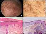
A 64-year-old Chinese male presented with multiple dark-brown papules on the scalp for 1 year. The lesions were asymptomatic and gradually increased in number over time. He was diagnosed as androgenetic alopecia (AGA) for a decade with no treatment except topical 5% minoxidil for only 1 month at 5 years ago. The patient was otherwise healthy and his family history was negative. Physical examination revealed discrete, 1- to 3-mm, flat-topped, brownish papules over the crown and vertex area of the scalp (Figure 1A). Dermoscopic examination revealed whitish scar-like centre surrounded by brownish dots or brown fine streaks radiating from the centre, located in a background hair and scalp alteration of typical AGA (Figure 1B). Histologically, deposition of pink amorphous material was identified in the papillary dermis with scattered melanophages in the expanded dermal papillae (Figure 1C). The deposits were highlighted by Congo red staining (Figure 1D). He was finally diagnosed with LA, then was treated with clobetasol propionate/all-trans retinoic acid compound ointment and achieved significant improvement in the appearance of the lesions after 2 months without any side effect.
Discussion
LA is a chronic pruritic skin disorder characterized by the deposition of amyloids in the papillary dermis. The precise pathogenesis of LA has yet to be clarified. To date, several medical conditions, including constant skin friction, genetic predisposition, virus infection, and environmental factors have been reported associated with LA.4 In addition, other comorbid diseases were also reported, such as atopic dermatitis, diabetes mellitus and renal insufficiency.
The most common distribution of LA is the extensor surfaces of extremities. Scalp involvement is rarely described in the literature. Kim et al2 reported a patient with papules over the left frontal hairline, extending onto the forehead. Frew et al3 described a patient diagnosed LA involving the central portion of the anterior scalp and lower limbs, and both patients were complained of pruritus. Interestingly, Bellinato et al5 reported a case of NA in a female patient presented with painful, ulcerated alopecic nodules on the vertex of the scalp. In the present study, the patient denied itching and taking any regular medication or therapy except for topical minoxidil because of AGA. We assume the lesions might be attributed to the long-term sun exposure of the scalp because of severe alopecia. Sun exposure has been proposed as one of the underlying causes of amyloidosis cutis dyschromica- an atypical clinical variants of PCA.1 Clinical differential diagnosis of this case includes lichen planopilaris, seborrheic dermatitis, verruca plana and epidermodysplasia verruciformis.
Dermoscopy may serve as a useful tool in confirming the clinical suspicion of PCA before skin biopsy. The features of LA are still been explored. Current notion suggests that LA presented with white central hubs or whitish scar-like centres (corresponding to the amyloid deposits or hyperkeratosis with acanthosis in pathology) surrounded by grey-brown pigmentation, mainly in the form of dots, globules or peppering (corresponding to the pigment incontinence and basal hyperpigmentation), which named “two-zone pattern”.6
Multiple treatment modalities, such as topical steroids, calcineurin inhibitors, systemic antihistamines, retinoids and various laser therapies have been tried in patients with PCA.1 However, there is no curative to date. Emerging biological agents and small molecule agents, such as dupilumab, IL-31 or JAK inhibitors might be the potential therapies for PCA, especially for generalized type.1,7,8
Conclusion
Primary cutaneous amyloidosis is a relatively common skin disorder characterized by extracellular deposition of amyloid materials that result from the degradation of various proteins in the skin. Besides typical clinical manifestations, some forms of PCA exhibit an unusual presentation on distribution, such as lichenoid papules on the scalp in the present case. We highlight the dermatologists’ awareness of atypical morphology or location of PCA, and suggest that dermoscopy could be a valuable tool in the diagnosis of the disease.
Consent Statement
Informed consent for publication of the case details and associated images was obtained from the patient and all procedures were performed in accordance with the Helsinki Declaration. Institutional approval was not required to publish the case details.
Funding
This study was supported by the National Natural Science Foundation of China (No. 81773311 and 82073459).
Disclosure
Xiangqian Li and Xinlv Huang are co-first authors for this study. The authors report no conflicts of interest in this work.
References
1. Hamie L, Haddad I, Nasser N, et al. Primary localized cutaneous amyloidosis of keratinocyte origin: an update with emphasis on atypical clinical variants. Am J Clin Dermatol. 2021;22(5):667–680. doi:10.1007/s40257-021-00620-9
2. Kim Y, Ioffreda MD, Chung CG. Lichen amyloidosis of the scalp and forehead. Dermatol Online J. 2017;23(11). doi:10.5070/D32311037276
3. Frew JW, Fallah H. Lichen amyloidosis involving the scalp. Australas J Dermatol. 2017;58(4):e260–e261. doi:10.1111/ajd.12582
4. Weidner T, Illing T, Elsner P. Primary localized cutaneous amyloidosis: a systematic treatment review. Am J Clin Dermatol. 2017;18(5):629–642. doi:10.1007/s40257-017-0278-9
5. Bellinato FRP, Sina S, Girolomoni G. Primary nodular localized cutaneous amyloidosis of the scalp associated with systemic lupus erythematosus. Arch Rheumatol. 2022;37(1):145–147.
6. Behera B, Kumari R, Mohan Thappa D, et al. Dermoscopic features of primary cutaneous amyloidosis in skin of colour: a retrospective analysis of 48 patients from South India. Australas J Dermatol. 2021;62(3):370–374. doi:10.1111/ajd.13662
7. Humeda Y, Beasley J, Calder K. Clinical resolution of generalized lichen amyloidosis with dupilumab: a new alternative therapy. Dermatol Online J. 2020;26(12):e34. doi: 10.5070/D32612051364
8. Chen J, Yang B. Tofacitinib for the treatment of primary cutaneous amyloidosis: a case report. Dermatol Ther. 2022. doi:10.1111/dth.15312:e15312
 © 2022 The Author(s). This work is published and licensed by Dove Medical Press Limited. The full terms of this license are available at https://www.dovepress.com/terms.php and incorporate the Creative Commons Attribution - Non Commercial (unported, v3.0) License.
By accessing the work you hereby accept the Terms. Non-commercial uses of the work are permitted without any further permission from Dove Medical Press Limited, provided the work is properly attributed. For permission for commercial use of this work, please see paragraphs 4.2 and 5 of our Terms.

