
Back to Journals » Infection and Drug Resistance » Volume 19
Levels of Toll-Like Receptor 2, Myeloid Differentiation Primary Response 88 and Mucin 5AC, Oligomeric Mucus/Gel-Forming in Bronchoalveolar Lavage Fluid for Predicting Severe Mycoplasma pneumoniae Pneumonia in Children
Authors Wang T, Li B, Xiao A, Ren R, Liu L, Xu Y, Wu X, Zhao Y, Yu Z, Geng J, Li S
Received 17 March 2026
Accepted for publication 7 July 2026
Published 21 July 2026 Volume 2026:19 609961
DOI https://doi.org/10.2147/IDR.S609961
Checked for plagiarism Yes
Review by Single anonymous peer review
Peer reviewer comments 2
Editor who approved publication: Dr Hazrat Bilal
Tuanjie Wang,1,* Baowen Li,2,* Aiju Xiao,3 Ruijuan Ren,1 Ling Liu,1 Yuping Xu,1 Xiangtao Wu,1 Yuanbo Zhao,1 Zhicai Yu,1 Jiahao Geng,1 Shujun Li1
1Department of PICU, The First Affiliated Hospital of Henan Medical University, Weihui City, People’s Republic of China; 2Department of PICU, Qinghai Provincial Women’s and Children’s Hospital, Xining City, People’s Republic of China; 3Department of Pediatric Internal Medicine, First Affiliated Hospital of Henan Medical University, Weihui City, People’s Republic of China
*These authors contributed equally to this work
Correspondence: Shujun Li, Department of PICU, The First Affiliated Hospital of Henan Medical University, No. 88, Jiankang Road, Weihui City, Henan Province, 453199, People’s Republic of China, Tel +86 13673520729, Email [email protected] Tuanjie Wang, Department of PICU, The First Affiliated Hospital of Henan Medical University, No. 88, Jiankang Road, Weihui City, Henan Province, 453199, People’s Republic of China, Tel +86 13673520729, Email [email protected]
Objective: This study aimed to investigate the value of measuring levels of toll-like receptor 2 (TLR2), myeloid differentiation primary response 88 (MyD88) and mucin 5AC, oligomeric mucus/gel-forming (MUC5AC) in bronchoalveolar lavage fluid (BALF) for assessing disease severity and predicting severe Mycoplasma pneumoniae pneumonia (SMPP) in children.
Methods: A total of 151 children with MPP were enrolled between July 2023 and February 2025. The patients with MPP were divided into SMPP (n = 60) and non-SMPP (n = 91) groups. Bronchoalveolar lavage fluid levels of TLR2, MyD88 and MUC5AC were measured via enzyme-linked immunosorbent assay. Clinical data and laboratory indices, including C-reactive protein (CRP), procalcitonin, serum amyloid A, interleukin-6, D-dimer and fibrinogen, were compared among groups. Diagnostic value was evaluated using receiver operating characteristic curves, and independent risk factors were identified via logistic regression.
Results: Multivariate logistic regression identified respiratory effort, CRP, lactate dehydrogenase, MyD88 and TLR2 as independent risk factors for SMPP (all p < 0.05). The combined prediction model achieved an area under the curve (AUC) of 0.950 (95% confidence interval: 0.912– 0.979), significantly outperforming any single biomarker. Toll-like receptor 2 alone showed the highest predictive value among individual BALF markers (AUC = 0.876).
Conclusion: Elevated levels of TLR2, MyD88 and MUC5AC in BALF are associated with SMPP. However, only TLR2 and MyD88 (along with clinical and laboratory indicators) were independent risk factors. The combined model shows good performance (AUC = 0.950) but requires external validation. These findings are associational and warrant further study.
Keywords: Mycoplasma pneumoniae, toll-like receptor 2, myeloid differentiation factor 88, mucin 5AC, child
Introduction
Mycoplasma pneumoniae (MP) is an important pathogen of community-acquired pneumonia in children, with a high incidence in school-aged and adolescent populations, accounting for approximately 10%–40% of cases.1,2 Most children have mild-to-moderate clinical symptoms, but in recent years, with the increase of macrolide-resistant strains and the complexity of host immune responses, some children’s conditions have progressed rapidly, and they may develop severe MP pneumonia (SMPP), complicated by pulmonary consolidation, atelectasis, bronchial mucus plugs and extrapulmonary complications, which seriously threaten children’s health and even endanger their lives.3,4
In recent years, with an increasing number of macrolide-resistant MP (MRMP) strains and the growing complexity of host immune responses, some children’s conditions have progressed rapidly. Notably, MRMP infection itself may induce a more intense or persistent inflammatory response, further contributing to immune dysregulation and disease severity, such as respiratory failure, acute respiratory distress syndrome and empyaema5 Severe cases are closely associated with excessive host immune responses (such as cytokine storm) and MRMP.6,7 The detection rate of MRMP is high in China and East Asia, and drug-resistant infections often lead to prolonged fever, extended hospital stays and increased use of systemic glucocorticoids and second-line antibiotics.7–9 In the process of the occurrence and development of SMPP, excessive activation of immune response and inflammatory factors is considered to be the key mechanism.3 Mucin 5AC, oligomeric mucus/gel-forming (MUC5AC) is a high molecular weight glycoprotein secreted by respiratory epithelial cells. It is one of the main components of airway mucus and can form viscous hydrogels, participating in the formation of mucus plugs and airway obstruction. Innate immune receptors play a core role in pathogen recognition and inflammatory amplification response.10 Toll-like receptor 2 (TLR2) can recognise the lipoprotein components of MP. Given that MP lacks a cell wall – a unique feature among bacteria – its cell membrane lipoproteins serve as major structural and virulence-associated components, acting as key pathogen-associated molecular patterns that interact with host innate immune receptors. Upon recognition by TLR2, the downstream myeloid differentiation primary response 88 (MyD88) signalling pathway is activated, further inducing the expression of various inflammatory factors, thereby participating in airway inflammation and tissue damage.11
Although clinical evidence for direct targeting of mucus plugs in children’s SMPP is limited, in vitro and animal experiments have shown that TLR2/MyD88 signalling can regulate MUC5AC secretion and participate in airway mucus hypersecretion and obstruction10,11 Studies have shown that MP infection can promote mucus secretion by activating TLR2 on airway epithelial cells, thereby upregulating MUC5AC mRNA and protein expression This process is particularly important in airway epithelial cells of patients with asthma, and TLR2 inhibitors can significantly reduce MUC5AC expression, suggesting that the TLR2 pathway plays a core role in the regulation of MUC5AC10 In addition, TLR2 activates the downstream MyD88 signalling pathway, inducing the release of inflammatory factors, such as tumour necrosis factor alpha (TNF-α), and amplifying the airway inflammatory response.11 Animal experiments further confirmed that TLR2-deficient mice showed reduced inflammatory response, decreased TNF-α levels and reduced lung tissue damage after MP infection.11
However, there is currently a lack of clinical evidence regarding the relationship between TLR2, MyD88 and MUC5AC in childhood SMPP. Furthermore, clinical assessment of SMPP severity and prognosis largely relies on serological markers, but since local pulmonary inflammation often precedes and is more severe than systemic changes, serological markers may not accurately reflect the local microenvironment of the lesion in real time. As an important tool for diagnosing and treating SMPP, bronchoalveolar lavage fluid (BALF) is obtained via fibreoptic bronchoscopy from the affected lung segment, providing a more direct reflection of pulmonary inflammatory infiltration and accumulation of secretions. Currently, there are few clinical studies on the diagnostic and prognostic value of TLR2, MyD88 and MUC5AC levels in BALF for childhood SMPP.
Therefore, this study explores the value of MUC5AC, TLR2 and MyD88 levels in BALF of children with MPP in disease assessment and prognosis prediction and to analyse their relationship with mucus plug formation. Simultaneously, by combining clinical indicators, a risk factor prediction model for SMPP is established to provide new evidence for the early identification and intervention of SMPP in children.
Research Methods
Research Participants
This is a single-centre prospective cohort study. The study participants were children hospitalised in the Department of Pediatrics at the First Affiliated Hospital of Xinxiang Medical University between July 2023 and February 2025 who were clinically and laboratory-diagnosed with MPP. This study strictly followed the Declaration of Helsinki guidelines. The screening and inclusion process for this study is detailed in Figure 1. The sample size for this study was estimated based on the events per variable (EPV) principle in logistic regression analysis. To ensure the stability and reliability of the multivariate logistic regression model, an EPV of ≥5–10 is typically required. After clinical pre-screening and collinearity testing, seven candidate variables – exhaustion of breathing, pulmonary consolidation, C-reactive protein (CRP), lactate dehydrogenase (LDH), TLR2, MyD88 and MUC5AC – were included in the multivariate logistic regression analysis. Theoretically, ≥35–70 patients with SMPP were required. This study actually included 60 patients with SMPP, meeting the sample size requirement for model construction.
 |
Figure 1 The Research Flowchart. Abbreviations: SMPP, severe Mycoplasma pneumoniae pneumonia; BALF, bronchoalveolar lavage fluid. |
Inclusion Criteria
(1) aged 0–14 years; (2) MPP diagnosed according to the latest Guidelines for the Diagnosis and Treatment of MP in Children (2023 Edition)1 as follows: clinical manifestations mainly of fever and irritating dry cough, lung signs, such as decreased breath sounds and dry and wet rales and chest radiological manifestations, such as alveolar inflammatory changes, combined with any one or two of the following – (i) MP antibody titre of a single serum sample ≥1:160 (particle aglutination method); MP antibody titre of two serum samples increases by ≥4 times during the course of the disease or (ii) MP DNA- or RNA-positive.
Exclusion Criteria
(1) patients with congenital cardiopulmonary diseases, immunodeficiency or chronic lung diseases; (2) patients who have received immunosuppressants or high-dose glucocorticoids before admission; (3) patients for whom informed consent from parents has not been obtained or whose information is incomplete; or (4) co-infection with adenovirus.
Grouping Method
The 151 children with MPP were divided into two groups according to the severity of their condition: the SMPP group (n = 60) and the non-severe MPP (non-SMPP) group (n = 91).
Criteria for severe illness:3 Patients exhibiting three out of the following five criteria: (1) dyspnoea or inspiratory retractions (aged <1 year: respiratory rate [RR] ≥ 50 bpm, heart rate [HR] ≥ 150 bpm; aged 1–5 years: RR ≥ 40 bpm, HR ≥ 140 bpm; aged >5 years: RR ≥ 30 bpm, HR ≥ 120 bpm); (2) oxygenation index < 250 mmHg or percutaneous oxygen saturation ≤93% when breathing air at rest; (3) altered consciousness (drowsiness, seizures or even coma); (4) elevated blood urea nitrogen ≥7.14 mmol/L; (5) hypotension or shock, requiring fluid resuscitation and vasoactive drug support.
Specimen Collection and Processing
All children enrolled in this study met the indications of the 2018 Chinese Guidelines for Flexible Bronchoscopy in Pediatrics,12 had unknown aetiologies or pulmonary consolidation and needed treatment or diagnosis. They had obtained the consent of their families and signed informed consent forms. All participants had venous blood collected on the day of admission to detect blood routine, inflammatory markers (CRP, procalcitonin [PCT], serum amyloid A [SAA], interleukin [IL]-6), coagulation function (D-dimer, fibrin degradation products [FDP]) and biochemical indicators (LDH, liver function, etc). All children underwent electronic bronchoscopy within 24–48 hours after admission, and bronchoalveolar lavage was performed in the lesion area. The lavage fluid was sterile saline (1 mL/kg, total amount ≤20 mL), and a recovery rate ≥40% was considered a qualified specimen. The lavage fluid was centrifuged at 1000 rpm for 10 min, and the supernatant was collected and stored at −80°C for later use. The median time from symptom onset (fever or cough) to BALF collection was 6 days (interquartile range [IQR]: 4–8 days) in the SMPP group and 5 days (IQR: 3–7 days) in the non-SMPP group (p = 0.08).
Detection Methods
The concentrations of MUC5AC, TLR2 and MyD88 in BALF were detected using enzyme-linked immunosorbent assay (ELISA). All kits used were purchased from Wuhan Fein Biotechnology Co., Ltd., Wuhan, China, with catalogue numbers EH1035 1, EH0300 2 and EH0240 3 for MUC5AC, TLR2 and MyD88, respectively. Experimental procedures were performed strictly according to the kit instructions. The routine laboratory tests included the following: complete blood count (white blood cell count, neutrophil percentage) was performed using a Sysmex XN-9000 fully automated haematology analyser (Sysmex Corporation, Tokyo, Japan); CRP, SAA, LDH and liver function tests were performed using an AU5800 fully automated immunoassay analyser (Beckman Coulter, Brea, California, USA); PCT and IL-6 tests were performed using an e801 fully automated immunoassay analyser (Roche Diagnostics, Zug, Switzerland); and D-dimer testing was performed using a CX-9000 fully automated coagulation analyser (Mindray Bio-Medical Electronics Co., Ltd., Shenzhen, China). All tests were performed by the Department of Laboratory Medicine at the First Affiliated Hospital of Xinxiang Medical University in strict accordance with standardised clinical operating procedures.
All laboratory tests, including ELISA measurements of BALF TLR2, MyD88 and MUC5AC, were performed by technicians who were blinded to the patients’ clinical grouping (SMPP vs non-SMPP). Clinical data and outcome assessments were recorded by attending physicians who were not aware of the BALF biomarker results at the time of data entry.
Observation Indicators
General Information
Age, gender, weight.
Clinical Data
(1) respiratory efforts – this refers to a significant increase in respiratory rate (judged by age: <1 year, ≥50 breaths/min; 1–5 years, ≥40 breaths/min; >5 years, ≥30 breaths/min), inspiratory three-recession sign (suprasternal notch, supraclavicular notch, intercostal retraction) or nasal flaring; (2) pericardial effusion – abnormal fluid echoes are confirmed in the pericardial cavity by echocardiography; (3) pulmonary consolidation – chest computed tomography or X-ray shows uniform increase in lung parenchyma density, air bronchograms are visible in the affected area and the consolidation involves one or more lung segments; (4) plastic bronchitis – endogenous bronchial tree-like gelatinous secretions are found and aspirated or removed from the airway during fibreoptic bronchoscopy; (5) switching to second-line antibiotics – this refers to the situation where the condition does not improve or progress after 72 hours of treatment with macrolide drugs (such as azithromycin) and the condition is switched to new tetracycline drugs (doxycycline, minocycline) or fluoroquinolone drugs according to the Guidelines for the Diagnosis and Treatment of MP in Children (2023 Edition).
Laboratory Indicators
Complete blood count (including white blood cell count, neutrophil percentage, neutrophil count, lymphocyte percentage), inflammatory markers (CRP, PCT, SAA, IL-6), coagulation function indicators (D-dimer, FDP), liver function (alanine aminotransferase [ALT], aspartate aminotransferase [AST], albumin, etc.) and LDH.
Bronchoalveolar Lavage Fluid Indicators
MUC5AC, TLR2 and MyD88 levels.
Statistical Analysis
Statistical analysis was performed using SPSS 26.0 software. Quantitative data were expressed as mean ± standard deviation ( ) or median (interquartile range), and comparisons between groups were performed using t-tests or Mann–Whitney U-tests. Categorical data were expressed as the number of cases and percentages, and comparisons between groups were performed using chi-square (χ2) tests or Fisher’s exact test. Receiver operating characteristic (ROC) curve analysis was used to analyse the predictive value of MUC5AC, TLR2, MyD88 and related clinical indicators for SMPP, and the area under the curve (AUC) was calculated. Univariate and multivariate logistic regression analyses were performed, and indicators with p < 0.05 and clear clinical significance in the univariate analysis were included in the multivariate logistic regression model. To minimise overfitting and comply with the EPV principle (requiring ≥5–10 events per candidate variable), a pre-specified variable selection strategy was adopted. First, all variables with p < 0.05 in univariate logistic regression were considered candidates. Second, clinically redundant or highly collinear variables (eg neutrophil percentage, neutrophil count, fever days, post-antibiotic fever days) were reviewed, and only the most clinically representative one was retained. Third, a collinearity diagnostic was performed using the variance inflation factor (VIF), with VIF < 5 as the threshold to exclude severe multicollinearity. Fourth, backward stepwise regression (likelihood ratio criterion, entry p < 0.05, removal p > 0.10) was applied to the final set of seven core variables (respiratory effort, pulmonary consolidation, CRP, LDH, TLR2, MyD88, MUC5AC) to identify independent risk factors. The final model included five variables (respiratory effort, CRP, LDH, MyD88, TLR2).
There were no missing data for the primary outcome or key exposure variables (TLR2, MyD88, MUC5AC, CRP, LDH and clinical indicators) in the enrolled 151 patients with MPP. For a small number of secondary laboratory parameters (<5% of all measurements, including complement C3 in 3 patients and FDP in 2 patients), missing values were not imputed; these patients were excluded only from analyses involving those specific variables.
Ethical Statement
The study has been approved by the Ethics Committee of the First Affiliated Hospital of Xinxiang Medical University (Approval No.: Ec-023-155; Approval Date: 13 June 2023). All guardians of the children participating in this study signed written informed consent forms.
Results
Comparison of Baseline Data and Observed Indicators Between the SMPP Group and the Non-Severe MP Pneumonia Group
This study included 91 patients in the non-SMPP group and 60 patients in the SMPP group. There were no statistically significant differences between the two groups in terms of age, weight, sex or respiratory efforts (p > 0.05). The SMPP group had a higher rate of developing respiratory distress, switching to second-line antibiotics, using gamma globulin, using albumin and using glucocorticoid than the non-SMPP group (p < 0.05). The SMPP group also had a higher fever peak, longer fever duration, longer hospital stay and longer fever duration after antibiotic use than the non-SMPP group (p < 0.05). The SMPP group had higher levels of neutrophils, neutrophil count, PCT, CRP, SAA, IL-6, fibrinogen, FDP, D-dimer, ALT, AST, LDH, TLR2, MyD88 and MUC5AC than the non-SMPP group and lower levels of lymphocytes, albumin and complement C3 (p < 0.05). The SMPP group also had a higher incidence of complications, such as pericardial effusion, coagulation dysfunction, pulmonary consolidation, pleural effusion and plastic bronchitis, than the non-SMPP group, with statistically significant differences (p < 0.05), as shown in Table 1 and Figure 2.
 |
Table 1 Comparison of Baseline Data and Observations Between SMPP Group and Non-SMPP Group in Children with MPP |
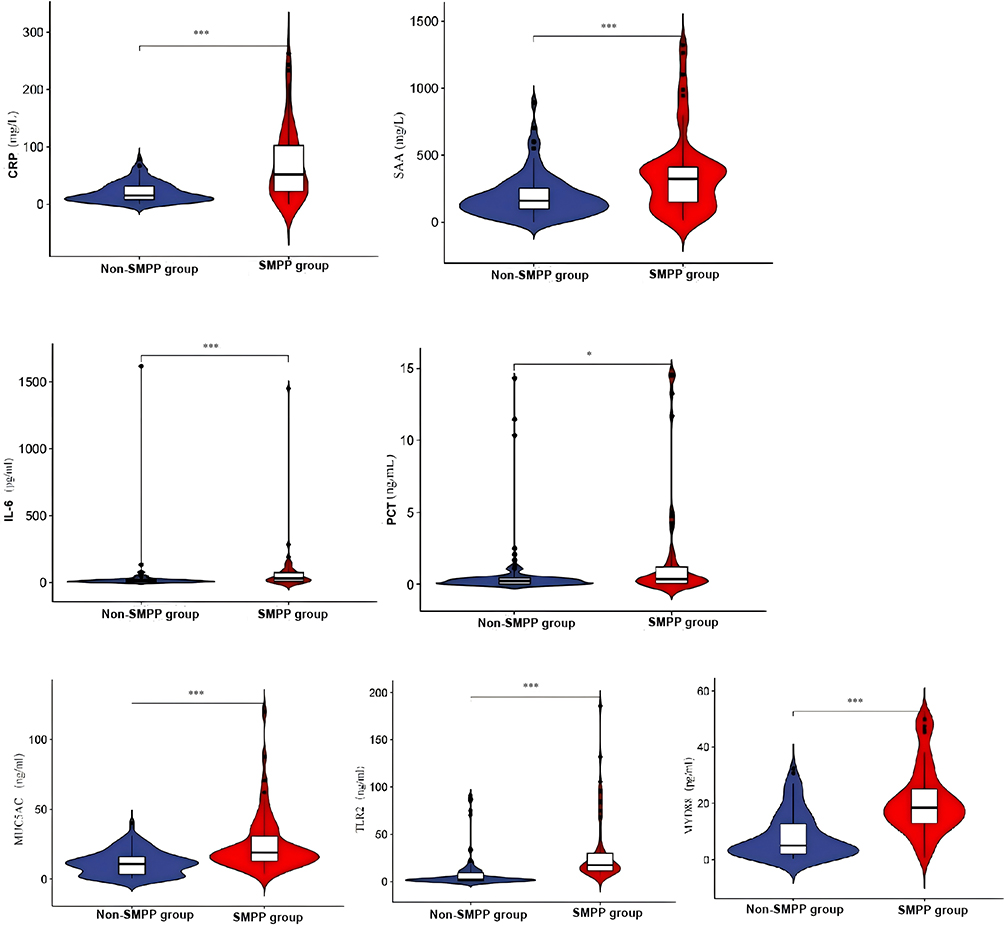 |
Figure 2 Comparison of inflammatory markers and observational indicators between the SMPP group and the non-SMPP group. ***indicates p<0.001, NS indicates P>0.05, * indicates P <0.05. Abbreviations: SMPP, severe Mycoplasma pneumoniae pneumonia; BALF, bronchoalveolar lavage fluid; CRP, C-reactive protein; PCT, procalcitonin; SAA, serum amyloid A; IL, interleukin; MUC5AC, Mucin 5AC, oligomeric mucus/gel-forming; TLR2, Toll-like receptor 2; MyD88, myeloid differentiation primary response. |
Logistic Regression Analysis of SMPP
Univariate logistic regression analysis was conducted with the development of SMPP as the outcome variable. The results showed that the following variables were significantly associated with this outcome: respiratory effort, pericardial effusion, coagulation dysfunction, pulmonary consolidation, pleural effusion, intrapulmonary complications, plastic bronchitis, switching to second-line antibiotics, use of glucocorticoids, drug resistance, length of hospital stay, peak fever, number of days of fever and duration of fever after antibiotic use, as well as certain blood routine indicators, inflammatory markers (CRP, PCT, SAA, etc)., biochemical and coagulation indicators (LDH, D-dimer, etc.) and BALF biomarkers (TLR2, MyD88, MUC5AC) (p < 0.05). Among these, the odds ratio for respiratory effort and certain inflammatory markers was >1, suggesting a positive impact on the development of SMPP; elevated levels of lymphocyte ratio and albumin may reduce the risk of SMPP. There were no statistically significant differences in factors such as gender (p > 0.05) (see Table 2).
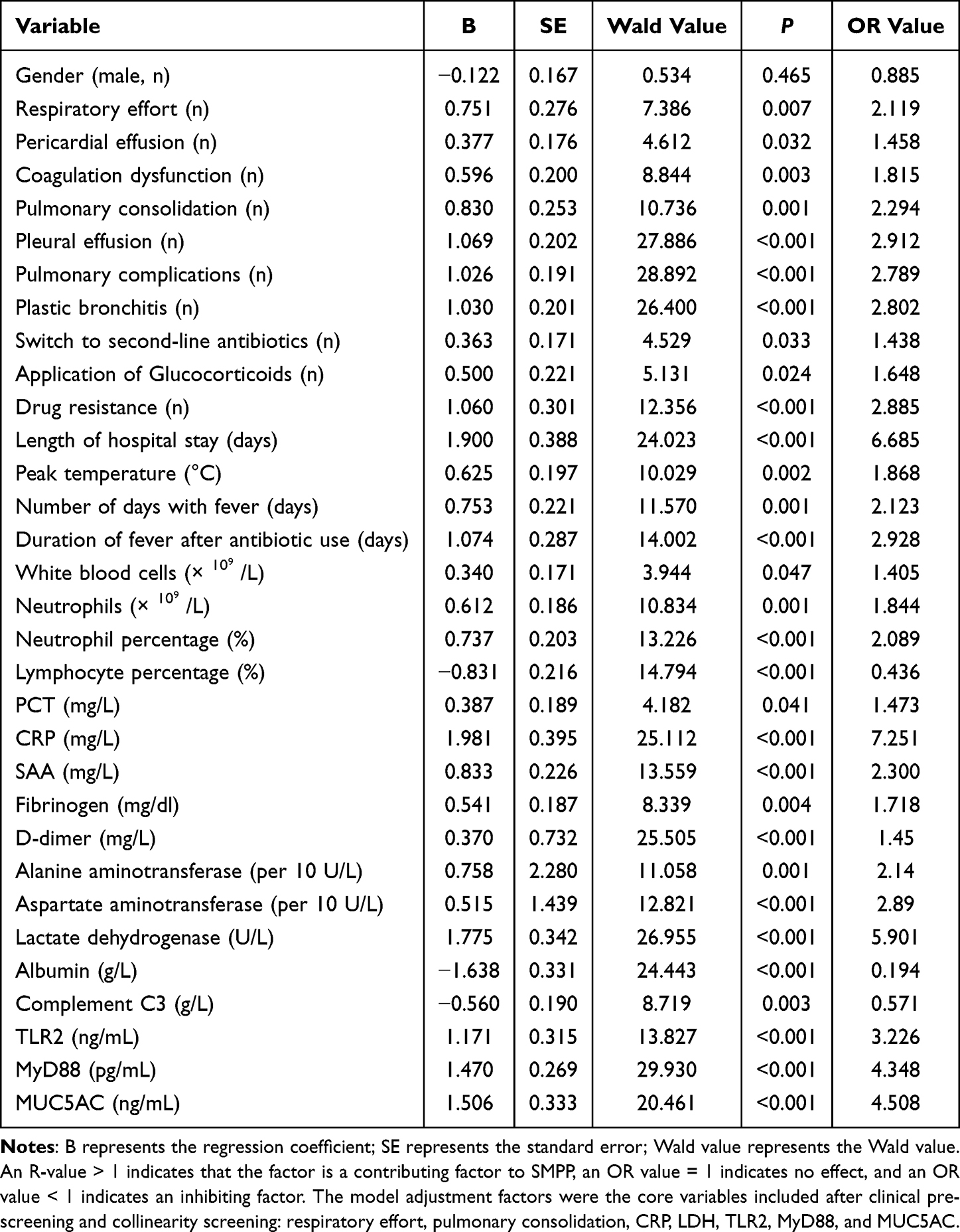 |
Table 2 Univariate Logistic Regression Analysis of the Severity of MPP in Children |
To meet the sample size-to-variable ratio requirement (EPV principle, requiring the number of positive events to be ≥5–10 times that of candidate variables) in multivariate logistic regression and to avoid model overfitting, pre-screening for clinical importance and checking for multicollinearity in indicators with statistically significant differences (p < 0.05) were performed in univariate analysis. Finally, seven core candidate variables with the highest clinical representativeness (exhaustion of breath, pulmonary consolidation, CRP, LDH, TLR2, MyD88 and MUC5AC) were selected for inclusion in the multivariate model. Multicollinearity was tested on these seven variables before they were included in the multivariate regression model. Specifically, the VIF for each variable was as follows: exhaustion of breath (1.37), pulmonary consolidation (3.63), CRP (2.44), LDH (5.13), TLR2 (1.65), MyD88 (3.81) and MUC5AC (3.00). All candidate variables had VIF values <10 (range 1.37–5.13) and corresponding tolerances >0.1 (range 0.19–0.73), clearly indicating the absence of severe multicollinearity among the included independent variables. Multivariate logistic regression analysis using backward stepwise regression revealed that respiratory effort, CRP, LDH, MyD88 and TLR2 were independent risk factors for SMPP (see Table 3).
 |
Table 3 Multivariate Regression Analysis of the Severity of MPP Disease in Children |
Construction and Validation of the SMPP Clinical Prediction Model
Based on the independent risk factors identified through multivariate logistic regression analysis, a joint risk model for predicting the occurrence of SMPP was constructed. The regression equation is as follows: Logit(P) = −7.403 + 2.996 × exhaustion of breathing + 0.044 × CRP + 0.008 × LDH + 0.135 × MyD88 + 0.033 × TLR2.
A multivariate forest plot was plotted based on the model (Figure 3A). Receiver operating characteristic curve analysis showed that the AUC for predicting SMPP by the combined prediction model was as high as 0.950 (95% confidence interval [CI]: 0.912–0.979), and its discrimination was significantly better than that of each individual detection indicator (p < 0.05) (Figure 3B). To evaluate the stability of the model, internal validation was performed using the bootstrap method (1000 resamples). The calibration curve (Figure 3C) showed that the predicted SMPP risk of the model fits well with the actual probability of occurrence, exhibiting high consistency. In addition, decision curve analysis (Figure 3D) showed that applying the combined model to guide clinical decision-making within a wide threshold probability range of 0.01–0.99 can bring considerable net benefits, confirming that the model has excellent clinical practical value (see Figure 3 and Table 4).
 |
Table 4 Value Analysis of Each Indicator in Differentiating Between Severe and Non-Severe Case Groups |
 |
Figure 3 (A) A multivariate forest plot; (B) ROC curve; (C) ROC curve; (D) Decision curve analysis. Abbreviations: ROC, Receiver operating characteristic; DCA, Decision Curve Analysis. |
Discussion
This study focused on the pathogenesis of SMPP in children with MPP, specifically exploring the clinical value of TLR2, MyD88 and MUC5AC levels in BALF. The results showed that TLR2, MyD88 and MUC5AC levels were significantly elevated in children with MPP, with even more pronounced increases in those with SMPP. Further analysis revealed that TLR2 had the highest predictive value (AUC = 0.876), and respiratory effort, CRP, LDH, MyD88 and TLR2 were independent risk factors for SMPP. Based on this, a combined predictive model comprising these five independent risk factors was constructed, achieving an AUC of 0.950 (95% CI: 0.912–0.979), significantly superior to the assessment performance of individual indicators. This result indicates that by combining clinical signs and symptoms, serological indicators reflecting systemic inflammation, and BALF biomarkers reflecting the local lung microenvironment, SMPP risk stratification can be achieved earlier and more accurately, providing a more specific basis for timely clinical intervention.
In recent years, the prevalence of drug-resistant MP strains has led to prolonged courses, increased complications and more severe cases of MPP in children. Studies have confirmed that excessive inflammatory response (cytokine storm) and immune imbalance are key mechanisms for the exacerbation of SMPP. In children with SMPP, inflammatory markers such as CRP, PCT, SAA and IL-6 are significantly elevated, reflecting the intense activation of the inflammatory response in the body.11,13–15 In addition, chemokines such as C-X-C motif chemokine ligand 10 and interferon gamma are significantly elevated in peripheral blood and BALF and are positively correlated with severity indicators, including length of hospital stay and duration of fever.14 Cytokine storm promotes the release of inflammatory mediators and chemokines via signalling pathways such as Janus kinase-signal transducer and activator of transcription 1 and nuclear factor kappa-light-chain-enhancer of activated B cells, creating a positive feedback loop that further exacerbates the pulmonary and systemic inflammatory response.11,14
Severe MPP is not limited to lung injury; it is often accompanied by multi-system damage. Several laboratory indicators, such as coagulation dysfunction (eg elevated D-dimer and FDP), abnormal liver function and elevated LDH, were higher in the SMPP group than in the non-severe group, suggesting that cytokine storm can lead to vascular endothelial damage, microthrombus formation and multi-organ damage.15 Inflammatory mediators, such as high-mobility group box 1, were also significantly elevated in severe and refractory MPP, further aggravating systemic inflammation and tissue damage.16
Studies have confirmed that MUC5AC is the main mucin component of airway mucus, and MP infection can significantly induce high expression of MUC5AC, leading to high secretion of airway mucus and mucus plug formation.10 In animal and cell models, MP upregulates the expression of MUC5AC and MUC5B and downregulates its inhibitor FOXA2 by activating the STAT3/STAT6-EGFR signalling pathway, ultimately promoting increased mucus secretion.17 In addition, inflammatory factors (such as IL-4, IL-6 and IL-13) are significantly elevated after MP infection, further promoting the excessive secretion of MUC5AC.17
This study showed that MUC5AC levels in children with SMPP were significantly higher than in the non-SMPP group and were closely associated with disease severity in univariate analysis. However, it is important to note that MUC5AC was not identified as an independent risk factor for SMPP in multivariate logistic regression. This may be because MUC5AC is a downstream effector of the TLR2/MyD88 signalling pathway, and its independent predictive weight is statistically covered by these upstream core mediators. Nevertheless, its elevated level still directly reflects local airway mucus hypersecretion and stasis, providing clinically valuable information for assessing mucus plug formation and the need for bronchoscopic lavage.18 Nevertheless, the increase in its expression level still directly reflects the deterioration of the local airway microenvironment. In addition, high-risk factors for mucus plug formation also include MRMP infection, strong inflammatory response (elevated CRP and LDH), allergic constitution and delayed use of glucocorticoids.19 The presence of mucus plugs often leads to prolonged hospitalisation, increased complications and poor prognosis.
Following infection with MP, its pathogen-associated lipoproteins are recognised by TLR2, activating MyD88-dependent downstream signalling pathways. This not only triggers the cascade release of pro-inflammatory factors but also directly induces the overexpression of MUC5AC in airway epithelial cells by activating downstream transcription factors.11,20,21 This TLR2/MyD88-mediated excessive immune response and the MUC5AC-driven airway hypersecretion state are mutually causal; the strong inflammatory response exacerbates mucus secretion, and the formation of mucus plugs makes it difficult to clear MP and its metabolites, continuously stimulating TLR2 receptors, thus forming a pathological positive feedback loop, ultimately leading to widespread pulmonary consolidation and airway obstruction in children with mild pulmonary angina pectoris (SMPP). Notably, although univariate analysis and ROC curves indicated that MUC5AC had high single-item predictive value for SMPP (AUC = 0.760), it did not remain an independent risk factor in multivariate logistic regression (p > 0.05). Nevertheless, the significant increase in MUC5AC level still intuitively and objectively reflects the severity of local airway mucus hypersecretion and stasis in the lesion and has irreplaceable reference value for clinical assessment of the risk of mucus plug formation and determination of the timing of bronchoscopic lavage. Animal and cell experiments have further confirmed that inhibiting TLR2 or MyD88 can significantly reduce inflammatory response and airway damage.11
Studies have shown that TLR2 and MyD88 are not only independent risk factors for SMPP but also that their expression levels in BALF are closely related to disease severity. Compared with traditional inflammatory markers such as CRP and LDH, TLR2 has a higher AUC and better sensitivity and specificity in predicting SMPP.11 Combining TLR2/MyD88 with conventional markers is expected to improve the early identification and risk stratification of SMPP. In addition, high expression of TLR2/MyD88 is also closely related to severe manifestations such as radiographic consolidation, atelectasis and pleural effusion, as well as multi-system damage.11,22
Limitations
This study has several limitations that should be acknowledged. First, this was a single-centre prospective cohort study with a relatively modest sample size (151 patients with MPP, including 60 cases of SMPP). The low incidence of certain binary clinical indicators (eg respiratory effort) in the non-severe group led to wide 95% CIs in multivariate logistic regression analysis, which may affect the precision of the effect estimates. Therefore, the clinical predictive weight and accurate effect sizes of these indicators require further validation through multicentre, large-sample studies.
Second, we only measured TLR2, MyD88 and MUC5AC levels in BALF at a single time point upon admission. Serial measurements during different disease stages (eg at diagnosis, after treatment, at discharge) were not performed. This lack of dynamic follow-up limits our ability to assess whether changes in these biomarkers correlate with clinical improvement or progression.
Third, although our combined prediction model showed excellent discrimination (AUC = 0.950), internal validation was performed only using the bootstrap method due to the absence of an external validation cohort. The generalisability of the model to other populations or centres remains to be established.
Fourth, although we used backward stepwise regression and the EPV criterion (60 events for up to seven candidate variables) to reduce overfitting, internal validation using the bootstrap method (1000 resamples) showed good calibration. However, external validation in an independent multicentre cohort is necessary to confirm the generalisability of our prediction model. The AUC of 0.950 may be optimistic due to overfitting, and we encourage future studies to validate the model in different populations.
Despite these limitations, our findings provide valuable insights into the role of BALF TLR2, MyD88 and MUC5AC as potential biomarkers for early identification of SMPP in children.
Further in-depth research into the molecular mechanisms linking the TLR2/MyD88 signaling pathway to MUC5AC overexpression in the airway epithelium of children with SMPP is warranted. Additionally, prospective multi-center studies with larger sample sizes and dynamic follow-up of BALF biomarkers are needed to validate our findings and to explore whether targeted inhibition of TLR2 or MyD88 could serve as a potential therapeutic strategy for SMPP.
Conclusion
This study demonstrates that BALF levels of MUC5AC, TLR2 and MyD88 are significantly elevated in children with MPP, with further increases in those developing SMPP. Among these, TLR2 and MyD88, along with clinical indicators including respiratory effort, CRP and LDH, were identified as independent risk factors for SMPP, whereas MUC5AC was not an independent predictor in multivariate analysis. The combined prediction model incorporating these factors achieved excellent discriminative ability (AUC = 0.950). These findings suggest that BALF TLR2, MyD88 and MUC5AC may serve as useful biomarkers for assessing disease severity; however, given the associational nature of this single‑centre study, external validation in independent cohorts is required before clinical application of the prediction model.
Data Sharing Statement
All data generated or analysed during this study are included in this article. Further enquiries can be directed to the corresponding author (Shujun Li).
Ethics Approval and Consent to Participate
This study was conducted in accordance with the Declaration of Helsinki and approved by the ethics committee of the First Affiliated Hospital of Xinxiang Medical University (Approval No.: Ec-023-155; Approval Date: June 13, 2023). All guardians of the children participating in this study have signed written informed consent forms.
Funding
Joint Construction Project of Henan Provincial Medical Science and Technology Research Program (LHGJ20240487).
Disclosure
The authors report no conflicts of interest in this work.
References
1. Respiratory Group of Pediatrics Branch of Chinese Medical Association, National Clinical Research Center for Respiratory Diseases, Editorial Board of Chinese Journal of Pediatrics. Evidence-based guidelines for the diagnosis and treatment of Mycoplasma pneumoniae pneumonia in children (2023). Chin J Pediatr. 2024;62(12):1137–13. doi:10.3760/cma.j.cn112140-20240722-00503
2. Butpech T, Tovichien P. Mycoplasma pneumoniae pneumonia in children. World J Clin Cases. 2025;13. doi:10.12998/wjcc.v13.i5.99149
3. Wu X, Lu W, Wang T, et al. Optimization strategy for the early timing of bronchoalveolar lavage treatment for children with severe Mycoplasma pneumoniae pneumonia. BMC Infect Dis. 2023;23(1):661. doi:10.1186/s12879-023-08619-9
4. Moynihan KM, Barlow A, Nourse C, et al. Severe Mycoplasma pneumoniae infection in children admitted to pediatric intensive care. Pediatr Infect Dis J. 2018;37(12):e336–e8. doi:10.1097/INF.0000000000002029
5. Méndez-Echevarría A, Calle-Miguel L, Miralbés S, et al. Increased severity of Mycoplasma pneumoniae infections in Spanish children. Pediatr Infect Dis J. 2024;43:1113–1119. doi:10.1097/INF.0000000000004461
6. Yuan L, Diao M, Zhou L. Analysis of the characteristics of mixed infections with Mycoplasma pneumoniae in children. Sci Rep. 2025;15.
7. Wang YS, Zhou YL, Bai G, et al. Expert consensus on the diagnosis and treatment of macrolide-resistant Mycoplasma pneumoniae pneumonia in children. World J Pediatr. 2024;20:901–914. doi:10.1007/s12519-024-00831-0
8. Kyi D, Xiao Y, Wang X, et al. Predominance of A2063G mutant strains in the Mycoplasma pneumoniae epidemic in children-a clinical and epidemiological study in 2023 in Wuhan, China. Inter J Infect Dis. 2024:107074. doi:10.1016/j.ijid.2024.107074
9. Chen B, Gao LY, Chu QJ, et al. The epidemic characteristics of Mycoplasma pneumoniae infection among children in Anhui, China, 2015–2023. Microbiol Spectrum. 2024;12.
10. Kraft M, Adler K, Ingram J, et al. Mycoplasma pneumoniae induces airway epithelial cell expression of MUC5AC in asthma. Eur Respir J. 2008;31:43–46. doi:10.1183/09031936.00103307
11. Chen M, Deng HJ, Zhao Y, et al. Toll-like receptor-mediated signaling induced by Mycoplasma pneumoniae. Front Cell Infect Microbiol. 2022;12.
12. Expert Group of Pediatric Respiratory Endoscopy Diagnosis and Treatment Technology, Talent Exchange Service Center of National Health Commission, Endoscopy Professional Committee of Pediatric Physicians Branch of Chinese Medical Doctor Association, Pediatric Respiratory Endoscopy Professional Committee of Endoscopy Physicians Branch of Chinese Medical Doctor Association, et al. Guidelines for flexible bronchoscopy in Chinese pediatrics (2018 Edition). Chin J Pract Clin Pediatrics. 2018;33(13):983–989.
13. Garne E, Stoll C, Clementi M. Evaluation of prenatal diagnosis of congenital heart diseases by ultrasound experience from 20 European registries. Ultrasound Obstet Gynecol. 2001;17(5):386–391. doi:10.1046/j.1469-0705.2001.00385.x
14. Zou Y, Huang F, Sun J, et al. The role of IFN-γ/CXCL10 axis in Mycoplasma pneumonia infection. Sci Rep. 2025;15.
15. Hu J, Ye Y, Chen X, et al. Insight into the pathogenic mechanism of Mycoplasma pneumoniae. Curr Microbiol. 2022;80. doi:10.1007/s00284-022-03103-0
16. Ding Y, Chu C, Li Y, et al. High expression of HMGB1 in children with refractory Mycoplasma pneumoniae pneumonia. BMC Infect Dis. 2018;18. doi:10.1186/s12879-018-3346-8
17. Hao Y, Kuang Z, Jing J, et al. Mycoplasma pneumoniae modulates STAT3-STAT6/EGFR-FOXA2 signaling to induce overexpression of airway mucins. Infect Immun. 2014;82:5246–5255. doi:10.1128/IAI.01989-14
18. Shi J, Chun L, Hao X, et al. [Dickkopf-1 inhibits the secretion of MUC5AC induced by Mycoplasma pneumoniae P1-C in mouse lung epithelial cells]. Sheng wu gong cheng xue bao. 2023;39(1):248–261. Chinese. doi:10.13345/j.cjb.220513
19. Zhang J, Wang T, Li R, et al. Prediction of risk factors of bronchial mucus plugs in children with Mycoplasma pneumoniae pneumonia. BMC Infect Dis. 2020;21.
20. Fan Y, Ding Y, Li Y, et al. Investigation of the relationship between community-acquired respiratory distress syndrome toxin and the high-mobility group box protein 1-toll-like receptors-myeloid differentiation factor 88 signaling pathway in Mycoplasma pneumoniae pneumonia. Italian J Pediatrics. 2021;48.
21. Shimizu T. Inflammation-inducing factors of Mycoplasma pneumoniae. Front Microbiol. 2016;7. doi:10.3389/fmicb.2016.00414
22. Fan Y, Ding Y, Li Y, et al. Investigation of the relationship between CARDS TX and the HMGB1-TLRs-MyD88 signaling pathway in MPP. Exp Ther Med. 2020;20(1):1–7. doi:10.3892/etm.2020.9163
 © 2026 The Author(s). This work is published and licensed by Dove Medical Press Limited. The
full terms of this license are available at https://www.dovepress.com/terms
and incorporate the Creative Commons Attribution
- Non Commercial (unported, 4.0) License.
By accessing the work you hereby accept the Terms. Non-commercial uses of the work are permitted
without any further permission from Dove Medical Press Limited, provided the work is properly
attributed. For permission for commercial use of this work, please see paragraphs 4.2 and 5 of our Terms.
© 2026 The Author(s). This work is published and licensed by Dove Medical Press Limited. The
full terms of this license are available at https://www.dovepress.com/terms
and incorporate the Creative Commons Attribution
- Non Commercial (unported, 4.0) License.
By accessing the work you hereby accept the Terms. Non-commercial uses of the work are permitted
without any further permission from Dove Medical Press Limited, provided the work is properly
attributed. For permission for commercial use of this work, please see paragraphs 4.2 and 5 of our Terms.