Back to Journals » International Journal of Nanomedicine » Volume 21
Intranasal Nano-Delivery Systems: Emerging Strategies for Central Nervous System Disease Therapeutics
Authors Gao T ![]() , Chu Q, Xing X, Liu Y, Fu S, Liu D
, Chu Q, Xing X, Liu Y, Fu S, Liu D
Received 14 December 2025
Accepted for publication 14 March 2026
Published 18 March 2026 Volume 2026:21 588836
DOI https://doi.org/10.2147/IJN.S588836
Checked for plagiarism Yes
Review by Single anonymous peer review
Peer reviewer comments 3
Editor who approved publication: Dr Kamakhya Prakash Misra
Tong Gao,1,* Qihui Chu,2,* Xiaomin Xing,1 Yuefen Liu,1 Shunli Fu,3 Donghua Liu1
1Department of Pharmacy, The Affiliated Hospital of Qingdao University, Qingdao, 266000, People’s Republic of China; 2Department of Pharmacy, Women and Children’s Hospital, Qingdao University, Qingdao, 266000, People’s Republic of China; 3Tumor Immunology and Cytotherapy of Medical Research Center, Shandong Provincial Key Laboratory of Clinical Research for Pancreatic Diseases, The Affiliated Hospital of Qingdao University, Qingdao, 266000, People’s Republic of China
*These authors contributed equally to this work
Correspondence: Shunli Fu; Donghua Liu, Email [email protected]; [email protected]
Abstract: The rising global incidence of central nervous system (CNS) diseases, exacerbated by the formidable blood-brain barrier (BBB) hindering effective drug delivery, necessitates novel therapeutic strategies. Nasal administration has emerged as a promising non-invasive route, bypassing the BBB via direct neural pathways (olfactory/trigeminal), systemic absorption, or lymphatic drainage. However, inherent nasal barriers like the mucus layer and epithelium limit its efficacy. This review distinguishes itself by integrating mechanistic insights into nasal transport pathways with the rational design of advanced nano-delivery systems. We first outline the challenges in CNS drug delivery and detail the nasal anatomy and transport pathways facilitating nose-to-brain delivery. Subsequently, we emphasize the critical properties required of advanced nano-carriers to improve mucosal penetration, prolong retention, and promote drug accumulation at cerebral injury sites. Following a detailed analysis of the advantages and limitations associated with nose-to-brain delivery, we consolidate recent advances in nasal nano-delivery systems for treating CNS disorders, emphasizing their capacity to improve brain-targeting efficiency, enhance therapeutic efficacy, reduce systemic toxicity, and enable previously undruggable CNS targets. Finally, we expand the discussion to encompass current challenges impeding clinical translation, including safety concerns, manufacturing scalability, and regulatory hurdles, while highlighting emerging trends such as artificial intelligence-driven formulation design. This comprehensive analysis aims to deepen the understanding of nasal-to-brain transport mechanisms and inform the future development of effective nasal formulations for improved neurological therapeutics.
Keywords: nasal-brain, intranasal delivery, nano-delivery systems, central nervous system diseases, blood-brain barrier
Introduction
In recent years, the incidence of brain diseases, including neurodegenerative disorders, cerebrovascular diseases, brain tumors, and neuro-psychiatric disorders, has risen significantly, imposing a heavy burden on society and healthcare systems.1 The development of central nervous system (CNS) therapeutics is associated with a notably high failure rates, attributable to a combination of biological, technical, clinical, and regulatory challenges. Among these, the blood–brain barrier (BBB) has garnered considerable attention as a primary obstacle to effective brain drug delivery.2–4 This dynamic interface, composed of endothelial cells, astrocytes, pericytes, neurons, and tight junctions, selectively permits essential nutrients while excluding exogenous substances, thereby constituting the principal impediment to brain drug delivery.5 Conventional oral administration often fails to achieve therapeutic concentrations in the brain and risks systemic toxicity. Although intraparenchymal, intracerebroventricular, or intrathecal injections enable direct CNS access, their invasiveness precludes long-term use.4 Hence, safe and effective brain-targeted strategies are urgently required.
The nasal cavity possesses unique anatomical features that accommodate diverse designs for CNS drug delivery. Intranasal administration has emerged as a non-invasive alternative that bypasses the BBB by facilitating direct drug transport from the nasal cavity to the brain, while minimizing systemic exposure.3,6 Several nasal formulations have now been approved for treating CNS conditions (mainly migraines and epilepsy), paving the way for a new era in neurological disease treatment.7,8 The representative drugs that have been approved for nasal administration to treat central nervous system diseases, or are currently in clinical trials, are shown in Tables 1 and 2. Common intranasal strategies, such as direct drug absorption or the use of permeation enhancers, exist, they often suffer from poor drug stability, rapid mucociliary clearance, and limited ability to target specific brain regions. In contrast, nano-delivery systems have emerged as a versatile platform capable of addressing these multifaceted barriers. Their tunable physicochemical properties and surface functionalization potential allow for enhanced drug stability, prolonged nasal residence, and improved mucosal penetration, thereby optimizing nose-to-brain transport while minimizing systemic side effects. To date, a range of advanced nanocarriers, including liposomes, polymeric nanoparticles, and nanogels, have been engineered to encapsulate therapeutic agents and incorporate targeting ligands. These systems facilitate efficient navigation across nasal barriers and have demonstrated promise in improving treatment outcomes for CNS disorders such as neurodegenerative diseases and gliomas.
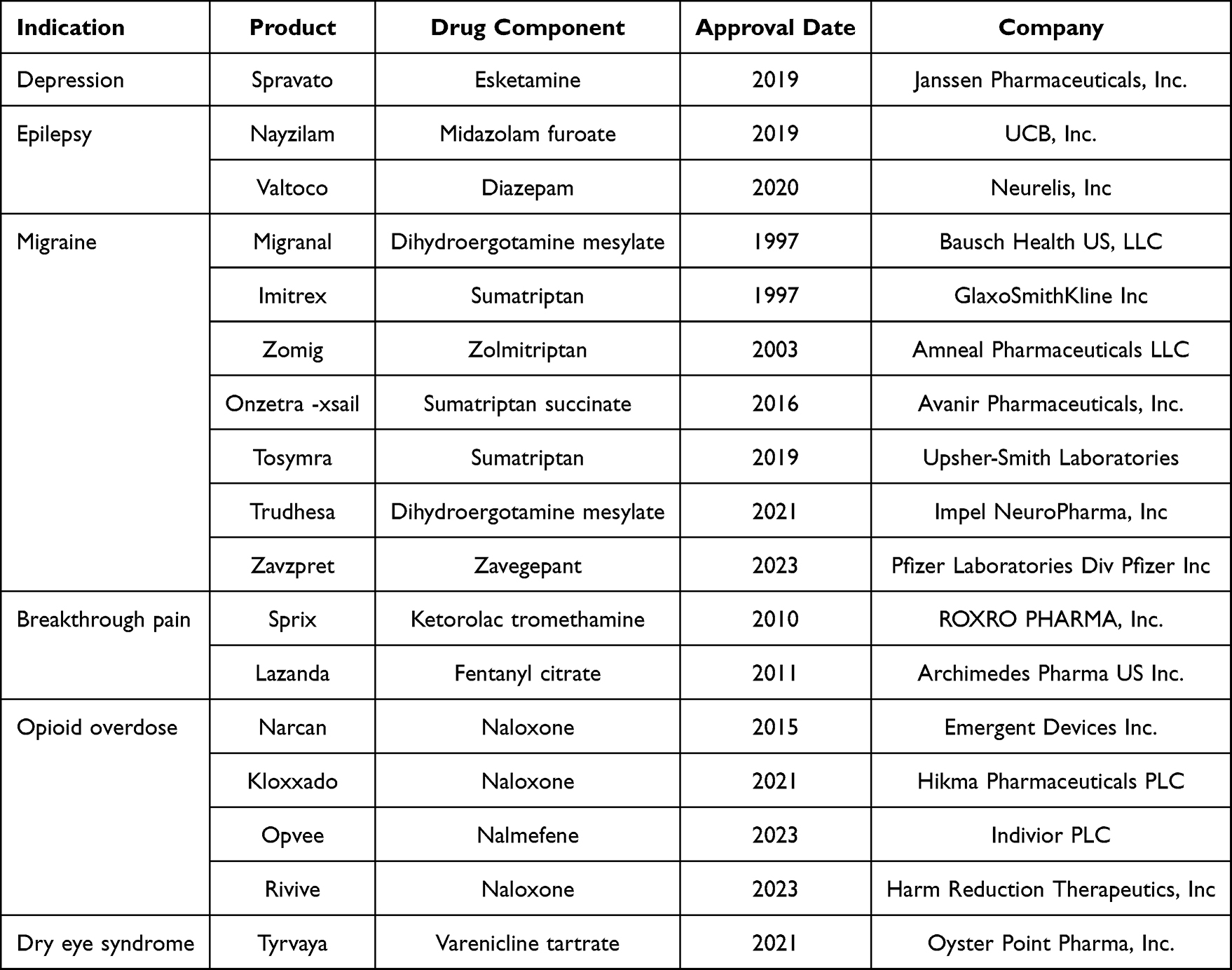 |
Table 1 The Approved Nasal Spray for the Treatment of Central Nervous System Diseases |
 |
Table 2 Clinical Trials Using the Nasal-to-Brain Drug Delivery Method for the Treatment of Central Nervous System Disease |
Distinct from existing reviews that have explored nose-to-brain drug delivery or nanocarrier applications in CNS diseases, this review integrates mechanistic insights into nasal transport pathways with the rational design of advanced nano-delivery systems. Furthermore, we expand the discussion to encompass current challenges impeding clinical translation, including safety concerns, manufacturing scalability, and regulatory hurdles, while highlighting emerging trends such as artificial intelligence-driven formulation design. First, we summarize therapeutic challenges in CNS disorders, comprehensively describe nasal anatomy, and elucidate the complex transport pathways enabling nose-to-brain delivery. Subsequently, we emphasize the essential properties of advanced nasal nano-delivery systems required to overcome mucosal and epithelial barriers while enhancing drug accumulation at cerebral injury sites. Following a detailed analysis of the advantages and challenges associated with nose-to-brain drug delivery, we consolidate recent advances in nasal nano-delivery systems for CNS therapeutics. Finally, we explore the challenges and future directions of intranasal nano-delivery systems. This integrated analysis aims to deepen the mechanistic understanding of transport pathways and inform the development of nasal formulations, ultimately guiding more effective therapeutic strategies for CNS disorders.
CNS Diseases and Challenges in Drug Delivery
Although nasal sprays are approved for treating some CNS diseases, drug delivery for these conditions remains a significant clinical challenge. The unique physiological structure of the CNS has caused most related drug development projects to fail, primarily due to complex, highly regulated barriers that prevent therapeutic agents from reaching brain targets. This challenge is particularly evident at the two critical blood-brain interfaces: the BBB and the blood-cerebrospinal fluid barrier (BCSFB). Even when therapeutic agents successfully bypass or penetrate the BBB and BCSFB through localized delivery, they still face the critical challenge of achieving sufficient diffusion from the entry point to specific target areas within brain tissue (Figure 1).
 |
Figure 1 (A) Representative central nervous system diseases. Schematic diagram of (B) the blood-cerebrospinal fluid barrier and (C) the blood-brain barrier. |
CNS Diseases
Neurodegenerative diseases are a broad category of disorders characterized by the progressive loss and functional impairment of neurons in specific CNS regions, ultimately leading to cognitive, motor, sensory, or autonomic decline. They primarily include Alzheimer’s disease (AD), Parkinson’s disease, Huntington’s disease, and amyotrophic lateral sclerosis.9,10 These diseases typically have an insidious onset, progress slowly, and currently lack curative treatments. Their core pathological mechanism involves the misfolding, aggregation, and deposition of specific proteins, forming characteristic neurotoxic inclusion bodies—such as β-amyloid (Aβ) protein, tau tangles, α-synuclein Lewy bodies, TDP-43 inclusions, and huntingtin aggregates—that disrupt normal cellular function.11 Currently, most neurodegenerative diseases can only be managed symptomatically or have their progression delayed; neuronal loss cannot be prevented or reversed. Effectively delivering therapeutic agents to affected CNS regions remains one of the greatest challenges in treating these disorders, compounded by brain barriers and dynamic pathological microenvironmental changes that increase delivery complexity and unpredictability.12,13
Glioblastoma is a common primary malignant intracranial brain tumor. Its growth compresses surrounding normal brain tissue, nerves, and blood vessels, causing increased intracranial pressure, focal neurological dysfunction, and risk of cerebral herniation. The tumor’s infiltrative growth pattern allows cells to diffuse along nerve fiber bundles and perivascular spaces, resulting in poorly defined boundaries with normal brain tissue.14,15 Furthermore, the inflammatory response present in the tumor microenvironment not only promotes the proliferation and survival of glioma cells but also influences the penetration and efficacy of therapeutic drugs by remodeling the microenvironment.16 Current treatment typically combines multimodal approaches including surgical resection, radiotherapy, and chemotherapy. Due to the brain’s structural heterogeneity, surgical resection often fails to remove all tumor tissue, and these unresectable cells drive the high recurrence rates and therapeutic resistance characteristic of glioblastoma.17,18 Drug treatment faces dual challenges: traditional administration of chemotherapeutic agents may cause systemic toxicity, while physiological changes in brain tumors, including pathological angiogenesis, extracellular matrix alterations, modified local immune composition, and the presence of the BBB, collectively hinder drug delivery to the tumor site.19,20
Ischemic stroke, an acute cerebrovascular event caused by obstruction of cerebral blood supply arteries, results in ischemia, hypoxic necrosis of brain tissue in the affected vascular territory, and corresponding neurological deficits. Accounting for over 80% of all strokes, its pathological process involves abnormal ion gradients, glutamate excitotoxicity, excessive reactive oxygen species production, and mitochondrial dysfunction.21,22 It is noteworthy that, following the restoration of blood perfusion in clinical treatment, some patients may develop cerebral ischemia/reperfusion injury, in which the sudden restoration of blood flow leads to a burst of reactive oxygen species (ROS) and an exacerbated inflammatory cascade, thereby aggravating the initial damage. This phenomenon not only expands the infarct size but also limits the ultimate benefits of thrombolytic therapy. Clinically, antithrombotic agents like aspirin and tissue plasminogen activator demonstrate therapeutic effects but carry bleeding risks due to non-specific distribution and short circulation times. Effectively delivering neuroprotective agents or reparative drugs to damaged brain tissue remains a major treatment challenge. Neuroprotective compounds must reach ischemic penumbral tissue within minutes to hours post-stroke to be effective, yet systemic administration delays BBB penetration. Consequently, overcoming BBB limitations to enable early diagnosis and treatment represents a critical future research direction for ischemic stroke.23,24
Traumatic brain injury refers to structural and functional impairment of brain tissue caused by external mechanical forces to the head, categorized into primary and secondary injuries. It features complex injury mechanisms, dynamic pathological evolution, heterogeneous clinical manifestations, and high long-term disability rates.25,26 Treatment centers on multidisciplinary management, emphasizing airway and circulatory support, intracranial pressure monitoring/control, hematoma evacuation, and complication prevention during the acute phase. The rehabilitation phase requires neuroreparative agents combined with physical, cognitive, and behavioral therapies.27,28 However, drug delivery faces significant challenges: while inflammation may partially open the BBB early post-injury, rapid BBB repair with upregulated efflux pumps subsequently hinders brain entry of large-molecule therapeutics like antibodies and neurotrophic factors. Concurrently, brain edema increases tissue pressure, restricting drug diffusion, while the injury site’s acidic, oxidative, and enzymatic microenvironment accelerates drug degradation. High-dose systemic administration risks systemic toxicity, underscoring the urgent need to develop targeted delivery methods that minimize side effects and enhance therapeutic precision.29,30
Barrier Limitations in Drug Delivery
BBB, a highly selective physiological interface, maintains cerebral homeostasis by regulating nutrient transport while preventing neurotoxic substances from entering brain parenchyma. As one of the most sophisticated biological barriers, it primarily comprises endothelial cells forming tight junctions that interact with perivascular astrocytes, pericytes, and microglia—collectively forming the neurovascular unit that mediates communication between the central and peripheral nervous systems.31,32 This barrier strictly controls molecular transit into the CNS, permitting only passive diffusion of oxygen, carbon dioxide, and lipophilic molecules (<400 Da) through the endothelium.33,34 Cerebral capillary permeability exhibits significantly reduced permeability compared to peripheral vessels, with over two orders of magnitude decrease for small water-soluble molecules and exceeding seven orders for larger molecules.35 Tight junctions restrict paracellular transport, necessitating carrier-mediated mechanisms for carbohydrates, amino acids, and hormones.36 Macromolecules like transferrin, insulin, and leptin undergo receptor-mediated transcytosis, while endothelial ion transporters regulate CNS ion concentrations.37 Crucially, ATP-binding cassette transporters such as P-gp actively efflux drugs, conjugated metabolites, and xenobiotics back into systemic circulation, reinforcing the barrier’s protective function.
BCSFB, while constituting a secondary barrier for drug delivery, exhibits greater permeability than the BBB. Comprising two distinct membranes—the choroid plexus epithelium and the arachnoid membrane (a multilayered fibroblastic envelope surrounding the brain)—the BCSFB features specialized choroidal tissue where ciliated ependymal cells line the ventricular surface for cerebrospinal fluid (CSF) production while enveloping fenestrated capillaries.38 Unlike the BBB, the BCSFB contains perforated vasculature without astrocytic involvement. This barrier critically regulates brain homeostasis through ion/nutrient modulation in cerebrospinal fluid, facilitating choroid plexus-brain signaling while restricting neurotoxic compound entry.39 Notably, BBB-impermeant molecules like sucrose, inulin, and albumin traverse the choroid plexus into CSF at rates inversely proportional to molecular weight.40 Drugs entering CSF may reach brain parenchyma via three routes: 1) systemic reabsorption followed by BBB crossing, 2) direct diffusion through ependymal lining, or 3) perivascular space migration. However, limited convective CSF flow within brain tissue substantially restricts perivascular infiltration into the parenchyma.41
Although therapeutic agents may circumvent or penetrate the BBB and BCSFB through localized delivery, they face the critical challenge of diffusing from entry sites to target regions within brain parenchyma. While neurotherapeutic research predominantly focuses on barrier crossing, intracerebral tissue penetration represents a frequently underestimated yet significant obstacle for CNS drug delivery.42 BBB-permeant lipophilic agents encounter renewed resistance when transitioning from endothelial membrane lipid bilayers to aqueous interstitial fluid. Drug diffusion through the brain’s extracellular space (ECS) is governed by cerebral blood flow, CSF dynamics, interstitial fluid movement, pH gradients, and extracellular matrix composition—while simultaneously constrained by therapeutic agents’ physicochemical properties including molecular size, surface charge, geometry, and molecular weight.43,44 Critically, heterogeneous ECS architecture and anisotropic diffusion patterns throughout brain regions further modulate net drug distribution. Pathological alterations in disease states exacerbate this challenge by modifying the ECS microenvironment, ECM organization, and neurovascular unit integrity. For instance, neuroinflammation induces vascular dysfunction, enzymatic dysregulation, and cellular damage that collectively reconfigure interstitial architecture.45
The Role of the Nasal-Brain Pathway in Drug Delivery
Overcoming barriers like the BBB to deliver therapeutics accurately, efficiently, and safely to brain lesions represents one of the most active research frontiers in neuroscience and drug development. The human nasal cavity comprises three functionally distinct regions—vestibular, respiratory, and olfactory—each with specialized cellular architecture that establishes direct neuroanatomical connections to the brain. This unique anatomical relationship enables brain-targeted delivery, positioning intranasal administration as a promising strategy for bypassing the BBB to deliver drugs directly to the CNS (Figure 2). Consequently, compared to conventional systemic or invasive localized delivery methods, this approach offers enhanced practicality, reduced systemic exposure, rapid onset of action, and superior CNS bioavailability.
 |
Figure 2 Schematic representation of the different pathways to the brain following intranasal administration. |
The Physiological Structure of the Nasal Cavity
The nasal vestibule, located at the nostril openings and extending to the lower end of the lateral nasal cartilage, serves as the supporting structure at the front of the nasal cavity. This relatively spacious region on the inner aspect of the cavity, with a surface area of approximately 0.6 cm2, is lined with a mucus layer and cilia responsible for mucus clearance.46 Its surface is covered by squamous epithelial cells and contains structures such as sebaceous glands, sweat glands, other mucous glands, and nasal hair follicles.47 Due to its limited blood flow, small surface area, and unique cellular composition, drug absorption in this area is highly restricted.48 However, nasal hairs play a crucial role by filtering airborne particles, causing most particulates entering with inhaled air to deposit in the anterior nasal vestibule. This mechanism prevents deeper penetration into the nasal cavity and respiratory system. Studies indicate that vestibular nasal hairs can block nearly all particulate matter larger than PM50, significantly contributing to respiratory health protection.49
The respiratory region, responsible for respiratory function and constituting the largest portion of the nasal cavity with an area of approximately 130 cm2, features the densest vascular network within the nasal passages.50 It comprises four primary cell types: basal cells, goblet cells, ciliated cells, and non-ciliated columnar cells. Basal cells act as reserve cells capable of differentiating into other types when needed.51 Goblet cells secrete mucoproteins, which combine with mucous gland secretions to form the mucus layer. This mucus traps inhaled molecules and transports them toward the pharynx, where the body swallows them, delivering the material to the gastrointestinal tract. Consequently, drugs must penetrate this mucus barrier to reach the epithelial cell surface for absorption.52 Furthermore, both ciliated and non-ciliated columnar cells possess extensive microvilli (and cilia in ciliated cells), a substantial surface area, and dense vascularization. These characteristics collectively establish the respiratory region as a critical site for systemic drug absorption into the circulatory system.51
The olfactory region, extending downward from the nasal cavity roof to the nasal septum and lateral wall, features mucosa with a pale yellow or whitish appearance. Covering approximately 10 cm2 (about 10% of the total nasal cavity area), this region contains roughly 50 million sensory receptor cells and represents the nasal compartment directly interfacing with the CNS.53 Its cellular composition primarily includes supporting cells, basal cells, and scattered microvillar cells, with interspersed olfactory sensory neurons that detect odor stimuli. Rich in olfactory neurons and trigeminal nerve endings, this area possesses neuronal central processes forming unmyelinated nerve fibers.54 These fibers converge into bundles traversing the submucosa, cross-anastomose to create olfactory fila, penetrate the cribriform plate, and ultimately synapse with the olfactory bulb. This unique neuroanatomical pathway establishes a direct conduit between the nasal cavity and the brain, conferring distinct advantages for targeted drug delivery to the CNS. Consequently, the olfactory region is extensively utilized in research exploring methods to bypass the BBB for direct CNS drug administration.55
The Route Through the Nasal-Brain Pathway
The olfactory pathway, comprising the olfactory epithelium, lamina propria, and olfactory bulb, serves as the primary conduit for nose-to-brain drug delivery. Following intracellular transport mechanisms, drugs initially interact with ciliary olfactory receptors on olfactory neuron dendrites, subsequently progressing toward the lamina propria and brain. Upon reaching the lamina propria, drugs traverse neural channels formed by olfactory ensheathing cells, penetrate the cribriform plate via axons and nerve bundles, and ultimately access the olfactory bulb and CSF.56,57 Diffusion within the CSF enables mixing with interstitial fluid for whole-brain distribution. Alternatively, drugs reaching the olfactory mucosa may directly enter the CNS through tight junctions between supporting cells or paracellular pathways between olfactory neurons and supporting cells.58 This facilitates drug absorption in the CNS, CSF, and olfactory bulb—a structure projecting to key brain regions including the piriform cortex, amygdala, and hypothalamus.59 Recognized as the most efficient route circumventing the BBB, this pathway achieves brain delivery within approximately 1–2 hours via intracellular transport, while extracellular routes enable access in as little as 30 minutes.60
The trigeminal nerve, the fifth cranial nerve with ophthalmic, maxillary, and mandibular branches, provides primary sensory innervation to the nasal cavity while establishing a direct conduit between the nasal cavity and brain.61 Through mucosal branches innervating the respiratory epithelium, this pathway enables direct drug delivery to the brainstem and other cerebral regions.62,63 Specifically, the ophthalmic and maxillary nerve branches—after traversing the pons and penetrating the cribriform plate—facilitate drug transport from nasal mucosa to the CNS, promoting drug distribution to both caudal and rostral brain regions.52,64 This pathway employs dual transport mechanisms: intracellular axonal trafficking and extracellular diffusion via perivascular spaces and perineural channels.65 Recognized as a significant alternative nose-to-brain delivery route, the trigeminal pathway exhibits slower transport kinetics compared to olfactory routes, with intracellular transport requiring 17–56 hours as documented in literature.66
The systemic absorption pathway primarily involves the respiratory region, where the mucosa’s dense capillary network and abundant blood flow facilitate drug molecule absorption into the circulatory system.67 Additional systemic entry may occur through the olfactory region’s lamina propria. However, drugs entering systemic circulation must subsequently cross the BBB to reach the CNS, prolonging therapeutic onset and limiting CNS drug delivery. While this process rapidly absorbs low-molecular-weight lipophilic compounds and highly permeable substances via capillary networks, the fraction reaching the CNS remains constrained by the BBB penetration requirement. Consequently, systemic delivery efficiency to the CNS remains severely limited due to this obligatory indirect pathway.51
Substances not entering the bloodstream may access nasal lymphatic vessels in the lamina propria. Beyond peripheral lymph nodes, evidence indicates nasal lymphatics connect to the brain. Although CSF primarily drains via arachnoid granulations, an alternative pathway drains it from the subarachnoid space through channels in the cribriform plate to nasal lymphatics and ultimately to cervical lymph nodes.68,69 PEGylated fluorescent microbead studies revealed direct connections between the subarachnoid space and lymphatics surrounding olfactory nerves where they traverse the cribriform plate to the nasal submucosa. Additionally, lymphatics around the olfactory bulb form an uninterrupted network functionally linked with nasal submucosal lymphatics.70 Recent research identifies the nasopharyngeal lymphatic plexus as a major conduit for CSF outflow to deep cervical lymph nodes; this plexus has unusual valves and short vessels without smooth muscle, whereas downstream deep cervical lymphatics possess typical semilunar valves, longer vessels, and smooth muscle to transport CSF.71 These findings suggest the nasal lymphatic pathway connects with intracranial CSF, providing a potential transport route.
Properties of Nanosystem for Nasal-Brain Delivery
The nasal-to-brain drug delivery route offers a highly advantageous pathway for brain-targeted administration. However, this approach is constrained by the limited surface area of the nasal cavity and the characteristics of the nasal mucosa, which can reduce effective drug absorption. Nano-based intranasal drug delivery is a current research hotspot in the pharmaceutical field, its core value lying in leveraging the unique anatomical connection between the nasal cavity and the brain to achieve direct brain targeting of drugs. This technology opens up new, non-invasive therapeutic possibilities for treating central nervous system disorders such as Alzheimer’s disease, Parkinson’s disease, and depression. To achieve efficient intranasal drug delivery, these nano delivery systems need to possess common characteristics and functionalities that align with the physiological features of the nasal-to-brain pathway, including aspects related to drug formulation and delivery device, nasal residence time, mucosal permeability, and intracranial transport properties.
Formulation and Delivery Equipment
To achieve intranasal drug administration, appropriate delivery devices are essential. Although the nasal cavity possesses a large mucosal surface area, nasal drug delivery is constrained by nasal anatomy and aerodynamics. Particles that are too large typically deposit in the anterior nasal region and are expelled or wiped away, while smaller particles may bypass the nose and enter the lungs.72 Pires et al found that droplets around 10 µm can readily traverse the nasal cavity to reach the lower respiratory tract, whereas 20 µm particles are more likely to be deposited in the anterior region and subsequently retained within the nasal cavity.73 Therefore, a suitable device is required to disperse intranasal formulations into appropriately sized particles for delivery to the posterior nasal region. This ensures droplet adhesion to the nasal mucosa and prevents their entrainment into the lower respiratory tract by the inhaled airstream.
Common dosage forms for nasal drug delivery systems include nasal drops, sprays, gels, and powders. In clinical practice, nasal drops and sprays are the most frequently used forms of intranasal administration. Nasal drops, while the simplest, suffer from inconsistent dosing, often leading to over- or under-administration, which increases side effects or reduces therapeutic efficacy. Compared to drops, nasal sprays offer accurate dosing, uniform drug distribution, and higher bioavailability, making them the most clinically utilized nasal drug delivery dosage form. A spray device consists of three main components: an actuator, a metering valve, and a drug solution bottle. The actuator and metering valve are key components responsible for spray formation. They significantly influence the droplet size, spray pattern, and plume geometry of the nasal spray, directly determining the deposition site and amount within the nasal cavity, thereby affecting nasal drug delivery efficiency.73 The orifice size and shape of the device’s actuator, along with the metering valve’s volume, can influence the spray pattern and plume geometry by affecting the compression force, spray velocity, and frictional forces of the liquid formulation. These factors ultimately alter drug deposition. Foo et al suggest that the plume angle significantly impacts the deposition of nasal sprays within the nasal cavity.74 Moraga-Espinoza et al found that as the plume angle decreases, the amount of drug deposited in the turbinate region gradually increases.75 Pires et al discovered that when the plume angle is 30°, drug deposition primarily occurs in the anterior nasal region, with a deposition rate approaching 90%. A smaller plume angle facilitates the spray traversing the nasal vestibule, reducing deposition in the anterior nasal region.76
Prolong Nasal Retention
Viscosity is a critical factor influencing the droplet size of nasal drug delivery systems. Higher formulation viscosity leads to larger droplet sizes, facilitating drug deposition within the nasal cavity. Furthermore, as the viscosity of the drug solution increases, the residence time of drug adhesion to the nasal mucosa is prolonged, thereby enhancing the likelihood of drug absorption.77 Simultaneously, highly viscous solutions can interfere with normal ciliary beating, inhibiting the nasal mucociliary clearance system and further increasing drug absorption rates across the nasal mucosa. In clinical applications, viscosity can be increased by incorporating mucoadhesive agents. These agents interact with mucus, prolonging the formulation’s residence time within the nasal cavity and consequently improving drug uptake.78 Common mucoadhesive agents include chitosan, polyacrylic acid, and carbomer.66
Among numerous mucoadhesive materials, natural polysaccharides (eg., chitosan) offer excellent biocompatibility and inherent bioadhesive properties. These can confer mucoadhesion to nanoparticles, thereby prolonging drug residence time and enhancing absorption. Sun Yu et al found that the residence time of a methotrexate solution in the nasal cavity was only 15 minutes. However, after increasing the formulation viscosity by adding chitosan, the nasal residence time extended to 1–2 hours. Nevertheless, excessively high viscosity can also hinder nasal drug absorption. Sridhar et al developed chitosan nanoparticles for the nasal delivery of selegiline to treat Parkinson’s disease. Following intranasal administration, the chitosan-based drug-loaded nanoparticles significantly increased the nasal residence time and permeation/absorption of the drug. Drug concentrations in the mouse brain and plasma were 20-fold and 12-fold higher, respectively, compared to the oral administration group. Additionally, improvements were observed in brain dopamine levels, catalase activity, and glutathione levels.79 Furthermore, a chitosan-based in situ gelling system was developed for the nasal delivery of vinpocetine (VIN) to the brain. Pharmacokinetic studies demonstrated significantly enhanced delivery of VIN to the brain, minimizing systemic exposure. Compared to conventional oral administration, the nasal delivery via the chitosan gel system nearly doubled the Cmax (P < 0.05) and AUC0-t (P < 0.05) of VIN in the brain. Histopathological examination of the nasal mucosa revealed no signs of irritation or toxicity, confirming its safety for intranasal administration.80
Promote Transmembrane Penetration
The nasal epithelium consists of a layer of pseudostratified columnar cells interconnected by tight junctions. Small hydrophobic molecules can be absorbed via permeation driven by a concentration gradient. Hydrophilic molecules traverse the lipid bilayer via selective transport systems, while large molecules and polar drugs require overcoming the tight junction structure and utilizing the paracellular transport pathway for absorption.81 Consequently, the bioavailability of most drugs following intranasal administration is low, primarily due to the significant barrier posed by the nasal epithelial permeability barrier.82 Enhancing the efficiency of nasal-to-brain delivery can be approached through two primary strategies. Firstly, permeation enhancers are the most used functional excipients in intranasal formulations. They promote drug penetration through the mucus layer and epithelial cell membranes, thereby improving the brain delivery efficiency of nanocarriers. Secondly, the binding of receptors expressed in the olfactory region to their specific ligands represents another crucial mechanism for facilitating drug transport across the permeability barrier, thereby increasing nose-to-brain delivery.
Permeation enhancers (PEs) increase the permeability of the nasal mucosal epithelium by modulating the phospholipid bilayer of cell membranes, promoting membrane fluidity, or opening tight junctions between epithelial cells to augment the paracellular pathway. This facilitates drug penetration through the mucus layer and epithelial cell membranes, thereby enhancing the brain delivery efficiency of nanocarriers.83,84 The selection of absorption enhancers depends on the drug’s structure and its impact on nasal physiology. Commonly used nasal mucosal permeation enhancers primarily include: surfactants (eg., laureth ethers, bile salts, fatty acids), polymeric enhancers (eg., chitosan, cyclodextrins, gelatin) and novel absorption enhancers (eg., alkyl glycosides, polyethylene glycol 15 hydroxystearate/Solutol HS15). However, surfactants can alter cell structure, leach proteins, and even damage the outer mucosal layer, causing significant irritation to the nasal mucosa. In contrast, some polymeric enhancers that act by opening tight junctions generally exhibit lower mucosal damage and higher safety profiles. Furthermore, novel absorption enhancers are widely employed in clinical intranasal formulations due to their potent permeation-enhancing effects and favorable safety characteristics.
To enhance the nasal-to-brain delivery of nanodrugs across the permeability barrier, the use of biorecognitive ligands is an excellent strategy. The most commonly employed targeting ligands are proteins with receptors in the olfactory region, primarily lactoferrin (Lf) and certain other glycoproteins.85,86 Liu et al constructed lactoferrin-conjugated PEG-PCL drug-loaded nanoparticles via maleimide-thiol chemistry for neural repair. Compared to unmodified nanoparticles, these rapidly traversed the nasal mucosa via lactoferrin receptor-mediated transport, achieving effective drug accumulation in mouse brain tissues including the cerebrum, cerebellum, and olfactory bulb.87 Several lectins, such as potato lectin (STL) and wheat germ agglutinin (WGA), have also been utilized to promote nose-to-brain drug delivery.88 Zhang et al prepared PEG-PLGA drug-loaded nanoparticles (PEG-PLGA-NPs) and modified their surface with potato lectin for targeting. Results demonstrated that intranasally administered STL-NPs rapidly bound to absorption sites in the nasal cavity, significantly increased the area under the curve (AUC) of the drug in the mouse brain, and improved cognitive impairment and spatial memory deficits compared to intravenously injected STL-NPs and free drug solution.89 Furthermore, borneol modification enhances nanoparticle brain penetration by downregulating the expression of ZO-1 and occludin in the nasal mucosa. Wang et al developed borneol-modified tanshinone IIA nanoparticles (Bo-TSA-NPs). With a particle size of approximately 160 nm, Bo-TSA-NPs significantly increased epithelial cell uptake via vesicle/caveolae-mediated endocytosis and micropinocytosis. Following intranasal (IN) administration, Bo-TSA-NPs markedly improved preventive efficacy in a rat model of cerebral ischemia/reperfusion injury, enhancing neurological function scores and reducing cerebral infarct volume.90
Promote Intracranial Transport
Following penetration through the mucus layer and epithelial cell membranes, the majority of drugs and their nanoformulations are transported to the CNS along the olfactory or trigeminal nerve pathways. The fundamental process involves cellular transport, encompassing both intracellular and extracellular routes. The intracellular transport pathway, also termed the intra-axonal neuronal pathway for drug transport, is an efficient route. However, it is characterized by a lengthy transport duration.91 Drugs are first internalized via endocytosis into olfactory sensory neurons (from the olfactory epithelium) or peripheral trigeminal neurons (from the respiratory epithelium). Subsequently, intracellular endocytic vesicles are transported to the projection sites of these neurons, where the drug is ultimately released via exocytosis. Specifically, the intracellular pathway delivers drugs from the olfactory nerve to the olfactory bulb and from the trigeminal nerve to the brainstem.92 Extracellular transport can occur through various mechanisms, all sharing a common principle: the movement of drugs within the fluid-filled spaces surrounding neurons via bulk fluid flow. Drugs traverse the nasal mucosal epithelium to reach the lamina propria. They are then conveyed along neuronal axons towards the CNS through the bulk flow of extracellular fluid, leading to further accumulation and enrichment within the brain.93,94 These transport mechanisms for intranasally administered drugs also provide the theoretical foundation for the design and preparation of nanoformulations.
Modifying the physicochemical properties of nanoformulations can enhance their efficiency for nasal-to-brain delivery. Particle size is the most significant and critical physicochemical factor influencing the nasal-to-brain delivery of drugs. Smaller nano-drug delivery systems encounter less resistance to permeation and migration during intranasal transport, facilitating easier passage to the brain via transcellular or paracellular pathways.95 Mistry et al investigated the efficacy of 100 nm and 200 nm nanoparticles for intranasal delivery. Their results indicated that, due to the constraint imposed by olfactory axonal diameter, the optimal particle size for nano-drug delivery systems transported along the olfactory pathway should be approximately 100 nm.96 Wang et al further confirmed through quantitative studies that smaller nanoparticles more readily achieve distribution across brain ventricles.97 Furthermore, the surface charge of nano-drug delivery systems also influences their brain transport efficiency. Gabal et al, in an intranasal administration study, found that cationic (positively charged) nanostructured lipid carriers (NLCs) exhibited higher plasma bioavailability than anionic (negatively charged) NLCs. However, the brain targeting efficiency of cationic NLCs was lower than that of anionic NLCs.98 Conversely, another fluorescence imaging study demonstrated that both cationic and anionic nanoparticles can migrate to the brain following intranasal administration. Cationic charge may favor transport via the trigeminal nerve pathway, while anionic charge appears more conducive to transport along the olfactory nerve pathway.99 The holistic impact and underlying mechanisms of nanoparticle surface charge (positive vs. negative) on the specific transport routes for nasal-to-brain delivery warrant further in-depth exploration.
Drug delivery characteristics are generally associated with surface coating and interactions with biological systems. Therefore, selecting appropriate ligands for surface modification of formulations can enhance brain delivery efficiency. Cell-penetrating peptides (CPPs), composed of short peptides, polycationic peptides, or amphipathic peptides, can mediate nanoparticle transport into the brain via the olfactory pathway.100 For instance, low-molecular-weight protamine (LMWP), a common CPP, plays a significant role in intranasal delivery. LMWP exhibits low toxicity and can itself serve as a targeting ligand to modify intranasal nanoparticles, enhancing their affinity for brain tissue.101 Xia et al prepared LMWP-modified PEG-polylactic acid nanoparticles. Cellular uptake results demonstrated significantly higher fluorescence intensity of LMWP nanoparticles in cells compared to unmodified nanoparticles. In vivo imaging experiments further revealed that LMWP nanoparticles were efficiently delivered to the brain along both olfactory and trigeminal nerve pathways.102 The human immunodeficiency virus transactivator of transcription (TAT) peptide is another widely utilized cell-penetrating peptide.103 The cell-penetrating property of the TAT peptide is attributed to the guanidinium groups of its arginine residues, which induce electrostatic and hydrogen bonding interactions. Yan et al employed cationic TAT peptide-modified PLGA nanoparticles (NPs) to deliver insulin to the brain. Compared to unmodified NPs, the modified NPs achieved a 6.5-fold higher drug quantity in the olfactory bulb.104
Application of Nanosystem for Nasal-Brain Delivery
Intranasal drug administration offers the significant advantage of bypassing the BBB to deliver drugs to the brain. Currently, the key bottleneck in intranasal-to-brain drug delivery lies in the low drug delivery efficiency to the brain. This inefficiency primarily stems from factors including poor nasal mucosal permeability of drugs, mucociliary clearance, short nasal residence time of drugs, and low bioavailability. Furthermore, achieving precise targeting to specific brain regions remains an additional challenge. Research on intranasal-to-brain nano-delivery systems, however, demonstrates their multiple advantages. These systems can protect encapsulated drugs from biological or chemical degradation. Moreover, surface modification can reduce mucociliary clearance, increase drug residence time in the olfactory region, enhance permeability and bioavailability, and improve active brain-targeting capabilities. In this review, we summarize eight classes of delivery systems, all showing promising potential for enhancing drug delivery from the nasal cavity to the brain (Figure 3 and Table 3).
 |
Table 3 Summary of Nanosystem for Nasal-Brain Delivery |
 |
Figure 3 Summary of nanosystem for nasal-brain delivery. |
Lipid-Based Nanoparticles
Liposomes
Liposomes are biological vesicles composed of a bilayer of phospholipids and cholesterol, enabling the encapsulation of hydrophilic compounds within an aqueous core and hydrophobic compounds within the lipid bilayer. Since both phospholipids and cholesterol are primary components of biological membranes, liposomes exhibit high biocompatibility, safety (characterized by non-toxicity and non-immunogenicity), and complete biodegradability.149 Furthermore, their versatile charge characteristics, adjustable particle size, and capacity for surface modification demonstrate broad application potential in drug delivery. Recent years have witnessed significant advances in utilizing liposomes for nasal-to-brain drug delivery.
Liposomes can accommodate diverse therapeutic agents to enhance nasal delivery efficiency and promote brain tissue accumulation. For instance, encapsulating the natural compound curcumin within cardiolipin liposomes (RCL) yields RCLs@CNPs. Intranasal administration delivers RCLs@CNPs to the brain via olfactory and trigeminal nerve pathways that bypass the BBB. Compared with intravenous delivery, this approach generated a 1.6-fold increase in fluorescence signal from Cy5-labeled RCLs@CNPs in mouse brain tissue. Thereafter, curcumin significantly alleviated AD symptoms in mice by inhibiting Aβ aggregation and preventing polarization of pro-inflammatory microglia toward the M1 phenotype.105 Liposomal encapsulation of protein-based drugs enhances their lipophilicity, prevents enzymatic degradation, and increases cerebral accumulation. Basic fibroblast growth factor (bFGF) shows great potential for preventing vascular dementia (VD). However, the BBB and low bioavailability of bFGF in vivo limit its application. Researchers confirmed that intranasal administration of bFGF-loaded liposomes significantly elevated hippocampal bFGF concentrations in VD mice. Once delivered, bFGF attenuated oxidative stress through regulation of apoptosis-related protein expression and activation of the PI3K/AKT/Nrf2 signaling pathway, thereby reducing neuronal apoptosis induced by repeated ischemia-reperfusion and ultimately ameliorating cognitive impairment in VD mice.106
Liposomes demonstrate significant advantages for genetic drug delivery. Targeted modification enhances their mucosal barrier penetration capacity, while encapsulation provides superior protection for genetic payloads during transit. glioblastoma exhibits malignancy closely associated with elevated expression of the proto-oncogene c-MYC. Wei et al encapsulated c-MYC-antagonizing siRNA in cationic liposomes and applied penetratin-derived peptides via post-insertion modification, constructing a nuclear-shell structured gene delivery system for intranasal brain targeting. This system inhibited c-MYC expression in intracranial tumors following nasal administration, elevated tumor cell apoptosis, and consequently extended the median survival time in glioblastoma model.107 Moreover, liposomes demonstrate utility in delivering nanoagents such as nanozymes to modulate lesional microenvironments. Shan et al developed the KLVFF@LIP-CeO2 therapeutic system by functionalizing liposomes with cerium dioxide nanozymes (CeO2) and surface-conjugating the Aβ-targeting peptide KLVFF. This dual-action platform leverages KLVFF for specific Aβ recognition and aggregation inhibition while employing CeO2 nanozymes to scavenge multiple reactive oxygen species. Following intranasal administration, KLVFF@LIP-CeO2 bypasses the BBB via olfactory and trigeminal neural pathways, achieving efficient targeting to AD lesions. The concomitant reduction of Aβ deposition and attenuation of oxidative stress collectively ameliorate AD pathology, consequently improving cognitive function in APP/PS1 transgenic mice.108
Lipid Nanoparticles
Lipid nanoparticles (LNPs) are lipid-based vesicles featuring a uniform lipid core. Conventional LNPs primarily comprise ionizable cationic lipids, polyethylene glycol (PEG)-modified lipids, cholesterol, and neutral helper lipids.150 This distinctive architecture enables efficient encapsulation and protection of nucleic acid therapeutics. Furthermore, LNPs can achieve targeted delivery to specific cells or tissues through compositional optimization and surface functionalization. Particularly, surface conjugation of BBB-traversing targeting ligands facilitates drug transport across the BBB for treating neurological disorders. These conjugation strategies hold critical implications for expanding therapeutic indications and enhancing treatment efficacy.151
LNPs demonstrate significant advantages for intranasal-to-brain delivery of nucleic acid therapeutics. Jia et al developed an innovative therapeutic strategy employing intranasal administration to efficiently deliver circSCMH1 RNA for repairing post-ischemic stroke brain injury. The authors demonstrated that following intranasal administration, circSCMH1@LNP1 enters the olfactory bulb via olfactory nerves and subsequently distributes to other brain regions. Compared with intravenous delivery, the intranasal route enhanced cerebral distribution of circSCMH1 while reducing nonspecific biodistribution in peripheral organs. A single intranasal dose of circSCMH1@LNP1 in photothrombosis stroke model mice promoted synaptic plasticity, vascular repair, neuroinflammation mitigation, and myelination, significantly improving sensorimotor and cognitive functions.109 Furthermore, targeted LNPs demonstrate promising efficacy in AD therapy. Gao et al developed an innovative intranasal delivery platform utilizing lactoferrin (Lf)-functionalized lipid nanoparticles (LNPs) co-encapsulating α-mangostin (α-M) and β-site amyloid precursor protein cleaving enzyme 1 (BACE1) siRNA (siB). Following intranasal administration, Lf functionalization enabled superior brain targeting via receptor-mediated transcytosis. Therapeutically, α-M reversed Aβ-induced downregulation of low-density lipoprotein receptors, enhancing microglial phagocytosis and autophagic degradation of Aβ, while siB effectively suppressed BACE1 expression to abolish Aβ production. In vivo murine studies revealed significant cognitive recovery, marked reduction in amyloid plaques, and alleviated neuroinflammation and oxidative stress.110
Nanoemulsion
Nanoemulsions are lipophilic systems composed of two immiscible phases (water and oil) stabilized by one or two emulsifiers. They primarily exist as water-in-oil (W/O) or oil-in-water (O/W) types. Lipophilic drugs dissolved in the oil phase can form nanoprecipitates upon release into the nasal cavity. These nanoprecipitates exhibit high specific surface area, enabling rapid dissolution rates.152 Furthermore, incorporating mucoadhesive agents into nanoemulsions overcomes mucociliary clearance, prolonging residence time at nasal absorption sites to enhance bioavailability. Their inherent lipophilicity, nasal mucosal permeability, and drug solubilizing capacity make them particularly effective for improving intranasal-to-brain drug delivery efficiency.
Glioblastoma represents the most malignant and prevalent primary brain tumor. Temozolomide (TMZ), an alkylating agent, is routinely employed against primary and recurrent high-grade gliomas. Luana et al engineered a lipid nanoemulsion (NTMZ) incorporating thermoresponsive Poloxamer 407 polymer to enhance brain-targeted TMZ delivery via intranasal administration. This nanoemulsion exhibits ideal nasal delivery properties—including sufficient TMZ loading, optimal nanosizing—while increasing nasal residence time, prolonging drug release, and enhancing permeation. Biodistribution studies revealed 2.8-fold higher TMZ concentrations in brain tissue following intranasal NTMZ administration compared to free drug treatment, with reduced systemic circulation. Pharmacodynamic evaluation demonstrated significant tumor growth suppression by NTMZ.111 Furthermore, RNA therapeutics have demonstrated significant efficacy in treating diverse diseases, including cancers, infectious diseases, and genetic disorders. However, their potential for neuropsychiatric disorders remains limited by the lack of effective CNS delivery systems. Gao et al developed a novel nanoemulsion platform—violacein-based nanoemulsions (PANEs)—engineered for brain-targeted circDYM delivery to ameliorate depression-like behaviors. PANEs successfully encapsulated circDYM via cationic lipid components while enhancing brain delivery efficiency in mice through their violacein phase. Compared to free circDYM and standard lipid nanoparticle formulations, the optimized PANE2-4 platform substantially enhanced circDYM brain delivery efficiency. In depression models, intranasal administration of PANE2-4-circDYM effectively alleviated depression-like behaviors.112
The review on the application of lipid-based nanoparticles for nasal-brain delivery was summarized as follows. Despite their potential, key challenges for intranasal lipid-based nanoparticles delivery include rapid mucociliary clearance, the selective penetration of the nasal mucosal barrier, and the precise targeting of specific brain regions. However, through rational design of surface properties and formulations, lipid-based nanoparticles offer a significant translational potential for non-invasive and direct brain delivery of neurotherapeutics, potentially revolutionizing treatment paradigms for neurological diseases.
Polymer-Based Nanoparticles
Polymer Nanoparticle
Polymeric nanoparticles are compact colloidal systems composed of natural or synthetic polymers, exhibiting highly tunable size ranges at the nanoscale.153 Therapeutic agents are dissolved, encapsulated, or conjugated within the polymeric matrix. Key advantages include surface hydrophobicity, high drug-loading capacity, and robust controlled-release capabilities. Modulating polymer properties enables precise adjustment of nanoparticle surface charge, drug-loading efficiency, and release kinetics. Furthermore, surface-functionalized polymeric nanocarriers evade immune recognition while achieving targeted binding to specific therapeutic sites.154 Incorporating mucoadhesive polymers into nanoparticle formulations is projected to prolong nasal mucosal residence time, thereby enhancing brain-targeted drug delivery efficiency.155
Poly(lactic-co-glycolic acid) (PLGA) is the most extensively utilized material for fabricating polymeric nanoparticles, with broad applications in intranasal drug delivery for treating CNS disorders. Shah et al prepared lamotrigine (LTG)-loaded PLGA nanoparticles (LTG-PNPs) via emulsion-solvent evaporation. Biodistribution and pharmacokinetic studies revealed a 15.8-fold increase in cerebral AUC of LTG following intranasal administration compared to free LTG. Furthermore, pharmacodynamic evaluation demonstrated that LTG-PNPs significantly prolonged seizure latency, extending it to 3.6 times that of the control group.113 Polymeric nanoparticles also enable co-delivery of therapeutic agents, with targeted modifications enhancing brain tissue penetration. Zhao et al developed borneol (Bo)/R8dGR peptide-modified PLGA nanoparticles co-loaded with curcumin and cisplatin (Cur/Cis), designated BoR-Cur/Cis-NPs for intranasal administration. Borneol functionalization improved nanoparticle brain permeation by downregulating ZO-1 and occludin expression in nasal mucosa, while R8dGR peptide modification conferred targeting specificity through binding to integrin αvβ3 receptors overexpressed on glioma cells. Following intranasal delivery, BoR-Cur/Cis-NPs alleviated hypoxia in the glioma microenvironment, reduced angiogenesis, and consequently prolonged survival in tumor-bearing mice.114 Beyond PLGA, various block copolymers are widely employed in polymeric nanoparticle fabrication for intranasal drug delivery. Specifically, Duan et al leveraged the high biocompatibility and in vivo stability of mPEG-PCL nanoparticles to develop a curcumin-encapsulated nanodelivery system, overcoming curcumin’s poor aqueous solubility and low bioavailability. The intranasal route enabled efficient brain-targeted delivery of curcumin while reducing systemic drug concentrations and adverse reaction incidence. By suppressing pro-inflammatory neuroinflammation and reprogramming microglia from pro-inflammatory to anti-inflammatory phenotypes in post-intracerebral hemorrhage mice, this approach attenuated neuronal pyroptosis and hematoma volume, ameliorated BBB damage, and promoted post-stroke neurological recovery.115
Polymeric Micelles
Polymeric micelles differ from polymeric nanoparticles in their formation through self-assembly of amphiphilic block copolymers, creating a hydrophobic core and hydrophilic shell.156 The hydrophobic core accommodates hydrophobic or poorly soluble drugs, enhancing drug stability, while the hydrophilic segment forms a stabilizing interface between the core and aqueous environment, conferring high kinetic stability to the nanosystem.157 Additionally, the external hydrophilic domain facilitates functionalization modifications to achieve active targeting and environmental responsiveness. Polymeric micelles further prolong nasal mucosal residence time by mitigating mucociliary clearance, thereby enhancing intranasal-to-brain delivery efficiency.158
Polymeric micelles enable intranasal-to-brain drug delivery, a process enhanced by ligand modification. Qi et al developed reactive oxygen species (ROS)-responsive micelles (PP-E micelles) loaded with etoposide (ECH) for nose-to-brain delivery targeting postoperative cognitive dysfunction (POCD). These micelles comprise a copolymer of poly (carboxybetaine methacrylate) segments and phenylboronic acid (PBA)-modified poly(2-(dimethylamino)ethyl methacrylate) segments. In POCD model mice, PP-E micelles recognize the betaine/GABA transporter-1 (BGT-1) on nasal mucosa, facilitating efficient transport across the epithelium via submucosal olfactory and trigeminal pathways with subsequent brain accumulation. By mitigating neuroinflammation and oxidative stress, this system remodels the cerebral microenvironment, synergistically promoting tissue repair and functional neurological recovery.116 Polymeric micelles also enable intranasal co-delivery of therapeutic agents. He et al engineered a multifunctional micellar system utilizing octenyl succinic anhydride (OSA)-modified starch (OSAS), co-loaded with selenium and folate to constitute FA-OSAS-SeNPs for synergistic intervention against neuroinflammatory disorders. This system demonstrated favorable safety and stability profiles. FA-OSAS-SeNPs significantly ameliorated cognitive dysfunction in neuroinflammatory mice while exerting neuroprotective effects through suppression of glial cell activation and attenuation of endoplasmic reticulum stress.117 Polymeric micelles further enable efficient intranasal-to-brain delivery of genetic therapeutics. Yang et al developed core-shell nanostructured HA/DP7-C micelles by encapsulating the cell-penetrating antimicrobial peptide DP7-C within a hyaluronic acid (HA) matrix for siRNA delivery. In vivo studies demonstrated that intranasally administered HA/DP7-C delivered siRNA to the CNS within hours via trigeminal nerve pathways, while HA-CD44 interactions enhanced tumor-specific accumulation. Successful intracellular delivery of anti-glioblastoma siRNA suppressed tumor progression, ultimately prolonging survival and reducing tumor volume in GL261 tumor-bearing mice.118
Dendrimers
Dendrimers are monodisperse macromolecules featuring dendritic architectures formed through iterative branching of oligomeric units linearly connected via branching points. These highly branched polymers typically comprise three structural components: a core, interior dendritic layers, and terminal surface groups. Through controlled generational growth, dendrimers undergo progressive radial expansion, evolving from open dendritic frameworks into closed three-dimensional globular nanostructures at higher generations.159,160 Their unique topology—characterized by internal cavities and surface-concentrated functional groups—combined with precisely tunable physicochemical properties, has established dendrimers as advanced drug delivery platforms extensively applied in pharmaceutical sciences.161
Polyamidoamine (PAMAM) dendrimers represent one of the most extensively studied dendritic macromolecules to date. Jia et al engineered multivalent bioadhesive nanoparticle clusters (BNPs-PAMAM) based on PAMAM dendrimers. This design not only enhances nasal mucosal adhesion but also prolongs drug residence time in the nasal cavity, enabling sustained drug release and efficient brain-targeted delivery while optimizing cerebral pharmacokinetics. By leveraging the direct nose-to-brain pathway, this platform achieves high-efficiency drug transport to the CNS, ensuring neuroprotective agents reach lesion sites prior to reperfusion—thus overcoming the narrow therapeutic window imposed by thrombotic events.119 Dendritic poly-L-lysine represents a synthetically engineered nanomaterial. Yang et al developed dendritic poly-L-lysine nanoparticles co-loaded with BACE1 siRNA and the autophagy activator rapamycin, further functionalized with Aleuria aurantia lectin (AAL) and Aβ-binding peptide KLVFF for intranasal AD therapy. The AAL lectin and KLVFF peptide confer enhanced nose-to-brain delivery efficiency and precise targeting to AD lesion sites. Upon reaching pathological regions, the nanoparticles release BACE1 siRNA and rapamycin to exert therapeutic effects. Experimental studies demonstrated that this system induces augmented autophagy in vitro and in vivo, downregulates BACE1 expression, reduces Aβ deposition, ameliorates neuronal pathology, and rescues memory deficits in AD model mice.120
The review on the application of polymer-based nanoparticles for nasal-brain delivery was summarized as follows. Polymer-based nanoparticles offer a versatile platform for intranasal brain delivery due to their tunable degradation, controlled drug release profiles, and potential for functionalization. However, their clinical translation faces challenges related to potential polymer toxicity, biocompatibility concerns, and the complexity of scalable, reproducible manufacturing. Their significant potential lies in enabling sustained, targeted delivery of neuroprotective agents directly to the central nervous system, offering a promising non-invasive strategy for managing chronic neurological disorders.
Extracellular Vesicles
Extracellular vesicles are nano- to micrometer-sized particles naturally released by cells, characterized by a bilayer lipid membrane structure. Based on their biogenesis and size, they are primarily classified into exosomes, microvesicles, and apoptotic bodies.162 Ubiquitously present in various body fluids, they carry bioactive molecules such as proteins, nucleic acids, and lipids, mediating intercellular communication. Leveraging their inherent source-cell tropism, low immunogenicity, excellent biocompatibility, and ability to cross biological barriers (eg., the blood-brain barrier), extracellular vesicles are being developed as next-generation drug delivery platforms.163 Specifically for intranasal-to-brain drug delivery, exosomes are a focal point of research aimed at enhancing targeting precision and delivery efficiency to the brain.164
Mesenchymal stem cell (MSC)-derived exosomes represent the most extensively utilized vehicles for intranasal-to-brain drug delivery, demonstrating therapeutic efficacy across multiple neurological disorders including AD, Parkinson’s disease, epilepsy, depression, and traumatic brain injury.165,166 Long et al demonstrated that human MSC-derived exosomes (positive for classical EV markers such as CD63 and CD81) administered intranasally penetrate the BBB within 6 hours, achieving precise targeting of hippocampal neurons and microglia. This system orchestrates tripartite therapeutic effects, suppressing neuroinflammation, providing neuronal protection, and promoting neuroregeneration, effectively mitigating seizure-induced hippocampal damage while preventing epilepsy-associated cognitive and memory deficits.121 Sun et al employed adipose-derived mesenchymal stem cell exosomes (ADSC-Exos) to treat depression-like behaviors in mice. Flow cytometry analysis showed that isolated ADSCs exhibited positive expression of Sca-1 and CD44, and negative expression of CD117 and CD31. Following intranasal administration, ADSC-Exos rapidly accumulated in brain tissue, exerting significant neuroprotective effects—including reduced neuronal apoptosis and enhanced autophagy—mediated through AMPK-mTOR pathway activation, ultimately mitigating neuroinflammation.122 Takuma Ikeda et al investigated the therapeutic role of intranasally administered exosomes in hypoxic-ischemic brain injury. Exosomes were isolated from cell supernatants via ultracentrifugation, and the high expression of CD9 and CD63 was detected by Western blot. Subsequently, administer intranasally at a dose of 3×10⁸ particles in 20 μL PBS or PBS alone. One hour post-administration, exosomes were predominantly detected in the olfactory bulb, subsequently distributing to the striatum and midbrain. Exosome-treated mice exhibited significantly improved cognitive function within 28 days post-injury, accompanied by markedly reduced apoptotic cells in the hippocampus and enhanced neuronal survival rates.123
Peng et al engineered a self-guided nanovehicle designated PR-EXO/PP@Cur by loading the curcumin phase into therapeutic MSC-derived exosomes. The exosomes was identified by flow cytometry to express CD29, CD44, and CD105, while they were negative for CD34 and CD45, which met the characteristics of MSCs. Following intranasal administration, PR-EXO/PP@Cur traverses multiple membrane barriers through autonomous navigation, achieving direct cytosolic drug release within target cells. With enhanced drug accumulation at pathological sites, this system executes tripartite synergistic therapy against PD complexity by reducing α-synuclein aggregation, promoting neuronal functional recovery, and mitigating neuroinflammation.124 Furthermore, MSC exosomes can be fused with other nanoparticles to form hybrid nanosystems for co-delivery applications. Jiang et al engineered a hybrid system (TSEL) by integrating MSC exosomes with liposomes to co-deliver two gene therapeutics: β-site amyloid precursor protein cleaving enzyme 1 (BACE1) siRNA (siBACE1) and triggering receptor expressed on myeloid cells 2 (TREM2) plasmid (pTREM2). Exosomes express proteins such as CD81, CD9, CD63, Calnexin, TSG101, and HSP70. Leveraging the exosomal homing capability and Angiopep-2 peptide modification, TSEL efficiently penetrates the BBB, facilitating targeted drug accumulation at AD lesion sites. Upregulated TREM2 expression reprograms microglia from pro-inflammatory M1 to anti-inflammatory M2 phenotypes, concurrently restoring Aβ phagocytic capacity and neurorepair functions. Additionally, siBACE1 suppresses Aβ plaque generation at its transcriptional origin by silencing BACE1, synergistically enhancing the therapeutic efficacy against AD pathology.125
The review on the application of extracellular vesicles for nasal-brain delivery was summarized as follows. Extracellular vesicles present a unique challenge for intranasal brain delivery due to complexities in isolation, standardization, and scalable manufacturing of these endogenous vesicles. Their inherent biocompatibility, low immunogenicity, and natural ability to cross biological barriers underpin their high translational potential, as they can efficiently deliver therapeutic cargo directly to the brain, offering a highly promising avenue for treating neurological diseases.
Hydrogel Delivery Systems
Hydrogels are three-dimensional crosslinked polymer networks synthesized from hydrophilic monomers via covalent or non-covalent interactions. They undergo phase transformation at the administration site, transitioning from a liquid state to a semi-solid gel state.80 These formulations exhibit a sol-gel transition in response to physiological environmental changes at the site of application. Administered as solutions, hydrogels form upon entering the nasal cavity due to stimuli (eg., pH or temperature) that induce conformational changes in the polymers. They exhibit pronounced sustained-release effects, significantly improving nasal residence time and enhancing nose-to-brain delivery efficiency. Consequently, hydrogels are frequently employed for drugs requiring sustained release to achieve therapeutic efficacy. Therefore, hydrogels hold promising application prospects for nose-to-brain delivery of therapeutic agents.167
Thermosensitive gels are widely employed in nose-to-brain delivery due to their rapid sol-gel transition upon contact with the nasal mucosa. This property confers excellent bioadhesion, significantly prolonging drug residence time on the mucosal surface and enhancing drug bioavailability.168 Poloxamers are poly(ethylene oxide)-poly(propylene oxide)-poly(ethylene oxide) (PEO-PPO-PEO) triblock copolymers. Upon temperature increase, the hydrophobic poly(propylene oxide) blocks within poloxamer micelles dehydrate and expand. Upon reaching a critical temperature, the micelles interact and form a network structure, resulting in gelation, most directly evidenced by a sharp increase in viscosity.169,170 Utilizing Poloxamer as the matrix, Xu et al developed a thermosensitive self-assembling hydrogel for intranasal drug administration. At body temperature, the hydrogel adheres to the nasal mucosa, enabling sustained drug release. The released drug enters the brain directly via the olfactory region, bypassing the BBB, facilitating rapid antidepressant effects. This system potentiated the antidepressant activity of the flavonoid compound icariin, while simultaneously reducing the required dosage and dosing frequency, thereby enhancing patient compliance.126
Poly(N-isopropylacrylamide) (PNIPAM) is a classic thermosensitive material. At lower temperatures, the three-dimensional network structure of PNIPAM contains numerous voids occupied by water molecules. These water molecules form hydrogen bonds with the amide groups, creating a hydration layer on the polymer surface. Upon temperature increase, the hydrogen bonds break, the isopropyl groups dehydrate, water content decreases, and the association of hydrophobic groups strengthens. This results in water expulsion and network contraction, leading to gel formation.171 Tang et al prepared a thermosensitive PNIPAM emulsion via free radical polymerization. They loaded this emulsion with magnolol nanocrystals (MAG-NCs) to form a microemulsion (MAG-NCs@emulsion). Subsequent natural cooling induced a self-gelation process, yielding the thermosensitive hydrogel drug delivery system MAG-NCs@Gel. Serving as an intranasal delivery platform, the thermosensitive MAG-NCs@Gel hydrogel facilitates drug transport across the BBB while enabling specific targeting of dopaminergic neurons. It effectively reverses mitochondrial dysfunction, thereby alleviating symptoms in the MPTP-induced Parkinson’s disease (PD) model.127
Chitosan itself lacks thermosensitivity. Thermosensitive behavior is typically imparted by blending it with sodium glycerophosphate. In this system, electrostatic attraction forms between the amino groups on chitosan and the phosphate groups of sodium glycerophosphate. Upon temperature increase, this electrostatic attraction is disrupted, leading to dehydration of the chitosan chains and subsequent gelation. Wang et al developed a thermosensitive hydrogel based on chitosan/β-glycerophosphate (CS/β-GP). They further optimized the formulation by adding 0.1% polyethylene glycol 400 (PEG400). This system serves as an intranasal smart sustained-release depot for efficient and targeted drug delivery. The delivery system undergoes rapid gelation at 34°C. Following intranasal administration, it bypasses hepatic and renal sequestration, overcomes the blood-spinal cord barrier (BSCB), and significantly enhances therapeutic efficacy for CNS injuries.128
Ion-sensitive in situ gels primarily utilize the reversible structural or conformational changes of certain polymeric materials in response to external ionic strength, enabling the transformation from solution to gel. The nasal cavity is rich in cations (eg., Na⁺, K⁺, Ca2⁺). When an ion-sensitive in situ gel is administered intranasally, these ions trigger a sol-gel transition, forming a drug depot with sustained-release functionality. Deacetylated gellan gum (DGG) is an anionic polysaccharide secreted by Sphingomonas elodea. Nasal fluid rich in cations (especially Ca2⁺) transforms DGG solution into a gel. Due to its mucoadhesive properties, the in situ gel prolongs mucosal residence time, maintaining its retention within the nasal cavity, thereby enhancing the bioavailability of intranasally administered drugs. Chen et al selected berberine (BRE) as the model drug. They successfully prepared BRE-loaded nano-gel (BRE-NG) by combining a BRE nanosuspension (BRE-NS) with 0.5% (m/v) gellan gum. BRE-NG exhibited favorable physicochemical properties, including an appropriate sol-gel transition temperature (Tsol-gel), high water retention capacity, suitable swelling index, and sustained-release behavior, along with excellent spreadability and bioadhesion. Owing to the direct nose-to-brain pathway, BRE-NG demonstrated significantly enhanced bioavailability within the brain.129
Schiff base bonds are dynamically reversible chemical linkages primarily based on dynamic interactions, including hydrogen bonding, electrostatic forces, dynamic metal coordination, hydrophobic interactions, supramolecular host-guest interactions, and imine bonds. Within Schiff base-based hydrogels, the recombination of internal dynamic bonds enables gelation. For instance, Andrew Lofts et al developed a sprayable, in situ-forming nanoparticle network hydrogel (NNH) based on Schiff base interactions for the intranasal delivery of lithium to the brain. This system consists of oxidized starch nanoparticles (SNPs) and carboxymethyl chitosan (CMCh), enabling in situ gel formation within the nasal cavity to prolong lithium release. By incorporating chelating agents such as citric acid, ethylenediaminetetraacetic acid (EDTA), and diethylenetriaminepentaacetic acid (DTPA), the hydrogel’s mechanical properties were enhanced, effectively extending lithium’s release profile within the brain. In an amphetamine-induced rat model of bipolar disorder, the lithium-chelated hydrogel (Li@EDTA-NNH) demonstrated significantly prolonged behavioral suppression compared to conventional lithium administration methods.130
Self-assembling peptide hydrogels constitute a specific class of hydrogels formed through non-covalent interactions between peptides, such as ionic bonds, hydrophobic interactions, hydrogen bonds, and van der Waals forces. The peptides spontaneously aggregate to form highly ordered nanoscale fibers, which subsequently entangle into a three-dimensional network structure. These hydrogels hold broad application prospects in the biomedical field. Huang et al synthesized the KPK protein-derived peptide (KSLSLSLGPASLSLSLK). By mixing a KPK sucrose solution with an equal volume of phosphate-buffered saline (PBS), they constructed a transparent self-assembling hydrogel capable of adhering to tube walls. Loaded with mesenchymal stem cell-derived extracellular vesicles (MSC-EVs) and administered intranasally, this peptide-based hydrogel facilitated the sustained and temperature-modulated release of MSC-EVs. This significantly prolonged the retention time of MSC-EVs and potentiated their therapeutic efficacy for AD.131
The review on the application of hydrogel delivery systems for nasal-brain delivery was summarized as follows. Hydrogel delivery systems face challenges for intranasal brain delivery primarily due to the precise formulation required to balance strong mucoadhesion with rapid nasal clearance, and potential impediment of drug diffusion from the gel matrix to the olfactory epithelium. Their transformative potential lies in providing prolonged residence time at the nasal mucosa, enabling sustained and localized release of therapeutics, which significantly enhances the window for direct nose-to-brain transport, particularly beneficial for chronic neurological conditions.
Micron Delivery System
Microspheres
In recent years, microspheres (MS), as a novel class of drug delivery carriers, have been widely applied in biomedical fields and beyond. Microspheres refer to particles with diameters on the microscale, typically composed of natural or synthetic polymers, exhibiting excellent biocompatibility and degradability.172 Research on particles within the micrometer range (1–1000 μm) for drug delivery has increased significantly in recent years. A key characteristic of micrometer-scale drug delivery systems is their ability to function locally, thereby holding promise for reducing the systemic toxicity and side effects associated with conventional administration. Zhu et al enveloped liposomes containing the natural anti-inflammatory drug Timosaponin B-II (TB) onto barium titanate nanoparticles (BTO), forming LTO@TB. Utilizing microfluidic technology and Schiff base reactions, they encapsulated LTO@TB within microspheres (MS) modified with aldehyde groups and methacrylate, constructing an ultrasound-responsive intranasal formulation designated MS@LTO@TB. The aldehyde groups on MS@LTO@TB spontaneously form amide bonds with the abundant amino groups in the nasal mucosa, enabling specific adhesion. Benefiting from its strong bioadhesion and efficient transmembrane transport capabilities, LTO@TB is continuously and non-invasively delivered to the brain following intranasal administration. Furthermore, under ultrasound stimulation, the delivered LTO@TB exerts an electro-pharmacological coupling effect within the brain, achieving non-invasive electrical stimulation of damaged neurons. MS@LTO@TB modulates microglial phenotype, restores electrical signal transmission between impaired neurons, remodels the inflammatory microenvironment, reduces neuronal apoptosis, activates the PI3K/AKT signaling pathway, and promotes axonal regeneration. In a mouse model of middle cerebral artery occlusion/reperfusion (MCAO/R), MS@LTO@TB also demonstrated unique capabilities in mitigating inflammation and promoting neuronal remodeling.132
Microneedles
Furthermore, microneedles represent an emerging transdermal drug delivery approach. Comprising a base and an array of microprojections typically ranging from 100 to 1000 μm in length, they facilitate the delivery of drugs and their formulations into the epidermal or dermal layers for therapeutic action. Analogous to the skin’s stratum corneum barrier, the nasal mucosal barrier constitutes one of the rate-limiting steps for nasal drug absorption. Consequently, utilizing microneedles for active intranasal administration holds promise for overcoming mucociliary clearance and the nasal mucosal barrier, thereby enhancing brain delivery.133,173 To facilitate nasal administration, Ruan et al developed a brush-shaped microneedle system. This system employs tannic acid-modified gelatin as the base and hyaluronic acid as the microneedle matrix. It dissolves completely within nasal mucosa within seconds, eliminating concerns regarding damage to nasal cilia or the resident microbial community. Through this innovative approach, lactoferrin-decorated, stigmasterol-reinforced cyclodextrin metal-organic frameworks (CD-MOFs) encapsulating huperzine A (HupA) were delivered directly and efficiently to the brain. This significantly enhanced the therapeutic efficacy in PC12 cell injury models induced by H2O2 and scopolamine. Additionally, this delivery system modulated the expression of two critical therapeutic targets in AD: it significantly attenuated the hyperphosphorylation of Tau protein and reduced Aβ levels. In summary, this ingenious device pioneers a novel pathway for efficient brain drug delivery.133
The review on the application of micron delivery systems for nasal-brain delivery was summarized as follows. Micron-scale delivery systems like microneedles and microparticles face challenges in intranasal brain targeting, including the limited nasal cavity volume restricting administration volume and the need for precise dosing to reach the olfactory region without causing irritation. However, their transformative potential lies in the ability to enhance mucosal adhesion for prolonged residence, physically bypass the mucociliary barrier (especially with dissolving microneedles), and serve as versatile carriers or depots for both small molecules and large biologics. These systems offer a promising strategy for non-invasive, direct nose-to-brain delivery, showing particular efficacy in preclinical models of neurodegenerative diseases like Alzheimer’s.
Protein Nanoparticles
Protein-based nanocarriers possess exceptional properties for targeted drug delivery, including highly ordered structure and symmetry, excellent biocompatibility and biodegradability, and optimal delivery dimensions. Notably, human serum albumin (HSA) features internal binding sites and surface functional groups enabling simultaneous carriage of multiple therapeutic moieties for multifunctional treatment. Furthermore, HSA can be engineered into drug-loaded nanoparticles under mild conditions through diverse methodologies.174 HSA-based drug delivery systems have garnered increasing attention in recent years owing to their biodegradability, non-immunogenicity, biocompatibility, and prolonged half-life.
Serum albumin nanoparticles demonstrate significant potential for enhancing brain accumulation via nose-to-brain drug delivery. In vivo studies reveal that intranasal administration of serum albumin-based R-flurbiprofen (R-FP) nanoparticles achieves a higher brain-to-plasma ratio compared to both intranasal and oral delivery of free R-FP solution. Moreover, at physiologically relevant brain concentrations, R-FP albumin nanoparticles significantly improved basal and maximal mitochondrial respiration in treated cells. Consequently, intranasal serum albumin-based nanoparticles represent a viable strategy for delivering therapeutic agents to the brain to mitigate mitochondrial dysfunction in AD.134 Zhang et al synthesized phenytoin sodium (PHT)-loaded bovine serum albumin-layered double hydroxide nanoparticles (BSA-LDHs-PHT) via coprecipitation-hydrothermal methods for seizure control. Biodistribution assays following intranasal administration demonstrated BSA-LDHs’ exceptional brain-targeting capability, with the BSA-LDHs-Cy5.5 group exhibiting twice the fluorescence intensity of the BSA-Cy5.5 group. This enhanced brain delivery efficiency arises from the hydroxide layers of LDH nanoparticles protecting payloads against enzymatic degradation, thereby improving drug release efficiency and reducing off-target effects, while concurrently enabling rapid brain access via extracellular transport through olfactory pathways.135
Furthermore, the physicochemical properties of albumin nanoparticles can be modulated through the incorporation of various chemical agents to enhance drug-loading capacity and intranasal-to-brain delivery efficacy. Barbara Luppi et al demonstrated that albumin nanoparticles complexed with native and hydrophilic β-cyclodextrin (β-CD) derivatives enable the development of mucoadhesive nasal formulations with enhanced drug permeation characteristics. Results indicated spherical nanoparticles with average sizes <300 nm, which maintained negative surface charges post-drug loading. Crucially, the presence of distinct β-cyclodextrins within the polymeric network not only influenced drug payload during nanoparticle fabrication but also differentially modulated mucoadhesive properties and drug permeation behavior.136 Gábor Katona et al optimized human serum albumin (HSA) nanoparticles loaded with meloxicam (MEL) using Quality by Design (QbD) methodology for intranasal-to-brain delivery. Results demonstrated that the incorporation of Tween 80 significantly enhanced MEL dissolution and permeability. In vivo studies confirmed trans-epithelial and axonal transport of nanoparticles, with intranasal administration achieving substantially elevated MEL brain concentrations compared to intravenous or oral delivery.137
The review on the application of protein nanoparticles for nasal-brain delivery was summarized as follows. Protein nanoparticles face significant challenges for nose-to-brain delivery, including the inherent instability and susceptibility to enzymatic degradation of proteins, as well as their typically high hydrophilicity, which hinders efficient transport across the nasal mucosal and olfactory epithelial barriers. Their major translational potential lies in their excellent biocompatibility, biodegradability, and the ease of precisely engineering their surface with functional ligands (eg., cell-penetrating peptides, targeting molecules) to enhance mucosal adhesion, promote cellular uptake, and achieve specific brain targeting. Furthermore, they serve as a versatile platform not only for delivering other therapeutic proteins but also for protecting and transporting sensitive biomolecules like peptides and nucleic acids directly to the central nervous system.
Inorganic Nanoparticles
Metallic-Based Nanoparticles
Despite favorable biocompatibility and biosafety of organic nanosystems, challenges persist including long-term storage instability, insufficient payload capacity, and batch-to-batch variability. Inorganic nanomaterials represent an alternative for drug delivery, offering enhanced stability and unique electromagnetic properties. Recently, inorganic nanocarriers fabricated from metals, metal oxides, and magnetic materials have gained traction for intranasal diagnosis and treatment of CNS disorders. Notably, metallic nanoparticles typically exhibit smaller dimensions than polymeric or lipid-based counterparts, thus conferring distinct advantages for traversing the BBB.175,176
Intranasal administration has gained significant attention as a strategy to bypass the BBB and deliver therapeutic agents directly to the brain. Utilizing this approach for metal nanoparticle delivery enables targeted imaging in specific brain regions. Gold nanorods (AuNRs) exhibit unique optical and biological properties due to their high aspect ratios, distinguishing them from other gold nanostructures.177,178 Han et al employed comprehensive analytical and imaging techniques to investigate the distribution of AuNRs in discrete murine brain regions and evaluated their potential as a CNS drug delivery platform. AuNRs functionalized with either the fluorescent dye Cyanine5 (Cy5) or the metal chelator diethylenetriamine pentaacetic dianhydride (DTPA anhydride)—conjugated to indium-111 via PEG spacers—facilitated optical and nuclear imaging, respectively. Rapid AuNR uptake in mouse brains was observed within 10 minutes post-intranasal administration, gradually decreasing over time. Autoradiography of sagittal brain sections revealed AuNR entry through the olfactory bulb, followed by dispersion to other brain regions within 1 hour. Critically, gold nanoparticles (AuNRs) were detected in glioblastoma after intranasal delivery, paving the way for their application as nose-to-brain drug carriers targeting a spectrum of CNS disorders.138 Furthermore, gold-iron nanocomposites demonstrate broad applicability in intranasal-to-brain drug delivery and cerebral imaging. Sukumar et al employed gold-iron oxide polymeric metallic nanoparticles to co-deliver miRNA for adjuvant chemotherapy of glioblastoma with temozolomide. In vivo fluorescence and magnetic resonance imaging (MRI) revealed efficient miRNA accumulation in murine brains following intranasal administration, leveraging the size advantages of metallic nanoparticles.139 Separately, Satoshi Seino engineered iron oxide-gold nanoparticle conjugates with additional surface modifications via Au-S bonds without compromising magnetic properties. Magnetization measurements confirmed migration of these PEGylated magnetic nanoparticles to the brain post-intranasal delivery. Moreover, targeted accumulation at specific sites was visualized using probe molecules immobilized at PEG terminals. This work paves the way for significant contributions to magnetic particle imaging of discrete brain regions.140
Non-Metallic Nanoparticles
Non-metallic nanoparticles possess unique physicochemical properties, including quantum confinement effects, enhanced surface-area-to-volume ratio, and tunable optoelectronic behavior, which confer exceptional adsorption capacity and catalytic activity. The physicochemical properties of inorganic nanoparticles are relatively stable, enabling them to protect drugs from degradation within the nasal environment—such as enzymatic degradation and damage from pH variations—thereby preserving drug activity and prolonging therapeutic duration. Furthermore, inorganic nanoparticles can serve as sustained- and controlled-release carriers for drugs. By modulating the structure and properties of the nanoparticles, they facilitate the slow release of drugs, prolonging therapeutic effect, reducing dosing frequency, and enhancing patient compliance.179,180
Mesoporous silica nanoparticles (MSN) possess a honeycomb-like porous structure containing hundreds of channels, endowing them with the potential to absorb molecules. Unlike conventional nanoformulations, MSN exhibit high surface area, large pore volume, and the additional advantage of tunable pore size within a very narrow size distribution.181 They also demonstrate exceptional physicochemical stability and drug-protective capabilities, which can reduce the degradation of peptides, proteins, and other therapeutics during nose-to-brain delivery, thereby enhancing brain delivery efficiency.182 Current research has successfully loaded ponatinib into MSN. Results demonstrate that the brain concentration of ponatinib in the MSN group was 8.9 times higher than that in the free drug group. This signifies a significant improvement in nose-to-brain delivery efficiency and demonstrates significant therapeutic advantages in the treatment of glioblastoma.141 The modifiable surface of MSNs confers advantages in promoting nasal mucosa adhesion and transcellular transport. For instance, Du et al developed a triple-functional nanoparticle (NADH@HMONs-AAL). This nanomaterial utilizes mesoporous silica as the core carrier, loaded with Nicotinamide Adenine Dinucleotide (NADH)—a therapeutic agent known to mitigate neural damage—and is surface-modified with the agglutinin protein (AAL). The AAL protein not only acts as a pore-capping agent to prevent drug leakage but also enhances the adhesion of NADH@HMONs to the nasal mucosa and facilitates their cellular internalization efficiency. Following intranasal administration, NADH@HMONs-AAL can directly enter the brain via the nose-to-brain pathway, traversing the olfactory nerve and cribriform plate. This non-invasive administration route overcomes the long-standing challenge of drug delivery imposed by the BBB, significantly enhancing drug uptake in the brain.142
Carbon-based nanomaterials, as a significant component of inorganic non-metallic materials, are widely employed in nose-to-brain drug delivery.183,184 Hu et al developed a photo-magnetically responsive intranasal delivery system composed of carbonized MIL-100 frameworks (CFs) and domperidone (DP), denoted as CFs@DP, which can reach the brain via nasal administration. In vivo studies demonstrated that after 10 days of treatment, an increase in DR density was observed in the prefrontal cortex and hippocampus of mice. Under dual stimulation from near-infrared (NIR) irradiation and catecholamine-induced complexation, the system disintegrated to release iron ions and DP. This subsequently triggered the upregulation of dopamine type 1 (D1) and type 2 (D2) receptors, as well as brain-derived neurotrophic factor (BDNF), resulting in antidepressant-like effects and enhanced cognitive function.143 Furthermore, Liu et al employed black phosphorus (BP) as both a drug carrier and an antioxidant, loading it with the tau protein aggregation inhibitor methylene blue (MB) to obtain BP-MB. Subsequently, a thermosensitive hydrogel was prepared by crosslinking carboxymethyl chitosan and aldehyde-modified Pluronic F127 (F127-CHO) micelles. The BP-MB nanocomposite was then incorporated into this hydrogel to yield BP-MB@Gel. BP-MB@Gel can be administered intranasally (IN), providing high nasal mucosal retention and controlled drug release. Following IN administration, BP-MB is sustainably released and delivered to the brain, exerting synergistic therapeutic effects. This occurs by inhibiting tau neuropathology, restoring mitochondrial function, and alleviating neuroinflammation, thereby promoting enhanced cognitive performance in a mouse model of AD.144
The review on the application of inorganic nanoparticles for nasal-brain delivery was summarized as follows. Inorganic nanoparticles face challenges in nose-to-brain delivery due to potential long-term toxicity, complex clearance pathways, and the need for sophisticated surface modifications to enhance biocompatibility and prevent aggregation in the nasal environment. However, their inherent properties such as controllable size, multifunctionality, and capability for simultaneous imaging (eg., MRI, CT) and therapy offer significant translational potential for theranostic applications in neurological disorders. Their surfaces can be precisely engineered with targeting ligands to facilitate transport across the nasal epithelial barrier, enabling direct and image-guided delivery of therapeutics to the brain.
Others
Beyond the aforementioned conventional carriers, the continuous advancements and contributions from diverse interdisciplinary fields have led to the emergence of innovative approaches for targeted drug transport in recent years. Furthermore, ingenious carrier designs have opened new avenues for efficient nose-to-brain drug delivery.
Crystals are structures composed of microscopic units (atoms, ions, molecules, etc) arranged in an ordered, periodic pattern. Crystalline nanoformulations (NCs) refer to drug delivery systems comprised of nano-sized crystals. Intranasally delivered nanocrystals can enhance drug absorption by increasing the contact area with the nasal mucosa and prolonging nasal residence time. They also facilitate drug permeation across the mucosal barrier, thereby improving brain delivery efficiency.145 Wu et al developed paeoniflorin nanocrystals (PA-NCs) suitable for intranasal administration in Parkinson’s disease treatment. These nanocrystals promote both mucosal permeation and bypassing of the BBB, leading to elevated brain concentrations of PA. PA-NCs also significantly enhanced the uptake and transport of PA across Calu-3 cells. Furthermore, intact NCs were internalized into Calu-3 cells. The elevated PA levels in the brain confirmed the targeting efficiency of intranasally delivered NCs.145 Tetrahedral framework nucleic acids (tFNAs), composed of nucleotides (each consisting of a base, a sugar, and a phosphate group), stand out due to their excellent biocompatibility and multiple internal and external modification sites. Zhang et al proposed a highly efficient nose-to-brain delivery system based on tetrahedral framework nucleic acids (tFNA, size ≈10 nm). Its high-density negatively charged surface (zeta potential ≈ −35 mV) enables mucoadhesion avoidance, facilitates rapid penetration through the mucus layer, and achieves broad distribution throughout the CNS along olfactory and trigeminal nerve axons. By loading the neuroactive peptide (NAP) for the treatment of sepsis-associated encephalopathy (SAE), it was demonstrated that tFNA-NAP modulates microglial activity, reduces neuronal damage, and exhibits no adverse effects in both short-term and long-term safety studies. This system pioneers a novel non-invasive therapeutic strategy for CNS diseases.146
Furthermore, carrier-free nanoparticles provide novel insights for the development of brain-targeted delivery systems, offering potential for multi-target therapy and demonstrating promising clinical translation prospects. Yang et al developed a carrier-free nanoregulator (NanoDS) through the self-assembly of donepezil and simvastatin for direct nose-to-brain delivery. This approach enables rapid and efficient penetration across the nasal epithelial barrier, facilitates drug release, and achieves multi-therapeutic outcomes. The nanoregulator enhances neurotransmitter transmission, reduces Aβ plaque deposition, reverses oxidative stress status, and remodels the pathological brain microenvironment, thereby restoring cognitive function and alleviating memory impairment.147 Chen et al constructed an intranasally administered, ROS-responsive, carrier-free gene delivery nanosystem, denoted FTBR-NAC. In this system, fingolimod and a biguanide compound were conjugated via a ROS-responsive linker to form FTBR nanoparticles carrying small interfering RNA targeting β-secretase 1 (siBACE1). The cationic nature of FTBR synergized with the mucolytic activity of N-acetylcysteine (NAC) to enhance brain delivery. Upon reaching the brain, FTBR responds to elevated ROS levels at pathological sites, releasing siBACE1 and fingolimod. Administration of FTBR-NAC ameliorated cognitive function in AD mice, demonstrating the high therapeutic efficiency of this relatively simple nanosystem.148
The review on the application of other nanoparticles for nasal-brain delivery was summarized as follows. The exploration of non-canonical nanocarrier architectures offers innovative solutions to overcome the limitations of conventional drug delivery systems through tailored physicochemical properties and programmable targeting strategies.
Challenges and Future Directions of Nasal-Brain Delivery
Despite the significant promise of nasal nano-delivery systems for CNS disease treatment, their clinical translation remains constrained by a series of complex and challenges, including concerns over long-term biological safety, difficulties in scalable and reproducible manufacturing, and the absence of clear regulatory frameworks for approval. Concurrently, the rapid advancement of cutting-edge computational approaches, particularly artificial intelligence (AI) and machine learning (ML), is poised to revolutionize this field. By enabling predictive modeling of nanoparticle interactions and accelerating the optimization of formulation parameters, these technologies offer new and powerful avenues to systematically address and overcome the existing translational barriers.
Current Challenges and Limitations
It must be emphasized that, to date, no endogenous nanocarrier or nano-system has been approved for clinical application, nor have any advanced to late-stage clinical trials. Despite encouraging preclinical data, translating nanocarrier-based nose-to-brain systems into clinical reality remains a significant challenge. First and foremost, even with nano-encapsulation techniques, nanoparticles administered intranasally must still overcome the complex nasal mucosal barrier. The viscoelastic layer of mucus can trap and rapidly clear particles via mucociliary clearance, substantially shortening the time available for drug absorption. Particle characteristics, such as size, surface charge, and hydrophobicity, greatly influence their interaction with mucus and their subsequent ability to permeate the epithelial barrier.77,185 Secondly, the safety profiles of nanomaterials intended for nasal administration remain incompletely understood. Potential concerns include nasal ciliary toxicity, where certain nanocarrier components may impair the function of the nasal ciliated epithelium, which is essential for normal mucociliary clearance. Nanoparticles may also trigger local or systemic immune responses; for example, lipid-based nanoparticles can elicit immunogenic reactions, potentially limiting their utility for repeat dosing. Concurrently, the long-term effects of nanomaterial accumulation in nasal tissues or the brain parenchyma remain largely unknown, necessitating rigorous long-term toxicological studies.186–188 Thirdly, translational and manufacturing hurdles impede the journey from bench to bedside. Scaling up laboratory-scale manufacturing processes to industrial production, while maintaining critical quality attributes such as particle size, encapsulation efficiency, and surface modification, remains technically challenging.189 Regulatory agencies have yet to establish comprehensive, standardized frameworks for evaluating the safety and efficacy of nose-to-brain nanomedicines, creating uncertainty for developers and delaying approval processes. Furthermore, anatomical and physiological variations in patients’ nasal passages—including differences in mucosal thickness, blood flow, and mucociliary clearance rates—lead to inconsistencies in drug absorption and brain targeting, thereby complicating dose standardization.190–192 Finally, current preclinical evaluation models often fail to accurately predict human responses. Traditional in vitro models lack the complexity of the native nasal epithelium, while animal models exhibit significant anatomical and physiological differences from humans, limiting their translational value. There is an urgent need to develop more advanced experimental models that can recapitulate the structure and function of the human nasal cavity.
Future Directions and Emerging Trends
Integrating AI and ML into the development of nanomedicine represents a paradigm shift in formulation science, offering powerful tools to surmount the aforementioned challenges. Recent advances underscore the transformative potential of data-driven approaches in the design of nasal nano-delivery systems.193 AI algorithms can explore the vast formulation space encompassing drug molecules, excipients, and synthesis parameters—an undertaking that is impractical to investigate manually. For instance, Ye et al have pioneered a smart theranostic paradigm that integrates high-fidelity brain organoid models, high-throughput screening (HTS/HCS), and AI to establish a closed-loop workflow of “data generation—intelligent prediction—rational design”. In this system, AI serves as the core engine, enabling data feature extraction, drug performance prediction, and rational design, thereby constructing an iterative optimization system that significantly shortens development cycles and enhances precision. This provides a novel solution for overcoming blood-brain barrier limitations and accelerating the development of brain-targeting nanomedicines.194 Furthermore, machine learning models can predict critical quality attributes—such as particle size, polydispersity, encapsulation efficiency, and stability—based on formulation composition and process parameters, facilitating rational design rather than empirical trial-and-error optimization. As an illustration, nose-to-brain delivery, while offering a non-invasive route to the central nervous system, has faced challenges in clinical translation due to low olfactory region deposition (ORD) rates. Addressing this, Zhai et al employed solid lipid nanoparticles (SLN) as model nanocarriers—chosen for their high biocompatibility and favorable drug-loading capacity—and prepared SLN-based nasal sprays (SLN-NS) with varying dispersion medium properties through high-throughput experimentation. By integrating machine learning, they systematically elucidated the three-tiered correlation mechanism linking dispersion medium properties, spray performance, and ORD, ultimately achieving precise ORD optimization and clarifying its regulatory mechanisms. This work provides a theoretical framework for the rational design of nose-to-brain delivery formulations.195 Collectively, the integration of AI and ML not only accelerates the optimization of nano-formulations but also establishes a data-driven paradigm that holds the key to overcoming the translational barriers confronting nose-to-brain nanomedicines.
Next-generation nanocarrier development will likely focus on biomimetic and cell-derived nanoparticles—such as those cloaked in cell membranes or derived from exosomes—offering enhanced biocompatibility, reduced immunogenicity, and improved targeting through natural ligand-receptor interaction. Stimuli-responsive “smart” nanocarriers that respond to endogenous (pH, enzymes, redox potential) or exogenous (temperature, magnetic field, ultrasound) stimuli could enable spatiotemporally controlled drug release at target brain region. Multi-functional theranostic platforms integrating therapeutic and diagnostic functions within a single nanocarrier could facilitate real-time monitoring of drug distribution and therapeutic response, enabling personalized treatment adjustment. The convergence of AI-driven formulation design, advanced manufacturing, and patient-specific data opens possibilities for personalized nose-to-brain medicine. Imaging and biomarker analyses could identify patient subgroups with favorable nasal absorption characteristics, enabling targeted prescribing. Three-dimensional printing and microfluidic manufacturing technologies may eventually enable on-demand production of patient-specific nanocarrier formulations optimized for individual anatomical and physiological profile. To accelerate clinical translation, concerted efforts are needed to establish standardized characterization protocols and develop regulatory science frameworks. Consensus on critical quality attributes and validated analytical methods will facilitate comparative evaluations and regulatory submission. Engagement between academic researchers, industry stakeholders, and regulatory agencies to establish clear pathways for nanomedicine approval, including specific guidance for nose-to-brain products, will be essential. Collaborative multi-center trials with harmonized protocols and endpoints will generate robust evidence for efficacy and safety, supporting regulatory approvals and clinical adoption.
Conclusion
Nasal-to-brain drug delivery represents a transformative paradigm for the treatment of neurological disorders, offering a non-invasive, efficient, and safe therapeutic avenue with distinct advantages over conventional routes. By enabling direct drug transport from the nasal cavity to the central nervous system, this approach effectively bypasses the blood–brain barrier and minimizes systemic adverse effects, addressing critical limitations of current pharmacotherapies. Such capabilities hold particular promise for diseases that have historically proven difficult to treat, including neurodegenerative disorders, gliomas, and neuropsychiatric conditions, where achieving therapeutic drug concentrations in the brain remains a formidable challenge. Despite these advantages, intrinsic barriers such as mucociliary clearance and epithelial tight junctions continue to limit the efficiency of nasal drug delivery. The emergence of nano-delivery systems has provided innovative strategies to overcome these obstacles. By encapsulating and protecting therapeutic agents, these nanocarriers leverage the nose-to-brain pathway to enhance drug stability, prolong nasal residence time, and improve mucosal penetration. Functionalized nanocarriers engineered from diverse materials, including liposomes, polymeric nanoparticles, and nanogels, can be tailored with targeting ligands to further improve brain-targeting efficiency, optimize therapeutic outcomes, and minimize off-target effects.
While nano-delivery systems have demonstrated significant therapeutic potential for CNS disorders, their clinical translation and broader application necessitate addressing several critical areas for future investigation. First, comprehensive studies on the toxicokinetics and in vivo biodistribution of both drugs and nanocarriers are essential to establish safety profiles and guide regulatory approval. Second, the development of novel nanocarrier design strategies, particularly those integrating stimuli-responsive release mechanisms and biomimetic functionalities, will enable more precise spatiotemporal control over drug delivery. Third, advances in intranasal delivery devices and functional materials are required to ensure reproducible, patient-friendly administration and to accommodate the anatomical and physiological variability among individuals. Looking forward, the convergence of nano-delivery systems with emerging technologies such as artificial intelligence and machine learning offers unprecedented opportunities to accelerate formulation optimization, predict in vivo behavior, and personalize treatment regimens. Addressing these research priorities will not only facilitate the clinical translation of nasal nano-delivery systems but also fundamentally reshape therapeutic paradigms for CNS diseases, ultimately improving patient outcomes and reducing the global burden of neurological disorders.
Funding
This work was supported by National Natural Science Foundation of China (82404526) and Natural Science Foundation of Shandong Province (ZR2024QH183 and ZR2025QC1717).
Disclosure
The author(s) report no conflicts of interest in this work.
References
1. Owolabi MO, Leonardi M, Bassetti C, et al. Global synergistic actions to improve brain health for human development. Nat Rev Neurol. 2023;19(6):371–33. doi:10.1038/s41582-023-00808-z
2. Liu HJ, Xu P. Strategies to overcome/penetrate the BBB for systemic nanoparticle delivery to the brain/brain tumor. Adv Drug Delivery Rev. 2022;191:114619. doi:10.1016/j.addr.2022.114619
3. Nance E, Pun SH, Saigal R, Sellers DL. Drug delivery to the central nervous system. Nature Rev Mater. 2022;7(4):314–331. doi:10.1038/s41578-021-00394-w
4. Pandit R, Chen L, Götz J. The blood-brain barrier: physiology and strategies for drug delivery. Adv Drug Delivery Rev. 2020;165–166:1–14. doi:10.1016/j.addr.2019.11.009
5. Banks WA. From blood-brain barrier to blood-brain interface: new opportunities for CNS drug delivery. Nat Rev Drug Discov. 2016;15(4):275–292. doi:10.1038/nrd.2015.21
6. Rawal SU, Patel BM, Patel MM. New drug delivery systems developed for brain targeting. Drugs. 2022;82(7):749–792. doi:10.1007/s40265-022-01717-z
7. Li D, Abreu J, Tepper SJ. A Brief Review of Gepants. Curr Pain Headache Rep. 2023;27(9):479–488. doi:10.1007/s11916-023-01142-1
8. Penovich PE, Rao VR, Long L, Carrazana E, Rabinowicz AL. Benzodiazepines for the treatment of seizure clusters. CNS Drugs. 2024;38(2):125–140. doi:10.1007/s40263-023-01060-1
9. Ilieva H, Vullaganti M, Kwan J. Advances in molecular pathology, diagnosis, and treatment of amyotrophic lateral sclerosis. BMJ. 2023;383:e075037. doi:10.1136/bmj-2023-075037
10. Stoker TB, Mason SL, Greenland JC, Holden ST, Santini H, Barker RA. Huntington’s disease: diagnosis and management. Pract Neurol. 2022;22(1):32–41. doi:10.1136/practneurol-2021-003074
11. Agnello L, Ciaccio M. Neurodegenerative diseases: from molecular basis to therapy. Int J Mol Sci. 2022;23(21):12854. doi:10.3390/ijms232112854
12. Lamptey RNL, Chaulagain B, Trivedi R, Gothwal A, Layek B, Singh J. A review of the common neurodegenerative disorders: current therapeutic approaches and the potential role of nanotherapeutics. Int J Mol Sci. 2022;23(3):1851. doi:10.3390/ijms23031851
13. Sweeney MD, Sagare AP, Zlokovic BV. Blood-brain barrier breakdown in Alzheimer disease and other neurodegenerative disorders. Nat Rev Neurol. 2018;14(3):133–150. doi:10.1038/nrneurol.2017.188
14. Politis A, Stavrinou L, Kalyvas A, Boviatsis E, Piperi C. Glioblastoma: molecular features, emerging molecular targets and novel therapeutic strategies. Crit Rev Oncol Hematol. 2025;212:104764. doi:10.1016/j.critrevonc.2025.104764
15. Schaff LR, Mellinghoff IK. Glioblastoma and other primary brain malignancies in adults: a review. JAMA. 2023;329(7):574–587. doi:10.1001/jama.2023.0023
16. Zeng WJ, Zhang L, Cao H, et al. A novel inflammation-related lncRNAs prognostic signature identifies LINC00346 in promoting proliferation, migration, and immune infiltration of glioma. Front Immunol. 2022;13. doi:10.3389/fimmu.2022.810572
17. Ma R, Taphoorn MJB, Plaha P. Advances in the management of glioblastoma. J Neurol Neurosurg. 2021;92(10):1103–1111. doi:10.1136/jnnp-2020-325334
18. McKinnon C, Nandhabalan M, Murray SA, Plaha P. Glioblastoma: clinical presentation, diagnosis, and management. BMJ. 2021;374:n1560. doi:10.1136/bmj.n1560
19. Rong L, Li N, Zhang Z. Emerging therapies for glioblastoma: current state and future directions. J Exp Clin Cancer Res. 2022;41(1):142. doi:10.1186/s13046-022-02349-7
20. Habeeb M, Vengateswaran HT, You HW, Saddhono K, Aher KB, Bhavar GB. Nanomedicine facilitated cell signaling blockade: difficulties and strategies to overcome glioblastoma. J Mat Chem B. 2024;12(7):1677–1705. doi:10.1039/D3TB02485G
21. Herpich F, Rincon F. Management of Acute Ischemic Stroke. Crit Care Med. 2020;48(11):1654–1663. doi:10.1097/CCM.0000000000004597
22. Paul S, Candelario-Jalil E. Emerging neuroprotective strategies for the treatment of ischemic stroke: an overview of clinical and preclinical studies. Exp Neurol. 2021;335:113518. doi:10.1016/j.expneurol.2020.113518
23. Mendelson SJ, Prabhakaran S. Diagnosis and management of transient ischemic attack and acute ischemic stroke: a review. JAMA. 2021;325(11):1088–1098. doi:10.1001/jama.2020.26867
24. Nong J, Glassman PM, Shuvaev VV, et al. Targeting lipid nanoparticles to the blood-brain barrier to ameliorate acute ischemic stroke. Mol Ther. 2024;32(5):1344–1358. doi:10.1016/j.ymthe.2024.03.004
25. Capizzi A, Woo J, Verduzco-Gutierrez M. Traumatic brain injury: an overview of epidemiology, pathophysiology, and medical management. Med Clin North Am. 2020;104(2):213–238. doi:10.1016/j.mcna.2019.11.001
26. Zhu L, Huang S, Chen W, Li K, Sheng J. Cytokines and related signaling pathways in traumatic brain injury. Front Immunol. 2026;17:1738589. doi:10.3389/fimmu.2026.1738589
27. Kaur P, Sharma S. Recent Advances in Pathophysiology of Traumatic Brain Injury. Curr Neuropharmacol. 2018;16(8):1224–1238. doi:10.2174/1570159X15666170613083606
28. Senior HE, Leung JH, Meehan B, et al. Interventions for fatigue management after traumatic brain injury. Cochrane Database Syst Rev. 2026;2(2):Cd006448. doi:10.1002/14651858.CD006448.pub2
29. Han L, Jiang C. Evolution of blood-brain barrier in brain diseases and related systemic nanoscale brain-targeting drug delivery strategies. Acta Pharmaceutica Sinica B. 2021;11(8):2306–2325. doi:10.1016/j.apsb.2020.11.023
30. Mohammed FS, Omay SB, Sheth KN, Zhou J. Nanoparticle-based drug delivery for the treatment of traumatic brain injury. Expert Opin Drug Delivery. 2023;20(1):55–73. doi:10.1080/17425247.2023.2152001
31. Armulik A, Genové G, Mäe M, et al. Pericytes regulate the blood-brain barrier. Nature. 2010;468(7323):557–561. doi:10.1038/nature09522
32. Sweeney MD, Ayyadurai S, Zlokovic BV. Pericytes of the neurovascular unit: key functions and signaling pathways. Nat Neurosci. 2016;19(6):771–783. doi:10.1038/nn.4288
33. Vanlandewijck M, He L, Mäe MA, et al. A molecular atlas of cell types and zonation in the brain vasculature. Nature. 2018;554(7693):475–480. doi:10.1038/nature25739
34. Katare P, Medhe TP, Nadkarni A, et al. Nasal drug delivery system and devices: an overview on health effects. ACS Chem Health Saf. 2024;31(2):127–143. doi:10.1021/acs.chas.3c00069
35. Groothuis DR. The blood-brain and blood-tumor barriers: a review of strategies for increasing drug delivery. Neuro Oncol. 2000;2(1):45–59. doi:10.1093/neuonc/2.1.45
36. Oldendorf WH. Brain uptake of radiolabeled amino acids, amines, and hexoses after arterial injection. A J Physiol. 1971;221(6):1629–1639. doi:10.1152/ajplegacy.1971.221.6.1629
37. Banks WA. Brain meets body: the blood-brain barrier as an endocrine interface. Endocrinology. 2012;153(9):4111–4119. doi:10.1210/en.2012-1435
38. Verhaege D, De Nolf C, Van Acker L, et al. Base barrier cells provide compartmentalization of choroid plexus, brain and CSF. Nat Neurosci. 2026;29:551–566. doi:10.1038/s41593-025-02188-7
39. Kaiser K, Bryja V. Choroid plexus: the orchestrator of long-range signalling within the CNS. Int J Mol Sci. 2020;21(13):4760. doi:10.3390/ijms21134760
40. Pardridge WM. Drug transport in brain via the cerebrospinal fluid. Fluids Barriers CNS. 2011;8(1):7. doi:10.1186/2045-8118-8-7
41. Tregub PP, Bystrov DA, Kushnir IA, Korsakova SA, Yurchenko SO, Salmina AB. Blood-brain barriers and drug pharmacokinetics: mechanisms and models. Eur J Pharmacol. 2025;1003:177872. doi:10.1016/j.ejphar.2025.177872
42. Henrich-Noack P, Nikitovic D, Neagu M, et al. The blood-brain barrier and beyond: nano-based neuropharmacology and the role of extracellular matrix. Nanomedicine. 2019;17:359–379. doi:10.1016/j.nano.2019.01.016
43. Wolak DJ, Thorne RG. Diffusion of macromolecules in the brain: implications for drug delivery. Mol Pharmaceut. 2013;10(5):1492–1504. doi:10.1021/mp300495e
44. Kadry H, Noorani B, Cucullo L. A blood-brain barrier overview on structure, function, impairment, and biomarkers of integrity. Fluids Barriers CNS. 2020;17(1):69. doi:10.1186/s12987-020-00230-3
45. Shi FD, Yong VW. Neuroinflammation across neurological diseases. Science. 2025;388(6753):eadx0043. doi:10.1126/science.adx0043
46. A IS, Rivas-Rodriguez F, Capizzano AA. Imaging anatomy of the nasal cavity and paranasal sinuses. Oral Maxillofac Surg Clin North Am. 2026;38(1):45–55. doi:10.1016/j.coms.2025.08.006
47. Liu DH, Chen H, Wong BJ. Anatomy and Physiology of the Nasal Valves. Otolaryngol Clin North Am. 2025;58(2):189–203. doi:10.1016/j.otc.2024.09.001
48. Drath I, Richter F, Feja M. Nose-to-brain drug delivery: from bench to bedside. Transl Neurodegener. 2025;14(1):23. doi:10.1186/s40035-025-00481-w
49. Jones N. The nose and paranasal sinuses physiology and anatomy. Adv Drug Delivery Rev. 2001;51(1–3):5–19. doi:10.1016/S0169-409X(01)00172-7
50. Harkema JR, Carey SA, Wagner JG. The nose revisited: a brief review of the comparative structure, function, and toxicologic pathology of the nasal epithelium. Toxicol Pathol. 2006;34(3):252–269. doi:10.1080/01926230600713475
51. Wei S, Zhai Z, Kong X, et al. The review of nasal drug delivery system: the strategies to enhance the efficiency of intranasal drug delivery by improving drug absorption. Int J Pharm. 2025;676:125584. doi:10.1016/j.ijpharm.2025.125584
52. D’Souza AA, DiFrancesco V, Yang A, Bleier BS, Amiji MM. Differential targeting of olfactory epithelium and respiratory epithelium in nose-to-brain drug delivery. Expert Opin Drug Delivery. 2026;23(1):17–36. doi:10.1080/17425247.2025.2568093
53. DeMaria S, Ngai J. The cell biology of smell. J Cell Biol. 2010;191(3):443–452. doi:10.1083/jcb.201008163
54. Crowe TP, Greenlee MHW, Kanthasamy AG, Hsu WH. Mechanism of intranasal drug delivery directly to the brain. Life Sci. 2018;195:44–52. doi:10.1016/j.lfs.2017.12.025
55. Seifelnasr A, Si X, Zhang JY, Luo MZ, Lei RL, Xi J. Improving nasal spray deposition: advances and strategies to overcome anatomical and physiological barriers. Expert Opin Drug Delivery. 2025;22(12):1895–1914. doi:10.1080/17425247.2025.2568086
56. Akel H, Ismail R, Csóka I. Progress and perspectives of brain-targeting lipid-based nanosystems via the nasal route in Alzheimer’s disease. Eur J Pharm Biopharm. 2020;148:38–53. doi:10.1016/j.ejpb.2019.12.014
57. Alberto M, Paiva-Santos AC, Veiga F, Pires PC. Lipid and polymeric nanoparticles: successful strategies for nose-to-brain drug delivery in the treatment of depression and anxiety disorders. Pharmaceutics. 2022;14(12):2742. doi:10.3390/pharmaceutics14122742
58. Marcello E, Chiono V. Biomaterials-enhanced intranasal delivery of drugs as a direct route for brain targeting. Int J Mol Sci. 2023;24(4):3390. doi:10.3390/ijms24043390
59. Zhang Y, Liu M, Wang Y, et al. Nasal nanotherapeutics for central nervous system disorders: bridging the translational gap in central nervous system drug delivery. Eur J Pharmacol. 2025;1003:177958. doi:10.1016/j.ejphar.2025.177958
60. Singh S, Shukla R. Nanovesicular-mediated intranasal drug therapy for neurodegenerative disease. AAPS Pharm Sci Tech. 2023;24(7):179. doi:10.1208/s12249-023-02625-5
61. Terrier LM, Hadjikhani N, Destrieux C. The trigeminal pathways. J Neurol. 2022;269(7):3443–3460. doi:10.1007/s00415-022-11002-4
62. Saha P, Kathuria H, Pandey MM. Intranasal nanotherapeutics for brain targeting and clinical studies in Parkinson’s disease. J Control Release. 2023;358:293–318. doi:10.1016/j.jconrel.2023.04.021
63. Selvaraj K, Gowthamarajan K, Karri V. Nose to brain transport pathways an overview: potential of nanostructured lipid carriers in nose to brain targeting. Artif Cells Nanomed Biotechnol. 2018;46(8):2088–2095. doi:10.1080/21691401.2017.1420073
64. Khatri DK, Preeti K, Tonape S, et al. Nanotechnological advances for nose to brain delivery of therapeutics to improve the parkinson therapy. Curr Neuropharmacol. 2023;21(3):493–516. doi:10.2174/1570159X20666220507022701
65. Boyetey MB, Choi Y, Lee HY, Choi J. Nanotechnology-based delivery of therapeutics through the intranasal pathway and the blood-brain barrier for Alzheimer’s disease treatment. Biomater Sci. 2024;12(8):2007–2018. doi:10.1039/D3BM02003G
66. Gänger S, Schindowski K. Tailoring formulations for intranasal nose-to-brain delivery: a review on architecture, physico-chemical characteristics and mucociliary clearance of the nasal olfactory mucosa. Pharmaceutics. 2018;10(3):116. doi:10.3390/pharmaceutics10030116
67. Xinchen Y, Jing T, Jiaoqiong G. Lipid-based nanoparticles via nose-to-brain delivery: a mini review. Front Cell Develop Biol. 2023;11:1214450. doi:10.3389/fcell.2023.1214450
68. Carare RO, Hawkes CA, Weller RO. Afferent and efferent immunological pathways of the brain. Anatomy, function and failure. Brain Behav Immun. 2014;36:9–14. doi:10.1016/j.bbi.2013.10.012
69. Creighton F, Bergmark R, Emerick K. Drainage patterns to nontraditional nodal regions and level IIB in cutaneous head and neck malignancy. Otolaryngol Head Neck Surg. 2016;155(6):1005–1011. doi:10.1177/0194599816662864
70. Spera I, Cousin N, Ries M, et al. Open pathways for cerebrospinal fluid outflow at the cribriform plate along the olfactory nerves. EBioMedicine. 2023;91:104558. doi:10.1016/j.ebiom.2023.104558
71. Mistry A, Glud SZ, Kjems J, et al. Effect of physicochemical properties on intranasal nanoparticle transit into murine olfactory epithelium. J Drug Targeting. 2009;17(7):543–552. doi:10.1080/10611860903055470
72. Boon M, Jorissen M, Jaspers M, et al. The influence of nebulized drugs on nasal ciliary activity. J Aerosol Med Pulm Drug Deliv. 2016;29(4):378–385. doi:10.1089/jamp.2015.1229
73. Pires A, Fortuna A, Alves G, Falcão A. Intranasal drug delivery: how, why and what for? J Pharm Pharm Sci. 2009;12(3):288–311. doi:10.18433/J3NC79
74. Foo MY, Cheng YS, Su WC, Donovan MD. The influence of spray properties on intranasal deposition. J Aerosol Med. 2007;20(4):495–508. doi:10.1089/jam.2007.0638
75. Moraga-Espinoza D, Warnken Z, Moore A, Williams RO, Smyth HDC. A modified USP induction port to characterize nasal spray plume geometry and predict turbinate deposition under flow. Int J Pharm. 2018;548(1):305–313. doi:10.1016/j.ijpharm.2018.06.058
76. Xu J, Tao J, Wang J. Design and application in delivery system of intranasal antidepressants. Front Bioeng Biotechnol. 2020;8:626882. doi:10.3389/fbioe.2020.626882
77. Qiu Y, Huang S, Peng L, et al. The nasal-brain drug delivery route: mechanisms and applications to central nervous system diseases. MedComm. 2025;6(6):e70213. doi:10.1002/mco2.70213
78. Ahmad K, Zhang Y, Chen P, Yang X, Hou H. Chitosan interaction with stomach mucin layer to enhances gastric retention and mucoadhesive properties. Carbohydr Polym. 2024;333:121926. doi:10.1016/j.carbpol.2024.121926
79. Sridhar V, Gaud R, Bajaj A, Wairkar S. Pharmacokinetics and pharmacodynamics of intranasally administered selegiline nanoparticles with improved brain delivery in Parkinson’s disease. Nanomedicine. 2018;14(8):2609–2618. doi:10.1016/j.nano.2018.08.004
80. Hard S, Shivakumar HN, Redhwan MAM. Development and optimization of in-situ gel containing chitosan nanoparticles for possible nose-to-brain delivery of vinpocetine. Int J Biol Macromol. 2023;253(Pt 6):127217. doi:10.1016/j.ijbiomac.2023.127217
81. Ghadiri M, Young PM, Traini D. Strategies to enhance drug absorption via nasal and pulmonary routes. Pharmaceutics. 2019;11(3):113. doi:10.3390/pharmaceutics11030113
82. Cassano R, Servidio C, Trombino S. Biomaterials for drugs nose-brain transport: a new therapeutic approach for neurological diseases. Materials. 2021;14(7):1802. doi:10.3390/ma14071802
83. Li Y, Li J, Zhang X, Ding J, Mao S. Non-ionic surfactants as novel intranasal absorption enhancers: in vitro and in vivo characterization. Drug Deliv. 2016;23(7):2272–2279. doi:10.3109/10717544.2014.971196
84. Taha E, Shetta A, Nour SA, Naguib MJ, Mamdouh W. Versatile nanoparticulate systems as a prosperous platform for targeted nose-brain drug delivery. Mol Pharmaceut. 2024;21(3):999–1014. doi:10.1021/acs.molpharmaceut.3c00588
85. Li L, Tan L, Zhang Q, et al. Nose-to-brain delivery of self-assembled curcumin-lactoferrin nanoparticles: characterization, neuroprotective effect and in vivo pharmacokinetic study. Front Bioeng Biotechnol. 2023;11:1168408. doi:10.3389/fbioe.2023.1168408
86. Pernet V, Joly S, Spiegel S, et al. Nogo-A antibody delivery through the olfactory mucosa mitigates experimental autoimmune encephalomyelitis in the mouse CNS. Cell Death Discov. 2023;9(1):290. doi:10.1038/s41420-023-01588-7
87. Liu Z, Jiang M, Kang T, et al. Lactoferrin-modified PEG-co-PCL nanoparticles for enhanced brain delivery of NAP peptide following intranasal administration. Biomaterials. 2013;34(15):3870–3881. doi:10.1016/j.biomaterials.2013.02.003
88. Huang Q, Chen X, Yu S, Gong G, Shu H. Research progress in brain-targeted nasal drug delivery. Front Aging Neurosci. 2023;15:1341295. doi:10.3389/fnagi.2023.1341295
89. Zhang C, Chen J, Feng C, et al. Intranasal nanoparticles of basic fibroblast growth factor for brain delivery to treat Alzheimer’s disease. Int J Pharm. 2014;461(1–2):192–202. doi:10.1016/j.ijpharm.2013.11.049
90. Wang L, Xu L, Du J, et al. Nose-to-brain delivery of borneol modified tanshinone IIA nanoparticles in prevention of cerebral ischemia/reperfusion injury. Drug Deliv. 2021;28(1):1363–1375. doi:10.1080/10717544.2021.1943058
91. Vedekhina T, Pavlova I, Svetlova J, Khomyakova J, Varizhuk A. Intracellular transport of monomeric peptides, (Poly)Peptide-based coacervates and fibrils: mechanisms and prospects for drug delivery. Int J Mol Sci. 2025;26(22):11015. doi:10.3390/ijms262211015
92. Wu DD, Salah YA, Ngowi EE, et al. Nanotechnology prospects in brain therapeutics concerning gene-targeting and nose-to-brain administration. iScience. 2023;26(8):107321. doi:10.1016/j.isci.2023.107321
93. Alabsi W, Eedara BB, Encinas-Basurto D, Polt R, Mansour HM. Nose-to-brain delivery of therapeutic peptides as nasal aerosols. Pharmaceutics. 2022;14(9):1870. doi:10.3390/pharmaceutics14091870
94. Ronaldson PT, Williams EI, Betterton RD, Stanton JA, Nilles KL, Davis TP. CNS drug delivery in stroke: improving therapeutic translation from the bench to the bedside. Stroke. 2024;55(1):190–202. doi:10.1161/STROKEAHA.123.043764
95. Naki T, Peter S, Alven S. Polymeric nanocarrier-based drug formulations for enhancing nose-to-brain delivery. Pharmaceutics. 2025;17(10):1242. doi:10.3390/pharmaceutics17101242
96. Mistry A, Stolnik S, Illum L. Nose-to-brain delivery: investigation of the transport of nanoparticles with different surface characteristics and sizes in excised porcine olfactory epithelium. Mol Pharmaceut. 2015;12(8):2755–2766. doi:10.1021/acs.molpharmaceut.5b00088
97. Wang B, Wang Q, Chen H, et al. Size-dependent translocation pattern, chemical and biological transformation of nano- and submicron-sized ferric oxide particles in the central nervous system. J Nanosci Nanotechnol. 2016;16(6):5553–5561. doi:10.1166/jnn.2016.11716
98. Gabal YM, Kamel AO, Sammour OA, Elshafeey AH. Effect of surface charge on the brain delivery of nanostructured lipid carriers in situ gels via the nasal route. Int J Pharm. 2014;473(1–2):442–457. doi:10.1016/j.ijpharm.2014.07.025
99. Bonaccorso A, Musumeci T, Serapide MF, Pellitteri R, Uchegbu IF, Puglisi G. Nose to brain delivery in rats: effect of surface charge of rhodamine B labeled nanocarriers on brain subregion localization. Colloids Surf B. 2017;154:297–306. doi:10.1016/j.colsurfb.2017.03.035
100. Uner BY, Demir A, Zhou P, Taskiran EZ, Wassenaar T. Peptide-nanoparticle platforms for antisense therapeutics: a coarse-grained modeling approach to brain delivery. Comput Biol Med. 2026;203:111479. doi:10.1016/j.compbiomed.2026.111479
101. Florendo M, Figacz A, Srinageshwar B, et al. Use of polyamidoamine dendrimers in brain diseases. Molecules. 2018;23(9):2238. doi:10.3390/molecules23092238
102. Xia H, Gao X, Gu G, et al. Low molecular weight protamine-functionalized nanoparticles for drug delivery to the brain after intranasal administration. Biomaterials. 2011;32(36):9888–9898. doi:10.1016/j.biomaterials.2011.09.004
103. Kanazawa T, Taki H, Tanaka K, Takashima Y, Okada H. Cell-penetrating peptide-modified block copolymer micelles promote direct brain delivery via intranasal administration. Pharm Res. 2011;28(9):2130–2139. doi:10.1007/s11095-011-0440-7
104. Yan L, Wang H, Jiang Y, et al. Cell-penetrating peptide-modified PLGA nanoparticles for enhanced nose-to-brain macromolecular delivery. Macromol Res. 2013;21(4):435–441. doi:10.1007/s13233-013-1029-2
105. Feng Q, Zhang X, Zhao X, et al. Intranasal delivery of pure nanodrug loaded liposomes for Alzheimer’s disease treatment by efficiently regulating microglial polarization. Small. 2024;20(50):e2405781. doi:10.1002/smll.202405781
106. Zhang M, Huang SS, He WY, Cao WJ, Sun MY, Zhu NW. Nasal administration of bFGF-loaded nanoliposomes attenuates neuronal injury and cognitive deficits in mice with vascular dementia induced by repeated cerebral ischemia‒reperfusion. Int J Nanomed. 2024;19:1431–1450. doi:10.2147/IJN.S452045
107. Hu Y, Jiang K, Wang D, et al. Core-shell lipoplexes inducing active macropinocytosis promote intranasal delivery of c-Myc siRNA for treatment of glioblastoma. Acta Biomater. 2022;138:478–490. doi:10.1016/j.actbio.2021.10.042
108. Shan Q, Zhi Y, Chen Y, et al. Intranasal liposomes co-delivery of Aβ-targeted KLVFF and ROS-responsive ceria for synergistic therapy of Alzheimer’s disease. Chem Eng J. 2024;494:10. doi:10.1016/j.cej.2024.153210
109. Jia Y, Xu L, Leng S, et al. Nose-to-brain delivery of circular RNA SCMH1-loaded lipid nanoparticles for ischemic stroke therapy. Adv Mater. 2025;37(19):e2500598. doi:10.1002/adma.202500598
110. Xu Y, Ye X, Du Y, et al. Nose-to-brain delivery of targeted lipid nanoparticles as two-pronged β-amyloid nanoscavenger for Alzheimer’s disease therapy. Acta Pharmaceutica Sinica B. 2025;15(6):2884–2899. doi:10.1016/j.apsb.2025.02.035
111. Michels LR, Fachel FNS, Schuh RS, et al. Nasal administration of a temozolomide-loaded thermoresponsive nanoemulsion reduces tumor growth in a preclinical glioblastoma model. J Control Release. 2023;355:343–357. doi:10.1016/j.jconrel.2023.01.070
112. Gao F, Zhang Z, Ju M, et al. Therapeutic delivery of circDYM by perillyl alcohol nanoemulsion alleviates LPS-induced depressive-like behaviors. Adv Sci. 2025;12(21):e2414559. doi:10.1002/advs.202414559
113. Shah P, Dubey P, Vyas B, et al. Lamotrigine loaded PLGA nanoparticles intended for direct nose to brain delivery in epilepsy: pharmacokinetic, pharmacodynamic and scintigraphy study. Artif Cells Nanomed Biotechnol. 2021;49(1):511–522. doi:10.1080/21691401.2021.1939709
114. Zhao X, Ni S, Song Y, Hu K. Intranasal delivery of Borneol/R8dGR peptide modified PLGA nanoparticles co-loaded with curcumin and cisplatin alleviate hypoxia in pediatric brainstem glioma which improves the synergistic therapy. J Control Release. 2023;362:121–137. doi:10.1016/j.jconrel.2023.08.048
115. Duan Z, Zhou W, He S, et al. Intranasal delivery of curcumin nanoparticles improves neuroinflammation and neurological deficits in mice with intracerebral hemorrhage. Small Methods. 2024;8(12):e2400304. doi:10.1002/smtd.202400304
116. Qi F, Liao Y, Ge C, et al. Intranasally delivered echinacoside micelles modulate mitochondrial fusion against postoperative cognitive dysfunction. Adv Funct Mater. 2025;35(41):2505499.
117. He Z, Li X, Yang Z, et al. Self-assembly of selenium-loaded octenyl succinic anhydride-modified starch nanomicelles for treating inflammation-related brain diseases. Chem Eng J. 2024;483:149168. doi:10.1016/j.cej.2024.149168
118. Yang Y, Zhang X, Wu S, et al. Enhanced nose-to-brain delivery of siRNA using hyaluronan-enveloped nanomicelles for glioma therapy. J Control Release. 2022;342:66–80. doi:10.1016/j.jconrel.2021.12.034
119. Jia Y, Kong X, Li R, et al. Enhanced nasal-to-brain drug delivery by multivalent bioadhesive nanoparticle clusters for cerebral ischemic reperfusion injury protection. Acta Biomater. 2025;194:411–427. doi:10.1016/j.actbio.2025.01.036
120. Yang X, Yang W, Xia X, et al. Intranasal delivery of BACE1 siRNA and rapamycin by dual targets modified nanoparticles for Alzheimer’s disease therapy. Small. 2022;18(30):e2203182. doi:10.1002/smll.202203182
121. Long Q, Upadhya D, Hattiangady B, et al. Intranasal MSC-derived A1-exosomes ease inflammation, and prevent abnormal neurogenesis and memory dysfunction after status epilepticus. Proc Natl Acad Sci USA. 2017;114(17):E3536–e3545. doi:10.1073/pnas.1703920114
122. Sun S, Rong J, Wang C, et al. Intranasal administration of exosomes derived from adipose mesenchymal stem cells ameliorates depressive-like behaviors and inhibits inflammation via AMPK/mTOR-mediated autophagy. J Affective Disorders. 2025;382:227–247. doi:10.1016/j.jad.2025.04.059
123. Ikeda T, Kawabori M, Zheng Y, et al. Intranasal administration of mesenchymal stem cell-derived exosome alleviates hypoxic-ischemic brain injury. Pharmaceutics. 2024;16(4):446. doi:10.3390/pharmaceutics16040446
124. Peng H, Li Y, Ji W, et al. Intranasal administration of self-oriented nanocarriers based on therapeutic exosomes for synergistic treatment of Parkinson’s disease. ACS Nano. 2022;16(1):869–884. doi:10.1021/acsnano.1c08473
125. Jiang S, Cai G, Yang Z, et al. Biomimetic nanovesicles as a dual gene delivery system for the synergistic gene therapy of Alzheimer’s disease. ACS Nano. 2024;18(18):11753–11768. doi:10.1021/acsnano.3c13150
126. Xu D, Lu YR, Kou N, Hu MJ, Wang QS, Cui YL. Intranasal delivery of icariin via a nanogel-thermoresponsive hydrogel compound system to improve its antidepressant-like activity. Int J Pharm. 2020;586:119550. doi:10.1016/j.ijpharm.2020.119550
127. Tan Y, Liu Y, Liu Y, et al. Rational design of thermosensitive hydrogel to deliver nanocrystals with intranasal administration for brain targeting in Parkinson’s disease. Research. 2021;2021:9812523. doi:10.34133/2021/9812523
128. Wang Y, Liu J, Li P, et al. Nasal delivery of engineered exosomes via a thermo-sensitive hydrogel depot reprograms glial cells for spinal cord repair. Adv Sci. 2025;12:e04486. doi:10.1002/advs.202504486
129. Chen Y, Liu Y, Xie J, et al. Nose-to-brain delivery by nanosuspensions-based in situ gel for breviscapine. Int J Nanomed. 2020;15:10435–10451. doi:10.2147/IJN.S265659
130. Lofts A, Abu-Hijleh F, Rigg N, et al. Nose-to-brain delivery of lithium via a sprayable in situ-forming hydrogel composed of chelating starch nanoparticles. J Control Release. 2025;378:831–846. doi:10.1016/j.jconrel.2024.12.063
131. Huang M, Zheng M, Song Q, et al. Comparative proteomics inspired self-stimulated release hydrogel reinforces the therapeutic effects of MSC-EVs on Alzheimer’s disease. Adv Mater. 2024;36(14):e2311420. doi:10.1002/adma.202311420
132. Zhu N, Wang F, Han Z, et al. Noninvasive method for achieving the regeneration of damaged nerves via ultrasonic nasal drops. Bioact Mater. 2025;49:342–361. doi:10.1016/j.bioactmat.2025.02.022
133. Ruan S, Li J, Ruan H, et al. Microneedle-mediated nose-to-brain drug delivery for improved Alzheimer’s disease treatment. J Control Release. 2024;366:712–731. doi:10.1016/j.jconrel.2024.01.013
134. Wong LR, Ho PC. Role of serum albumin as a nanoparticulate carrier for nose-to-brain delivery of R-flurbiprofen: implications for the treatment of Alzheimer’s disease. J Pharm Pharmacol. 2018;70(1):59–69. doi:10.1111/jphp.12836
135. Zhang J, Zuo H, Fu Y, et al. Intranasal delivery of phenytoin loaded layered double hydroxide nanoparticles improves therapeutic effect on epileptic seizures. J Nanobiotechnol. 2024;22(1):144. doi:10.1186/s12951-024-02405-8
136. Totoki T, Ogawa K, Ozeki T. Inclusion complex of carbamazepine and hydroxypropyl-β-cyclodextrin enhances nose-to-brain delivery via improved solubility. Eur J Pharm Biopharm. 2026;218:114942. doi:10.1016/j.ejpb.2025.114942
137. Katona G, Balogh GT, Dargó G, et al. Development of meloxicam-human serum albumin nanoparticles for nose-to-brain delivery via application of a quality by design approach. Pharmaceutics. 2020;12(2):97. doi:10.3390/pharmaceutics12020097
138. Han S, Wang JT, Yavuz E, et al. Spatiotemporal tracking of gold nanorods after intranasal administration for brain targeting. J Control Release. 2023;357:606–619. doi:10.1016/j.jconrel.2023.04.022
139. Sukumar UK, Bose RJC, Malhotra M, et al. Intranasal delivery of targeted polyfunctional gold-iron oxide nanoparticles loaded with therapeutic microRNAs for combined theranostic multimodality imaging and presensitization of glioblastoma to temozolomide. Biomaterials. 2019;218:119342. doi:10.1016/j.biomaterials.2019.119342
140. Seino S, Ikehata H, Tanabe M, et al. Investigating the efficacy of nasal administration for delivering magnetic nanoparticles into the brain for magnetic particle imaging. J Control Release. 2024;367:515–521. doi:10.1016/j.jconrel.2024.01.027
141. Sánchez-Dengra B, Alfonso M, González-Álvarez I, Bermejo M, González-Álvarez M, Martínez-Máñez R. Intranasal administration of molecular-gated mesoporous nanoparticles to increase ponatinib delivery to the brain. Nanomedicine. 2023;18(25):1799–1813. doi:10.2217/nnm-2023-0131
142. Du X, Zhao G, Zhou Y, et al. A Triple‐Role Nano‐Therapy by NADH@HMONs‐AAL for precision treatment of cognitive dysfunction induced by neuroinflammation through the nose‐brain pathway. Adv Funct Mater. 2024;34(37). doi:10.1002/adfm.202402129
143. Hu J, Lian Z, Weng Z, et al. Intranasal delivery of near-infrared and magnetic dual-response nanospheres to rapidly produce antidepressant-like and cognitive enhancement effects. Adv Mater. 2024;36(31):e2405547. doi:10.1002/adma.202405547
144. Liu Y, Tan Y, Cheng G, et al. Customized intranasal hydrogel delivering methylene blue ameliorates cognitive dysfunction against Alzheimer’s disease. Adv Mater. 2024;36(19):e2307081. doi:10.1002/adma.202307081
145. Wu C, Li B, Zhang Y, et al. Intranasal delivery of paeoniflorin nanocrystals for brain targeting. Asian J Pharm Sci. 2020;15(3):326–335. doi:10.1016/j.ajps.2019.11.002
146. Zhang G, Gao Y, Yao L, et al. Tetrahedral framework nucleic acids as an efficient nasal‐to‐brain delivery carrier via neural transport pathways. Adv Funct Mater. 2025;35(30). doi:10.1002/adfm.70574
147. Yang W, Shi Y, Zhang Y, et al. Intranasal carrier-free nanomodulator addresses both symptomatology and etiology of Alzheimer’s disease by restoring neuron plasticity and reprogramming lesion microenvironment. ACS Nano. 2024;18(43):29779–29793. doi:10.1021/acsnano.4c09449
148. Chen Y, Yang X, Li J, et al. A nasally administrated reactive oxygen species-responsive carrier-free gene delivery nanosystem for Alzheimer’s disease combination therapy. J Control Release. 2025;381:113604. doi:10.1016/j.jconrel.2025.113604
149. Antimisiaris SG, Marazioti A, Kannavou M, et al. Overcoming barriers by local drug delivery with liposomes. Adv Drug Delivery Rev. 2021;174:53–86. doi:10.1016/j.addr.2021.01.019
150. Kon E, Ad-El N, Hazan-Halevy I, Stotsky-Oterin L, Peer D. Targeting cancer with mRNA-lipid nanoparticles: key considerations and future prospects. Nat Rev Clin Oncol. 2023;20(11):739–754. doi:10.1038/s41571-023-00811-9
151. Thi TTH, Suys EJA, Lee JS, Nguyen DH, Park KD, Truong NP. Lipid-based nanoparticles in the clinic and clinical trials: from cancer nanomedicine to COVID-19 vaccines. Vaccines. 2021;9(4):359. doi:10.3390/vaccines9040359
152. Nirale P, Paul A, Yadav KS. Nanoemulsions for targeting the neurodegenerative diseases: alzheimer’s, Parkinson’s and Prion’s. Life Sci. 2020;245:117394. doi:10.1016/j.lfs.2020.117394
153. Bolon M, Fieux M, Monge C, Richard S. Polymeric nanoparticles for efficient nose-to-brain delivery. Nanoscale. 2025;17(31):17947–17979. doi:10.1039/D5NR00870K
154. Lu Y, Wang JT, Li N, et al. Intranasal administration of edaravone nanoparticles improves its stability and brain bioavailability. J Control Release. 2023;359:257–267. doi:10.1016/j.jconrel.2023.06.001
155. Maher R, Moreno-Borrallo A, Jindal D, Mai BT, Ruiz-Hernandez E, Harkin A. Intranasal polymeric and lipid-based nanocarriers for CNS drug delivery. Pharmaceutics. 2023;15(3):746. doi:10.3390/pharmaceutics15030746
156. Ferreira MD, Duarte J, Veiga F, Paiva-Santos AC, Pires PC. Nanosystems for brain targeting of antipsychotic drugs: an update on the most promising nanocarriers for increased bioavailability and therapeutic efficacy. Pharmaceutics. 2023;15(2):678. doi:10.3390/pharmaceutics15020678
157. Bolon M, Marie M, Megy S, et al. Polymeric nanoparticles as a platform for nose-to-brain delivery. J Mat Chem B. 2025;13(37):11674–11686. doi:10.1039/D5TB00947B
158. Sipos B, Csóka I, Budai-Szűcs M, et al. Development of dexamethasone-loaded mixed polymeric micelles for nasal delivery. Eur J Pharm Sci. 2021;166:105960. doi:10.1016/j.ejps.2021.105960
159. Chis AA, Dobrea C, Morgovan C, et al. Applications and limitations of dendrimers in biomedicine. Molecules. 2020;25(17):3982. doi:10.3390/molecules25173982
160. Li H, Zha S, Li H, Liu H, Wong KL, All AH. Polymeric dendrimers as nanocarrier vectors for neurotheranostics. Small. 2022;18(45):e2203629. doi:10.1002/smll.202203629
161. Kirkby M, Sabri AHB, Holmes A, Moss GPJ, Scurr D. PAMAM dendrimers as mediators of dermal and transdermal drug delivery: a review. J Pharm Pharmacol. 2024;76(10):1284–1300. doi:10.1093/jpp/rgae080
162. Geng TJ, Ding L, Liu MY, et al. Preservation of extracellular vesicles for drug delivery: a comparative evaluation of storage buffers. J Drug Deliv Sci Technol. 2025;107:106850. doi:10.1016/j.jddst.2025.106850
163. Soliman HM, Ghonaim GA, Gharib SM, et al. Exosomes in Alzheimer’s disease: from being pathological players to potential diagnostics and therapeutics. Int J Mol Sci. 2021;22(19):10794. doi:10.3390/ijms221910794
164. Gotoh S, Kawabori M, Fujimura M. Intranasal administration of stem cell-derived exosomes for central nervous system diseases. Neural Regen Res. 2024;19(6):1249–1255. doi:10.4103/1673-5374.385875
165. Rajput A, Pingale P, Dhapte-Pawar V. Nasal delivery of neurotherapeutics via nanocarriers: facets, aspects, and prospects. Front Pharmacol. 2022;13:979682. doi:10.3389/fphar.2022.979682
166. Wang Y, Niu H, Li L, et al. Anti-CHAC1 exosomes for nose-to-brain delivery of miR-760-3p in cerebral ischemia/reperfusion injury mice inhibiting neuron ferroptosis. J Nanobiotechnol. 2023;21(1):109. doi:10.1186/s12951-023-01862-x
167. Pina Costa C, Nižić Nodilo L, Silva R, et al. In situ hydrogel containing diazepam-loaded nanostructured lipid carriers (DZP-NLC) for nose-to-brain delivery: development, characterization and deposition studies in a 3D-printed human nasal cavity model. Int J Pharm. 2023;644:123345. doi:10.1016/j.ijpharm.2023.123345
168. Agrawal M, Saraf S, Saraf S, et al. Stimuli-responsive In situ gelling system for nose-to-brain drug delivery. J Control Release. 2020;327:235–265. doi:10.1016/j.jconrel.2020.07.044
169. Gadhave D, Gupta A, Khot S, Tagalpallewar A, Kokare C. Nose-to-brain delivery of paliperidone palmitate poloxamer-guar gum nanogel: formulation, optimization and pharmacological studies in rats. Ann Pharm Fr. 2023;81(2):315–333. doi:10.1016/j.pharma.2022.08.010
170. Choudhary AJ, Mahajan SS, Majumdar AS. Nose to brain delivery of flurbiprofen from a solid lipid nanoparticles-based thermosensitive in-situ gel. Neurosci Appl. 2024;3:104062. doi:10.1016/j.nsa.2024.104062
171. Raghuwanshi VS, Joram Mendoza D, Browne C, et al. Effect of temperature on the conformation and functionality of poly(N-isopropylacrylamide) (PNIPAM)-grafted nanocellulose hydrogels. J Colloid Interface Sci. 2023;652(Pt B):1609–1619. doi:10.1016/j.jcis.2023.08.152
172. Su Y, Zhang B, Sun R, et al. PLGA-based biodegradable microspheres in drug delivery: recent advances in research and application. Drug Deliv. 2021;28(1):1397–1418. doi:10.1080/10717544.2021.1938756
173. Ahmed Saeed Al-Japairai K, Mahmood S, Hamed Almurisi S, et al. Current trends in polymer microneedle for transdermal drug delivery. Int J Pharm. 2020;587:119673. doi:10.1016/j.ijpharm.2020.119673
174. Lin J, Yu Z, Gao X. Advanced noninvasive strategies for the brain delivery of therapeutic proteins and peptides. ACS Nano. 2024;18(34):22752–22779. doi:10.1021/acsnano.4c06851
175. Mansour A, Eldin MH, El-Sherbiny IM. Metallic nanomaterials in Parkinson’s disease: a transformative approach for early detection and targeted therapy. J Mat Chem B. 2025;13(12):3806–3830. doi:10.1039/D4TB02428A
176. Saadh MJ, Khidr WA, Alfarttoosi KH, et al. Metal nanoparticles as a promising therapeutic approach for prostate cancer diagnosis and therapy: a comprehensive review. Med Oncol. 2025;42(4):83. doi:10.1007/s12032-025-02633-4
177. Jahangiri-Manesh A, Mousazadeh M, Taji S, et al. Gold nanorods for drug and gene delivery: an overview of recent advancements. Pharmaceutics. 2022;14(3):664. doi:10.3390/pharmaceutics14030664
178. Hajebi S, Chamanara M, Nasiri SS, et al. Advances in stimuli-responsive gold nanorods for drug-delivery and targeted therapy systems. Biomed Pharmacother. 2024;180:117493. doi:10.1016/j.biopha.2024.117493
179. Yanar F, Carugo D, Zhang X. Hybrid nanoplatforms comprising organic nanocompartments encapsulating inorganic nanoparticles for enhanced drug delivery and bioimaging applications. Molecules. 2023;28(15):5694. doi:10.3390/molecules28155694
180. Zimina TM, Sitkov NO, Gareev KG, et al. Biosensors and drug delivery in oncotheranostics using inorganic synthetic and biogenic magnetic nanoparticles. Biosensors. 2022;12(10):789. doi:10.3390/bios12100789
181. Park J, Choi SW, Cha BG, Kim J, Kang SJ. Alternative activation of macrophages through Interleukin-13-loaded extra-large-pore mesoporous silica nanoparticles suppresses experimental autoimmune encephalomyelitis. ACS Biomater Sci Eng. 2021;7(9):4446–4453. doi:10.1021/acsbiomaterials.1c00946
182. Benko F, Kristó K, Sovány T. Mesoporous silica nanoparticles as drug delivery systems. Pharmaceuticals. 2025;18(9):1392. doi:10.3390/ph18091392
183. Bagheri B, Surwase SS, Lee SS, et al. Carbon-based nanostructures for cancer therapy and drug delivery applications. J Mat Chem B. 2022;10(48):9944–9967. doi:10.1039/D2TB01741E
184. Kalita R, Sarma A, Baruah H, Zaman A, Goswami D. Nose to brain delivery of curcumin loaded therapeutic nanostructures for neurodegenerative diseases. Biopharm Drug Dispos. 2026. doi:10.1002/bdd.70021
185. Li X, Wang XR, Tong F, Li HM, Gao HL, Liu T. Challenges and strategies for nose-to-brain delivery in treating neurological disorders. Expert Opin Drug Delivery. 2026;23(2):313–332. doi:10.1080/17425247.2025.2583254
186. Brako F, Boateng J. Transmucosal drug delivery: prospects, challenges, advances, and future directions. Expert Opin Drug Delivery. 2025;22(4):525–553. doi:10.1080/17425247.2025.2470224
187. Zhang XY, Chow S, Chan HW, Chow SF. Overcoming translational barriers in nose-to-brain drug delivery for clinical applications. J Pharmaceut Sci. 2026;115(2):104156. doi:10.1016/j.xphs.2026.104156
188. Pickering AJ, Lamson NG, Marand MH, Hwang W, Straehla JP, Hammond PT. Layer-by-layer polymer functionalization improves nanoparticle penetration and glioblastoma targeting in the brain. ACS Nano. 2023;17(23):24154–24169. doi:10.1021/acsnano.3c09273
189. Joyce P, Allen CJ, Alonso MJ, et al. A translational framework to DELIVER nanomedicines to the clinic. Am Ceram Soc Bull. 2025;104(3).
190. Delfino CDC, Pereira MCD, Oliveira MD, et al. Scaling nanopharmaceutical production for personalized medicine: challenges and strategies. J Nanopart Res. 2025;27(4). doi:10.1007/s11051-025-06333-y
191. Herdiana Y. Bridging the gap: the role of advanced formulation strategies in the clinical translation of nanoparticle-based drug delivery systems. Int J Nanomed. 2025;20:13039–13053. doi:10.2147/IJN.S554821
192. Senanayake D, Yapa P, Dabare S, Munaweera I. Precision targeting of the CNS: recent progress in brain-directed nanodrug delivery. RSC Adv. 2025;15(32):25910–25928. doi:10.1039/D5RA03578C
193. Vengateswaran HT, Habeeb M, Ahmed R, et al. Integrating artificial intelligence for design, optimization and pharmacokinetic prediction in nanoparticle based drug delivery. J Drug Deliv Sci Technol. 2026;115:107805. doi:10.1016/j.jddst.2025.107805
194. Ye R, Zhang YP, Xu W, et al. AI and organoid platforms for brain-targeted theranostics. Theranostics. 2026;16(2):876–897. doi:10.7150/thno.123243
195. Zhai ZZ, Wang WH, Wang GL, et al. Rationalizing nose-to-brain drug delivery: machine learning-guided optimization and mechanistic elucidation of olfactory deposition for nasal sprays. J Control Release. 2026;392:114667. doi:10.1016/j.jconrel.2026.114667
 © 2026 The Author(s). This work is published and licensed by Dove Medical Press Limited. The
full terms of this license are available at https://www.dovepress.com/terms
and incorporate the Creative Commons Attribution
- Non Commercial (unported, 4.0) License.
By accessing the work you hereby accept the Terms. Non-commercial uses of the work are permitted
without any further permission from Dove Medical Press Limited, provided the work is properly
attributed. For permission for commercial use of this work, please see paragraphs 4.2 and 5 of our Terms.
© 2026 The Author(s). This work is published and licensed by Dove Medical Press Limited. The
full terms of this license are available at https://www.dovepress.com/terms
and incorporate the Creative Commons Attribution
- Non Commercial (unported, 4.0) License.
By accessing the work you hereby accept the Terms. Non-commercial uses of the work are permitted
without any further permission from Dove Medical Press Limited, provided the work is properly
attributed. For permission for commercial use of this work, please see paragraphs 4.2 and 5 of our Terms.