
Back to Journals » International Journal of General Medicine » Volume 18
Integration of Surgical and Coagulation Risk Factors for Predicting Postoperative Pulmonary Embolism in Thoracic Surgery: A Multi-Center Retrospective Study
Authors Li J, Wang X, Mei L, Liu Q, Dai F, Zhou J, Chen J
Received 13 July 2025
Accepted for publication 21 October 2025
Published 8 November 2025 Volume 2025:18 Pages 6821—6832
DOI https://doi.org/10.2147/IJGM.S548989
Checked for plagiarism Yes
Review by Single anonymous peer review
Peer reviewer comments 2
Editor who approved publication: Dr Woon-Man Kung
Jianfeng Li,1 Xintian Wang,2 Longyong Mei,2 Qingsong Liu,2 Fuqiang Dai,2 Jie Zhou,3 Junying Chen2
1Department of Cardiothoracic Surgery, the First Affiliated Hospital of Chongqing Medical University, Chongqing, People’s Republic of China; 2Department of Thoracic Surgery, Daping Hospital, Army Medical University, Chongqing, People’s Republic of China; 3Department of General Surgery, the Traditional Chinese Medicine Hospital of Shizhu, Chongqing, People’s Republic of China
Correspondence: Junying Chen, Department of Thoracic Surgery, Daping Hospital, Army Medical University, Chongqing, 400042, People’s Republic of China, Email [email protected] Jie Zhou, Department of General Surgery, the Traditional Chinese Medicine Hospital of Shizhu, Chongqing, 409100, People’s Republic of China, Email [email protected]
Introduction: Postoperative pulmonary embolism (PE) is a severe and potentially fatal complication following thoracic surgery. Existing prediction methods often lack accuracy and timeliness. This study aimed to develop an early and reliable multifactorial prediction model for PE using multicenter data to identify high-risk patients.
Methods: We retrospectively analyzed data from 977 patients who underwent pulmonary surgery at three medical centers. Independent risk factors for PE were identified, and a logistic regression model was constructed and validated both internally and externally.
Results: Significant predictors included older age, upper lobe lesions, open thoracic surgery, longer surgical duration, greater intraoperative blood loss, and elevated D-dimer and fibrinogen levels. The model demonstrated excellent discrimination, with AUC values of 0.97, 0.95, and 0.94 in the training, internal validation, and external validation sets, respectively. Calibration curves showed strong consistency between predicted and observed outcomes (p > 0.05). In the external validation cohort, risk stratification based on the 85th percentile of estimated risk effectively distinguished between high-risk and low-risk groups.
Conclusion: This predictive model, integrating surgical and coagulation related factors, shows strong potential for early PE detection and clinical utility. Further prospective studies are warranted to confirm its effectiveness in improving patient outcomes.
Keywords: postoperative pulmonary embolism, risk predictive model, pulmonary resection, coagulation biomarkers, multi-center study
Introduction
Pulmonary embolism (PE) is a common and life-threatening condition in thoracic surgery, with an incidence of approximately 1%-5%,1 posing a substantial burden on both patients and healthcare systems.2–4 As one of the most frequent yet preventable causes of perioperative mortality, PE has a death rate as high as 30%.5 Even with appropriate treatment, mortality still approaches 8%.6 Standard treatment typically includes anticoagulant therapy and respiratory support such as mechanical ventilation. However, the nonspecific nature of PE symptoms—ranging from being asymptomatic to presenting with acute respiratory failure or shock—can result in delayed diagnosis and treatment.7 Common manifestations include sudden chest pain, dyspnea, and hemoptysis. Multiple clinical risk factors have been implicated in PE development, including advanced age, diabetes, obesity, malignancy, recent surgery, trauma, and prolonged immobility.8
Despite this knowledge, specific risk factors for PE in patients undergoing pulmonary surgery remain poorly characterized. Notably, the incidence of PE following thoracic surgery is significantly higher than other surgical procedures. Previous studies have demonstrated an increased risk and mortality of PE after lung cancer surgery, exceeding that of abdominal or pelvic operations.9,10 Postoperative PE is often misinterpreted as a normal physiological response postoperatively, contributing to diagnostic delays and worsened clinical outcomes.11,12 Therefore, a deeper understanding of PE risk factors specific to thoracic surgery is essential for improving patient prognosis.
Postoperative pulmonary embolism is a multifactorial complication influenced by both patient-related and surgery-related factors. Traditional determinants, such as advanced age, obesity, and comorbid cardiopulmonary disease, are well-established contributors.13 In thoracic surgery specifically, several unique risk factors have been implicated. Open thoracotomy is associated with prolonged operative duration, increased tissue trauma, and greater postoperative immobility compared with minimally invasive approaches, thereby facilitating venous stasis and hypercoagulability.14 Additionally, surgeries performed on different lobectomies may result in varying rates of thrombosis. From a biomarker perspective, D-dimer and fibrinogen play central roles in coagulation and fibrinolysis. Elevated postoperative levels of these markers reflect an activated coagulation cascade, which has been strongly associated with increased susceptibility to thromboembolic events in thoracic surgical patients.15 These mechanistic insights provide the rationale for integrating both surgical characteristics and coagulation markers into risk prediction models for postoperative PE.
Most current PE risk prediction models have been developed in the context of general surgery or orthopedic surgery.16,17 However, these models are rarely tailored to thoracic surgery populations.18 While some clinical variables—such as age, surgical procedure, and intraoperative blood loss—are suspected to correlate with PE risk, prior studies have been limited by small sample sizes, single-center data, and insufficient consideration of multiple variables. Moreover, widely used tools like the Caprini risk score were not originally designed for thoracic surgery patients and thus lack predictive accuracy in this population.19 These limitations hinder thoracic surgeons from identifying high-risk individuals and from implementing timely preventive interventions.
The aim of this study was to integrate surgical characteristics (eg, surgical approach and lobe location) with coagulation indicators through multicenter data collection to systematically summarize the characteristics and risk factors of PE after thoracic surgery. We planned to construct a predictive model based on a nomogram and comprehensively evaluate the model’s predictive effectiveness through a combination of internal validation (bootstrap method) and external validation (multicenter datasets). We anticipate this tool will facilitate early risk identification and guide personalized preventive strategies for postoperative PE.
Methods
Data Source
Patients undergoing pulmonary surgery were retrospectively enrolled from three centers between January 2019 and December 2024. The study was approved by the institutional ethics committees of the Army Medical Center of the Chinese People’s Liberation Army (Daping Hospital) (No. 2025–176), the First Affiliated Hospital of Chongqing Medical University (No. CY2025–206), and the Traditional Chinese Medicine Hospital of Shizhu (No. 2025–019). Informed consent was waived due to the retrospective nature of the study. All procedures conformed to the ethical principles outlined in the Declaration of Helsinki (as revised in 2013).20
Inclusion criteria were as follows: (1) male or female patients aged 20–75 years; (2) postoperative histopathological diagnosis of lung cancer or benign tumors; (3) at least one postoperative contrast-enhanced chest CT or pulmonary angiography within 30 days after surgery for PE evaluation; and (4) complete clinical documentation. Exclusion criteria included: (1) poor cardiopulmonary function or inability to tolerate surgery due to other organ dysfunction; (2) preoperative pulmonary embolism or lower limb deep vein thrombosis; (3) long-term use of anticoagulants or antiplatelet drugs (eg, for atrial fibrillation); and (4) coexisting coagulation disorders.
Data were collected and analyzed including age, gender, BMI, smoking status, hypertension, diabetes, predicted percentage of the forced expiratory volume (FEV1%), tumor location, clinical stage of tumor, pathologic type of tumor, coagulation parameters, type of operation (open or thoracoscopic surgery), operation duration, blood loss, blood transfusion status, D-dimer, fibrinogen, platelet counts on postoperative days 3. Upper lobe lesions were defined as tumors located in the right or left upper lung lobe excluding those spanning multiple lobes. Open thoracotomy was defined as a traditional surgical approach involving a thoracotomy incision rather than minimally invasive methods like video-assisted thoracoscopic surgery (VATS). Postoperative D-dimer and fibrinogen levels were measured using turbidimetric Immunoassay, with reference ranges of 0–500 µg/L for D-dimer and 2.0–4.0 g/L for fibrinogen.
Treatment Options
All patients underwent preoperative contrast-enhanced chest CT, cardiopulmonary function tests, and blood analysis. Surgical procedures included video-assisted thoracoscopic surgery (VATS), open thoracotomy, sublobectomy, lobectomy, and bronchial or vascular sleeve resection. After surgery, the patients were encouraged to cough and expectorate to promote drainage and pulmonary re-expansion and were instructed to early activities.
Patients were closely monitored for symptoms and signs suggestive of PE. If a patient developed unexplained chest pain, dyspnea, or decreased oxygen saturation, an enhanced CT or pulmonary angiography was promptly performed. Confirmed PE cases were managed with ECG and vital sign monitoring, respiratory support (oxygen or mechanical ventilation), and anticoagulation therapy as indicated (admitting to ICU if necessary).
Model Construction and Validation
Multivariate logistic regression was used to identify independent predictors of PE. The Storm Clinical Statistics Platform (version 4.2.3; https://medsta.cn/software) based on R was employed to construct the predictive model. Data from Daping Hospital were randomly divided into a training set (70%) and an internal validation set (30%) using a seed value of 1234. A nomogram was developed using the training cohort data.
Model performance was evaluated using receiver operating characteristic (ROC) analysis, calibration curves, and decision curve analysis (DCA). External validation was conducted using data from the other two centers.
We also assessed the model’s risk stratification ability by dividing the external validation cohorts into high-risk and low-risk groups based on the 85th percentile of predicted risk.21 Clinical and surgical characteristics were compared between subgroups.
Statistical Analysis
Data analysis was performed using SPSS version 26.0. Continuous data were presented as mean ± standard deviation (SD) and analyzed by the two-tailed t-test or rank sum test. Categorical data were reported as frequency and percentage (%) and were analyzed by either chi-square test or Fisher’s exact test. A tow-tailed p-value <0.05 was considered statistically significant.
Results
Clinical and Demographical Characteristics
This study included 977 patients from three medical centers (Figure 1). Cohort A (Daping Hospital) contributed 533 cases and served as the primary dataset for model development and internal validation (Table 1). Cohort B (First Affiliated Hospital of Chongqing Medical University, n = 318) and Cohort C (Traditional Chinese Medicine Hospital of Shizhu, n = 126) were used for external validation (Table 2).
 |
Table 1 Demographic, Clinical, and Pathological Factors in the Main Center (Daping Hospital) |
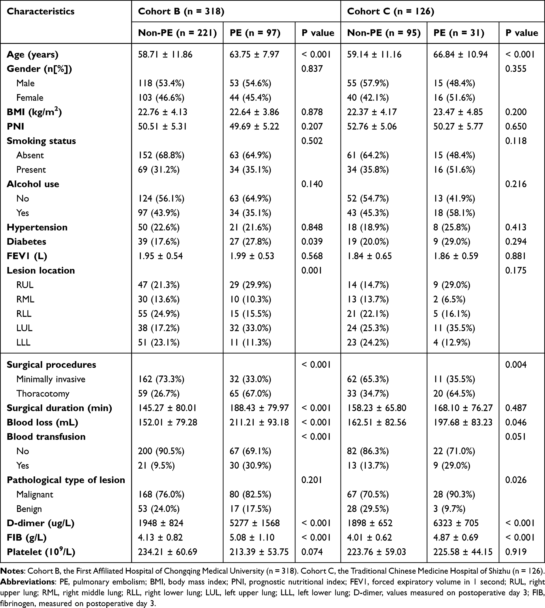 |
Table 2 Demographic, Clinical, and Pathological Factors in the Other Two Centers |
 |
Figure 1 Study design and profile. |
In cohort A, compared to the non-PE group, patients in the PE group were older (64.15± 9.07 vs 61.38± 10.73, p = 0.003), greater proportion distributed in the upper lobe lung (right upper: 35.0% vs 22.3%; left upper: 31.8% vs 22.1%; p < 0.001), more proportion of open thoracotomy (65.0% vs 33.0%, p < 0.001), longer operative duration (177.96 ± 72.08 min vs 157.43± 74.63 min, p = 0.004), more blood loss (210.40 ± 53.96 mL vs 161.66 ± 45.86 mL, p < 0.001), more perioperative blood transfusions (26.1% vs 17.6%, p = 0.024), higher proportion of malignancies (87.3% vs 72.9%, p < 0.001), higher D-dimer level (5820 ± 1441 ug/L vs 2023 ± 449 ug/L, p < 0.001), and higher FIB level (5.04 ± 0.95 g/L vs 4.03 ± 0.86 g/L, p < 0.001). There were no significant differences between the two groups in terms of gender, BMI, prognostic nutritional index (PNI), smoking history, alcohol consumption, hypertension, diabetes history, and baseline pulmonary function (Table 1).
Similar trends were observed in Cohorts B and C. Compared with non-PE patients, those with PE were older, more frequently underwent open thoracotomy, experienced more blood loss and transfusions, and exhibited elevated D-dimer and fibrinogen levels (Table 2).
Risk Factor Identification and Model Construction
Risk factors for PE were identified using univariate and multivariate logistic regression analyses with PE occurrence serving as the dependent variable. Seven independent risk factors were identified: advanced age, upper lobe lesion location, open thoracotomy, longer surgical duration, greater intraoperative blood loss, and elevated postoperative D-dimer and fibrinogen levels (Table 3). Feature importance ranking identified D-dimer and fibrinogen as the strongest predictors of PE (Figure 2).
 |
Table 3 Univariate and Multivariate Analysis of Pulmonary Embolism After Pulmonary Surgery |
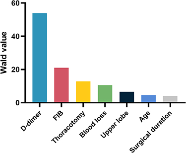 |
Figure 2 Ranking of risk factors for pulmonary embolism after pulmonary surgery. |
Based on these factors, a nomogram was developed to predict PE risk (Figure 3A). The results of the training and internal validation sets demonstrated excellent discrimination, with AUC of 0.97 (95% CI: 0.95–0.99) and 0.95 (95% CI: 0.92–0.97), respectively (Figure 3B and E). Similarly, calibration curves and Hosmer–Lemeshow (H-L) goodness-of-fit tests indicated strong accordance of nomogram model with the groups of training and internal validation (p = 0.232 and p = 0.182, respectively) (Figure 3C and F). Furthermore, the clinical decision curve analysis (DCA) showed the curve was generally close to the upper right corner and high net benefit across a wide range of threshold probabilities, confirming the model’s clinical utility (Figure 3D and G).
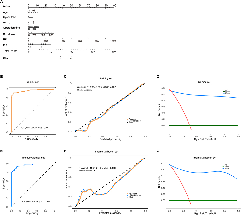 |
Figure 3 Construction and preliminary validation of a predictive model for pulmonary embolism after pulmonary surgery. (A) nomogram to predict pulmonary embolism. (B) the AUC curve for the training set. (C) the calibration curve and Hosmer–Lemeshow test for the training set. (D) the clinical decision curve analysis for the training set. (E) the AUC curve for the internal validation set. (F) the calibration curve and Hosmer–Lemeshow test for the internal validation set. (G) the clinical decision curve analysis for the internal validation set. |
External Validation and Risk Stratification
External validation using Cohorts B and C confirmed the model’s performance, with an AUC of 0.94 (95% CI: 0.91–0.96) and good calibration (p = 0.918) (Figures 4A and B). The decision curve similarly supported the model’s applicability in clinical decision-making (Figure 4C).
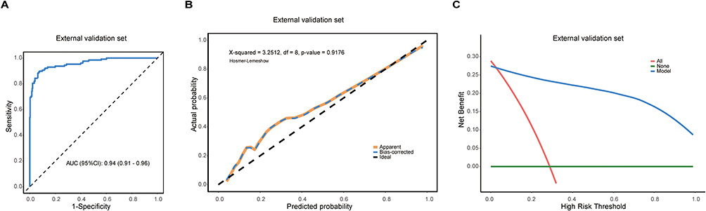 |
Figure 4 External validation of a predictive model for pulmonary embolism after pulmonary surgery. (A) the AUC curve for the external validation set. (B) the calibration curve and Hosmer–Lemeshow test for the external validation set. (C) the clinical decision curve analysis for the external validation set. |
To evaluate risk stratification, patients in the external validation cohorts were classified as high-risk or low-risk based on the 85th percentile of predicted risk. Patients in the high-risk group had PE probabilities >15%. Compared with the low-risk group, the high-risk group included older individuals, more frequent upper lobe involvement, longer operative times, greater blood loss, and higher postoperative D-dimer and fibrinogen levels. Open thoracotomy was also significantly more common among high-risk patients (Figure 5).
 |
Figure 5 Comparison of clinical characteristics between high-risk and low-risk patients in the external validation group. Patients in the external validation cohort were categorized as high-risk or low-risk according to the 85th percentile of the anticipated risk. Compared with the low-risk group, patients in the high-risk group were older (A), showed no significant difference in gender distribution (B), underwent more open thoracotomy (C), had more upper lobe lesions (D), experienced longer operative durations (E), had more intraoperative blood loss (F), and exhibited higher postoperative D-dimer (G) and fibrinogen levels (H). ***p < 0.001. |
Discussion
Postoperative PE is a potentially fatal complication following surgery, caused primarily by venous thrombus migration into the pulmonary arterial system. As a leading cause of sudden postoperative death and respiratory failure, PE affects approximately 1–5% of thoracic surgery patients, with mortality rates ranging from 15–30%.5 This poses a substantial burden on both patient outcomes and healthcare systems. The nonspecific clinical presentation of PE often leads to misdiagnosis or delayed recognition, underscoring the importance of identifying predictive risk factors and implementing timely intervention strategies.
In this multicenter retrospective study of 977 patients undergoing pulmonary resection, we identified seven independent risk factors for postoperative PE: advanced age, upper lobe lesions, open thoracotomy, prolonged operative duration, increased intraoperative blood loss, elevated D-dimer, and elevated fibrinogen levels. Based on these variables, we constructed a nomogram prediction model that demonstrated excellent discrimination (AUC 0.94–0.97) and good calibration across both internal and external validation cohorts. These findings offer a valuable tool for early PE risk prediction in thoracic surgery patients, especially when combining dynamic coagulation monitoring with assessment of surgical trauma.
Among these predictors, D-dimer and fibrinogen emerged as the most influential. This supports prior evidence that hypercoagulability plays a central role in PE development.22,23 Importantly, our model integrated surgical variables with coagulation biomarkers, extending beyond traditional assessments. Compared with previous studies that emphasized individual risk factors, our approach provides a more comprehensive framework to support perioperative decision-making.24 For clinicians, this model may facilitate early identification of high-risk patients and implementation of personalized preventive strategies.
A novel finding of our study was the significant association between upper lobe lesions and postoperative PE, which has not been reported in earlier literature.25 We hypothesize that this relationship may relate to the unique anatomical characteristics of the upper lobes and the complexity of surgical manipulation in these regions.26 Upper lobe resections are typically associated with longer operative times, increased surgical trauma, and more extensive lymphatic and vascular disruption, particularly during open surgery.27 After upper lobectomy, vascular stumps may also be anatomical factors contributing to thrombus formation.28 Postoperative hemodynamic alterations and impaired lymphatic drainage may further promote venous thrombus formation. Previous studies have also shown that upper lobes have more complex venous return and richer blood supply,29 which may increase thrombosis risk post-resection. These findings suggest that surgical trauma to upper lobes may be a critical factor in PE development, warranting additional mechanistic research.
We also observed that patients with lung cancer had significantly higher PE rates compared with those with benign diseases. This aligns with prior studies and reflects the greater surgical trauma and blood loss typically associated with lung cancer surgery, as well as the slower postoperative recovery in these patients.30,31 Moreover, malignancies are known to induce a hypercoagulable state through tumor-related secretion of procoagulant factors such as tissue factor and tumor necrosis factor, further increasing thrombotic risk.32 This dual influence—surgical and tumor-related—may explain the elevated PE incidence in cancer patients.
Our findings reinforce the clinical utility of D-dimer and fibrinogen as critical biomarkers for the early identification of patients at risk for postoperative PE. In particular, elevated D-dimer levels demonstrated a strong and statistically significant association with PE onset, consistent with previous studies indicating its high sensitivity in detecting thromboembolic events.33,34 D-dimer, a degradation product of cross-linked fibrin, reflects ongoing activation of the coagulation and fibrinolytic systems. Its elevation in the postoperative setting may signal subclinical thrombus formation, allowing for earlier risk stratification and timely intervention before clinical PE develops.35
Fibrinogen, an acute-phase reactant and a key factor in clot formation, was also identified as an independent predictor in our model. Higher postoperative fibrinogen levels have been shown to enhance blood viscosity and promote a prothrombotic state, especially in the context of surgical trauma and inflammation.36,37 Our results suggest that combining D-dimer and fibrinogen measurements provides additive predictive value, reflecting both fibrin turnover and the systemic procoagulant response.
Given the high AUC values observed, our model demonstrates strong discriminatory power, suggesting it can reliably predict the risk of postoperative pulmonary embolism in thoracic surgery patients. However, this raises a valid concern regarding the potential for overfitting. Overfitting occurs when a model is excessively tailored to the training data, capturing noise rather than underlying trends, which may compromise its generalizability to independent cohorts.38 Future studies should aim to validate the model with larger, more diverse prospective cohorts. A broader dataset would help to assess the generalizability of the model across different populations, surgical procedures, and institutional settings, reducing the risk of overfitting.39 Additionally, cross-validation techniques can be implemented during model development to ensure its stability and performance across various subsets of data.40 By applying these strategies, future research can further refine the model and enhance its clinical applicability, ensuring that it performs well in real-world settings. A major strength of this study is the use of multicenter data to validate the model, enhancing its generalizability across diverse clinical settings. The model demonstrated robust performance not only in the training cohort but also in independent external validation cohorts, suggesting it may be applicable across different regions and institutions. The addition of a risk stratification system further enhances the model’s clinical value by enabling the identification of high-risk patients who may benefit from intensified perioperative surveillance and prophylactic anticoagulation.
Despite these strengths, our study has several limitations. First, the retrospective design may introduce selection bias despite efforts to control confounding variables. Second, although external validation was performed, prospective clinical trials are still needed to confirm the model’s effectiveness in routine practice. Third, we did not incorporate other potential contributors to PE risk, such as genetic predisposition, systemic inflammation, or tumor microenvironment changes, which may affect the model’s predictive precision. Lastly, our model relies on postoperative day 3 values of coagulation markers for risk assessment, which may delay timely interventions, particularly for high-risk patients who might benefit from earlier preventive measures. Earlier measurements of coagulation markers within the first 24–48 hours post-surgery, may allow for more prompt identification of at-risk patients. Future studies should consider integrating multi-omics data (eg, genomics, metabolomics) to improve model sensitivity and specificity.
Conclusion
In conclusion, we developed and validated a multifactorial prediction model for postoperative PE in thoracic surgery patients based on clinical and surgical characteristics as well as coagulation biomarkers. This model provides a valuable tool for early risk stratification and may inform individualized preventive strategies. With further prospective validation and refinement, the model has the potential to become an integral part of clinical decision-making in thoracic surgical care.
Statement of Patient Data Confidentiality
All patient data were anonymized to ensure confidentiality, and no personally identifiable information was included in the analysis or the manuscript.
Data Sharing Statement
The datasets generated and analyzed during this study are available from the corresponding author (Junying Chen, E-mail: [email protected]) on reasonable request.
Disclosure
The authors report no conflicts of interest in this work.
References
1. Kolokotroni SMJSC. Deep vein thrombosis and pulmonary embolism following lung resection. 2020;2020:5.
2. Wendelboe AM, Raskob GE. Global burden of thrombosis: epidemiologic aspects. Circul res. 2016;118(9):1340–1347. doi:10.1161/CIRCRESAHA.115.306841
3. Keller K, Hobohm L, Ebner M, et al. Trends in thrombolytic treatment and outcomes of acute pulmonary embolism in Germany. Eur Heart J. 2020;41(4):522–529. doi:10.1093/eurheartj/ehz236
4. Raskob GE, Angchaisuksiri P, Blanco AN, et al. Thrombosis: a major contributor to global disease burden. Arteriosclerosis Thrombosis Vasc Biol. 2014;34(11):2363–2371. doi:10.1161/ATVBAHA.114.304488
5. Temgoua MN, Tochie JN, Noubiap JJ, et al. Global incidence and case fatality rate of pulmonary embolism following major surgery: a protocol for a systematic review and meta-analysis of cohort studies. Syst Rev. 2017;6(1):240. doi:10.1186/s13643-017-0647-8
6. Cormican D, Morkos MS, Winter D, Rodrigue MF, Wendel J, Ramakrishna H. Acute perioperative pulmonary embolism-management strategies and outcomes. J Cardiothoracicvascul Anesthesia. 2020;34(7):1972–1984. doi:10.1053/j.jvca.2019.11.018
7. Konstantinides SV, Meyer G, Becattini C, et al. 2019 ESC guidelines for the diagnosis and management of acute pulmonary embolism developed in collaboration with the European Respiratory Society (ERS): the task force for the diagnosis and management of acute pulmonary embolism of the European Society of Cardiology (ESC). Europ resp J. 2019;54(3). doi:10.1183/13993003.01647-2019.
8. Douma RA, Kamphuisen PW, Büller HR. Büller HR: acute pulmonary embolism. Part 1: epidemiology and diagnosis. Nat Rev Cardiol. 2010;7(10):585–596. doi:10.1038/nrcardio.2010.106
9. Du H, Chen J. Occurrence of venous thromboembolism in patients with lung cancer and its anticoagulant therapy. Zhongguo fei ai za zhi. 2018;21(10):784–789. doi:10.3779/j.issn.1009-3419.2018.10.09
10. Stein PD, Matta F. Acute pulmonary embolism. Curr Prob Cardiol. 2010;35(7):314–376. doi:10.1016/j.cpcardiol.2010.03.002
11. van Maanen R, Trinks-Roerdink EM, Rutten FH, Geersing GJ. Geersing GJ: a systematic review and meta-analysis of diagnostic delay in pulmonary embolism. European J General Pract. 2022;28(1):165–172. doi:10.1080/13814788.2022.2086232
12. Mansella G, Keil C, Nickel CH, et al. Delayed diagnosis in pulmonary embolism: frequency, patient characteristics, and outcome. Respiration. 2020;99(7):589–597. doi:10.1159/000508396
13. van Lier F, van der Geest PJ, Hol JW, Leebeek FW, Hoeks SE. Risk modification for postoperative pulmonary embolism: timing of postoperative prophylaxis. Thrombosis Res. 2012;129(4):e14–17. doi:10.1016/j.thromres.2011.11.033
14. Spiezia L, Cuzzolin M, Elssy H, et al. Post-operative hypercoagulable whole blood profiles in patients undergoing open thoracotomy vs video-assisted thoracoscopic surgery. Blood Transfusion. 2021;19(2):144–151. doi:10.2450/2020.0040-20
15. Tian B, Song C, Li H, et al. The significance of perioperative coagulation and fibrinolysis related parameters after lung surgery for predicting venous thromboembolism: a prospective, single center study. J Thoracic Dis. 2018;10(4):2223–2230. doi:10.21037/jtd.2018.03.174
16. Black SR, Howard JT, Chin PC, Starr AJ. Toward a more robust prediction of pulmonary embolism in trauma patients: a risk assessment model based on 38,000 patients. J of Orthop Trauma. 2016;30(4):200–207. doi:10.1097/BOT.0000000000000484
17. Liu K, Qian D, Zhang D, Jin Z, Yang Y, Zhao Y. A risk prediction model for venous thromboembolism in hospitalized patients with thoracic trauma: a machine learning, national multicenter retrospective study. World J Emerg Surg. 2025;20(1):14. doi:10.1186/s13017-025-00583-w
18. Li Y, Liu Z, Chen C, et al. Risk factors and potential predictors of pulmonary embolism in cancer patients undergoing thoracic and abdominopelvic surgery: a case control study. Thrombosis J. 2022;20(1):80. doi:10.1186/s12959-022-00442-7
19. Ke L, Cui S, Yang M, et al. Validation of a modified caprini risk assessment model in lung cancer patients undergoing surgery: results of a multicenter cross-sectional observational study. J Surg Oncol. 2022;125(5):933–942. doi:10.1002/jso.26794
20. Malik AY, Foster C. The revised declaration of Helsinki: cosmetic or real change? J R Soc Med. 2016;109(5):184–189. doi:10.1177/0141076816643332
21. Wu J, Ding V, Luo S, et al. Predictive model to guide brain magnetic resonance imaging surveillance in patients with metastatic lung cancer: impact on real-world outcomes. JCO Precis Oncol. 2022;6(6):e2200220. doi:10.1200/PO.22.00220
22. Rezania S, Puskarich MA, Petrusca DN, Neto-Neves EM, Rondina MT, Kline JA. Platelet hyperactivation, apoptosis and hypercoagulability in patients with acute pulmonary embolism. Thrombosis Res. 2017;155:106–115. doi:10.1016/j.thromres.2017.05.009
23. Yang G. Nie SJJoTD: risk factors for pulmonary embolism: a case-control study. 2025. 2025;17(3):1552–1560.
24. Mahajan A, Esper S, Oo TH, et al. Development and validation of a machine learning model to identify patients before surgery at high risk for postoperative adverse events. JAMA Network Open. 2023;6(7):e2322285–e2322285. doi:10.1001/jamanetworkopen.2023.22285
25. Selten K, Durak K, Kersten A, Kalverkamp S. First ultrasound-assisted thrombolysis for pulmonary embolism after lung surgery. Thoracic Cardiovascul Surg Rep. 2023;12(1):e14–e16. doi:10.1055/s-0043-1762551
26. Rakovich G. Thoracoscopic S1 segmentectomy, right upper lobe: alternative posterior approach. Multimedia Manual Cardiothoracic Surg. 2024,;2024.
27. Ueda K, Murakami J, Tanaka T, Nakamura T, Yoshimine S, Hamano K. Postoperative complications and cancer recurrence: impact on poor prognosis of lower lobe cancer. Ann Thorac Surg. 2020;109(6):1750–1756. doi:10.1016/j.athoracsur.2019.12.061
28. Song CY, Kimura D, Fukuda I, Tsushima F, Sakai T, Tsushima T. Tsushima T: chest radiotherapy after left upper lobectomy may be a risk factor for thrombosis in the pulmonary vein stump. J Cardiothoracic Surg. 2022;17(1):154. doi:10.1186/s13019-022-01902-x
29. Koç ZP, Burma O, Uysal A, Mitil HA. An experimental lymphoscintigraphy study in an acute unilateral deep venous thrombosis model. Lymphatic Res Biol. 2013;11(2):76–80. doi:10.1089/lrb.2012.0026
30. Li YP, Shen L, Huang W, et al. Prevalence and risk factors of acute pulmonary embolism in patients with lung cancer surgery. Semin Thromb Hemost. 2018;44(4):334–340. doi:10.1055/s-0037-1612625
31. Li Y, Shang Y, Wang W, Ning S, Chen H. Lung cancer and pulmonary embolism: what is the relationship? A review. J Cancer. 2018;9(17):3046–3057. doi:10.7150/jca.26008
32. Caine GJ, Stonelake PS, Lip GY, Kehoe ST. The hypercoagulable state of malignancy: pathogenesis and current debate. Neoplasia. 2002;4(6):465–473. doi:10.1038/sj.neo.7900263
33. Kearon C, de Wit K, Parpia S, et al. Diagnosis of pulmonary embolism with d-dimer adjusted to clinical probability. New Engl J Med. 2019;381(22):2125–2134. doi:10.1056/NEJMoa1909159
34. Righini M, Van Es J, Den Exter PL, et al. Age-adjusted D-dimer cutoff levels to rule out pulmonary embolism: the ADJUST-PE study. JAMA. 2014;311(11):1117–1124. doi:10.1001/jama.2014.2135
35. An TJ, Engstrom SM, Oelsner WK, Benvenuti MA, Polkowski GG, Schoenecker JG. Elevated d-dimer is not predictive of symptomatic deep venous thrombosis after total joint arthroplasty. J Arthroplasty. 2016;31(10):2269–2272. doi:10.1016/j.arth.2016.02.059
36. Lip GY, Andreotti F, Fauchier L, et al. Bleeding risk assessment and management in atrial fibrillation patients. executive summary of a position document from the European heart rhythm association [EHRA], endorsed by the European Society of Cardiology [ESC] working group on thrombosis. Thrombosis Haemostasis. 2011;106(6):997–1011. doi:10.1160/TH11-10-0690
37. Kattula S, Byrnes JR, Wolberg AS. Wolberg AS: fibrinogen and fibrin in hemostasis and thrombosis. Arteriosclerosis Thrombosis Vasc Biol. 2017;37(3):e13–e21. doi:10.1161/ATVBAHA.117.308564
38. Steyerberg EW, Vickers AJ, Cook NR, et al. Assessing the performance of prediction models: a framework for traditional and novel measures. Epidemiology. 2010;21(1):128–138. doi:10.1097/EDE.0b013e3181c30fb2
39. bar O, Neimark D, Zohar M, et al. Impact of data on generalization of AI for surgical intelligence applications. Sci Rep. 2020;10(1):22208. doi:10.1038/s41598-020-79173-6
40. Yates LA, Aandahl Z, Richards SA, Brook BW. Cross validation for model selection: a primer with examples from ecology. 2022;2022.
 © 2025 The Author(s). This work is published and licensed by Dove Medical Press Limited. The
full terms of this license are available at https://www.dovepress.com/terms
and incorporate the Creative Commons Attribution
- Non Commercial (unported, 4.0) License.
By accessing the work you hereby accept the Terms. Non-commercial uses of the work are permitted
without any further permission from Dove Medical Press Limited, provided the work is properly
attributed. For permission for commercial use of this work, please see paragraphs 4.2 and 5 of our Terms.
© 2025 The Author(s). This work is published and licensed by Dove Medical Press Limited. The
full terms of this license are available at https://www.dovepress.com/terms
and incorporate the Creative Commons Attribution
- Non Commercial (unported, 4.0) License.
By accessing the work you hereby accept the Terms. Non-commercial uses of the work are permitted
without any further permission from Dove Medical Press Limited, provided the work is properly
attributed. For permission for commercial use of this work, please see paragraphs 4.2 and 5 of our Terms.